Int. J. Trop. Vet. Biomed. Res. Vol. 2 (2) : 28-33; November 2017
www.jurnal.unsyiah.ac.id/IJTVBR E-ISSN : 2503-4715
Pediatric Vaginal Discharge
Fitria Salim1,2 and Sitti Hajar1,2
1
Dermatology and Venereology Department Faculty of Medicine 2
Syiah Kuala University/dr. zainoel Abidin Hospital, Banda Aceh, Indonesia
Email for correspondence: [email protected]
Abstract
Vaginal discharges are commonly seen in the pediatric population, approximately more than 50% of all pediatric genital complaints. Normal vaginal secretion is usually thin, and clear to white with a variable amount, while malodorous, abnormal consistency of vaginal discharge accompanied by blood, pain, pruritus, or dysuria is usually pathologic. Many factors can cause vaginal discharge such as anatomy of genital area that more susceptible to inflammation and infection, numerous organisms that including those associated with the sexually transmitted diseases or child sexual abuse, also mechanical and chemical substances, as well as poor hygiene. Recurrent vaginal discharge can be very distressing to children, especially if associated with discomfort and the most common cause is vulvovaginitis. The management of vaginal discharge is based on underlying causes, either by initiating proper therapeutic or teaching the children about good hygiene.
Keywords: etiology, pediatric, vaginal discharge
Background
Vaginal discharge is the most frequent gynecological disorder encountered in pediatrics or pre-pubertal girls and can cause repeated clinical episodes with numerous etiological factors. The incidence is unknown; however, the most typical age of referral is between 3–10 years. The presence of discharge is not necessarily abnormal, this symptom may represent the
vagina’s response to changes in estrogen
levels, and the pediatrician need only reassure the patient and her parents. In most circumstances, the age of the patient, her pubertal status, and whether she has ever had sexual intercourse are essential elements in sorting out the cause of the discharge.
The common cause of vaginal itching and discharge in young girls include chemicals, yeast or parasite (pinworm) infection, vaginitis and a foreign body. The chemicals such as perfumes and dyes in detergents, softeners, creams or ointments may irritate the vagina or the skin around. Vaginitis that suspicious from sexual transmitted vaginal infection, however, sexual abuse must be considered and addressed. The foreign body such as toilet
paper or crayon remains in the vagina that causes infection. A useful way of approaching the diagnosis of discharge is by categorizing the patient as being pre-pubertal of post-pre-pubertal. In the pre-pre-pubertal age group, discharge is associated with vulva-vaginitis. In the post-pubertal age group, discharge may be physiologic or may associate with cervicitis or vaginitis.
In utero, the vaginal epithelium of the neonate is stimulated by maternal hormones that cross the placenta into the fetal circulation. After delivery, these hormone levels fall rapidly, and the parents may note a thick, grayish-white, mucoid discharge from the neonate’s vagina. In many instances, the discharge is blood tinged or even grossly bloody. No treatment is needed, and the discharge usually resolves by ten days of age. Non-specific vaginal discharge is usually treated by removing the irritant, sitz baths and education about proper hygiene and increasing air flow to the area, but infection must treat with specific drugs.
highly anxious, particularly if the symptoms have been present for several weeks or months. Vaginal discharge has been associated with pelvic infection, lack of cleanliness and sexual abuse, so knowing the cause is very important to determine the appropriate and accurate therapy.
Pediatric Genital Anatomy and Physiology
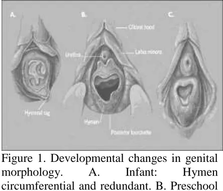
The newborn girl is affected by maternal estrogens that cross the placenta. Vaginal discharge is common in newborns, as are long hymenal tags that frequently are visible, extending outside the vestibule (Figure 1). The vaginal epithelium also demonstrates the estrogen effect. The epithelium contains glycogen, and vaginal
secretions are acidic. The newborn’s vagina
is approximately 4 cm long. The cervix is much larger than the uterine corpus (ratio 3:1). Even after maternal circulating estrogen concentrations decline, the infant girl maintains relatively high endogenous production of gonadal estrogens. Hymenal tissue, sensitive to estrogen, remains thick, redundant, and elastic throughout infancy. Most commonly, the hymen surrounds the vaginal orifice and appears circumferential. In some infants, the hymen extends outward in a sleeve-like configuration. The clitoral hood is prominent, and the urethra may be obscured by redundant hymenal tissue.
The preschool and school-age child (ages 3 to 6 years) that as the hypothalamic-pituitary-gonadal axis becomes suppressed, the hormonal effects on the genital structures recede. The labia become flattered and the clitoris less prominent. The labia minor throughout infancy and childhood are rudimentary, appearing as an extension of the clitoral hood, and extend only about one-third the length of the labia major. In this age group, the hymen becomes thinner and may be translucent, although some children maintain a thickened, redundant hymenal configuration. The hymen tissue often recedes from the anterior vaginal orifice,
leaving a “crescentic” appearance. The
vaginal epithelium becomes thin, and the vaginal pH is alkaline. A vaginal swab shows ovoid epithelial cells.
Sexual Maturity Rating (SMR) staging or Tanner staging is based on breast development, and pubic hair finds in older school-age and preteen children (ages 7 to 12 years). Changes in the genital tissues are usually visible before breast buds form and before pubic hair growth begins. The mons becomes fuller, as do the labia major. The labia minor continue to develop, although it is only years after menarche that the labia minor becomes an entirely separate layer and join at the posterior commissure. The vestibular mucosa forms a cobblestoned, rough appearance. The hymen thickens and often develops fimbriations. The vagina elongates to about 8 cm, the uterine corpus enlarges, and the vaginal mucosal thickens. A milky vaginal discharge may be noted, vaginal secretions become acidic, and lactobacilli become the predominant organism. A vaginal swab shows polygonal epithelial cells. Physiologic leucorrhea commonly precedes menses by 3 to 6 months.
Figure 1. Developmental changes in genital
morphology. A. Infant: Hymen
circumferential and redundant. B. Preschool through school age: Rudimentary labia minor, thinner hymen. C. Early puberty: Labia minor develop, hymen thickens.
Pediatric Gynaecological Examination
what the examiner is doing and engaging the child in distracting conversation.
The genital examination of the newborn girl naturally follows the hip
examination when the baby’s hips are flexed
and adducted. Placing the hands on each thigh, the examiner can use his or her thumbs to separate the labia major. A second technique, labial traction, allows better visualization of the introitus and consists of gently grasping the labia with thumb and forefinger and gently pulling the labia toward the examiner. If the redundant hymen obscures the orifice, the examiner may drop a little warm water or saline into the introitus to assure that the hymen is perforate. The genital examination follows the evaluation of the hips and the diaper area skin and is followed by a brief anal examination. For toddlers, the brief genital-anal examination incorporates into a diaper change on the table. It is easiest to examine the genital area while the child is sitting in a modified frog-leg position on a diaper in her
parent’s lap.
For preschoolers, clinicians may
move the child’s legs into the frog-leg
position or like “butterfly wings” with the
soles of her feet together allow the genital examination. Another option is to have the parent sit on the examination table with the child on her lap. Labial separation and labial traction allow good visualization of the entire introitus. The prone knee-chest position is useful when the examiner needs to see the vaginal vault because of concern for a foreign body.
School-age and preteen children may be embarrassed, ask the parent to stay by the
child’s head. Drapes always should be used,
place the child in the frog-leg position and move the crotch area of the underpants to the side. This maneuver often allows an adequate view, and many children are more comfortable with this method. The clinician
can enlist the child’s intellect and interest in
the examination by explaining, with illustrations, what the genital area normally looks like and why it is helpful to know if each of the three openings in that area of the body appears normal. This active engagement assists the child in mastering the medical examination process.
Causes of Pediatric Vaginal Discharge
Vaginal discharge is a common symptom in young girls and signifies a vulvar or vaginal inflammation that may be caused by physical, chemical, or infectious agents. Most cases are caused by primary irritants or poor hygiene. Bacterial were found in only one-third of young girls who presented with vulva vaginitis. The other causes included suspected sexual abuse (5%), foreign body (3%), labial adhesions (3%), and vaginal agenesis (2%). Nonspecific factors should be considered a variant of atopic dermatitis, because they are highly sensitive to a variety of irritants, including soaps, poor hygiene, and tight-fitting clothing.
1. Irritants
Irritants are one of the most common causes vulva soreness and vaginal discharge. Vulva dermatitis has been reported as a result of using soap or bubble bath, douches, spermicides, latex and playing in a sandpit, as well as poor hygiene with prolonged contact with urine and feces against the skin. Masturbation and restrictive clothing also can cause vaginal discharge. Avoidance of the irritative agent should lead to resolution of symptoms. Allergic contact dermatitis may develop as a result of prolonged exposure to irritant substances, such as perfumes and clothing dyes. Accurate allergy patch testing may help to identify the culprit.
2. Infection or infestation
Vaginal discharge is a common form of infection/infestation manifestations. The most common main symptom occurs in girls by 62-92%. Vaginal discharge can be clear, yellowish or greenish accompanied by an unpleasant odor, the reddish skin around the genital area, pain, itching, and dysuria. Infectious organisms or infestations may include:
Non-specific bacterial
Non-specific bacterial vulva vaginitis is the most commonly found with mixed bacterial flora. Normal vaginal flora in prepubertal girls include Diphtheroids,
epidermidis; if one of these organisms is found in an asymptomatic child, antibiotic treatment is not appropriate. Vaginal cultures will be reported as non-specific skin flora or will show mixed anaerobes or coliforms from the gut. Enteric pathogens,
Escherichia coli, Salmonella sp, and
Shigella sp; the latter two organisms are known to cause bloody vaginitis. Rarely, vaginitis may be the earlier or more prominent sign, with only mild diarrhea. The presence of leukocytes on the smear of the discharge is sensitive but not specific for bacterial infection. Poor personal hygiene is a common trigger factor, as the onset of symptoms usually occurs when the child has responsibility for her anal hygiene; for example, on first attending nursery or school.
Specific bacterial
The most common specific bacterial is the group A beta-hemolytic Streptococcus. This organism has been isolated in 11–18% of vaginal aspirates in various studies.It has been suggested that the epidemiology is related to an upper respiratory tract infection.The onset can be quite acute, with a serous purulent vaginal discharge, which may be associated with dysuria and an inflamed vulva, a dramatic scarlet, well-demarcated dermatitis of the vulva or perianal tissues. Haemophilus influenzae is the second most common cause and more likely to have recurrent symptoms of vulva vaginitis. The most common are biotype II, which was isolated in 57% of isolates in one study. These bacterial infections are likely to be caused by self-inoculation as a child carries bacteria from her nose and mouth via her hand to her vulva.
Gonorrhea in children is a vaginal infection and rarely is associated with ascending infection caused by Neisseria
gonorrhoeae. The vaginal discharge
typically is profuse and green. The diagnosed more often in children who present with a complaint of vaginal discharge than in those who present for evaluation of sexual abuse. Chlamydia trachomatis infection may be asymptomatic or present with a clear vaginal discharge. Both Chlamydia and gonorrhea may be
transmitted perinatal from an infected mother. Gonorrhea typically presents with signs, but asymptomatic carriage of perinatal acquired Chlamydia has been documented to 34 months of age.
Candida
This organism is a very uncommon cause. However, there tends to be some other predisposing factor in association with the presence of Candida albicans; for example, a recent course of antibiotics, diabetes or the wearing of nappies. It has been suggested that vaginitis associated with
Candida is more likely to be related to sexual abuse. The symptoms are similar to adult women, and there are pruritus and a white, curd-like discharge. Inflammation of the vulva and perineal area and white plaques adherent to the vagina often occur.
Protozoa
Pinworms (Enterobius vermicularis) may cause another type of irritant vulvitis. Although pinworms do not reside in the vulva, they can crawl from the anus to the vulvar introitus and cause intense pruritus. The interview should identify pruritus as the prominent symptom. The diagnostic test is an anal pinworm preparation with sellotape.
Trichomonas vaginalis is one of the
protozoan causes of vaginal discharge in children. The discharge is yellowish or greenish, foamy, malodor sometimes dysuria. This organism is a sexually transmitted infection that can be transmitted through perinatal and sexual abuse in children.
Virus
Vaginal discharge in children can be caused by viruses, however, the frequency fewer than bacteria or fungi infection. Herpes simplex virus and human papilloma virus is the cause of the genital disease that transmission can be perinatal or sexual contact. Serum antibody titers from blood and Tzank test for multinucleated giant cells in vaginal cytology can be done to confirm the diagnosis.
3. Foreign body
is small bits of toilet paper, although larger objects such as coins, bottle caps, or small toys may be inserted by the child. If the foreign body is visible and the patient is cooperative, a plastic ear curette can be used to hook it and pull it out. Irrigation also may be used to flush out the foreign body. A soft feeding tube or red rubber catheter is child has denied inserting anything.
4. Other
Vulva skin conditions can also present with vulva irritation and soreness. Vaginal discharge is usually a less prominent feature, especially if the skin is traumatized due to scratching. Atopic eczema can affect up to 15% of young children, and vulva symptoms are not uncommon. Lichen sclerosis usually presents with itching and soreness, and vaginal discharge is unusual unless there is a secondary infection but bleeding can occur from purpura and blister formation. It is essential to make the correct diagnosis as the traumatized appearance of the skin can raise suspicions of sexual abuse. Other uncommon causes include fistula, ectopic ureter, polyps, neoplasms. Malignancies such as rhabdomyosarcoma and endodermal sinus tumors can also cause discharge or bleeding and require an intravaginal examination under anesthesia, with biopsy of suspicious lesions. A study of 24 girls younger than six years who underwent such an examination for bleeding or discharge identified six patients with malignancies.
Treatment of Pediatric Vaginal Discharge
The management of pediatric vaginal discharge depends on the causes. Leucorrhea caused by non-specific vulva vaginitis or irritant no specialized therapies, only need to maintain the cleanliness of the genital area, such as the use of cotton underwear, avoid nylon-based clothing,
avoid tight clothes, clean the genito-anal area from the front to the back after urination and washing hands after doing the activities. Improved hygiene with daily or twice-daily sitz baths as well as avoidance of bubble bath, soap, and shampoo in the bathtub are also suggested as management techniques. Barrier creams such as nappy creams are useful, as are emollients to protect the vulva skin from further irritation, also the use of mild or moderate strength steroid cream may be necessary for short periods.
Vaginal discharge with intense and worse itching at night should get therapy for pinworms as empiric treatment is recommended. If the vaginitis persists, a follow-up in 2 weeks should be arranged, a culture of the inner labia can be obtained at that time. Antibiotics should be used when a pure growth of a specific pathogenic organism has been identified. The treatment for most strains of bacterial infections is sensitive to penicillin. However the resistance is increasing; therefore, clinicians should be guided by sensitivity test results. Erythromycin is a suitable alternative for a girl who is allergic to penicillin. Relapse can occur in up to a third of treated individuals. Topical antibiotics are no use for treatment of vaginal infection. If the child presents with a profuse green discharge or has blood-streaked or foul-smelling discharge, culture for respiratory and enteric pathogens and gonococcus should be obtained without waiting two weeks. A positive culture for gonorrhea should prompt an evaluation for sexual abuse. When cultures are negative and significant vaginitis persists, the clinician should consider infection with
prepubertal girls, such as tumors, skin conditions, malformations, and trauma sometimes need a biopsy of suspicious lesions to determine the accurate diagnosis and give effective therapy.
Conclusion
Pediatric vaginal discharge is very common, and many factors can be the causes. The proper examination and cultures are the key to make the diagnosis that may lead to a particular therapy. Many times, the problem is nonspecific and related to poor hygiene or chemical irritants but sometimes still need antibiotic for infection cause. If the adequate treatment has been given, but symptoms persist, it is important to check for foreign bodies in the vagina. The presence of trauma in the genital area there should be suspicious possible sexual abuse.
References
Antunes DE; Araujo S; Ferreira GP; Cunha CS; Costa AV; et al. Identification of Clinical, Epidemiology and Laboratory risk Factor for Leprosy Reaction for Leprosy Reactions during and after Multidrug Theraphy. Dermatology Sanitria, Hospital de Clinicas, Faculdade de Medicine, Universidade Federal de Uberlandia. Brasil. 2013. Vol 7. 108:901-908. McGreal S, Wood P. Recurrent vaginal
discharge in children. Journal Pediatric Adolescent Gynecologic. 2013. 26 (4): 205-208.
Randelovic G, Mladenovic V, Ristic L, et al.
Microbiological aspects of
vulvovaginitis in prepurbetal girls. Eur J Pediatr 2012; 171: 1203. Sanfillippo JS. Vaginal discharge—time for
a reappraisal. J Pediatr Adolesc Gynecol 2013; 26:203-204.
Yilmaz AE, Celik N, Soylu G, et al.
Comparison of clinical and
microbiological features of