E-ISSN: 2347-5129 P-ISSN: 2394-0506
(ICV-Poland) Impact Value: 5.62 (GIF) Impact Factor: 0.549 IJFAS 2017; 5(2): 33-37 © 2017 IJFAS
www.fisheriesjournal.com Received: 08-01-2017 Accepted: 09-02-2017
Ajay S Khandagale Department of Biochemistry, College of Fisheries, KVAFSU, Mangaluru, Karnataka, India
Likhitha Mundodi
Department of Chemistry, P.A. College of Engineering, Mangaluru, Karnataka, India
Balladka Kunhanna Sarojini Department of
Industrial Chemistry, Mangalore University, Mangalagangothri, Mangaluru, Karnataka, India
Correspondence Ajay S Khandagale Department of Biochemistry, College of Fisheries, KVAFSU, Mangaluru, Karnataka, India
Email: [email protected]
Isolation and characterization of trypsin from fish
viscera of Oil Sardine (
Sardinella longiceps
)
Ajay S Khandagale, Likhitha Mundodi and Balladka Kunhanna Sarojini
Abstract
The development of the fisheries industry has led to the increased interest in the full utilization of non-edible seafood fish viscera as a large source of unique digestive enzymes especially the proteases. We report the purification and biochemical characterization of a trypsin from the viscera of Oil Sardine (Sardinella longiceps) using anion-exchange and soybean trypsin inhibitor (SBTI) affinity
chromatography. Final enzyme preparation was homogeneous in SDS-PAGE and the molecular weight of the purified enzymes was estimated to be 24,000 Da. The enzyme activity was optimum at 60 °C for hydrolysis of benzoyl-dl-arginine-p-nitroanilide (BAPNA). The purified enzyme maximal activity was
observed at pH 8.0. Isolated trypsin was strongly inhibited by SBTI and N-p-tosyl-1-lysine chloromethyl ketone (TLCK) which are the specific inhibitors of trypsin and was markedly inhibited by the serine-protease inhibitor PMSF. Results signify that Oil sardine viscera are useful source for production of enzymes that could be used as a biotechnological tool in various industries.
Keywords: Oil Sardine (Sardinella longiceps), fish viscera, trypsin, protease inhibitors
1. Introduction
The limited biological resources, increased environmental concerns and the development of the fisheries industry have lead to the increased interest in the full utilization of seafood processing wastes. Fish viscera are non-edible parts produced in large quantities as byproduct by the fishery industries. These resources however are recognized as potential source of unique
digestive enzymes especially the proteases [1]. The most important class of proteolytic enzymes
isolated includes the aspartate protease (pepsin) and serine proteases (trypsin, chymotrypsin and elastase). The acidic proteases isolated from viscera display high activity between pH 2.0 and 4.0, while alkaline digestive proteases, such as trypsin are most active between pH 8.0 and 10.0. Trypsin is a very useful enzyme in detergent production, leather processing, chemical modifications industry and food processing industry due to its stable nature under harsh conditions such as high temperatures (50-60 °C), high pH, presence of surfactants and
oxidizing agents [2]. Additionally these enzymes also serve as a useful tool for basic research.
The presence of several types of proteolytic enzymes in sardine and related species has been
widely reported [2]. Acidic, neutral and alkaline proteinases have been found in Monterey
sardine (Sardinops sagax caerulea) muscle [3]. Castillo-yanez et al. [4]isolated trypsin from the
pyloric caeca of M. sardine viscera with an optimal pH of 2.5. Purified trypsin from true
sardine (Sardinops melanostictus) exhibited optimal activity at pH 8 and temperature of 60°C.
Similarly, Bougatef et al. [2] reported the purification of trypsin from the viscera of sardine
(Sardina pilchardus); the enzyme showed an optimal temperature and pH of 60 °C and 8
respectively. Further, Khaled et al. [5] isolated and characterized trypsin from viscera of
sardinelle (Sardinella aurita) with an optimal pH and temperature of 8 and 55 °C respectively.
In this study, we report the purification, basic biochemical information and kinetic
characterization of a trypsin from the viscera of Oil Sardine (Sardinella longiceps) from
Arabian Sea Mangalore, India.
2. Materials and methods 2.1 Chemicals
(SBTI), benzoyl-dl-arginine-p-nitroanilide (BAPNA), sodium
dodecyl sulfate (SDS), acrylamide, ammonium persulphate,
N,N,N’,N’-tetramethyl ethylenediamine (TEMED),
Coomassie Brilliant Blue R-250, Sephadex G-75 and diethylaminoethyl (DEAE)-cellulose cyanogen bromide (CNBr)-activated Sepharose 4B, Folin-Ciocalteu’s phenol reagent were all purchased from Sigma Chemical Company (St. Louis MO, USA). All other reagents used were of analytical grade.
2.2 Sample collection and crude visceral extract preparation
Internal organs from Oil Sardine (Sardinella longiceps) were
obtained from the fishing boats landed in ‘Bunder area’, Mangalore, India. The sample collected was packed in a polyethylene bag, kept in ice with the sample/ice ratio of 1:2 (w/w), and transported to the laboratory. Within one hour the viscera were collected and then stored in sealed cryogenic sample storage containers at -20 °C until they were used for enzyme extraction. About 100gm of tissue was weighed and diluted 1:2 (w/v) using buffer A (10mM Tris-HCl, pH 8 containing 1mM CaCl2) and then homogenized. Further the homogenized sample was stirred continuously for two hour using magnetic stirrer at 4 °C. The sample was then
centrifuged at 10,000 x g for 30 minutes and the resulting
supernatants were collected and used as a crude extract.
2.3 Purification of trypsin from the crude visceral extract The crude extract was subjected to ammonium sulfate precipitation (30-70% saturation) under ice bath. The pellet obtained was dissolved in 10ml of buffer A and dialyzed against 10 volumes of buffer A overnight. Dialyzed sample was concentrated by lyophilization and applied to DEAE-cellulose column (2.2 × 18 cm) pre-equilibrated with buffer A. There after the column was washed (0.8 mL/min) until absorbance at 280 was less than 0.05. Following washing, the column was eluted with linear gradient of NaCl in buffer A (0.0 - 0.5M, 5 ml fractions). Tubes with trypsin activity were pooled, dialyzed against buffer A and concentrated. The concentrate following protein estimation was applied to SBTI-Sepharose 4B column (1 × 5 cm) prepared according to the manufacturer's instructions. The column was washed (buffer A, 0.5 ml/min flow rate) until the absorbance was less than 0.005. Further, the elution was carried out using 5mM HCl with a flow rate of 1 mL/min (1.5 mL fractions) into tubes containing 0.5 ml of buffer B (100 mM tris-HCl pH 8.5). The tubes with trypsin activity were pooled, dialyzed
against 10 volumes of buffer A and stored (-20oC) for further
characterization.
2.4 Trypsin activity assay
Amidase activity of isolated trypsin was determined
according to the methods of Erlanger et al. [4] and Castillo-hydrolysis units (U) was calculated with following equation.
U= A (410)/min × final volume of mixture (ml) ×1000
8800 ×0.01 (ml)
Where 8800 is the p-nitro aniline molar extinction coefficient. One unit of activity is defined as the amount of enzyme required to release 1 μmol of p-nitro aniline/minute. Values are represented as mean of three independent experiments.
2.5 Effect of pH and temperature on the activity of trypsin Trypsin activity was assayed over the pH range of 4.0 to 12.0 at 30 °C for 20 minutes. The following buffer systems were used: 100 mM sodium acetate buffer (pH 3.0-6.0); 100mM phosphate buffer (pH 6.0-8.0); 100 mM Tris-HCl buffer (pH 8.0-9.0); and 100 mM glycine-NaOH (pH 10.0-12.0). For thermal stability, the enzyme sample was incubated at different temperatures (20, 30, 40, 50, 60, 70, and 80 °C) for 60minutes in a temperature-controlled water bath. Thereafter, the treated samples were cooled on ice water. The residual activity was assayed using BAPNA as a substrate at pH 8.0 and 30 °C for 30 minutes.
2.6 Effect of NaCl on the activity of trypsin
The activity was assayed in the presence of varying concentrations of NaCl (0-30 % (w/v)) in the reaction mixture. The residual activity was determined at 30°C and pH 8.0 for 30 minuntes.
2.7 Effects of enzyme inhibitors.
Trypsin inhibition by different protease inhibitors was studied
according to the method of Klomklao et al.[1]. The enzyme
solution was incubated with equal volume of proteinase inhibitor solution to obtain the final concentration designated (1 mg/mL soyabean trypsin inhibitor, 5mM TLCK, 5 mM PMSF and 2 mM EDTA). The purified enzyme was pre-incubated with each inhibitor for 30 minutes at 25 °C, and then the remaining enzyme activity was initiated by adding BAPNA. The native activity of the enzyme in absence of the inhibitors was considered as 100 %.
2.8Determination of molecular weight by SDS- PAGE.
Sodium dodecyl sulfate-polyacrylamide gel electrophoresis (SDS-PAGE) was performed according to the method of
Laemmli [7]. Protein solutions were mixed at a 1:1 (v/v) ratio
with the SDS-PAGE sample buffer (0.125 M Tris-HCl at pH
6.8, 4 % SDS, 20 % glycerol, and 10 % β-mercaptoethanol)
and boiled for 3 minutes. The samples (20 μg) and molecular
weight markers (6 µl) were loaded on the gel made of 4 % stacking and 12.5 % separating gels and subjected to electrophoresis at a constant current of 15 mA per gel. After electrophoresis, the gels were stained with 0.1 % Coomassie Brilliant Blue R-250 in 50 % methanol and 7 % acetic acid. Distained with 7 % acetic acid and 50 % methanol.
2.9 Kinetic studies
The Michaelis-Menten constant (Km) maximum velocity
(Vmax) and catalytic constant (kcat) were evaluated. The initial
velocity of the enzymatic reaction was evaluated at 30°C by
varying BAPNA concentration between 0.01 to 2 mM. Km and
Vmax were evaluated by non-linear regression analysis after
plotting velocity against substrate concentration, using Prism3
computer program. Turnover number or (kcat) was obtained by
dividing Vmax by enzyme molar concentration, which was
estimated, using molecular weight determined by SDS-PAGE.
2.10 Protein determination.
The protein concentration was measured by the method of
3. Results and Discussion
The summary of purification of trypsin from oil sardine is
given in Table-1. Earlier report by Castillo-yanez et al. [4]
used ammonium sulfate precipitation (30-70 %) for removal of unwanted proteins of high molecular weight from
Monterey sardine Sardinops sagax caerulea pyloric caeca.
They obtained a 1.3-fold increase in specific activity of trypsin following this procedure. The present study followed the same strategy as an initial step to remove other contaminating proteins in the crude extract and to make the source more concentrate and rich in trypsin. As expected the present study obtained an increase of 1.7-fold purity following ammonium sulfate precipitation. This concentrated sample was loaded on to DEAE-cellulose column for further
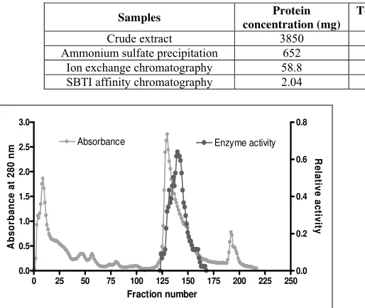
purification. The unbound and initial fractions (1 to 115) contained high concentrations of other proteins without trypsin activity. A linear gradient of NaCl (0.0 to 0.05 M) was applied from fractions 116 and above. Protein elution from fraction 125 -165 showed trypsin activity (Fig.1). This stepwise elution leads to a significant increase in purity by
14.7 fold. Wang et al.[9] chromatographed the trypsin from
the intestine of Hybrid tilapia (Oreochromis x O. aureus)
using DEAE-sephadex and obtained 11.2-fold purification. Fractions pooled from the DEAE- Cellulose further were applied to SBTI-affinity chromatography, which lead to a
23.5-fold purity. Similar strategy was used by Khantaphant et
al. [10] to obtain 13-fold purity from pyloric caeca of
brownstripe red snapper (Lutjanus vitta).
Table 1: Summary of purification of trypsin from Oil Sardine.
Samples Protein
Ammonium sulfate precipitation 652 2542.8 3.9 28.9 1.7
Ion exchange chromatography 58.8 1981.56 33.7 22.5 14.7
SBTI affinity chromatography 2.04 109.54 53.7 1.24 23.5
0 25 50 75 100 125 150 175 200 225 250
Fig 1: Ion exchange chromatography. Equilibrate buffer A. elution with linear gradient with NaCl concentration increment from 0 to 0.5
M in equilibration buffer (fractions 115 -220).
The molecular weight of the purified enzyme was estimated by SDS-PAGE as 24 KDa (Fig. 2), which was in the range of
20-30 KDa for trypsin purified from various fish species [11].
The molecular weight of Oil Sardine (Sardinella longiceps)
trypsin was similar to those reported for other fish trypsin,
such as sardine(Sardina pilchardus) [2], Monterey sardine [12],
Tongol tuna [1], Nile tilapia [13], Brownstripe red snapper [10].
Further the SDS-PAGE under reducing conditions demonstrated a single band indicating the complete purity and monomeric nature of the enzyme.
The effect of temperature on enzyme activity was determined by assaying proteolytic activity at different temperatures (Fig. 3). The enzyme was active between the temperatures of 40 and 70 °C, with an activity maximum/optimum of 60 °C. Similar studies by others authors demonstrated that trypsin
from sarda sarda pyloric ceca had an optimal temperature of
60 °C [14]. While trypsin from Sardinelle, jacopever and
elkhorn sculpin pyloric ceca showed optimum activity within
the range of 50-60 °C [15].
Fig 2: SDS-PAGE of purified trypsin from Oil Sardine (Sardinella longiceps) lane 2, standard markers lane 1.
In thermal stability determination the purified trypsin was highly stable below 50 °C, but the activity sharply decreased when the temperature was raised above 60 °C (Fig. 3). The enzyme activity was almost completely lost at 80 °C. This loss in enzyme activity could be due to the denaturation of the
enzyme [9]. Published reports indicated that trypsin isolated
from red snapper [10] and smooth hound had an optimum
stability below 40°C and decrease in the activity above 50°C
[16].
The effect of pH on enzyme activity was determined over a pH range of 4.0-12.0. The purified enzyme was active between pH 7.0 and 11.0, and the maximal activity of the enzyme was observed at pH 8.0 (Fig. 4). Decrease in activity was found at high acidic and high alkaline pH because most enzymes undergo irreversible denaturation in these conditions
[17]. The isolated trypsin was very stable in a broad pH range
(7.0 -10.0), but it was unstable below and above pH 6.0 and 11.0 respectively (Fig. 4). After incubating at pH 4.0, the trypsin lost complete activity. The present study results were in agreement with other reports which indicate trypsin to be
active at alkaline conditions [11]. The studies published by
Bougatef et al. [2] and Khaled et al.[5] also showed that the
optimum pH of trypsin was between pH 8 to10. Further
reports in sarda sarda [14], intestine and pyloric ceca of
spotted goatfish [18], carp [11] and the pyloric ceca of rainbow
trout [19] demonstrated that these enzymes are alkaline stable.
3 4 5 6 7 8 9 10 11 12
Fig 4: Optimum pH and pH stability of purified trypsin from Oil Sardine (Sardinella longiceps).
In the presence of increasing concentration of NaCl, a continuous decrease in trypsin activity was observed (Fig. 5). The decrease in activity could be described by the salting out phenomenon. An increase in the ionic strength causes a reduction in enzyme activity by an enhanced hydrophobic interaction between protein chains and competition for water
by the ionic salts, leading to enzyme precipitation [14].
0 5 10 15 20 25 30 35
Fig 5: Effect of NaCl concentrations on activities of purified trypsin from Oil Sardine (Sardinella longiceps).
Purified trypsin from Oil Sardine was strongly inhibited (100 %) by SBTI and TLCK (Fig. 6) which are the specific inhibitors of trypsin and was markedly inhibited by the serine-protease inhibitor PMSF (76.2 % inhibition). EDTA, which chelates the metal ions required for the enzyme, partially lowered trypsin activity (7.5 % inhibition). No loss of activity was observed with β-mercapto ethanol. Removal of calcium ion might affect the enzyme structure, resulting in some loss
of activity [20]. The present study results suggest that trypsin
most likely required metal ions as cofactors for activity. Therefore, these observations confirm that the purified enzyme is a serine proteinase, mostly likely a trypsin. Kinetic
data for purified trypsin from Oil Sardine (Sardinella
longiceps) are summarized in table 3. Km and kcat for
hydrolysis of BAPNA were 0.0206 and 1.19 s-1 respectively.
The catalytic efficiencies (kcat/Km) were found to be 57.76s-1
mM-1(Table 2).
TLCK (5mM) PMSF (5mM) EDTA (2mM) SBTI (1mM) Benzimidine (5mM)
0
Table 2: Kinetic parameters of Oil Sardine (Sardinella longiceps) trypsin and other fish trypsins using BAPNA as a substrate.
Trypsins Km(mM) kcat (s-1) kcat / Km(s-1 mM-1)
Oil Sardine (Sardinella longiceps) 0.0206 1.19 57.76
Brownstripe red snapper (Lutjanus vitta)* 0.507 4.71 9.27
New Zealand hoki (Macruronus novaezelandiae)* 0.06 0.33 5.5
Chum salmon (Oncorhynchus keta)* 0.029 2.29 79.03
Anchovy (Engraulis japonicus)* 0.05 1.52 30.7
Monterey sardine (Sardinops sagax caerulea)* 0.05 2.12 41.0
Bigeye snapper (Priacanthus macracanthus)* 0.31 1.06 3.4
* Comparative data given are reported from Khantaphant and Benjakul [10]
5. Conclusion
The present study was aimed to isolate trypsin from visceral
waste of Oil Sardine (Sardinella longiceps) by standard
methods. Isolated enzyme hydrolyzes specific synthetic substrate BAPNA with molecular weight 24 kDa, inhibited by specific trypsin inhibitors and optimal conditions of reaction for trypsin. Hence the purified protease from viscera of Oil
Sardine (Sardinella longiceps) is classified as trypsin.
Purification fold obtained was 23.5. Isolated trypsin exhibited the highest hydrolytic activity towards BAPNA at 60 °C and
pH 7-9. Therefore viscera of Oil Sardine (Sardinella
longiceps) are found to be potential source for the isolation of
industrially important enzyme trypsin. The methods adopted for the isolation of trypsin are efficient enough to scale up the process.
6. Acknowledgement
Thanks to MoES, Govt. of India for the financial assistance under the Research Grant (No. MoES/11-MRDF/1/63/P/08 dated 26/03/09).
7. References
1. Klomklao S, Benjakul S, Visessanguan W. Comparative
studies on proteolytic activity of spleen extracts from three tuna species commonly used in Thailand. Journal of Food Biochemistry. 2004; 28:355-372.
2. Bougatef A, Souissi N, Fakhfakh N, Ellouz-Triki Y,
Nasri M. Purification and characterization of trypsin from
the viscera of sardine (Sardina pilchardus). Food
Chemistry. 2007; 102:343-350.
3. Lugo-Sanchez ME, Pacheco-Aguilar RP,
Yepiz-Plascencia G. Catalytic activities of crude enzyme fractions from Monterey sardine, Journal of Food Science. 1997; 62:976-979.
4. Castillo-yanez FJ, Pacheco-Aguilar R, Garcia-Carreno
FL, Navarrete-De Toro MA. Isolation and characterization of trypsin from pyloric caeca of
Monterey sardine (Sardinops sagax caerulea).
Comparative Biochemistry and physiology. 2005; 140:91-98.
5. Khaled HB, Bougatef A, Balti R, Triki-Ellouz Y, Souissi
N. Isolation and characterization of trypsin from
sardinelle (Sardinella aurita) viscera. Journal of the
science of food and agriculture. 2008; 88:2654-2662.
6. Erlanger BF, Kokowski N, Cohen W. The precipitation
and properties of two new chromogenic substrate of trypsin. Archives of biochemistry biophysics. 1961; 95:271-278.
7. Laemmli UK. Cleavage of structure proteins during the
assembly of the head of bacteriophage T4. Nature. 1970; 277:680-685.
8. Lowry OH, Rosebrough NJ, Fan AL, Randall RJ. Protein
measurement with Folin phenol reagent. Journal of
Biochemistry. 1951; 193:256-275.
9. Wang Q, Gao Z, Zhang N, Shi Y, Xie X, Chen Q.
Purification and characterization of trypsin from intestine
of Hybrid tilapia (Oreochromis x O. aureus). Journal of
Agricultural Food Chemistry. 2010; 58:655-659.
10. Khantaphant S, Benjakul S. Purification and
characterization of trypsin from pyloric caeca of
brownstripe red snapper (Lutjanus vitta). Food
Chemistry. 2010; 120:658-664.
11. Simpson BK. Digestive Proteinases from Marine
Animals. In Seafood Enzymes: Utilization and Influence on Postharvest Seafood Quality; Haard NF, Simpson BK. Eds. Marcel Dekker. New York. 2000, 531-540.
12. Lugo-Sanchez ME, Pacheco-Aguilar R, Yepiz-Plascencia
G. Catalytic activities of crude enzyme fractions from Monterey sardine. Journal of Food Science. 1997; 62:976-979.
13. Bezerra RS, Lins EJF, Alencar RB, Paiva PMG, Chaves
MEC, Coelho LCBB et al. Alkaline proteinase from
intestine of Nile tilapia (Oreochromis niloticus), Process
Biochemistry. 2005; 40:1829-1834.
14. Klomklao S, Benjakul S, Visessanguan W, Kishimura H,
Simpson BK. 29kDa trypsin from pyloric ceca of Atlantic
bonito (Sarda sarda): Recovery and characterization.
Journal of Agricultural Food Chemistry. 2007; 55:4548-4553.
15. Kishimura H, Hayashi K, Miyashita Y, Nonami Y.
Characteristics of trypsins from the viscera of true
sardine (Sarsinops melanostictus) and the pyloric ceca of
arabesque greenling (Pleuroprammus azonus). Food
Chemistry. 2007; 97:65-70.
16. Bougatef A, Balti R, Nasri R, Jellouli K, Souissi N, Nasri
M. Biochemical properties of anionic trypsin acting at high concentration of NaCl purified from the intestine of
a carnivorous fish: smooth hound (Mustelus mustelus).
Journal of Agricultural Food Chemistry. 2010; 58:5763-5769.
17. Benjakul S, Morrissey MT. Protein hydrolysates from
Pacific whiting solid wastes. Journal of Agricultural Food Chemistry. 1997; 45:3423-3430.
18. Souza AAG, Amaral IPG, Espirito SAR, Carvalho LJ,
Bezerra RS. Trypsin-like enzyme from intestine and
pyloric caeca of spotted goatfish (Pseudupeneus
maculatus). Food Chemistry. 2007; 100:1429-1434.
19. Kristjansson MM. Purification and characterization of
trypsin from the pyloric caeca of rainbow trout (Oncorhynchus mykiss). Journal of Agricultural Food
Chemistry. 1991; 39:1738-1742.
20. Klomklao S, Benjakul S, Visessanguan W, Kishimura H,
Simpson BK. Purification and characterization of trypsin
from the spleen of Tongol tuna (Thunnus tonggol).