A Fossa Canina Abscess Originated From First Maxillary Primary Molar: A Case Report Management
Abses Fosa Canina Yang Berasal Dari Gigi Molar Satu Desidui: Laporan Kasus Dalam Perawtannya
Abdul Rochim1
1
Oral and Maxillofacial Surgery department Faculty of Dentistry Jember University
Abstract
A possible complication of fossa canina infections is reactive thrombosis of the vena angular is, which can lead to cavernous sinus phlebothrombosis. According to the literature there are different opinions about the treatment protocol of fossa canina abscesses. This study reports the
unusual case of odontogenic infection in children. A four years old girl patient arrived at a dental clinic with her mother and sister. The patient presented with massive swelling and redness in the region of left eye. According to the subjective data obtained from her mother and sister, the swelling occurs in the morning, six hours before being taken to the dentist. We suspect that the left maxillary first primary molar caused the case. Management of this case is open bur and there was bleeding from the pulp without pain. After 4 min, swelling in the lower eyelid decreased. The condition is getting better after 39 min before the patient took the drug. The patient still get medicine for healing efforts.
Keyword: Fossa canine abscess, open bur
Abstrak
Pada umumnya infeksi rongga mulut penyebab utamnya ialah infeksi odontogen yang bnrsumber dari pulpa gigi, jaringan periodontal atau jaringan peri koronal. Penelitia ini melaporkan kasus infeksi odontogen yang tidak umum pada anak-anak. Pasien anak berumur 5 tahun berkelamin wanita datang keklinik gigi bersama ibunya. Pasien ini menderita bengkak dan warna merah pada daerah infra orbital da menyebar sampai kelopak mata bawah. Pada allo anamneses, pembengkakan terjadi di pagi hari, enam jam sebelum dibawa ke dokter gigi. Saat pemeriksaan klinis intraoral kami menemukan karies pada proksimal (distal) dari gigi susu molar pertama kiri. Penanganan kasus ini adalah dengan open bur (trepanasi) dan terjadi pendarahan dari ruang pulpa tanpa rasa sakit. Setelah 4 menit, bengkak pada kelopak mata bawah berkurang, dan setelah 39 menit keadaannya menjadi h baik saat pasien menunggu di apotik untuk mengambil obat. Pasien tetap mendapatkan obat untuk upaya penyembuhan.
Kata kunci: Abses fosa kanina, fase serous, penanganan
Introduction
Canine fossa abscess is one of the odontogenic infections which is located in the small space between the elevator labii
superior and the levator anguli oris muscles. The common etiologi from infected root canals of pre molars and especially those canines of the maxilla are considered to be responsible for the development of abscesses Correspondence: Abdul Rochim, Oral and Maxillofacial Surgery department Faculty of Dentistry Jember University, e -mail:
Abdul Rochim, A Fossa Canina Abscess Originated From First Maxillary Primary Molar…
of the canine fossa. This study reports the unusual case of canine fossa abscess in the children.
Case Report
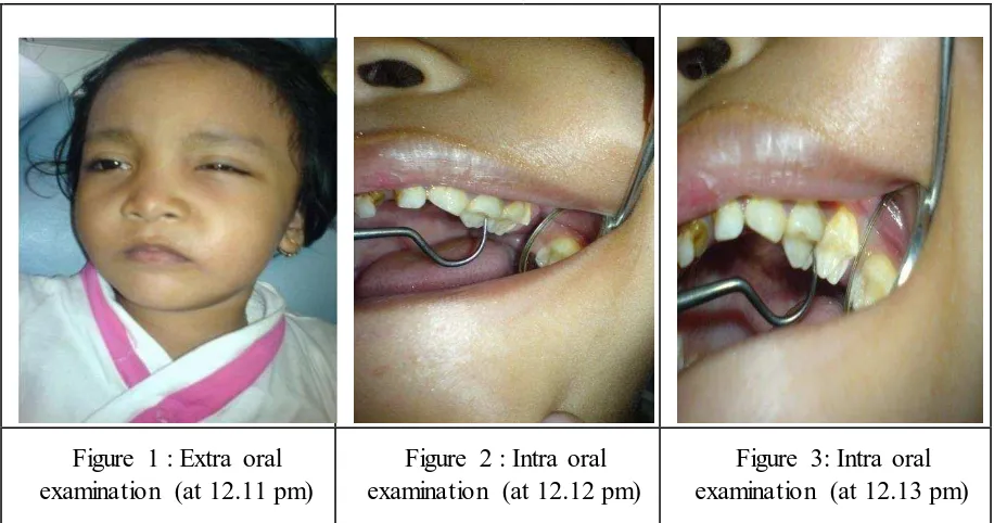
A 4 years old girl patient referring to our clinic with complaints of swelling and pain in her left maxillary with her mother and her
sister. The patient presented localized swelling and redness in infra orbitalis region spreading toward the lower eyelid. According to the subjective data obtained from her mother and sister, the sweling occurs in the morning, six hours before being taken the dentist. We suspect the caries on the proximal (distal) of the left maxillary first
primarry molar by intra oral examinatio.
Figure 1 : Extra oral examination (at 12.11 pm)
Figure 2 : Intra oral examination (at 12.12 pm)
Figure 3: Intra oral examination (at 12.13 pm)
Management Patient
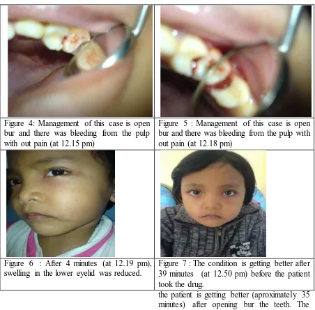
Having performed the assessment, the following interventions were carried out. Management of this case is open bur and there was bleeding from the pulp without
pain. After 4 min, swelling in the lower eyelid decreased. The condition is getting better after 39 min before the patient took the drug. The patient still get medicine for healing efforts.
[image:2.595.84.542.217.458.2]
Abdul Rochim, A Fossa Canina Abscess Originated From First Maxillary Primary Molar…
Discussion
Odonfectogenic infection can occur
locally or spread rapidly. The presence of necrotic teeth will cause the bacteria to penetrate from the pulp chamber to the apicalis. Apicalis foramen of the pulp can not drain the infected pulp. In adition, the infection spreads rapidly into the space or other tissues close to the necrotic tooth structure. This study reports unusul case of odontogenic infection. We find the case of Canine fossa abscess in serous phase. Generally, this phase lasts aproximately 36
hours, and is characterized by local
inflammatory edema, hyperemia or redness with elevated temperature, and pain. In this case, the canine fossa abscess develops rapidly than usual aproximately 7-8 hours and
the patient is getting better (aproximately 35 minutes) after opening bur the teeth. The patient still get medicine for healing effort.
Conclution
The study report concludes that the canine fossa abscess develops faster than usual (aproximately 7-8 hours) and patient is getting better (aproximately 35 minutes) after opening bor the teeth.
References
1. Fragiskos, F.D. Oral Surgery: Odonto genic infection. Chapter 9. New York.: Springer Berlin Heidelberg. 2007.
Figure 4: Management of this case is open bur and there was bleeding from the pulp with out pain (at 12.15 pm)
Figure 5 : Management of this case is open bur and there was bleeding from the pulp with out pain (at 12.18 pm)
Figure 6 : After 4 minutes (at 12.19 pm), swelling in the lower eyelid was reduced.
Figure 7 : The condition is getting better after 39 minutes (at 12.50 pm) before the patient took the drug.
[image:3.595.89.541.79.524.2]Abdul Rochim, A Fossa Canina Abscess Originated From First Maxillary Primary Molar…
2. Lopez Piriz, R, Aguilar, L. Gimenes, M.J. Management of Odontogenic Infection of Pulpa and Periodontal Origin. Med Oral Patol Oral Cir Bucal, 12:E154-159