Received 19 April 1999; received in revised form 16 July 1999; accepted 14 September 1999
Abstract
Potato plants contain calystegines in leaves, stems, flowers, fruits and roots. Calystegines A3and B2are the main constituents.
Highest concentrations were measured in sprouts emerging from the tubers. In 3 mm long sprouts, 3.3 mg total calystegines per g fresh mass were detected. Dormant tubers directly after harvest contain less calystegines in all parts than sprouting tubers. Flowers and young leaves are the aerial plant tissues with the highest calystegine concentration, i.e. 150mg total calystegines per
g fresh mass. Calystegine levels did not rise when sprouts were wounded. Tropinone application to sprouts and aerial tissues lead to an accumulation of pseudotropine and not to tropine. That indicates that stereospecific tropinone reduction is active in potato. © 2000 Elsevier Science Ireland Ltd. All rights reserved.
Keywords:Potato;Solanum tuberosum; Calystegine accumulation; Tropane alkaloid biosynthesis; Tropinone reductase
1. Introduction
Calystegines, a class of nortropane alkaloids, have been described in potato (Solanum tuberosum L.) leaves and tuber skins [1]. However, higher concentrations were measured in sprouting pota-toes [2].
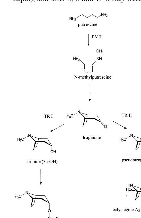
Calystegines possess considerable activities as glycosidase inhibitors [3]. The inhibitory activity depends on the number and position of hydroxyl groups on the nortropane skeleton and varies with specific glycosidases. Up to now 14 different ca-lystegine structures have been published (Fig. 1), among them N-methyl derivatives, which have tropane skeletons. Most of the calystegines were isolated from Solanaceae [3].
We became interested in calystegines because of our research on tropane alkaloid biosynthesis. It is assumed that calystegines originate from the
tropane alkaloid pathway [4] (Fig. 2). In order to prove this hypothesis plant tissues were investi-gated for high biosynthetic activity. Potato tissues of several developmental stages were analysed.
Here are presented detailed calystegine measure-ments in potatoes of the cultivar Liu and first attempts to investigate the biosynthetic pathway by feeding the metabolite tropinone.
2. Material and methods
2.1. Plant and tissue culti6ation
Potatoes, cultivars Adretta and Arkula (early tuberisation) and cultivar Liu (later tuberisation), were purchased from a local provider in early September, and it was ensured that they were freshly harvested and not treated with sprouting inhibitors. They were kept dark at 4°C. Under these conditions they did not sprout within 8 months. For sprouting induction tubers were transferred to room temperature and kept dark. * Corresponding author. Tel.: +49-345-552-5765; fax: +
49-345-552-7213.
E-mail addresses: [email protected] (R. Keiner), [email protected] (B. Dra¨ger)
Fig. 1. Calystegines inS.tuberosum.
After the onset of sprouting, samples were taken every second day.
Potato tubers cut longitudinally show a ring of vascular tissue 0.5 – 1.5 cm below the skin. The storage tissue inside this ring, termed ‘pith’, was examined. When the outside cork layer of the tubers was cut off by a scalpel B1 mm in
thick-ness, it was termed ‘skin’ and when it was cut off with a scalpel 2 mm in thickness we called it ‘peel’. Potatoes, either sprouting with up to 3 mm long sprouts or directly from the cold room were planted in April into the soil of the medicinal plant garden at the institute in Halle. Samples were taken every fifth day until aerial leaves ap-peared. Thereafter plants of three different sizes were analysed.
Some of the sprouting potatoes were planted in pots and kept at room temperature. They grew faster with longer internodia than outside. The stems were used for feeding experiments with tropinone.
Tobacco leaves (Nicotiana tabacum) were ob-tained from flowering green house plants of the medicinal plant garden.
2.2. Feeding experiments with tropinone
Tropinone (Aldrich, Deisenhofen, Germany) was 99% pure according to the manufacturer’s declaration. The purity was confirmed by GC-analysis. Further, tropine or pseudotropine were not detected. More than 0.5% tropine or pseu-dotropine would have been clearly visible by GC. Ten potato sprouts of length=3 cm were cut off the tuber. The endings were dipped (5 mm immer-sion depth) into a solution of 5 mM tropinone, buffered with HCl to pH 7. After 24 h the endings were thoroughly washed with water to remove tropinone solution at the outside and extracted.
The experiment was repeated three times. Five whole leaves of each, potato and tobacco were treated with the same tropinone solution (3 cm immersion depth). Twelve aerial stems of plants with leaves (:15 cm in length) were dipped into 5
[image:2.612.289.524.292.654.2]mM buffered tropinone solution (3 cm immersion depth), and after 3, 8 and 18 h they were cut into
method, strong acidic cation exchange resin I LAB (Merck, Darmstadt, Germany) was used instead of Dowex 50 WX8. Each analysis used 1 – 5 g fresh mass.
Isolated calystegines A3, A5, B1, B2, B3, B4 and
C1, N-methyl-calystegine B2 (gifts from Dr N.
Asano, Kanasawa) were available as reference compounds for GC. Instead of deoxynojirimycin [5], isolated calystegine B2 was taken for
calibra-tion of the GC method and azobenzene (100 mg/
ml) was used as an internal standard. Calystegines were derived with silylating agents TMCS/HMDS as described before [5].
The detection limit for calystegines in extracts was :1 mg/ml sample and reliable quantification
was possible with \10 mg/ml sample. With the
sample preparation procedure described above the limit of quantification results as 2 – 5 mg calystegi-nes/g fresh mass of tissue depending on the fresh mass used per sample.
Identity of calystegines in the potato extracts was further confirmed by GC-MS analysis in com-parison to reference compounds. The TMS-deriva-tives of the calystegines give typical fragmentation patterns [3,5].
2.4. Extraction and analysis of tropane alkaloids
After feeding tropinone tissues were ho-mogenised in H2O and the filtrate was evaporated
to B1 ml. Fifty microlitres of ammonia were
added and the volume was adjusted to 1.0 ml with water. The extracts were loaded onto columns with 1.0 g Extrelut (Merck, Darmstadt). After 15 min the columns were eluted with 2×3 ml CHCl3
and 1×3 ml CHCl3/MeOH 85:15. The eluate was
concentrated to B1.0 ml, diluted to 1.0 ml with
ethyl acetate/MeOH 1:1 and used for GC and TLC analysis.
GC for tropinone, tropine and pseudotropine was performed as described previously [6], with some alterations: column DB5, 30 m, 0.32 mm ID,
was carried out on silica gel plates (0.2 mm layer, 60 mm particle size, Merck, Darmstadt, Germany), solvent system: CHCl3/MeOH/NH3 44:9:1, with
double development. For detection the Dragen-dorff reagent was used with the variation accord-ing to Munier [7] usaccord-ing tartaric acid instead of acetic acid. The sensitivity for tropine and pseu-dotropine was 0.1 mg, visible as purple zone on a light yellow background.
2.5. Statistical treatment of data
In order to quantify calystegines, between three and six independent samples of each tissue were extracted. S.D. are always given in bars, in Fig. 4 they are sometimes so low that they are hardly visible. In exceptional cases (sprouts in soil, Fig. 3), when only two tissue samples were available, no S.D. is given.
The extract of each sample was analysed by GC usually twice, with 1 ml and 2 ml injection volume to account for the different concentrations of calystegines.
Data were examined for outliers. An outlyer was defined as a single value that deviated more than 30% from the average calculated over all values. The limit of 30% was set after calculating several suspected outliers by the method of Nal-imov. Single data with about 30% difference from the arithmetic average turned out to be outliers. These values were eliminated before calculation of S.D.
3. Results and discussion
3.1. Calystegine distribution in tubers and sprouts
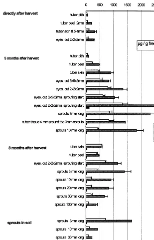
Fig. 3. Calystegine levels in tubers and sprouts.
content in corresponding developmental stages. To reduce the number of samples, only cultivar Liu was examined in more detail.
After harvesting the tubers were completely dor-mant [8]. They did not sprout even if they were kept at room temperature for several weeks. The dormant tubers were examined for calystegines (Fig. 3). For subsequent experiments, tubers stored at 4°C were examined monthly. After 3 months of storage single sprouts appeared when tubers were transferred to room temperature for 2 weeks. After 5 months of storage at 4°C, transfer
to room temperature resulted in intensive sprout-ing after 1 week. These tissues were examined (Fig. 3).
Further sprouting tissues were analysed after 8 months of storage at 4°C. At that time (April) tubers were also planted in soil and the growing plants were used for further measurements.
Calystegine A3, B2and B4were the most
promi-nent and could be quantified in all tissues given in Fig. 3. The presence of further calystegines was noted, and calystegines A5, B1 and B3 were
com-ter 5 months of cold storage (Fig. 3). Sprouting start was defined by the appearance of white shoot tip primordia of B1 mm size in the brown cork.
All of the tissues measured at that time con-tained more calystegines than dormant tubers, e.g. the tuber skins without eyes at sprouting start contained three times more calystegines than 5 months earlier.
Eight months after harvesting the calystegine accumulation in the sprouting eyes and small sprouts was approximately half of that after 5 months. Sprout growth was faster than after 5 months of storage. As sprout growth proceeds, the calystegine level decreases on a fresh mass basis. Longer sprouts contain more water than 3-mm sprouts (Fig. 5), but this alone is not the reason for the decrease in calystegine concentration. Ca-lystegine accumulation decreases as sprouts elon-gate and does not parallel growth on a dry mass basis either.
In potatoes planted in the soil (Fig. 3) calystegi-nes in the 3-mm sprouts reached about the same levels as in sprouts kept in the dark at room temperature, but thereafter the calystegine levels decreased more sharply in the soil than at room temperature in the air. It is not clear whether the decrease is due to the mechanical influence of the soil or to the colder temperature. They could have been excreted to attract micro-organisms as as-sumed by Tepfer et al. [9].
The ratio of calystegines B2 to A3 was rather
constant in shoots, but in pith and skin the rela-tion was changed in favour of calystegine A3(Fig.
3), which is suspected to be a metabolite (Fig. 2). The large variation in calystegine levels in pota-toes may explain the divergent values in the litera-ture: Nash et al. [1] reported 0.01% of fresh mass calystegines in potato peels, while Asano et al. [10] found 3.4 – 7 mg/g fresh mass in the whole tuber which indicates that the potatoes were probably taken from the stage of dormancy.
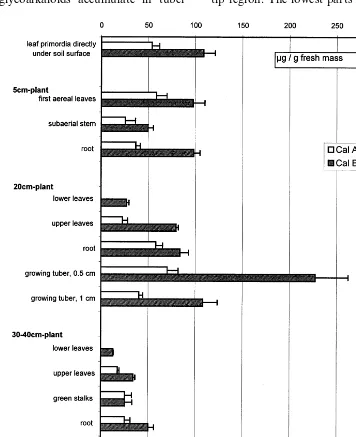
sprouting tubers. Calystegine B4could be detected,
but was too low to be quantified reliably. Only the levels of calystegines A3 and B2 are given.
The leaf primordia directly under the soil sur-face (Fig. 4) show a decreased calystegine level compared with the sprouts in the soil (Fig. 3).
The first aerial leaves keep the calystegine level of leaf primordia, and the roots of the small plants contain about the same concentration and pattern of calystegines (Fig. 4).
While the calystegine level in the roots remains rather constant in 20-cm plants, the leaves gradu-ally lose calystegines. Younger leaves contain more calystegines than older leaves, that has been shown for leaves of other Solanaceae as well [2]. Plants of 20 cm begin to form tubers, which contain the highest concentrations of calystegines at this stage of development.
Plants of 30 – 40 cm (Fig. 4) were flowering and lower leaves had begun to wilt. The tubers had almost reached their final size at that stage. Flow-ers extracted as a whole including petals had the highest calystegine levels recorded in aerial parts of the plants.
Besides the values given in Fig. 4 more tissues of older plants were examined, e.g. yellow leaves, wilted leaves, yellow stems, whole green fruits. In these organs calystegine levels were too low to be measured, i.e. B5 mg/g fresh mass.
All calystegine concentrations are given on a fresh mass basis, but dry mass was determined for each sample. Typical values for each type of tissue are given in Fig. 5. Other dry mass per fresh mass relations were similar for the respective tissues. The dry mass proportion in aerial parts of the plant is lower than in tubers, but this alone does not account for the lower calystegine content per fresh mass. On a dry mass basis calystegine con-tent in aerial parts is lower as well.
Calystegine concentrations are partially parallel to those of the glycoalkaloids. Tuber tissues known for high accumulation of glycoalkaloids are skins (up to 640 mg/g fm), eyes and sprouts (:5 mg/g
fm), the glycoalkaloid concentrations being highly dependent on the cultivar. Tuber pith contains only low amounts of glycoalkaloids (up to 60mg/g fm), the same is true for calystegines. In aerial parts of the potato plants again the flowers con-tain the highest concentrations of glycoalkaloids (up to 5 mg/g fm), but on the whole, aerial parts, leaves in particular, contain more glycoalkaloids than tubers while calystegine concentrations are highest in particular tuber tissues. Another differ-ence is that glycoalkaloids accumulate in tuber
skins when tubers are illuminated [11]. Calystegi-nes in skins do not rise with illumination, and when tubers are left to sprout under illuminations the sprouts are green, stunted and contain less calystegines that the equivalent sprouts in the dark (data not shown) .
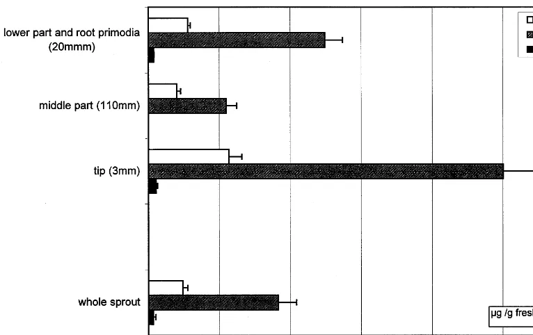
3.3. Distribution within the sprouts
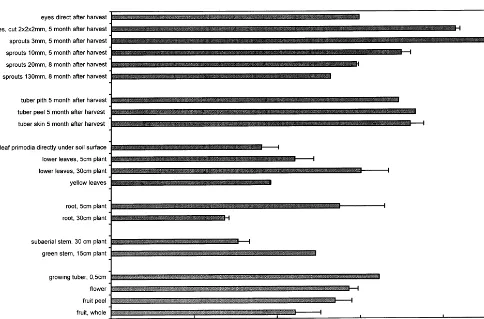
Because upper leaves always contain more ca-lystegines than leaves from lower parts of the plants, sprouts were dissected and analysed (Fig. 6).
[image:6.612.100.456.240.677.2]Calystegines accumulate predominantly in the tip region. The lowest parts of the sprouts contain
Fig. 5. Percentage of dry mass in typical potato tissues.
higher calystegine levels probably due to higher concentration in root primordia. Higher calystegi-nes concentrations in tips and root tips were re-peatedly measured with sprouts \100 mm, but in
sprouts of 20 mm no difference could be found between tips and middle parts.
Calystegines seem to concentrate in young meristematic tissues. This leads to the question of the biological significance of calystegine accumula-tion. They could be protecting compounds as dis-cussed for other glycosidase inhibitors [12], and we wondered whether they are inducible like phy-toalexins. We tested the response to mechanical stress by wounding sprouts with a scalpel. The surface of 3 cm long sprouts, 4 mm in diameter was scratched vertically to 1 – 2 mm depth. The sprouts were analysed every day up to 6 days after wounding and showed no alteration of calystegine content. Six days after wounding the scratch had formed a brown cork and side sprouts began to proliferate. Thus wounding had an effect on plant growth but not on calystegine accumulation.
Under these conditions potato phytoalexins have been described to accumulate within 24 h
[13]. Typical phytoalexins in response to tuber wounding are the sesquiterpenes rhishitin, lubimin, and solavetivone, the compounds are hardly de-tectable in healthy tissues. Wounding also leads to a higher accumulation of glycoalkaloids [11]. In our experiment calystegines are no phytoalexins in the sense that they appear or rise in concentration after biotic or abiotic stress. If calystegines serve as defence compounds they are constitutive pro-tectants for young tissues. Experiments with insects to investigate feed deterrence are on the way.
3.4. Tropinone as metabolite
Fig. 6. Distribution of calystegines in long sprouts.
non-metabolised tropinone by TLC and GC. Tropine could not be detected. In control tissues none of the metabolites could be found. Tobacco leaves fed with tropinone solution showed tropinone in GC and TLC, but neither pseu-dotropine nor tropine. Here nicotine was visible on TLC (results not shown).
After 3 h of tropinone application to potato stems, tropinone was detected in the lowest section (:4 cm) only. After 8 h tropinone was found in
the lowest and in the next higher part, but no pseudotropine or tropine could be found. After 18 h tropinone was detectable in all sections, and in the lowest section pseudotropine, but no tropine, was identified. In our view the pseudotropine con-centration is the result of the formation minus the further metabolism to calystegines, so that pseu-dotropine accumulates, because with the high sub-strate supply the tropinone reduction is very active.
In summary there is transport of tropinone in tobacco and potato tissues, but tropinone is only reduced to pseudotropine in potato sprouts, stems and leaves. The reduction is stereospecific, with only pseudotropine, not tropine accumulating. This is in accordance with the finding of two separate tropinone reductases in other tropane alkaloid containing Solanaceae [6,14]. The tropine forming enzyme (TR I, Fig. 2) leads to hyoscyamine and scopolamine, which have never
been found in S. tuberosum. It is suspected that the pseudotropine forming enzyme (TR II) takes part in calystegine biosynthesis.
These experiments are taken as first evidence that tropinone and pseudotropine are metabolites for the biosynthesis of calystegines in potato. The next aim is to elucidate the subsequent steps and to prove the metabolic pathway by characterisa-tion of the enzymes involved.
Acknowledgements
We wish to thank Dr Naoki Asano, Kanasawa, Japan, for the gift of isolated calystegines. Techni-cal assistance by Ursula Ko¨del is gratefully ac-knowledged. R.K. is grateful for a grant from the Landesgraduiertenfo¨rderung Sachsen-Anhalt.
References
[1] R.J. Nash, M. Rothschild, E.A. Porter, A.A. Watson, R.D. Waigh, P.G. Waterman, Calystegines in Solanum
and Datura species and the death’s-head hawk-moth (Acherontia atropus), Phytochemistry 34 (1993) 1281 – 1283.
[2] B. Dra¨ger, A. van Almsick, G. Mrachatz, Distribution of calystegines in several Solanaceae, Planta Med. 61 (1995) 577 – 579.
of tropinone in D. stramonium root cultures by two specific reductases, Phytochemistry 37 (1994) 391 – 400. [7] A. Baerheim Svendsen, R. Verpoorte in:
Chromatogra-phy of alkaloids, Part A, Thin-layer chromatograChromatogra-phy, Journal of Chromatography Library, Vol. 23, Elsevier, Amsterdam, 1983, pp. 91 – 101.
[8] J.J.J. Wiltshire, A.H. Cobb, A review on the physiology of potato tuber dormancy, Ann. Appl. Biol. 129 (1996) 553 – 569.
[9] D. Tepfer, A. Goldmann, N. Pamboukdjian, M. Maille, A. Lepingle, D. Chevalier, J. De´narie, C. Rosenberg, A Plasmid ofRhizobium meleloti41 encodes catabolism of two compounds from root exudate ofCalystegia sepium, J. Bacteriol. 170 (1988) 1153 – 1161.
K. Mengel, D.J. Pilbeam (Eds.), Nitrogen Metabolism of Plants. In: Proceedings of the Phytochemical Society of Europe, vol. 33, Clarendon Press, Oxford, 1992, pp. 271 – 282.
[13] A. Kumar, S.J. Jadhav, D.K. Saluhnke, Phytoalexins, in: D.K. Salunkhe, S.S. Kadam, S.J. Jadhav (Eds.), Potato: Production, Processing and Products, CRC Press, Boca Raton, FL, 1991, pp. 247 – 267.
[14] K. Nakajima, T. Hashimoto, Y. Yamada, Two tropinone reductases with different stereospecificities are short-chain dehydrogenases evolved from a common an-cestor, Proc. Natl. Acad. Sci. U.S.A. 90 (1993) 9591 – 9595.