ω-ALKYNYL FATTY ACIDS: SURROGATES TO STUDY PROTEIN
ADDUCTION BY ENDOGENOUSLY GENERATED LIPID ELECTROPHILES
By
William Norris Beavers Jr.
Dissertation
Submitted to the Faculty of the Graduate School of Vanderbilt University
in partial fulfillment of the requirements for the degree of
DOCTOR OF PHILOSOPHY in
Chemistry August, 2015 Nashville, Tennessee
Approved:
Lawrence J. Marnett, Ph.D.
Brian O. Bachmann, Ph.D.
H. Alex Brown, Ph.D.
Sean S. Davies, Ph.D.
Ned A. Porter, Ph.D.
DEDICATION
To my wife Liz, and our daughters Pearson, Kate, and Nora. None of this would have been possible without your unconditional love and endless support.
ACKNOWLEDGEMENTS
First I would like to thank my mentor Larry Marnett. Larry has provided me with every resource necessary to be a successfully scientist. I have been able to do any experiment I can dream. He has also shown a lot of patience. We have suffered many set backs and delays getting to the point where we are currently with this project, but he has always been patient when others might have chosen to “cut our losses”, and move in a different direction. Larry has also struck the delicate balance between pushing me hard to succeed, and giving me the freedom to drive my project in a direction I saw fit; allowing me to make mistakes and fix those mistakes. It has made the process full of learning pains at times, but I feel well prepared for future scientific challenges.
I would like to thank the other members of my committee as well. Brian Bachmann, Alex Brown, Sean Davies, and Ned Porter have spent many hours discussing my project with me, reading my reports, and critiquing my presentations. Your comments and suggestions have been invaluable to me in my preparation to become an independent scientist.
Vanderbilt has created a culture where a chemistry graduate student can easily find assistance in other departments. I would like to thank the many members of the Vanderbilt community who have taken time to discuss various aspects of my project with me including Aaron Bowman, Alan Brash, Cathy Clarke, Simona Codreanu, Josh Fessel, David Harrison, Annet Kirabo, Dan Liebler, Stokes Peebles, Jack Roberts, Claus Schneider, and Bing Zhang.
The Marnett laboratory has a long-standing relationship with the Porter laboratory. As such, I have spent a lot of time in the Porter laboratory. First I would like to further thank Ned for taking the time to discuss various aspects of radical chemistry with me. Keri Tallman has generated an arsenal of tools that I have been fortunate to have access to, and would like to thank her greatly for the steady supply of these probes as well as all of our conversations on various aspects of my project. I would also like to thank Connor Lamberson, Libin Xu, and Huiyong Yin for help and discussions over the years.
I have been fortunate to work with the various fantastic core facilities around campus. I would like to thank Don Stec of the small molecule NMR core. I would like to thank Rob Carnahan and Matt Goff of the Vanderbilt Antibody and Protein Resource. I would like to thank Wade Calcutt, Julie Coleman, Dave Hachey, Salisha, Hill, and Kristie Rose of the Mass Spectrometry Resource Center. An extra thank you is deserved by Kristie Rose, for without her hard work and expertise, there is no way we would have made as much progress on this project as we have.
I have also had the opportunity to get a lot of teaching experience. I would like to thank Larry for giving me the opportunity to guest lecture in his Foundations of Chemical Biology course. I have also been fortunate to work with the Vanderbilt Program for Talented Youth. I would like to thank Michelle Sulikowski and Brittany Allison for first introducing me to the program. I would also like to thank all of those at PTY who have help with the courses I was a part of including Sarah DeLisle, David Dunn, Megan Parker-Peters, and Gem
Thomerson. It was a fantastic experience to design and implement courses to get young, gifted students interested in science as a career.
The administrative staff at Vanderbilt has been extremely helpful. Leigh Clayton, Stephen Doster, Sandra Ford, Celeste Gouldman, Magda Paszewska, Amanda Renick-Beech, Leigh Thompson, and Mary Veazy have made my life much easier through their help with scheduling, submitting paperwork, and submitting grants. Anne Lara deserves a special thank you. Without Anne’s help, I would have been lost trying to accomplish anything. She is truly the factor that allows the Marnett laboratory to interface with the rest of the world efficiently.
I have been fortunate to work with some fantastic colleagues during my time in the Marnett lab. Thank you to all of the lemmings for comments and discussions about my project. I would also like to thank Chris Aluise, Jeannie Camarillo, Brenda Crews, Kelsey Duggan, Kebreab Ghebreselasie, Phil Kingsley, Shalley Kudalkar, Joe Manna, Michelle Mitchener, Yuki Shimozu, Sarah Shuck, Jody Ullery, Orrette Wauchope, James Wepy, and Matt Windsor specifically for work that we have done together or conversations we have had during my time in the lab. I want to thank Colleen Lawrence, who was my mentor when I first joined the lab, and introduced me to alkynyl lipid electrophiles. I would also like to thank Jim Galligan. Jim joined the laboratory shortly after Colleen left, and has helped with my day-to-day mentorship on the things needed to be successful. I have also had the opportunity to mentor several talented rotation students during my time in the Marnett laboratory. Thank you to Esha Dalvie and Nick Shelburne for being eager to learn and work hard, and also
being patient with me as I learned to mentor young scientists. Finally, I would like to thank our lemming from afar, Carol Rouzer. Larry once told me, “Carol improves everything she touches”. This statement could not be truer. Carol, I thank you for all of your comments and patience over the years as I have struggled with learning manuscript and grant writing.
I have had ample funding to pursue my project and attend scientific conferences without restriction. I would like to thank the Vanderbilt Institute for Chemical Biology Fellowship and the Hercules Fellowship from the Department of Chemistry for funding the start of my graduate career. I would like to thank the Vanderbilt Center in Molecular Toxicology Training Grant from the National Institute of Environmental Health Sciences (T32 ES007028) for two years of funding. Finally, I would like to thank the American Heart Association for two years of fellowship funding (13PRE17270009).
I have had many great mentors that have helped me grow, mature, and develop a desire for an academic life over the years. I would like to thank Joe Spicer, my high school chemistry teacher, and Mike Krepich, my high school Latin teacher for being wonderful role models during my formative years. I would like to thank my mentor at Old Dominion University, Craig Bayse, and my mentor at Northeastern University, Sunny Zhou, for helping me develop as a young scientist. I would like to thank Paul Morrison, Jim Lee, and Loren Walensky at the Dana-Farber Cancer Institute. The experience I gained working with them prepared me to be successful in graduate school.
I have an outstanding support network of family and friends, and am fortunate that there are too many to name here individually. They have always believed in me, even when my own faith has waivered, and for that I am truly grateful. I would like to specifically thank my parents. I watched my Dad get up every morning and go to a job he did not enjoy to support his family, and never complained about it. In thirty-three years, I can count on one hand the number of sick days he took, which has been a strong influence on how I approach work every morning. My Mom always encouraged us to explore and pushed us academically. She spent hours exploring in the woods and helping us with homework as children. During the summers she would indulge our curiosities with countless trips to the museums in Washington, D.C. (several hours of driving, in terrible traffic, with three rowdy boys in the back seat). These interactions with my parents have contributed greatly to my choice to pursue science as a career. I truly love coming into lab every day. What I enjoy most is that I get to be curious, learn new things every day, and think about things nobody else has.
Finally, I have to thank the people who have provided me with the daily support that I needed to make it through graduate school. I do not think I can put into words how much I appreciate the love and support my wife Liz and daughters Pearson, Kate, and Nora have given me. You have kept me motivated when I would have otherwise lost focus. You have kept me grounded when my ego would have swelled. You have been my escape when I needed one. I love you very much. Thank you.
TABLE OF CONTENTS
Page
DEDICATION ... ii
ACKNOWLEDGEMENTS ... iii
LIST OF TABLES ... x
LIST OF FIGURES ... xi
LIST OF ABBREVIATIONS ... xv
Chapter I. INTRODUCTION ... 1
Immune Response, Inflammation, and Disease ... 1
Macrophages ... 1
Inflammation and disease ... 4
Oxidation of Polyunsaturated Fatty Acids ... 9
Polyunsaturated fatty acids ... 9
Radical mechanism of lipid oxidation ... 10
Autoxidation ... 11
Cyclooxygenases ... 15
Lipoxygenases ... 18
Lipid Electrophiles ... 21
Generation of lipid electrophiles ... 21
Reactivity of lipid electrophiles ... 24
Cellular consequences of lipid electrophile adduction ... 26
Studying lipid electrophiles in disease models ... 29
Exogenously generated lipid electrophiles ... 30
Endogenously generated lipid electrophiles ... 34
Dissertation Aims ... 37
II. ω-ALKYNYL LIPID SURROGATES FOR POLYUNSATURATED FATTY ACIDS: FREE RADICAL AND ENZYMATIC OXIDATIONS ... 40
Introduction ... 40
Materials and Methods ... 42
Results ... 53
Discussion ... 69
III. ENDOGENOUSLY GENERATED LIPID ELECTROPHILES TARGET MITOCHONDRIAL PROTEINS IN ACTIVATED
RAW264.7 MACROPHAGES ... 76
Introduction ... 76
Materials and Methods ... 79
Results ... 88
Discussion ... 115
IV. PATHWAYS OF PEROXIDATION: STUDIES TO DETERMINE ROUTES OF LIPID OXIDATION AND ELECTROPHILE FORMATION ... 125
Introduction ... 125
Materials and Methods ... 130
Results ... 136
Discussion ... 149
Future Directions ... 156
V. SUMMARY/FUTURE VISIONS ... 157
Summary ... 157
Future Visions ... 164
Rotenone generation of lipid electrophile protein adducts ... 164
Identification of the site of modification of Sod2 ... 168
aLA in vivo ... 171
REFERENCES ... 174
LIST OF TABLES
Table Page
II-1 Kinetic comparison of AA and aAA with COX and LOX enzymes ... 56 II-2 1H-NMR chemical shifts and coupling constants of the COX-2 aAA
metabolite with m/z = 315.2 ... 63 II-3 1H-NMR chemical shifts and coupling constants of the COX-2 aAA
metabolite with m/z = 331.2 ... 63 III-1 Wikipathway enrichment for the 192 proteins in the most
differentially expressed class as determined by Webgestalt ... 103 III-2 Wikipathway enrichment for the 76 proteins in the most
differentially adducted class as determined by Webgestalt ... 106 IV-1 R/S ratios for esterified hydroxy polyunsaturated fatty acids ... 138 IV-2 Comparison of protein adduction and esterified HODEs and HETEs in
macrophages with indomethacin and giripladib treatment ... 153
LIST OF FIGURES
Figure Page
I-1 Reactive species generated by leukocyte activation ... 3
I-2 Common fatty acids ... 9
I-3 Radical mechanism of lipid oxidation ... 10
I-4 General mechanism of PUFA oxidation ... 11
I-5 Autoxidation products of LA ... 13
I-6 Autoxidation of AA generates F2α-isoprostanes ... 15
I-7 COX metabolism of AA ... 17
I-8 5-LOX metabolism of AA ... 21
I-9 Structures of some commonly studied lipid electrophiles ... 22
I-10 Adducts formed on amino acids by lipid electrophiles ... 25
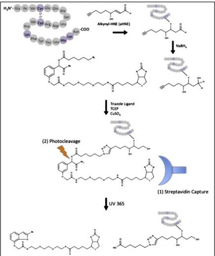
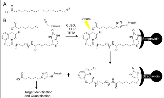
I-11 Analysis of lipid electrophile adducted proteins using click chemistry, affinity purification, and UV-cleavable biotin ... 33
II-1 Autoxidation products of LA, aLA, AA, and aAA ... 53
II-2 Chiral analysis of 13-c,t-HODE ... 54
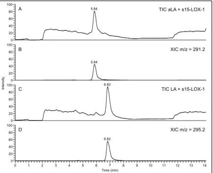
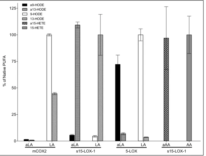
II-3 s15-LOX-1 metabolism of LA, aLA, AA, and aAA ... 57
II-4 Soybean 15-LOX metabolite profiles of LA and aLA ... 59
II-5 Comparison of the metabolite profiles of aLA and LA with s15-LOX-1 ... 60
II-6 COX-2 in vitro metabolite profiles of aAA and AA ... 61
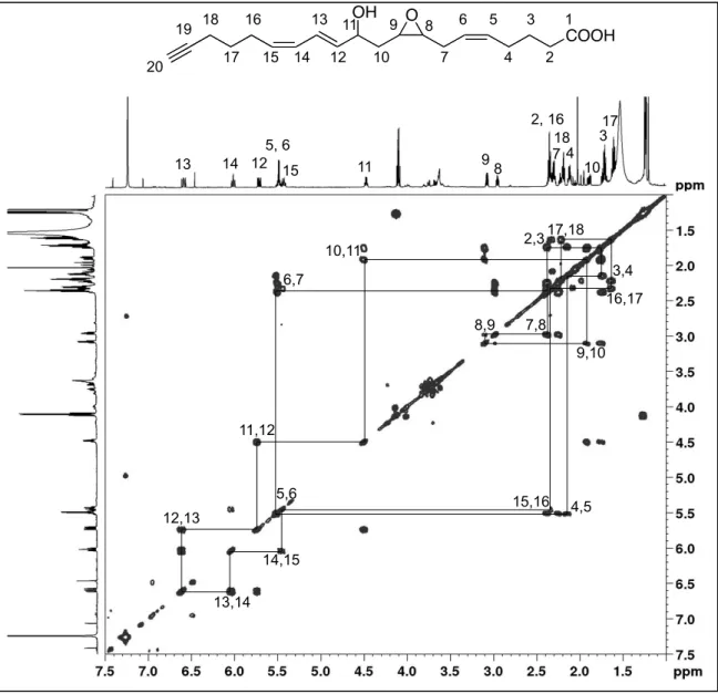
II-7 1H-1H-COSY spectrum of the COX-2 aAA metabolite with m/z = 315.2 ... 64
II-8 1H-1H-COSY spectrum of the COX-2 aAA metabolite with m/z = 331.2 ... 65
II-9 Michaelis-Menten plots and relevant kinetic parameters
for COX-2 metabolism of AA and aAA ... 67
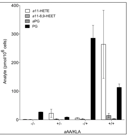
II-10 Activated macrophages generate aAA metabolites ... 68
II-11 Proposed mechanism of COX-2 oxygenation of aAA ... 71
III-1 Click chemistry scheme for protein adduction ... 89
III-2 Extent of aLA incorporation into phospholipid pools ... 90
III-3 Total amounts of aLA and LA in RAW264.7 macrophages before and after aLA incorporation ... 91
III-4 aAA is biosynthesized from aLA ... 92
III-5 Total amounts of aAA and AA in RAW264.7 macrophages before and after aLA incorporation ... 93
III-6 KLA activation induces lipid electrophile protein adduction ... 94
III-7 SILAC workflow ... 96
III-8 Analysis of expression and adduction changes during KLA-activation of macrophages ... 98
III-9 Frequency counts for proteome and adductome replicates ... 99
III-10 Spearman correlation coefficients relating the proteome and adductome replicates to each other ... 101
III-11 Complex V of the electron transport chain is the most differentially expressed pathway in activated macrophages ... 104
III-12 Complex V of the electron transport chain is the most differentially adducted pathway in activated macrophages ... 106
III-13 MitoTEMPO modulates KLA-induced protein adduction, while TEMPOL does not ... 108
III-14 MitoTEMPO modulates the amount of adducted protein affinity purified in aLA-incorporated macrophages ... 109
III-15 Western blot confirmation of protein targets of lipid electrophile adduction ... 111
III-16 Activity changes associated with lipid electrophile adduction of
mitochondrial proteins ... 113
III-17 Superoxide dismutase 1 and 2 expression in mitochondrial and cytosolic isolates ... 114
III-18 Absolute Sod2 activity in unactivated and activated RAW264.7 macrophage mitochondria ... 115
IV-1 Enzymatic and autoxidation products of LA ... 126
IV-2 Kinetic and thermodynamic autoxidation products of LA ... 128
IV-3 Quantification of positional and geometric isomers of esterified hydroxy polyunsaturated fatty acids ... 137
IV-4 Chiral standards and cell extracts of LA oxidation products ... 139
IV-5 Chiral standards and cell extracts of AA oxidation products ... 140
IV-6 Chiral analysis of abundant ω-alkynyl fatty acid metabolites ... 140
IV-7 Click blot and PG analysis for MitoTEMPO treated macrophages ... 141
IV-8 Click blot and PG analysis for indomethacin treated macrophages ... 142
IV-9 Click blot and PG analysis for COX-inhibited macrophages with and without PG supplementation ... 144
IV-10 Click blot and PG analysis for giripladib treated macrophages ... 145
IV-11 Quantification of esterified HODEs in indomethacin and giripladib treated macrophages ... 147
IV-12 Quantification of esterified HETEs in indomethacin and giripladib treated macrophages ... 148
IV-13 Inhibitor effects on PG synthesis and protein adduction by lipid electrophiles in macrophages ... 155
V-1 Rotenone induced protein adduction in RAW264.7 macrophages ... 165
V-2 Rotenone does not activate RAW264.7 macrophages ... 166
V-3 MitoTEMPO reduces lipid electrophile protein adduction
induced by rotenone ... 167 V-4 Rotenone induces lipid electrophile protein adducts
in HEK-293 cells ... 169 V-5 Sod2 is adducted by lipid electrophiles in rotenone treated
HEK-293 cells ... 170 V-6 Expression and purification of Sod2-His6 ... 171 V-7 Subcellular fractionation of Sod2-His6 ... 171 V-8 Lipid electrophile protein adduction in aLA-incorporated
mice treated with AngII ... 173
LIST OF ABBREVIATIONS
1-AG 1-arachidonylglycerol
2-AG 2-arachidonylglycerol
5-LOX arachidonate 5-lipoxygenase
8-LOX arachidonate 8-lipoxygenase
9-LOX linoleate 9-lipoxygenase
12-LOX arachidonate 12-lpoxygenase
13-LOX linoleate 13-lipoxygenase
15d-PGJ2 15-deoxy-Δ12,14-prostaglandin J2
15-LOX arachidonate 15-lipoxygenase
a11-8,9-HEET ω-alkynyl 11-hydroxy-8,9-epoxy eicosatrienoic acid a11-HETE ω-alkynyl 11-hydroxy eicosatetraenoic acid
AA arachidonic acid
aAA ω-alkynyl arachidonic acid
AD Alzheimer’s disease
aF2α-isoP ω-alkynyl F2α-isoprostane
aHETE ω-alkynyl hydroxy eicosatetraenoic acid aHNE ω-alkynyl 4-hydroxy-2-nonenal
aHODE ω-alkynyl hydroxy octadecadienoic acid aHPETE ω-alkynyl hydroperoxy eicosatetraenoic acid
aLA ω-alkynyl linoleic acid
ALS amyotrophic lateral sclerosis
aONE ω-alkynyl 4-oxo-2-nonenal
aPG ω-alkynyl prostaglandin
aPGD2 ω-alkynyl prostaglandin D2
aPGE2 ω-alkynyl prostaglandin E2
aPGG2 ω-alkynyl prostaglandin G2
ATP adenosine triphosphate
BHT butylated hydroxy toluene
BODIPY boron-dipyrromethene
BSA bovine serum albumin
COX cyclooxygenase
COX-1 cyclooxygenase 1
COX-2 cyclooxygenase 2
cPLA2 cytosolic phospholipase A2
DAMP damage-associated molecular pattern
DMEM Dulbecco’s modified Eagle medium + glutamax
DMSO dimethyl sulfoxide
DNA deoxyribonucleic acid
ESI electrospray ionization
ETC electron transport chain
F2α-isoP F2α-isoprostane
FLAP arachidonate 5-lipoxygenase activating protein
GAPDH glyceraldehyde-3-phosphate dehydrogenase
GRP78 78 kDa glucose-regulated protein
GSH glutathione
hCOX2 human cyclooxygenase 2 (purified)
HEK-293 human embryonic kidney 293 cells
HETE hydroxy eicosatetraenoic acid
HHT 12-hydroxy heptadecatrienoic acid
Hmox1 heme oxygenase-1
HNE 4-hydroxy-2-nonenal
HODE hydroxy octadecadienoic acid
HPETE hydroperoxy eicosatetraenoic acid HPLC high-performance liquid chromatography
HPODE hydroperoxy octadecadienoic acid
HSA human serum albumin
HSP90 heat shock protein 90
IFNγ interferon gamma
IκB inhibitor of nuclear factor kappa-light-chain-enhancer of activated B cells
IKK inhibitor of nuclear factor kappa-light-chain-enhancer of activated B cells kinase
IKK-α inhibitor of nuclear factor kappa-light-chain-enhancer of activated B cells kinase, alpha subunit
IKK-β inhibitor of nuclear factor kappa-light-chain-enhancer of activated B cells kinase, beta subunit
IKK-γ inhibitor of nuclear factor kappa-light-chain-enhancer of activated B cells kinase, gamma subunit
IL-1β interleukin-1 beta
IL-6 interleukin-6
IL-10 interleukin-10
IL-12 interleukin-12
In radical initiator
iNOS inducible nitric oxide synthase
IsoP isoprostane
Keap1 Kelch-like ECH-associated protein 1
KETE oxo eicosatetraenoic acid
KLA Kdo2-lipid A
KODE oxo octadecadienoic acid
L lipid radical
LA linoleic acid
LC/MS/MS liquid chromatography tandem mass spectrometry
LDL low-density lipoprotein
lk12LOX porcine leukocyte-type 12-lipoxygenase
LOO lipid peroxyl radical
LOX lipoxygenase
LPS lipopolysaccharide
LTA4 leukotriene A4
LTB4 leukotriene B4
LTC4 leukotriene C4
LTC4S leukotriene C4 synthase
LTD4 leukotriene D4
LTE4 leukotriene E4
MeOAMVN 2,2’-azobis(4-methoxy-2,4-dimethylvaleronitrile)
mRNA messenger ribonucleic acid
MS mass spectrometry
MudPIT multidimensional protein identification technology
m/z mass to charge ratio
NF-κB nuclear factor kappa-light-chain-enhancer of activated B cells
NMBHA N-methyl benzohydroxamic acid
NMR nuclear magnetic resonance
NP-HPLC normal-phase high-performance liquid chromatography
Nrf2 nuclear factor erythroid 2-related factor 2 NSAID nonsteroidal anti-inflammatory drug
Nu nucleophile
oCOX1 ovine cyclooxygenase 1
ONE 4-oxo-2-nonenal
PAMP pathogen-associated molecular pattern
PBS phosphate buffered saline
PD Parkinson’s disease
PG prostaglandin
PGA2 prostaglandin A2
PGD2 prostaglandin D2
PGE2 prostaglandin E2
PGF2α prostaglandin F2α
PGG2 prostaglandin G2
PGH2 prostaglandin H2
PGI2 prostacyclin
PGJ2 prostaglandin J2
PIN1 peptidyl-prolyl cis/trans isomerase A1 plt12LOX human platelet-type 12-lipoxygenase
PPh3 triphenyl phosphine
PRR pattern recognition receptor
PUFA polyunsaturated fatty acid
r15LOX1 rabbit reticulocyte 15-lipoxygenase 1
RNS reactive nitrogen species
ROS reactive oxygen species
RP-HPLC reverse-phase high-performance liquid chromatography
s15-LOX-1 soybean arachidonate 15-lipoxygenase-1
SCX strong cation exchange
SDS-PAGE sodium dodecyl sulfate polyacrylamide gel electrophoresis
SIRT3 sirtuin 3
SN substantia nigra
Sod1 superoxide dismutase 1
Sod2 superoxide dismutase 2
SRM selected reaction monitoring
TBTA tris[(1-benzyl-1H-1,2,3-triazol-4-yl)methyl]amine
TCEP tris(2-carboxyethyl)phosphine
TFE 2,2,2-trifluoroethanol
TNFα tumor necrosis factor alpha
TXA2 thromboxane A2
Chapter I
INTRODUCTION
Immune Response, Inflammation, and Disease
Macrophages
As the sesquicentennial anniversary of Dr. Élie Metchnikoff’s discovery of the macrophage approaches4, macrophages have become one of the most highly studied immune cells in the human body. In 2014 nearly 10,000 publications listed in PubMed contained the keyword macrophage. Dr.
Metchnikoff was studying nutrient uptake in cells when he noticed a cell type that would engulf particulates. He coined the term phagocytes, or “eating cell” as a name for these cells5. Dr. Metchnikoff was not the first to observe this phenomenon. Dr. Robert Koch noted anthrax inside granulomas, but misinterpreted what he saw as the bacterium invading the host cell, and not the host cell engulfing the bacterium5. Dr. Metchnikoff noticed that these cells were also involved in the first line of host defense (innate immunity), as they would phagocytose yeast cells that were foreign to the host. These new cells “ate”
different amounts of material and he named them accordingly: microphages or
“little eaters” later to be renamed neutrophils and macrophages or “big eaters”5. Dr. Metchnikoff, along with Dr. Paul Ehrlich received the 1908 Nobel Prize in
Physiology or Medicine for their discoveries of innate immunity and adaptive immunity respectively5.
Since their discovery, macrophages have been identified in nearly every body system. They have various names depending on the tissue type including Kupfer cells in the liver6, microglia in the central nervous system7, osteoclasts in bones8, Langerhans cells in skin and mucosa9, alveolar macrophages in lungs10, monocytes, or macrophages in an early differentiation stage, are abundant in the bone marrow and blood11, histiocytes in the lymph nodes12, Hofbauer cells in the placenta13, and many more. Macrophages are a critical component of the immune system, with functions as varied as their names and locations. The functions of macrophages begin in early embryo development with the removal of apoptotic cells allowing formation of organs and limbs, and continues to protect the organism until death14.
Macrophages are primarily involved in innate immunity, but can also participate in adaptive immunity10, 15. Innate immunity is the nonspecific host response to a variety of pathogenic stimuli, generating an acute inflammatory response (M1-polarization) through a series of pattern recognition receptors (PRR)16. The PRRs have evolved to recognize molecules unique to broad classes of pathogens, and easily distinguished from host molecules called pathogen-associated molecular patterns (PAMPs)17. One example of a PAMP is lipopolysaccharide (LPS), which is on the outer coat of all Gram-negative bacteria, and initiates inflammatory signaling through toll-like receptor 4, a macrophage PRR18, 19. PAMP signaling stimulates phagocytosis to engulf the
pathogen; generates reactive oxygen species (ROS)20 and reactive nitrogen species (RNS)21 to kill the pathogen; and synthesizes a variety of chemokines and cytokines, including prostaglandins (PGs)22 leukotrienes23, interleukin-1 beta (IL-1β)24, interferon gamma (IFNγ)25, interleukin-6 (IL-6)24, interleukin-12 (IL-12)26, and tumor necrosis factor alpha (TNFα)27. These chemokines and cytokines activate and recruit neighboring leukocytes by signaling that a pathogen has been detected, exacerbating the immune response (Figure 1)15, 28, 29.
Figure 1. Reactive species generated by leukocyte activation. Leukocyte activation by PAMPs as well as pro-inflammatory cytokines and chemokines results in the production of reactive oxygen species and reactive nitrogen species. These highly reactive molecules can damage both host and pathogen cellular macromolecules including carbohydrates, proteins, and nucleic acids. Reprinted by permission from Macmillan Publishers Ltd: Taghizadeh, K, et al.
Nature Protocols (2008), 3; 1287. Copyright 20081.
Even though largely considered a pro-inflammatory cell, macrophages also play a role in resolving inflammation and repairing tissue damage30, 31. Macrophages have another set of PRRs that recognize and respond to damage- associated molecular patterns (DAMPs), molecules associated with host tissue damage. Unlike PAMPs, DAMPs initiate a noninfectious inflammatory signal (M2- polarization), meaning it is not designed to kill an invading pathogen, but to repair damaged tissue by engulfing and recycling damaged cells32, 33. M2-polarized macrophages also emit a host of cytokines and small molecules including interleukin-430, interleukin-10 (IL-10)26, 34, interleukin-1330, interleukin-1 receptor antagonist35, transforming growth factor beta36, and PGs37.
Inflammation and disease
Due to their prominent role in inflammatory responses, many macrophage studies have been focused on inflammatory signaling pathways. Inflammation is a complex process involving a variety of cells types38. The process of inflammation starts with some initiating factor, which can range from cellular injury to invasion by a pathogen, discussed in depth above. In addition, many environmental factors can induce inflammatory signaling including cigarette smoke39, 40 and non-nutrient transition metals41, 42. The cardinal signs of inflammation were first described by Celsus nearly two millennia ago. Calor (heat), rubor (redness), tumor (swelling), and dolor (pain), are relatively simplistic, but the molecular processes underlying these signs are anything but simple43. Inflammation involves signaling cascades with multiple redundancies as
well as feed back and feed forward mechanisms. Both proteins and small molecules are generated that play prominent roles in cellular signaling and cellular consequences both intended and unintended44.
One of the major inflammatory signaling pathways is the nuclear factor kappa-light-chain-enhancer of activated B cells (NF-κB) pathway, named after the ultimate transcriptional regulator of the pathway45. When a PRR recognizes a molecule associated with a pathogen, a series of phosphorylation events is induced, resulting in the phosphorylation of the inhibitor of NF-κB (IκB) kinase (IKK) complex46-48. The IKK complex consists of two catalytic subunits, IKK-α and IKK-β, and one regulatory subunit, IKK-γ49. IκB binds the two subunits of NF-κB, preventing them from translocating to the nucleus and initiating transcription50, 51. IKK phosphorylates IκB, targeting it for proteasomal degradation, releasing NF- κB to the nucleus to induce transcription52, 53. NF-κB regulates the synthesis of many pro-inflammatory enzymes that generate chemokines including cyclooxygenase-2 (COX-2)54 and inducible nitric oxide synthase (iNOS)55 as well as the pro-inflammatory cytokines TNFα56 and IL-657. NF-κB signaling also results in the up regulation of many of the anti-apoptotic, pro-survival proteins including B-cell lymphoma-extra large58, B-cell lymphoma-2 related protein59, cellular inhibitors of apoptosis60, and superoxide dismutase 2 (Sod2)61, 62.
Inflammatory signaling has many feed back loops that help to regulate signaling, preventing a transition to chronic inflammation. One of the major anti- inflammatory signaling pathways is the Kelch-like ECH-associated protein 1 (Keap1) and nuclear factor erythroid 2-related factor 2 (Nrf2) pathway63, 64. Under
normal cellular conditions, Keap1 binds to the transcription factor Nrf2, facilitating its ubiquitination by Cullin-3, leading to its turnover and preventing translocation to the nucleus65, 66. Keap1 is cysteine rich, and ROS generated during inflammatory signaling can bind to cysteines67. Cysteine modification results in Keap1 conformational changes that prevent ubiquitination of Nrf2, allowing its release and translocation to the nucleus. Nrf2 binds to the antioxidant response elements of its target genes, inducing transcription of genes that facilitate survival of oxidative stress68-70. Target genes of Nrf2 include heme oxygenase-1 (Hmox1) to generate the anti-inflammatory CO71, glutamate-cysteine ligase to synthesize the cellular antioxidant glutathione (GSH)72, and glutathione S-transferase to catalyze the conjugation of GSH to reactive electrophiles resulting in detoxification73.
Many diseases can be linked to inflammation playing a role in etiology or progression including cancer, neurodegenerative diseases, and cardiovascular disease. Virchow first noted the linkage between cancer and leukocyte infiltration, years before the discovery of the macrophage74. Studies have confirmed a link between inflammation and cancer through the presence of macrophages in the tumor microenvironment, and the dependence on macrophages for tumor survival75-77. Human breast cancer and ovarian cancer cells become more invasive when co-incubated with macrophages. Invasiveness is reduced to control levels when TNFα signaling is reduced by treatment with TNFα antibodies78. A mouse model for hepatocellular carcinoma shows that inhibition of NF-κB signaling or TNFα signaling increases tumor cell apoptosis79. Human
mammary tumor cells are four-fold more motile when they are within 20 µm of a macrophage80. IKK-β knockout mice showed a 75% reduction in tumors over wild-type mice when treated with azoxymethane, a known pro-carcinogen for colorectal cancer81. All of these studies strongly indicate a correlation between macrophage infiltration, inflammation, and various forms of cancer.
Many neurodegenerative diseases have been linked to inflammation as well, including Alzheimer’s disease (AD)82, Amyotrophic Lateral Sclerosis (ALS) or Lou Gehrig’s Disease83, and Parkinson’s Disease (PD)84. Accumulation of amyloid-beta peptide, a hallmark of AD, has been shown to induce ROS in microglia leading to increased inflammation in the brain85. Superoxide dismutase 1 (Sod1) transcript expression increases, but protein expression is reduced in sporadic ALS (90% of patients)86. Sod1 is an important ROS defense protein, converting superoxide anion, a molecule generated in high amounts and a precursor to most ROS, to the much less reactive H2O2. A contributing factor to familial ALS (10% of patients) is mutations in the Sod1 gene. Identified mutations linked to ALS include A4V87, H46R88, and G93A89. PD results from neuronal death in the substantia nigra (SN)90. Significantly higher levels of resting microglia are present in the SN of the mature brain compared to other regions.
The increased numbers of microglia make the SN particularly susceptible to ROS and RNS generation91. Nitric oxide generation, a direct result of inflammatory signaling has been implicated in the SN neuronal death of PD92. These studies show that inflammation mediated ROS production is a major contributor to neurodegenerative diseases.
Atherosclerosis has long been associated with chronic inflammation and leukocytes38, 93. It was first discovered that macrophages are a major component of atherosclerotic plaques in porcine aorta94. Accumulation of lipoprotein causes monocytes, progenitors of macrophages, to infiltrate the intima of blood vessels.
They begin to ingest large amounts of lipid and protein and differentiate to a special class of macrophages called foam cells. Foam cell formation is visualized as fatty streaks in early atherosclerotic lesions, and foam cells make up most of the mass of a mature atherosclerotic lesion95. As the lesion grows, other leukocytes are recruited96. If M1-polarized macrophages are recruited, then the lesion will grow, as will the risk of rupture, resulting in myocardial infarction or stroke97, 98. This recruitment is through C-C chemokine receptor 2, which when knocked out make mice resistant to atherosclerosis99. Both cyclooxygenases (COXs)100 and lipoxygenases (LOXs)101, enzymes that generate lipid mediators of inflammation, are over expressed in atherosclerotic lesions. However, M2- polarized macrophage recruitment will result in the repair of the lesion102.
The study of inflammatory signaling has led to an appreciation of the many potent small molecule signals generated by cells involved. One major class of molecules generated during inflammation is oxidized polyunsaturated fatty acids (PUFAs). A diverse class of these species are generated during inflammation with a larger ranges of cellular consequences reported.
Oxidation of Polyunsaturated Fatty Acids
Polyunsaturated fatty acids
Fatty acids, named because they are composed of a carboxylic acid and a hydrophobic tail, can be saturated, monounsatured, and polyunsaturated. These terms refer to the number of double bonds in the fatty acids, with saturated having zero, monounsaturated having one, and polyunsaturated having two or more. Fatty acids are named generically X:Y (ω-Z) where X is the number of carbons in the fatty acid, Y is the number of double bonds, and Z is the number of carbons between the last double bond and the end of the hydrophobic tail (Figure 2). Most of double bonds in fatty acids are in the cis conformation, with a single methylene group separating multiple double bonds in PUFAs. The number and orientation of these double bonds have a tremendous influence on signaling action as well as the chemistry of oxidation.
HO O
HO O HO
O
HO O
HO O HO
O
Docosahexanoic Acid 22:6(ω-3) Arachidonic Acid 20:4(ω-6) alpha Linolenic Acid 18:3(ω-3)
Linoleic Acid 18:2(ω-6) Oleic Acid 18:1(ω-9)
Palmitic Acid 16:0
Figure 2. Common fatty acids. Common unsaturated: palmitic acid; monounsatured: oleic acid; and polyunsaturated fatty acids: linoleic acid, alpha linolenic acid, arachidonic acid and docosahexanoic acid.
Radical mechanism of lipid oxidation
PUFA oxidation follows the common radical mechanism of initiation, propagation, and termination (Figure 3). As mentioned above, most PUFAs have at least one bis-allylic position (Figure 4). The hydrogen atoms at the bis-allylic position are easily abstractable and the resulting pentadienyl radical is delocalized across five carbons. This hydrogen abstraction results from the action of a cellular oxidant as is the case for autoxidation, or in a more controlled fashion by enzymes such as the COXs and LOXs. The resulting radical reacts with molecular oxygen forming a peroxyl radical that can propagate the chain reaction, and/or terminate to nonradical products depending on microenvironment conditions103-105.
In! LH L!
LOO!
LOOH
+ "
"
"
InH + O2
+ L!
Initiation
Propagation
LOO! + LH L! + LOO! " nonradical
products Termination
Figure 3. Radical mechanism of lipid oxidation. General scheme for the radical mechanism of lipid (LH) oxidation. First the reaction is initiated by some initiating species (In) generating a lipid radical (L). Reaction of the lipid radical with molecular oxygen to form a lipid peroxyl radical (LOO). The peroxyl radical can function as the initiator for another lipid molecule, propagating the reaction. Finally, termination of the peroxyl radical to nonradical products.
Autoxidation
Non-enzymatic abstraction of the bis-allylic hydrogen is called autoxidation, which is uncontrolled lipid peroxidation. As expected with an uncontrolled reaction, a much more diverse array of lipid peroxidation products will be formed. While autoxidation can occur with both mono and polyunsaturated fatty acids, I will focus on linoleic acid (LA) and arachidonic acid (AA) here.
H H
O
O H H
+
OH O O 2
Oxidant
Figure 4. General mechanism of PUFA oxidation. First an oxidant abstracts a bis-allylic hydrogen resulting in a pentadienyl radical (initiation). The pentadienyl radical can react with molecular oxygen forming the peroxyl radical. The peroxyl radical can then abstract a bis- allylic hydrogen from an adjacent PUFA, generating a new pentadienyl radical (propagation), but also terminating itself to the hydroperoxide (termination).
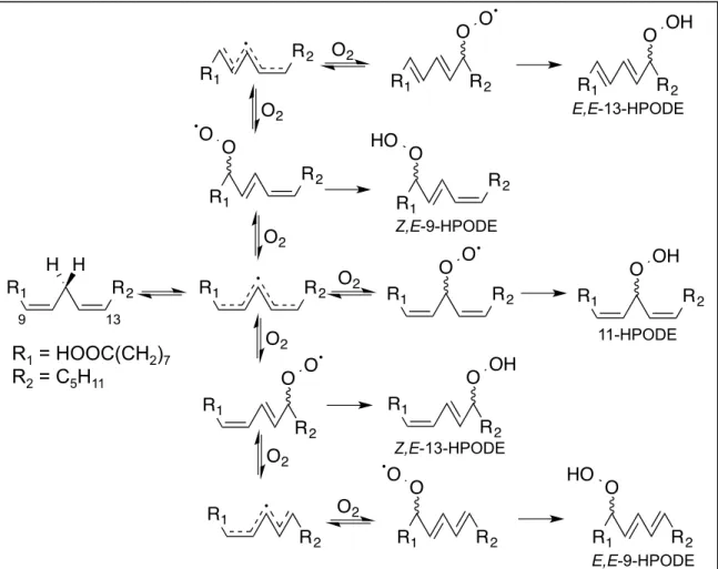
The autoxidation of LA is one of the simpler PUFA oxidation profiles to study due to its structure. With only two double bonds, LA only has one set of abstractable hydrogen atoms. Oxygen can add to the 9-, 11-, or 13- carbons generating a conjugated diene peroxyl radical for 9- and 13- positional isomers or a bis-allylic peroxyl radical for the 11- positional isomer106. Oxygen addition is a reversible process, and the peroxyl radical can be regenerated107. The rate constant for the reverse reaction of the 11-peroxyl radical is orders of magnitude greater than the rate constant for the formation of both the Z,E-9-peroxyl radical and Z,E-13-peroxyl radical. These rate constants indicate that unless trapped immediately, the 11-peroxyl radical will decompose quickly, giving all 9- or 13- peroxyl radicals108. 11-Hydroperoxy octadecadienoic acid (11-HPODE) is rarely seen except in cases where a good hydrogen donor is present. This was demonstrated in the presence of α-tocopherol, a good hydrogen donor, where a mixture of 1:1:1, 9-:11-:13-HPODE was generated by autoxidation109.
Isomerization of the double bond allylic to the peroxyl radical also occurs in the 9- and 13- positional isomers. In the presence of a good hydrogen donor, the peroxyl radical will be trapped generating the Z,E-9-HPODE or Z,E-13- HPODE in equal amounts. As mentioned above, oxygen addition is reversible.
After regeneration of the peroxyl radical, oxygen can add at either position potentially resulting in a second allylic bond isomerization, generating E,E-9- HPODE and E,E-13-HPODE. When no good hydrogen donor is present, the E,E:Z,E-HPODE ratio will be 4:1, designating Z,E-HPODE as the kinetic product, and E,E-HPODE as the thermodynamic product (Figure 5)110, 111.
In addition to the various positional and geometric isomers generated, HPODE is also a chiral molecule. During autoxidation, oxygen can add from either side of the molecule generating both the R and S enantiomers. Analysis of R/S ratios is one of the most accurate methods to determine in vivo if lipid peroxidation is occurring by an enzymatic or non-enzymatic process because microenvironment factors like hydrogen donor ability do not play a role in the
R1 R2 H H
R1 R2 R1 R2 O O O2
O2
O2
R1
R2 O O R2 R1
O O
R2 R1
HO O
R1
R2 O OH
R1 R2 O OH O2
O2
R1
R2 R2 R1
O2
O2
R2 O O R1
R1 O O
R2
R2 O OH R1
R1 HO O
R2 Z,E-13-HPODE
9 13
Z,E-9-HPODE
E,E-9-HPODE E,E-13-HPODE
11-HPODE
R1 = HOOC(CH2)7 R2 = C5H11
Figure 5. Autoxidation products of LA. After bis-allylic hydrogen abstraction, the pentadienyl radical of LA can react at three different positions, generating three distinct peroxyl radicals.
The peroxyl radicals can be quenched generating HPODE, or they can revert to the pentadienyl radical. The pentadienyl radical can then react with molecular oxygen at any of the three original positions, reforming a peroxyl radical. With each reversal of the peroxyl radical, double bond isomerization from cis to trans will occur. If allowed to reverse a sufficient amount of times, trans double bonds will form preferentially as they are a lower energy state.
stereochemistry observed. Therefore an R/S ratio of 1 indicates an autoxidation process, whereas a ratio greatly skewed to one enantiomer over the other suggests an enzymatic process112-114.
AA oxidation generates a much more complex autoxidation profile than LA due to it having 3 bis-allylic positions at the 7-, 10-, and 13-carbons. A single oxygen addition generates six positional isomers: E,Z,Z,Z-5-hydroperoxy eicosatetraenoic acid (E,Z,Z,Z-5-HPETE), Z,E,Z,Z-8-HPETE, Z,E,Z,Z-9-HPETE, Z,Z,E,Z-11-HPETE, Z,Z,E,Z-12-HPETE, and Z,Z,Z,E-15-HPETE103. As with LA, bis-allylic peroxides are rarely observed and a mixture of R and S enantiomers are formed from autoxidation115. Additionally, little HPETE is observed with more than one trans double bond because the rate of cyclization is estimated to be greater than the rate of reversal of the peroxyl radical116. The additional reactive positions of AA also allow for endoperoxide formation by cyclization, resulting in F2α-isoprostanes (F2α-isoP), products structurally similar to prostaglandin F2α (PGF2α), and a host of other cyclized products117-119. F2α-isoPs have been used as an in vivo biomarker for the identification of increased oxidative stress and lipid peroxidation120, 121. The mixture of isoprostanes generated is extremely complex, with four hydroxyl positional isomers possible: 5-, 8-, 12-, and 15-F2α- isoP, with the 5- and 15-positional isomers the most abundant (Figure 6)119. Each series of isoprostane has five chiral centers, resulting in eight unique enantiomers for each120.
Cyclooxygenases
PUFAs can also be oxygenated in a much more controlled fashion by enzymes that direct hydrogen abstraction as well as oxygen addition to get a specific product profile with defined isomers and stereochemistry. One such set of enzymes are the COXs, which use a heme-bound iron to catalyze hydrogen abstraction122. Two enzymes comprise this class: COX-1123, which is the constitutively expressed species, and COX-2124, which is the species induced in response to an inflammatory stimulus. Both enzymes are capable of oxygenating LA125, AA125, as well as other PUFAs of both the ω-6 and ω-3 families126. COX-2 also exclusively oxygenates neutral ester and amide derivatives of AA, including 2-arachidonoylglycerol and arachidonylethanolamine127. The result of these oxygenation events are potent and diverse signaling molecules involved in a host
HO
HO OH
COOH HO
HO
OH
COOH HO
HO
OH
COOH
HO
HO OH
COOH 5-series F2α-isoprostane 8-series F2α-isoprostane
12-series F2α-isoprostane 15-series F2α-isoprostane
Figure 6. Autoxidation of AA generates F2α-isoprostanes. The F2α-isoprostanes are a series of autoxidation AA metabolites that are structurally similar to prostaglandins. The mechanism of formation of isoprostanes is similar to that of prostaglandins, but because they are formed non-enzymatically, they have a mixture of positional isomers and enantiomers.
of cellular functions including vascular dilation and constriction, platelet aggregation, and mucosal regeneration128.
Due to the potent signaling capabilities of COX metabolites, the enzymes were the focus of pharmacology long before their existence and products were known. Historically, extract of willow bark was given to patients to treat pain and inflammation. The active ingredient in willow bark is salicylic acid, which when acetylated by Felix Hoffman in 1897, created aspirin, a pharmaceutical agent still used today129. Inhibition of the COX enzymes is the basis of a multibillion-dollar set of drugs including the nonsteroidal anti-inflammatory drugs (NSAIDs) ibuprofen, naproxen, celecoxib, and indomethacin130-132. With the prominent role of COXs in generating cell signaling molecules, it is no wonder that COX expression has been linked to a host of disease including cancer133, cardiovascular disease134, rheumatoid arthritis135, and neurodegenerative diseases136.
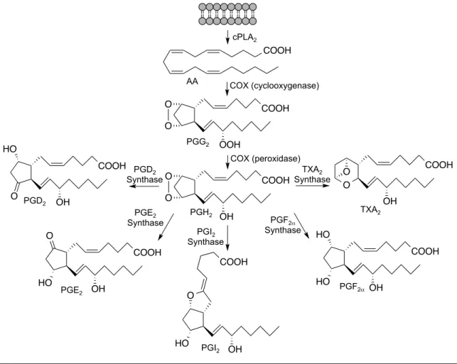
The COX enzymes are structural homodimers that act as functional heterodimers to bis-dioxygenate and cyclize AA, released from the lipid bilayer by cytosolic phospholipase A2 (cPLA2)137, 138. Oxygenation and cyclization occur in the cyclooxygenase active site forming PGG2 (Figure 7). The peroxide of PGG2 is then reduced in the peroxidase active site to give PGH2. PGH2 is the precursor to the signaling molecules PGE2, PGD2, PGF2α, PGI2, and
122 O
O
COOH
OOH
O O
COOH
OH COOH
OH HO
O
COOH
OH O
HO
HO OH
COOH
OH HO
HO O
O O
OH
COOH COOH
COOH AA
PGG2
PGH2
PGI2 PGD2
PGE2 PGF2α
TXA2 PGD2
Synthase
PGE2 Synthase
PGI2 Synthase
PGF2α
Synthase TXA2 Synthase cPLA2
COX (cyclooxygenase)
COX (peroxidase)
Figure 7. COX metabolism of AA. The COX enzymes consist of two active sites that act on free AA released from the lipid bilayer by cPLA2. The cyclooxygenase active site bis- dioxygenates and cyclizes AA to form PGG2. PGG2 is then reduced at the peroxidase active site to PGH2. PGH2 is the precursor to other eicosanoids including PGD2, PGE2, PGI2, PGF2α, and TXA2, which are generated by their respective synthases.
The prostaglandins have a panoply of signaling roles, including both vasodilation and constriction, muscle contraction, as well as the pain and fever associated with inflammation128. Additionally, COXs can generate several monooxygenated arachidonic acid species including 11(R)-HETE and 15(S)-HETE139. Aspirin, a COX inhibitor that acetylates Ser530, does not abolish the activity of COXs, but shifts the product profile from one generating PGH2 to one generating 15(R)- HETE140. Many studies have used COX variants to determine the residues responsible for the cyclooxygenase and peroxidase activity as well as inhibitor binding to the COX enzymes139, 141-143. LA is also a substrate for the COX enzymes. Since LA only has one bis-allylic carbon, cyclized LA products are not generated. The major products generated by COX metabolism of LA are the monooxygenated products at the 9- and 13-carbons, 9-HPODE and 13-HPODE respectively125.
Lipoxygenases
Like the COXs, LOXs represent a controlled mechanism of lipid oxidation, with both LA and AA being potential substrates. LOXs can act on both free144 and esterified145 fatty acids. Despite having an active role in inflammatory signaling, LOXs have not been successful drug targets like the COXs. In fact there is only one approved drug targeting 5-LOX, Zileuton146, which has a limited patient population because of its generation of a cytotoxic compound, 5-oxo eicosatetraenoic acid (5-KETE)147. Even though they have not been
pharmacological targets, 5-LOX148, 12-LOX149, and 15-LOX150 inhibitors as well as pan-LOX inhibitors151 have been generated for research purposes.
There are many LOX enzymes that are named differently depending on the organism and the oxygenation position of the fatty acid substrate, but all of them use a non-heme iron for their chemistry. LOXs first discovered to oxygenate LA were named 9-LOX and 13-LOX indicating that they add oxygen to the 9- or 13-carbon generating 9-HPODE or 13-HPODE respectively152. Conversely, LOXs first discovered to oxygenate AA are named 5-LOX, 8-LOX, 12-LOX, or 15-LOX indicating oxygenation of AA at the 5-, 8-, 12-, or 15-carbon, generating 5- HPETE, 8-HPETE, 12-HPETE, or 15-HPETE respectively153. Much substrate overlap can be seen between these groups of LOX enzymes. For example soybean 15-LOX-1 functions as a 15-LOX when AA is the substrate, but a 13- LOX when LA is the substrate154, 155. Another example is potato 5-LOX, which functions as a 5-LOX when AA is the substrate, but a 9-LOX when LA is the substrate156.
While not quite as vast as COX metabolism, LOXs generate a host of biologically active metabolites. 15-HPETE and 15-hydroxy eicosatetraenoic acid (15-HETE) generated by 15-LOX, have been shown to be angiostatic and angiogenic respectively157. Increased levels of LOX-derived 13-hydroxy octadecadienoic acid (13-HODE) results in increased growth in hepatocellular carcinoma158. However, the most studied class of metabolites resulting from LOX metabolism are the leukotrienes159 (Figure 8), which result from 5-LOX metabolism of AA160. The first step in leukotriene biosynthesis is the release of
AA from the lipid bilayer by cPLA2161. 5-LOX is an interesting PUFA metabolizing enzyme because it requires a second protein, 5-LOX activating protein (FLAP), to metabolize AA162. The mechanism by which FLAP confers activity to 5-LOX is not fully understood, but it is know that FLAP does not have enzymatic activity of its own, and it is hypothesized that FLAP delivers AA to 5-LOX163. During leukotriene biosynthesis, AA is oxygenated by 5-LOX to 15(S)-HPETE, reduced in the cell medium to 15(S)-HETE, then oxidized again by 5-LOX to leukotriene A4 (LTA4)164, 165. LTA4 is then further oxidized by LTA4 hydrolase to leukotriene B4
(LTB4)166 or conjugated to GSH by LTC4 synthase (LTC4S) generating leukotriene C4 (LTC4)167. The GSH of LTC4 can be further degraded sequentially losing glutamate to form leukotriene D4 (LTD4), and then glycine to form leukotriene E4 (LTE4)168. Leukotrienes are pro-inflammatory in their signaling, and have been implicated in many diseases associated with inflammation169 including rheumatoid arthritis170, irritable bowel syndrome171, and asthma172. The only drug targeting LOXs, Zileuton, inhibits 5-LOX and thus leukotriene biosynthesis, and has been implemented as a treatment for many of the diseases associated with leukotriene biosynthesis171, 173.
Lipid Electrophiles
Generation of lipid electrophiles
Further oxidation and degradation of the oxidized lipid species mentioned above results in the generation of various chain length α,β-unsaturated carbonyls and free aldehydes. These lipid species are electrophilic, reacting with
COOH
COOH O OH
COOH OH
O COOH
OH OH
COOH
COOH OH
S
NH O
NH O
OH O
H2N OH
O
COOH OH
S
NH2 O
NH O
OH COOH
OH
S
NH2 O
OH cPLA2
AA
15(S)-HPETE
15(S)-HETE
5-LOX
5-LOX H2O
LTA4
LTB4 LTC4
LTD4
LTE4
LTC4S
Figure 8. 5-LOX metabolism of AA. 5-LOX catalyzes the addition of molecular oxygen to AA released from the lipid bilayer by cPLA2 to generate 15(S)-HPETE. 15(S)-HPETE is quickly reduced to 15(S)-HETE in the cellular milieu. 5-LOX also oxidizes 15(S)-HETE to LTA4. LTA4 can be converted to LTB4 by LTA4 hydrolase or conjugated to GSH to form LTC4 by LTC4
synthase. The GSH of LTC4 can be degraded to LTD4 then LTE4 by losing glutamate and glycine sequentially.
nucleophilic groups of cellular macromolecules forming adducts, and potentially altering function. I will focus on adducts formed on nucleophilic amino acid residues in proteins. By no means has the entire electrophile pool been discovered, and thus we don’t know exactly how they are formed. However, the chemistry of formation has been identified for several of the more highly studied lipid electrophiles.
Several mechanisms have been proposed to explain the formation of these reactive lipid species. Both 4-hydroxy-2-nonenal (HNE) and 4-oxo-2- nonenal (ONE) can be generated from both LA and AA metabolism174. LOX175, 176 and COX-2177 derived 15-HPETE can be converted to ONE. Additionally, COX-2 metabolism of 5-HETE generated by 5-LOX can result in HNE formation178. LA can be converted to both HNE179, 180 and ONE180, 181 by autoxidation in vitro.
HO O
HO O O
O
O O
O OH
levuglandin E2 13-oxo octadecadienoic acid 15-oxo eicosatetrenoic acid
4-hydroxy-2-nonenal 4-oxo-2-nonenal
15-deoxy-Δ12,14-prostaglandin J2
O
COOH
O O
OH
COOH
Figure 9. Structures of some commonly studied lipid electrophiles.