Activity of Chondroitin Sulfate D and E
Thesis by
Kuang-Wei Yang
In Partial Fulfillment of the Requirements for the degree of
Doctor of Philosophy
CALIFORNIA INSTITUTE OF TECHNOLOGY Pasadena, California
2018
(Defended 23 March 2018)
2018 Kuang-Wei Yang All Rights Reserved
ACKNOWLEDGEMENTS
First and foremost, I would like to thank my advisor, Dr. Linda Hsieh-Wilson, for her constant support and guidance. Without Linda, I would not have been able to finish such exciting projects. I would also like to thank my committee members, Dr. Dennis Dougherty, Dr. William Goddard, and Dr. Sarah Reisman, for their insightful comments and suggestions.
I would like to thank Dr. Andrew Almond at University of Manchester, my collaborator for the NMR structural study. Our discussion of science and world news is certainly enjoyable. Also, I need to thank Dr. Scott Virgil, Dr. David VanderVelde, Dr. Mona Shahgholi and Dr. Jeffery Pelton for their assistance. Additionally, I would like to thank all Hsieh-Wilson lab members, past and present, for your scientific input and your day-to-day companionship.
Last but not least, I would like to thank my mom and dad. Thank you for always supporting and believing in me and thank you for your unconditional love. I hope I have made you proud.
Chondroitin sulfate are ubiquitously expressed linear, sulfated polysaccharides that play critical roles in neuronal development and regeneration growth factor signaling, morphogenesis, and virus invasion. The diverse sulfation patterns presented by chondroitin sulfate has been suggested to regulate its activity, but the structural complexity and heterogeneity have hampered the understanding of structure-activity relationship. Therefore, we envisioned that chemically synthesized chondroitin sulfate oligosaccharide may provide a unique opportunity to specifically study the functions of sulfation patterns.
Here, we report the synthesis of a CS-D and CS-E tetrasaccharide in a step-efficient manner. By generating a disaccharide precursor from hydrolysis of polysaccharides, we were able to streamline the synthesis and reduce the number of steps by one-third comparing to the traditional synthesis without losing versatility of the synthetic route and functionality of the final product. With the structurally defined molecules, we were able to determine the NMR solution structure of CS-D and CS-E. In this work, we accomplished the first structural study of CS-D tetrasaccharide and the most thorough study of CS-E to date. Furthermore, we also discovered the existence of a second conformer in CS-D, which is the first time for such behavior to be observed experimentally in chondroitin sulfate. The electrostatic potential surface constructed based on the NMR structure presented unique structural features that may allow proteins to interact specifically.
The CS-D and CS-E tetrasaccharide, along with a CS-D disaccharide, was investigated for their neuritogenic activity. We discovered that the CS-D tetrasaccharide specifically stimulates dendritic growth whereas the CS-E tetrasaccharide preferentially promoted axonal growth, revealing the potential critical role chondroitin sulfate with specific sulfation patterns may play in the nervous system. The lack of activity of the CS-D disaccharide suggested that the minimum motif required for activity of CS-D is a tetrasaccharide.
Acknowledgements………...iii
Abstract ………iv
Table of Contents………...v
List of Illustrations and/or Tables……….…vi
Chapter 1: Introduction to Glycosaminoglycans ... 1
Chapter 2: Chemically Synthesis of the CS-D and CS-E Tetrasaccharides ... 15
Appendix for Chapter 2: Relevant Spectral Data ... 67
Chapter 3: NMR Solution Structure of the CS-D and CS-E Tetrasaccharides ... 111
Appendix for Chapter 3: Relevant Data for Structure Determination ... 158
Chapter 4: Neuritogenic Activity of CS-D and CS-E ... 166
Page
F1.1 Structure of the five GAG families ... 3
F1.2 Structure of the four major sulfation pattern of CS ... 3
F1.3 Structure of Arixtra ... 5
F1.4 Biosynthesis of CS, DA HS and heparin ... 5
F1.5 The brush-like structure of HA-PG complex ... 7
T2.1 Disaccharide composition analysis of commercially available polysaccharides ... 16
F2.1 Target CS oligosaccharides ... 18
S2.1 Retrosynthesis of the CS-D tetrasaccharide and disaccharide ... 19
S2.2 Synthesis of the key disaccharide intermediate ... 21
S2.3 Synthesis of the disaccharide donor ... 22
T2.2 Optimization of O-acetate deprotection ... 23
S2.4 Synthesis of diol 21 ... 23
T2.3 Optimization of trichloroacetamide reduction ... 24
S2.5 Synthesis of the CS-D disaccharide ... 25
S2.6 Synthesis of the disaccharide acceptor ... 25
T2.4 Optimization of the coupling reaction ... 26
S2.7 Synthesis of the CS-D tetrasaccharide ... 27
S2.8 Retrosynthesis of the CS-E tetrasaccharide ... 36
S2.9 Synthesis of the CS-E disaccharide acceptor ... 28
T2.5 Optimization of the coupling reaction ... 30
S2.10 Synthesis of the CS-E tetrasaccharide ... 31
S2.11 Derivatization towards the CS-A and CS-C tetrasaccharides .... 31
F3.1 Schematic representation of the torsion angles of interest ... 114
F3.2 Spherical mapping of pyranose conformations represented by the Cremer-Pople polar coordinates ... 114
F3.4 Distribution of glycosidic angles of CS-D and CS-E ... 118
T3.1 Average glycosidic torsion angles of the CS-D tetrasaccharide 119 T3.2 Average glycosidic torsion angles of the CS-E tetrasaccharide 119 T3.3 Population of puckers of the CS-D tetrasaccharide ... 119
T3.4 Population of puckers of the CS-E tetrasaccharide ... 120
T3.5 Experimental coupling constants of CS-D ... 122
T3.6 Experimental coupling constants of CS-E ... 122
T3.7 Comparison of coupling constants of CS-D ... 125
T3.8 Comparison of coupling constants of CS-E ... 125
T3.9 Coupling constants of most abundant puckers of CS-D ... 125
T3.10 Average glycosidic torsion angles in the calculated CS-E structure ... 129
F3.5 Ensemble of the top 25 structures of CS-E ... 129
F3.6 The GalNAc2-GlcA1 linkage of the calculated structure of CS-D ... 130
T3.11 Average glycosidic torsion angles in the calculated CS-D N structure ... 134
F3.6 Ensemble of the top 25 structures of CS-D N ... 135
T3.12 Average glycosidic torsion angles in the calculated CS-D I structure ... 135
F3.6 Ensemble of the top 25 structures of CS-D I... 135
T3.13 Low-energy conformers of CS-D ... 137
T3.13 Low-energy conformers of CS-E ... 137
F3.8 Free energy landscape contour maps of CS-D and CS-E ... 138
F3.9 Comparison of inter-residual interaction between CS-D and CS-E ... 143
F3.10 Structure of the NMR based GalNAc2-GlcA1 linkage of CS-D I ... 144
F3.11 Structure of the low-energy conformer in CS-D... 145
F3.13 Structure of the low-energy conformer in CS-E ... 147
F3.14 Structure of the low-energy conformer in CS-E ... 147
T3.15 Comparison of glycosidic torsion angles in the present study with selected published CS structures ... 148
F3.14 Electrostatic potential surface of CS-D and CS-E ... 149
TA3.1 Ring puckering definitions ... 159
TA3.2 Inter-residual NOE restraints of CS-D N ... 160
TA3.3 Inter-residual “no-NOE” restraints of CS-D N ... 161
TA3.4 Inter-residual NOE restraints of CS-D I ... 161
TA3.5 Inter-residual “no-NOE” restraints of CS-D I... 162
TA3.6 Inter-residual NOE restraints of CS-D I ... 163
TA3.7 Inter-residual “no-NOE” restraints of CS-D I... 164
F4.1 Representative images of CS-D and CS-E stimulated neurite outgrowth ... 171
F4.2 Quantitative analysis of neuritogenic activity of CS-D and CS-E ... 172
C h a p t e r 1
INTRODUCTION TO GLYCOSAMINOGLYCANS
C h a p t e r 1
INTRODUCTION TO GLYCOSAMINOGLYCANS
The glycosaminoglycan family
Glycosaminoglycans (GAGs) are a diverse class of polysaccharides that are covalently attached to core proteins, forming proteoglycans (PGs). After GAGs are synthesized in the Golgi apparatus, GAGs are inserted into the plasma membrane, secreted into the extracellular matrix (ECM), or stored in intracellular granules. Although GAGs were once thought of as simply structural components of connective tissue, they are now known to mediating a myriad of cellular processes including but not limited to neuronal development1-3, neuroregeneration4-5, cancer metastasis6-9 and viral invasion10-12. Structurally speaking, GAGs are linear polysaccharides composed of repeating disaccharide units. The combination of variable length, disaccharide constituent and sulfation pattern of GAGs creates extraordinary structural diversity that enables them to interact with a wide range of biological molecules and participate in diverse biological activities.
Families of GAGs
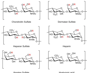
Based on the disaccharide constituents, five families of GAGs (figure 1.1) have been established, namely chondroitin sulfate (CS), dermatan sulfate (DS), heparin and heparan sulfate (HS), keratan sulfate (KS) and hyaluronic acid (HA). CS is the most abundant family in brain and in cartilage13 and participates in numerous biological processes including neuronal development and regeneration5, growth factor signaling14,
morphogenesis15, and virus infection16. It is composed of repeating glucuronic acid (GlcA)- N-acetylgalactosamine (GalNAc) disaccharide units joined by β(1→3) and β(1→4)
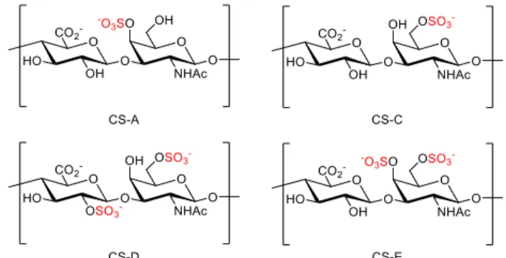
linkages, respectively. There are four major sulfation patterns of CS: CS-A, -C, -D, and -E (figure 1.2) which contain sulfate groups at the 2-O position of GlcA or the 4-O or 6-O positions of GalNAc. Although 3-O sulfation of GlcA is known as well17-18, it is less common. The expression of different CS sulfation pattern is regulated spatiotemporally by the corresponding sulfotransferases (STs), however, little is known about the exact mechanism of regulation.
Figure 1.1. Structures of the five GAG families.
Figure 1.2. Structures of the four major sulfation pattern of CS.
The next GAG family, DS, is the predominant GAG present in skin. DS has been reported to play roles in coagulation19, cardiovascular disease20, and bacterial invation21. It is composed of repeating iduronic acid (IdoA)-GalNAc disaccharide units joined by β(1→3) and β(1→4) linkages, which is identical to CS except for only one different stereocenter orientation at C5 of uronic acid. DS can be sulfated at the 2-O of GlcA, 4-O, or 6-O of GalNAc like CS. However, the 2-O sulfation of GlcA and 4-O sulfation of GalNAc are more common in DS22.
Together heparin and HS constitute the most well studied family of GAGs that are particularly important in anticoagulation23, cell growth and development24, angiogenesis25-
26, and viral invasion27-28. Arixtra (figure 1.3), a methylated heparin pentasaccharide, is an anticoagulant medication that has a market of ~$500 million. Heparin and HS are both composed of alternating α(1→4) N-acetylglucosamine (GlcNAc) and β(1→4) GlcA units or alternating α(1→4) GlcNAc and α(1→4) IdoA units. They also share the same available positions for modifications, namely the sulfation at the 3-O and 6-O position of GlcNAc, sulfation of the 2-O position of GlcA or IdoA and N-sulfation or N-acetylation. Although heparin and HS are structurally related, they are still different in some key aspects29. Heparin is only produced by connective tissue type mast cells and has a smaller size (7 – 20 kDa), while HS is expressed ubiquitously and larger in size (10 – 70 kDa). Although both IdoA and GlcA are present in both heparin and HS, the IdoA content is markedly higher in heparin (>70%) than in HS (30% - 50%). The extent of sulfation is also different in that the number of sulfates/disaccharide ratio is 0.8 – 1.8 in HS and 1.8 – 2.6 in heparin.
Heparin thereby possesses the highest negative charge density of any known biological
macromolecule30. Additionally, N-sulfation is much more common in heparin (>80%) than in HS (40% - 60%).
Figure 1.3. Structure of Arixtra.
CS, DS, heparin and HS are linked to the core proteins with the tetrasaccharide linker GlcA-Gal-Gal-Xylose (Xyl). As shown in figure 1.4, after the linker unit was covalently linked to a serine residue of the core protein, addition of the first hexosamine commits the GAG chain to either CS and DS (GalNAc) or HS and heparin (GlcNAc).
Subsequent chain elongation, epimerization, deacetylation (HS and heparin), and sulfation of the GAG chains then generates the corresponding GAGs.
Figure 1.4. Biosynthesis of CS, DA, HS, and heparin.
KS, the last family of sulfated GAGs, is composed of repeating disaccharide units of galactose (Gal)-GlcNAc joined by β(1→4) and β(1→3) linkages, respectively. KSs can be classified in three categories, KS-I, -II, and -III, based on the GAG-core protein linkage31: KS-I is composed of N-linked chains that are abundant in cornea. KS-II is composed of chains O-linked through GalNAc and identified in cartilage. KS-III is extended from O-linked mannose and has been isolated from brain tissue. KS is related to maintaining the proper hydration levels of the cornea, which is relevant to keep the transparency of the tissue32. Aside from the principal function in cornea, KS also participates in developmental biology, cellular signaling, and migration, like CS, DS, and HS32. Both GlcNAc and Gal in KS can be sulfated at C6, but GlcNAc sulfation is most abundant33. Since KS only have two sulfation sites, the structural diversity of KS is significantly less than that of CS and HS.
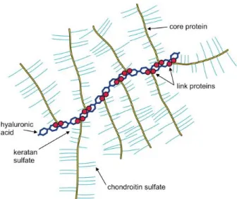
HA is the only family of GAGs that is not sulfated. Interestingly, it is also the only GAG family that does not covalently bind to core proteins to form PGs. Instead, HA binds to PGs though link proteins. A single HA chain can be linked with multiple PGs and form a brush-like structure (figure 1.5). Normally, HA are composed of 2,000–25,000 disaccharide units of β(1→3) GlcA β(1→4) GlcNAc, which corresponds to polysaccharides with relative molecular masses of 106 -107 and polymer lengths of 2–
25 μm. HA is abundant not only in soft connective tissues, but also in epithelial and neural tissues. Due to the size and anionic nature of HA, it has a unique capacity in retaining water and is important to retaining moisture in the skin and lubricating movable parts of the body, such as joints and muscles.
Figure 1.5. The brush-like structure of HA-PG complex.
Tools to study the role of sulfation
Among the modifications of GAG chains, namely epimerization, deacetylation, and sulfation, sulfation is the most important, as sulfation alone can give rise to hundreds of possible tetrasaccharide structures. Also, the sulfate groups provide a charged surface which allow GAGs to electrostatically interact with proteins. Indeed, GAGs are known to bind to several hundreds of proteins, and the sulfation pattern of GAGs allow selective interactions with different molecules34. For example, in the well-studied HS-fibroblast growth factor (FGF) interaction, the binding of particular FGFs requires specific sulfation patterns. While FGF-2 requires 2-O-sulfation but not 6-O-sulfation for HS binding, FGF- 10 has the reverse preference, and FGF-1 requires both 2-O-sulfation and 6-O-sulfation35. Signaling molecules in the central nervous system (CNS) such as semaphoring 3A36, midkine37-38, pleiotrophin39, and neurotrophins40 have also been reported to preferentially bind to CS with specific sulfation patterns .
While diverse sulfation patterns allow GAGs to be formed and interact specifically with different proteins, complexity of the sulfation pattern also hampers the understanding of the structure-activity relationship (SAR) of GAGs. Although a number of tools are available, not all them allow the study of strictly defined sulfation motif.
Genetic and biochemical approaches such as known-down of STs or digestion of polysaccharide by lyases have established important biological implications for GAGs.
However, STs do not function independently and the removal of one ST may lead to global changes of the sulfation profile, making it difficult to specify the activity of a particular sulfation pattern. On the other hand, while GAG lyases can be used to study the function of each GAG family, the impact of specific sulfation patterns cannot be determined. GAG polysaccharides and oligosaccharides can also be isolated from natural sources. However, although polysaccharides enriched in a sulfation patterns are available, they are still far from homogeneous. The oligosaccharides obtained by partial digestion of polysaccharides also suffer from heterogeneity due to the nature of lyases and the source polysaccharides.
Furthermore, the inherent sulfation motifs in available polysaccharides and selectivity of the available digestion enzymes limits the variety of sulfation patterns available for study.
Our group envisioned that chemical synthesis can provide a powerful solution to these challenges as the length, sulfation pattern, and stereochemistry are all fully under control38,41-43. Aside from the ability to obtain defined oligosaccharides, the freedom of chemical synthesis also facilitates the generation of GAG-conjugated tools that are otherwise unavailable. For example, microarrays conjugated with GAG oligosaccharides allows rapid identification of GAG-protein interaction38,44. Glycopolymers with GAG
oligosaccharides attached to a polymeric backbone enable the study of specific sulfation patterns in a multivalent and polysaccharide-like context42-43,45-46. GAG oligosaccharides conjugated to immunogenic carrier proteins allow the generation of the most sulfation pattern-specific antibodies4,38,44,47.
In the present study, we achieved streamlined chemical synthesis of CS-D and CS- E tetrasaccharides. Next, we successfully calculated the solution structure of the tetrasaccharides based on nuclear magnetic resonance (NMR) data. This is the first NMR structural study of a CS-D tetrasaccharide and the most thorough study of a CS-E tetrasaccharide to date. Neuritogenic activity in hippocampal neurons of the tetrasaccharides was also investigated. We discovered that the CS-D tetrasaccharide specifically stimulates dendritic outgrowth and the CS-E tetrasaccharide specifically stimulates axonal outgrowth.
References
(1) Sugahara, K.; Mikami, T.; Uyama, T.; Mizuguchi, S.; Nomura, K.; Kitagawa, H., Recent advances in the structural biology of chondroitin sulfate and dermatan sulfate. Curr.
Opin. Struct. Biol. 2003, 13 (5), 612.
(2) Bovolenta, P.; Fernaud-Espinosa, I., Nervous system proteoglycans as modulators of neurite outgrowth. Progress in Neurobiology 2000, 61 (2), 113.
(3) Yamaguchi, Y.; Inatani, M.; Matsumoto, Y.; Ogawa, J.; Irie, F., Roles of Heparan Sulfate in Mammalian Brain Development: Current Views Based on the Findings from Ext1 Conditional Knockout Studies. Glycosaminoglycans in Development, Health and Disease 2010, 93, 133.
(4) Brown, J. M.; Xia, J.; Zhuang, B.; Cho, K. S.; Rogers, C. J.; Gama, C. I.; Rawat, M.;
Tully, S. E.; Uetani, N.; Mason, D. E.; Tremblay, M. L.; Peters, E. C.; Habuchi, O.; Chen,
D. F.; Hsieh-Wilson, L. C., A sulfated carbohydrate epitope inhibits axon regeneration after injury. Proc Natl Acad Sci U S A 2012, 109 (13), 4768.
(5) Laabs, T.; Carulli, D.; Geller, H. M.; Fawcett, J. W., Chondroitin sulfate proteoglycans in neural development and regeneration. Current Opinion in Neurobiology 2005, 15 (1), 116.
(6) Afratis, N.; Gialeli, C.; Nikitovic, D.; Tsegenidis, T.; Karousou, E.; Theocharis, A. D.;
Pavao, M. S.; Tzanakakis, G. N.; Karamanos, N. K., Glycosaminoglycans: key players in cancer cell biology and treatment. Febs Journal 2012, 279 (7), 1177.
(7) Knelson, E. H.; Nee, J. C.; Blobe, G. C., Heparan sulfate signaling in cancer. Trends in Biochemical Sciences 2014, 39 (6), 277.
(8) Kumar, A. V.; Gassar, E. S.; Spillmann, D.; Kiesel, L.; Yip, G. W.; Gotte, M., Specific sulfation patterns in heparan sulfate promote a proinvasive phenotype of breast cancer cells via upregulation of Wnt and MAPK signaling. Experimental and Clinical Endocrinology
& Diabetes 2013, 121 (3).
(9) Sanderson, R. D., Heparan sulfate proteoglycans in invasion and metastasis. Seminars in Cell & Developmental Biology 2001, 12 (2), 89.
(10) Aquino, R. S.; Park, P. W., Glycosaminoglycans and infection. Frontiers in Bioscience-Landmark 2016, 21, 1260.
(11) Leistner, C. M.; Gruen-Bernhard, S.; Glebe, D., Role of glycosaminoglycans for binding and infection of hepatitis B virus. Cellular Microbiology 2008, 10 (1), 122.
(12) Jinno, A.; Park, P. W., Role of glycosaminoglycans in infectious disease. Methods Mol Biol 2015, 1229, 567.
(13) Hascall, V. C.; Sajdera, S. W., Physical Properties and Polydispersity of Proteoglycan from Bovine Nasal Cartilage. Journal of Biological Chemistry 1970, 245 (19), 4920.
(14) Kim, S. H.; Turnbull, J.; Guimond, S., Extracellular matrix and cell signalling: the dynamic cooperation of integrin, proteoglycan and growth factor receptor. Journal of Endocrinology 2011, 209 (2), 139.
(15) Tanaka, M.; Maeda, N.; Noda, M.; Marunouchi, T., A chondroitin sulfate proteoglycan PTP zeta/RPTP beta regulates the morphogenesis of Purkinje cell dendrites in the developing cerebellum. Journal of Neuroscience 2003, 23 (7), 2804.
(16) Banfield, B. W.; Leduc, Y.; Esford, L.; Visalli, R. J.; Brandt, C. R.; Tufaro, F., Evidence for an Interaction of Herpes-Simplex Virus with Chondroitin Sulfate Proteoglycans during Infection. Virology 1995, 208 (2), 531.
(17) Kinoshita-Toyoda, A.; Yamada, S.; Haslam, S. M.; Khoo, K. H.; Sugiura, M.; Morris, H. R.; Dell, A.; Sugahara, K., Structural determination of five novel tetrasaccharides containing 3-O-sulfated D-glucuronic acid and two rare oligosaccharides containing a beta- D-glucose branch isolated from squid cartilage chondroitin sulfate E. Biochemistry 2004, 43 (34), 11063.
(18) Sugahara, K.; Tanaka, Y.; Yamada, S.; Seno, N.; Kitagawa, H.; Haslam, S. M.; Morris, H. R.; Dell, A., Novel sulfated oligosaccharides containing 3-O-sulfated glucuronic acid from king crab cartilage chondroitin sulfate K - Unexpected degradation by chondroitinase ABC. Journal of Biological Chemistry 1996, 271 (43), 26745.
(19) Fernandez, J. A.; Petaja, J.; Griffin, J. H., Dermatan sulfate and LMW heparin enhance the anticoagulant action of activated protein C. Thrombosis and Haemostasis 1999, 82 (5), 1462.
(20) Kovanen, P. T.; Pentikainen, M. O., Decorin links low-density lipoproteins (LDL) to collagen: A novel mechanism for retention of LDL in the atherosclerotic plaque. Trends in Cardiovascular Medicine 1999, 9 (3-4), 86.
(21) Schmidtchen, A.; Frick, I. M.; Bjorck, L., Dermatan sulphate is released by proteinases of common pathogenic bacteria and inactivates antibacterial alpha-defensin. Molecular Microbiology 2001, 39 (3), 708.
(22) da Costa, D. S.; Reis, R. L.; Pashkuleva, I., Sulfation of Glycosaminoglycans and Its Implications in Human Health and Disorders. Annual Review of Biomedical Engineering, Vol 19 2017, 19, 1.
(23) Liu, J.; Pedersen, L. C., Anticoagulant heparan sulfate: structural specificity and biosynthesis. Applied Microbiology and Biotechnology 2007, 74 (2), 263.
(24) Perrimon, N.; Bernfield, M., Specificities of heparan sulphate proteoglycans in developmental processes. Nature 2000, 404 (6779), 725.
(25) d'Angelo, I.; Oliviero, O.; Ungaro, F.; Quaglia, F.; Netti, P. A., Engineering strategies to control vascular endothelial growth factor stability and levels in a collagen matrix for angiogenesis: The role of heparin sodium salt and the PLGA-based microsphere approach.
Acta Biomaterialia 2013, 9 (7), 7389.
(26) Folkman, J.; Taylor, S.; Spillberg, C., The Role of Heparin in Angiogenesis. Ciba Foundation Symposia 1983, 100, 132.
(27) Hilgard, P.; Stockert, R., Heparan sulfate proteoglycans initiate dengue virus infection of hepatocytes. Hepatology 2000, 32 (5), 1069.
(28) O'Donnell, C. D.; Shukla, D., The Importance of Heparan Sulfate in Herpesvirus Infection. Virol Sin 2008, 23 (6), 383.
(29) Esko, J. D.; Kimata, K.; Lindahl, U., Proteoglycans and Sulfated Glycosaminoglycans.
In Essentials of Glycobiology, nd; Varki, A.; Cummings, R. D.; Esko, J. D.; Freeze, H. H.;
Stanley, P.; Bertozzi, C. R.; Hart, G. W.; Etzler, M. E., Eds. Cold Spring Harbor (NY), 2009.
(30) Shriver, Z.; Capila, I.; Venkataraman, G.; Sasisekharan, R., Heparin and heparan sulfate: analyzing structure and microheterogeneity. Handbook of Experimental Pharmacology 2012, (207), 159.
(31) Funderburgh, J. L., Keratan sulfate biosynthesis. IUBMB Life 2002, 54 (4), 187.
(32) Funderburgh, J. L., Keratan sulfate: structure, biosynthesis, and function.
Glycobiology 2000, 10 (10), 951.
(33) Prydz, K., Determinants of Glycosaminoglycan (GAG) Structure. Biomolecules 2015, 5 (3), 2003.
(34) Esko, J. D.; J, H. P.; Linhardt, R. J., Proteins That Bind Sulfated Glycosaminoglycans.
In Essentials of Glycobiology, rd; Varki, A.; Cummings, R. D.; Esko, J. D.; Stanley, P.;
Hart, G. W.; Aebi, M.; Darvill, A. G.; Kinoshita, T.; Packer, N. H.; Prestegard, J. H.;
Schnaar, R. L.; Seeberger, P. H., Eds. Cold Spring Harbor (NY), 2015; pp 493.
(35) Ashikari-Hada, S.; Habuchi, H.; Kariya, Y.; Itoh, N.; Reddi, A. H.; Kimata, K., Characterization of growth factor-binding structures in heparin/heparan sulfate using an octasaccharide library. Journal of Biological Chemistry 2004, 279 (13), 12346.
(36) Dick, G.; Tan, C. L.; Alves, J. N.; Ehlert, E. M. E.; Miller, G. M.; Hsieh-Wilson, L.
C.; Sugahara, K.; Oosterhof, A.; van Kuppevelt, T. H.; Verhaagen, J.; Fawcett, J. W.; Kwok, J. C. F., Semaphorin 3A Binds to the Perineuronal Nets via Chondroitin Sulfate Type E Motifs in Rodent Brains. Journal of Biological Chemistry 2013, 288 (38), 27384.
(37) Tamura, J.; Tsutsumishita-Nakai, N.; Nakao, Y.; Kawano, M.; Kato, S.; Takeda, N.;
Nadanaka, S.; Kitagawa, H., Synthesis and interaction with midkine of biotinylated chondroitin sulfate tetrasaccharides. Bioorganic & Medicinal Chemistry Letters 2012, 22 (3), 1371.
(38) Gama, C. I.; Tully, S. E.; Sotogaku, N.; Clark, P. M.; Rawat, M.; Vaidehi, N.; Goddard, W. A., 3rd; Nishi, A.; Hsieh-Wilson, L. C., Sulfation patterns of glycosaminoglycans encode molecular recognition and activity. Nat Chem Biol 2006, 2 (9), 467.
(39) Maeda, N.; Fukazawa, N.; Hata, T., The binding of chondroitin sulfate to pleiotrophin/heparin-binding growth-associated molecule is regulated by chain length and oversulfated structures. Journal of Biological Chemistry 2006, 281 (8), 4894.
(40) Rogers, C. J.; Clark, P. M.; Tully, S. E.; Abrol, R.; Garcia, K. C.; Goddard, W. A., 3rd; Hsieh-Wilson, L. C., Elucidating glycosaminoglycan-protein-protein interactions using carbohydrate microarray and computational approaches. Proc Natl Acad Sci U S A 2011, 108 (24), 9747.
(41) Tully, S. E.; Mabon, R.; Gama, C. I.; Tsai, S. M.; Liu, X.; Hsieh-Wilson, L. C., A chondroitin sulfate small molecule that stimulates neuronal growth. Journal of the American Chemical Society 2004, 126 (25), 7736.
(42) Oh, Y. I.; Sheng, G. J.; Chang, S. K.; Hsieh-Wilson, L. C., Tailored glycopolymers as anticoagulant heparin mimetics. Angewandte Chemie, International Edition in English 2013, 52 (45), 11796.
(43) Sheng, G. J.; Oh, Y. I.; Chang, S. K.; Hsieh-Wilson, L. C., Tunable heparan sulfate mimetics for modulating chemokine activity. Journal of the American Chemical Society 2013, 135 (30), 10898.
(44) Tully, S. E.; Rawat, M.; Hsieh-Wilson, L. C., Discovery of a TNF-alpha antagonist using chondroitin sulfate microarrays. Journal of the American Chemical Society 2006, 128 (24), 7740.
(45) Rawat, M.; Gama, C. I.; Matson, J. B.; Hsieh-Wilson, L. C., Neuroactive chondroitin sulfate glycomimetics. Journal of the American Chemical Society 2008, 130 (10), 2959.
(46) Lee, S. G.; Brown, J. M.; Rogers, C. J.; Matson, J. B.; Krishnamurthy, C.; Rawat, M.;
Hsieh-Wilson, L. C., End-functionalized glycopolymers as mimetics of chondroitin sulfate proteoglycans. Chemical Science 2010, 1 (3), 322.
(47) Gama, C. I.; Hsieh-Wilson, L. C., Chemical approaches to deciphering the glycosaminoglycan code. Current Opinion in Chemical Biology 2005, 9 (6), 609.
C h a p t e r 2
Chemical synthesis of the CS-D and CS-E tetrasaccharides
C h a p t e r 2
CHEMICAL SYNTHESIS OF THE CS-D AND CS-E TETRASACCHARIDES
Introduction
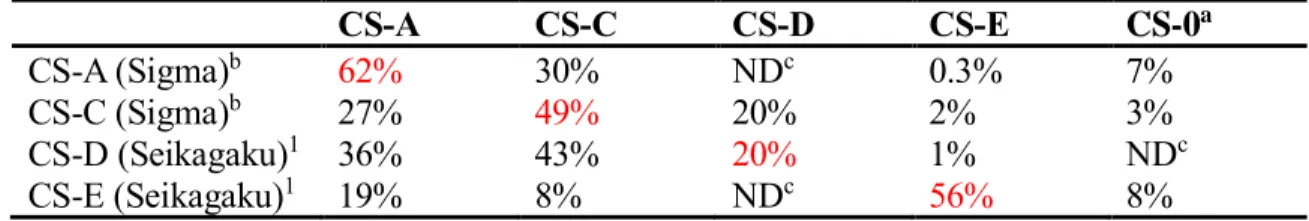
While the sulfation patterns of CS are essential to regulate specific biological functions, the chemical complexity and inherent heterogeneity of CS polysaccharides have hampered the detailed study of SAR. Although CS polysaccharides enriched in a specific sulfation motif are commercially available, the polysaccharides are far from homogeneous, as shown in table 2.1. While CS-A, CS-C, and CS-E have around 50% of the desired sulfation motifs, the CS-D polysaccharide contains only 20% of the D motif, highlighting the need for methods to obtain homogeneous CS-D.
CS-A CS-C CS-D CS-E CS-0a
CS-A (Sigma)b 62% 30% NDc 0.3% 7%
CS-C (Sigma)b 27% 49% 20% 2% 3%
CS-D (Seikagaku)1 36% 43% 20% 1% NDc
CS-E (Seikagaku)1 19% 8% NDc 56% 8%
Table 2.1. Disaccharide composition analysis of commercially available polysaccharides.
Suppliers are given in parenthesis. aUnsulfated chondroitin. bUnpublished data provided by Dr. Sheldon Cheung. cNot detectable.
Available methods to prepare chondroitin sulfate oligosaccharide include enzymatic digestion, chemoenzymatic synthesis, and organic synthesis. The digestion approach2-5 offers a straightforward way to obtain multiple CS oligosaccharides with a few steps. The sulfation pattern of the product oligosaccharide can also be tuned by the choice of enzyme and reactant polysaccharide to some extent. However, separation of the product oligosaccharides is challenging in that high purity samples are not always available.
Furthermore, the available oligosaccharide structures are still limited by the available selectivity of enzymes and the source structure. The unsaturated reducing end generated by chondroitinase digestion may also negatively impact the SAR determination.
While chemoenzymatic synthesis of heparin oligosaccharide has been more extensively studied6-7, the chemoenzymatic synthesis of homogeneous CS oligosaccharide was not reported until recently8. Although the chemoenzymatic approach offers shorter synthetic routes and higher overall yields, available structures are still limited by available enzyme specificity9.
Organic synthesis of CS oligosaccharides is still the most powerful method, as virtually any desired CS structure can be prepared, even those with non-natural motifs10. However, the synthetic routes are usually long and challenging. The first reported synthesis of CS-D tetrasaccharide took 38 steps in total11, highlighting the need for improved methods to access CS oligosaccharides. The synthesis of the CS-A, CS-C, and CS-E tetrasaccharide previously reported by our group10,12, while modular and efficient, still required 27 steps to obtain the CS-E tetrasaccharide. Traditionally, the synthesis of CS begins with monosaccharide precursors
In this study, we took advantage of the selective acid hydrolysis of CS polysaccharide first reported by Levene13 and Meyer and Davidson14 and improved by the Jacquinet group15 to rapidly access disaccharide starting material, as opposed to the traditional CS synthesis that begins with monosaccharide precursors. Part of the protecting group strategy developed by the Jacquinet group15-16 was also adapted. The disaccharide starting material significantly streamlined the synthesis by reducing the number of steps required to obtain our target
molecule, the CS-D and CS-E tetrasaccharides, by about one-third. We also installed an allyl group at the reducing end of the tetrasaccharides to serve as a versatile chemical handle, which has yet to be done for CS-D. Although a CS-D tetrasaccharide with a dithiolane moiety has been reported17, no description of source, preparation, or characterization is available in the publication.
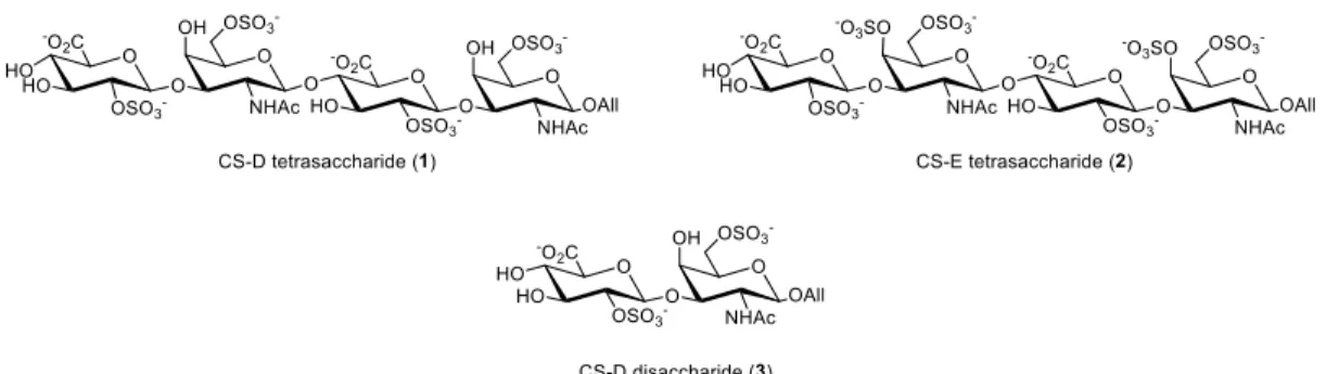
To study the 3-D structure and neuritogenic activities of the CS-D and CS-E sulfation motifs, we chose to chemically synthesize the CS-D tetrasaccharide (1) and CS-E tetrasaccharide (2). The CS-D disaccharide (3) was also synthesized to determine the minimum length of the CS-D motif required for activity. The CS-E disaccharide has been synthesized in a previous study and reported to have no significant neuritogenic activity12, and hence is not included in the present study.
Figure 2.1. Target CS oligosaccharides. All = allyl.
Synthetic Design of the CS-D tetrasaccharide and disaccharide
To synthesize the desired CS-D disaccharide and tetrasaccharide, we decided to utilize the polysaccharide digestion method13-14,16 to obtain the key disaccharide intermediate 7. The key disaccharide intermediate will be further derivatized to the corresponding disaccharide donor 5 and acceptor 6 for the coupling reaction. The CS-D disaccharide 4 can
also easily be derivatized from the disaccharide donor 5. We chose to install an allyl group at the reducing end of the oligosaccharides as a convenient chemical handle for conjugation to proteins, small molecules, or surfaces, as previously developed by our group12,18. To ensure proper stereochemistry, regioselectivity, and orthogonality, the protecting groups were carefully chosen to facilitate the synthesis.
Scheme 2.1. Retrosynthesis of the CS-D tetrasaccharide and disaccharide. All = allyl, Me = methyl, Lev = levulinyl, Bz = benzoyl, MCA = chloroacetyl, TCA = trichloroacetyl, Ph = phenyl, Ac = acetyl.
For the most important step in the synthesis, the coupling reaction, the Schmidt trichloroacetimidate method19-20 was adopted for its mild reaction conditions, good stereocontrolling ability, and convenient donor preparation. To ensure β-glycoside formation, the neighboring participating group trichloroacetamide has been reported to be a powerful stereocontrolling auxiliary. Also, trichloroacetamides can be easily reduced to acetamides by tributyltin hydride in one step and was therefore chosen to be installed at the C2 position of GalNAc. 2-naphthylmethyl group was used to temporarily protect the C1
position of GalNAc for the selective and convenient deprotection with 2,3-Dichloro-5,6- dicyano-1,4-benzoquinone (DDQ).
Benzoyl groups was used to protect the free hydroxyl groups of CS for its robustness because they were removed at the latest stage of the synthesis. The C4 position of GlcA is the glycosidic linkage site so a levulinyl group that can be selectively removed by hydrazine hydrate was used. The levulinyl group can also be removed by the same condition used to remove the benzoyl groups, saving one step at the final stage. For the sulfation sites of CS- D, the C2 position of GlcA, and C6 position of GalNAc, chloroacetyl groups that can be selectively cleaved by thiourea were installed. Finally, a methyl group was chosen to mask the carboxylic acid that can deprotected along with other ester groups in the final stage.
Synthesis of the CS-D disaccharide donor
Starting from the rigorous acid hydrolysis, chondroitin polysaccharide was digested by sulfuric acid to produce the GlcA-GalNAc disaccharide with all the sulfates and acetyl groups removed. Esterification of the carboxylic acid with dilute methanolic HCl then rendered the methyl ester 8. N-Trichloroacetylation was achieved by pertrichloroacetylation with an excess of trichlroacetyl chloride followed by methanolysis of the O-trichloroacetyl esters. Purification of compound 9 was difficult, presumably because of the high polar and the degraded products from hydrolysis. However, further experiments showed that this product doesn’t need to be completely purified at this stage.
The first differentiation of the hydroxyl groups was achieved by the selective formation of the 4,6-benzilidene acetal in the presence of three other hydroxyl groups. The following peracetylation then gave the temporary protection to the rest of the hydroxyl
groups. Selective deprotection of the anomeric acetate with hydrazine acetate followed by treatment with 1,8-diazabicyclo[5.4.0]undec-7-ene (DBU) and trichloroacetonitrile gave the α-acetimidate 7 exclusively under kinetic control.
Scheme 2.2. Synthesis of the key disaccharide intermediate 7. Me = methyl, TCA = trichloroacetyl, Ph = phenyl, Ac = acetyl.
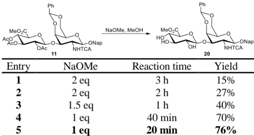
Glycosylation of 7 with 2-naphthalenemethanol by catalytic Lewis acid boron trifluoride etherate gave the β-glycoside exclusively with good yield. 2,3,4-O-acetate deprotection was carried out under Zemplén transesterification condition with sodium methoxide. We tried several base loadings and reaction times after we found that the trichloroactamide could also be deprotected by sodium methoxide. The best result was obtained at 20 min duration and 0.33 equivalence of base per acetate (table 2.2). Preferential 2,3-isopropylidene formation of the triol 19 could be achieved by 2-methoxypropene and camphorsulfonic acid (CSA). Treatment of 13 with levulinic acid, dicyclohexylcarbodiimide (DCC), and 4-dimethylaminopyridine (DMAP) afforded 14 with excellent yield. The isopropylidene acetal was then removed by mild acid hydrolysis. Since removal of benzylidene acetal was observed, we quenched the reaction prematurely to avoid tetraol
formation and recovered starting material. Therefore, the apparent yield 55% can be increased to an overall yield of 81% with two extra hydrolysis of recovered 14.
Scheme 2.3. Synthesis of the disaccharide donor 5. Me = methyl, Lev = levulinyl, Bz = benzoyl, MCA = chloroacetyl, TCA = trichloroacetyl, Ph = phenyl, Ac = acetyl, Nap = 2- naphylmethyl.
Regioselective benzoylation of the 3-hydroxyl group was achieved by benzoyl cyanide without forming 2-O-benzoate. Chloroacetic anhydride was then used to mask the 2-O-sulfation site. After a rigorous hydrolysis, the benzylidene acetal was removed to give diol 17. Selective chloroacetylation of the primary alcohol was achieved by slow addition of chloroacetic anhydride at low temperature. The last hydroxyl group was then protected by benzoyl chloride, affording the fully protected disaccharide 19. Consecutive DDQ and
trichloroacetonitrile/DBU treatment then gave the disaccharide donor 5 activated at the C1 position. The CS-D disaccharide 3 and disaccharide acceptor 6 were then synthesized from the donor 5.
Entry NaOMe Reaction time Yield
1 2 eq 3 h 15%
2 2 eq 2 h 27%
3 1.5 eq 1 h 40%
4 1 eq 40 min 70%
5 1 eq 20 min 76%
Table 2.2. Optimization of O-acetate deprotection of 12. Me = methyl, Ac = acetyl, TCA = trichloroacetyl, Ph = phenyl, Nap = 2-naphthylmethyl.
Synthesis of the CS-D disaccharide
The disaccharide donor 5 was first treated with allyl alcohol and trimethylsilyl trifluoromethanesulfonate (TMSOTf) to give the allyl glycoside 21.
Interestingly, the 6-O-chloroacetate was deprotected upon the use of excess allyl alcohol, but the 2-O-chloroacetate remained intact. Both allyl glycoside 21 and 22 can be transformed into the diol 23 by thiourea.
Scheme 2.4. Synthesis of diol 21. Me = methyl, TCA = trichloroacetyl, MCA = chloroacetyl, Lev = levulinyl, Bz = benzoyl, All = allyl.
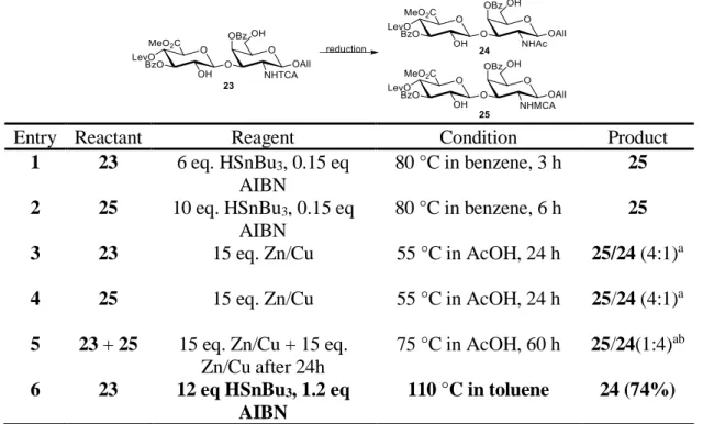
Initial attempts to reduce the trichloroacetyl groups were unsuccessful due to the formation of partially reduced byproduct 25. Both tributyltin hydride/
azobisisobutyronitrile (AIBN) and Zn/Cu conditions were explored (table 2.3). The best result was obtained with a high equivalence of tributyltin hydride and AIBN at elevated temperature in toluene.
Entry Reactant Reagent Condition Product
1 23 6 eq. HSnBu3, 0.15 eq AIBN
80 °C in benzene, 3 h 25 2 25 10 eq. HSnBu3, 0.15 eq
AIBN
80 °C in benzene, 6 h 25 3 23 15 eq. Zn/Cu 55 °C in AcOH, 24 h 25/24 (4:1)a 4 25 15 eq. Zn/Cu 55 °C in AcOH, 24 h 25/24 (4:1)a 5 23 + 25 15 eq. Zn/Cu + 15 eq.
Zn/Cu after 24h
75 °C in AcOH, 60 h 25/24(1:4)ab 6 23 12 eq HSnBu3, 1.2 eq
AIBN
110 °C in toluene 24 (74%)
Table 2.3. Optimization of trichloroacetamide reduction. Me = methyl, Ac = acetyl, TCA = trichloroacetyl, Lev = levulinyl, Bz = benzoyl, All = allyl. aProduct ratio is determined by LC-MS. b Unidentified side product was found.
Complete sulfation was then achieved by a large excess of sulfur trioxide trimethylamine complex in 48 h. A two-step saponification process was then carried out with sequential addition of LiOH/H2O2 and NaOH to avoid potential β-elimination at the GlcA21, affording the desired CS-D disaccharide 3.
Scheme 2.5. Synthesis of the CS-D disaccharide 3. Me = methyl, Ac = acetyl, TCA = trichloroacetyl, Lev = levulinyl, Bz = benzoyl, All = allyl
Structure of the CS-D disaccharide 3 was confirmed by NMR (see appendix for chapter 2). The doublet for the anomeric proton of GalNAc and GlcA has a J value of 7.7 and 8.0 Hz, respectively, demonstrating the β-glycosidic bond. The C-2 proton of GlcA and the C-6 protons of GalNAc are at 4.07 ppm and 4.16 – 4.21 ppm, respectively, indicating the sulfation sites22.
Synthesis of the CS-D tetrasaccharide
The allyl glycoside 21 was first transformed to the disaccharide acceptor 6 by treatment of hydrazine hydrate, which selectively removed the levulinyl group. With the donor and acceptor in hand, we first attempted the coupling reaction with TMSOTf at room temperature. However, the donor was transformed into the oxazoline and was unable to react with the acceptor. We then tried the reaction with different Lewis acids, catalyst loading, equivalence of acceptor, temperature, and reaction time (table 2.4), but the desired tetrasaccharide was still not observed. Trimethylsilyl ether 28 was observed with TMSOTf treatment in elevated temperature (entry 3). High Bu2BOTf loading with long reaction time transformed the oxazoline to the hydrolyzed and decomposed product as determined by LC- MS (entry 10).
Scheme 2.6. Synthesis of the disaccharide acceptor 6. Me = methyl, TCA = trichloroacetyl, MCA = chloroacetyl, Lev = levulinyl, Bz = benzoyl, All = allyl.
Entry Donor 5 Catalyst Condition Product 1 1.5 eq TMSOTf (0.2 eq) DCM, RT, 1 h 26 + 6 2 1.2 eq TMSOTf (0.2 eq) DCM, RT, 3 h 26 + 6 3 1.2 eq TMSOTf (0.2 eq) DCE, 40 °C, 3 h 26 + 6 + 7 4 1.2 eq TBSOTf (0.2 eq) DCM, RT, 3 h 26 + 6 5 1.2 eq TBSOTf (0.2 eq) DCE, 40 °C, 3 h 26 + 6 6 1.2 eq BF3·Et2O (1.0 eq) DCE, 40 °C, 3 h 26 + 6 7 1.2 eq Bu2BOTf (0.1 eq) DCM, -45 °C
to -20 °C, 3h
26 + 6 + 5 8 1.2 eq Bu2BOTf (0.1 eq) DCM, RT, 3 h 26 + 6 9 1.2 eq Bu2BOTf (0.3 eq) DCE, 40 °C, 3 h 26 + 6 10 1.5 eq Bu2BOTf (0.3 eq +
0.6 eq after 12 h)
DCE, 40 °C, 16 h 6 + byproductsa 11 1.2 eq TMSOTf (0.2 eq) DCM, RT, 2 h, in
glove box
25 (40%) + 6 12 1.2 eq TMSOTf (0.2 eq) DCM, RT, 2 h, in
Schlenk flask
25 (58%) +6
Table 2.4. Optimization of the coupling reaction. Me = methyl, MCA = chloroacetyl, TCA
= trichloroacetyl, Lev = levulinyl, Bz = benzoyl, All = allyl, TMS = trimethylsilyl. a- Oxazoline derived byproducts as determined by the disappearance of oxazoline spot on TLC or peak in HPLC.
Since the initial attempts were all carried out with the presence of 4Å molecular sieves (entry 1-10), we next tried the reaction without molecular sieves in a glove box to avoid moisture. To our delight, the desired tetrasaccharide was observed and isolated (entry 11). Further attempts to carry the reaction with a Schlenk flask in a common hood were also successful (entry 12). While molecular sieves are commonly used in glycosidation reactions to prevent hydrolysis of the acetimidates resulted from trace amount of water, their slightly basic nature of may be the cause of oxazoline formation. Fortunately, by achieving strictly
dry conditions with Schlenk flasks in the absence of molecular sieves, we were able to prevent both hydrolysis and oxazoline formation.
After obtaining the tetrasaccharide 26, thiourea treatment then selectively removed the chloroacetyl groups, exposing the sulfation sites. Radical-mediated reduction of the N- trichloroacetyl group with tributyltin hydride and AIBN provided 29. Complete sulfation was achieved by a large excess of sulfur trioxide trimethylamine complex in 48 h in one step without partially sulfated product as determined by HPLC. Sequential addition of LiOH/H2O2 and NaOH resulted in complete ester hydrolysis and gave the target CS-D tetrasaccharide 1.
Scheme 2.7. Synthesis of the CS-D tetrasaccharide 1. Me = methyl, MCA = chloroacetyl, TCA = trichloroacetyl, Lev = levulinyl, Bz = benzoyl, All = allyl.
Structure of the CS-D tetrasaccharide 1 was also confirmed by NMR (see appendix for chapter 2). The doublet for the anomeric protons of the GalNAc and GlcA units have J values of 8.4, 8.0, 7.3, and 7.6 Hz, all indicating the β-glycosidic bond of each linkage. The C-2 protons of GlcA are at 4.06 and 4.12 ppm, confirming the 2-O-sulfation. The C-6 protons
of GalNAc are at 4.16-4.19 ppm and 4.20 – 4.22 ppm, also fully consistent with the chemical shift of 6-O-sulfated C-6 protons22.
Synthetic design of the CS-E tetrasaccharide
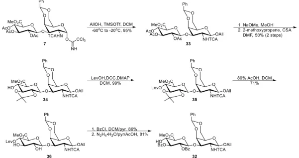
The Schmidt glycosidation and the neighboring participating group trichloroacetamide was adopted for the synthesis of CS-E tetrasaccharide as well. Since we do not attempt to synthesize oligosaccharides longer than tetrasaccharides in this study, we envisioned that the key disaccharide intermediate can be directly used as the disaccharide donor, which saved a great amount of material. The sulfation sites, the C4 and C6 position of GalNAc were already protected with a benzylidene acetal that can be selectively removed.
The rest of the hydroxyl groups can stay protected with acetyl groups and do not require further differentiation. If longer oligosaccharides are desired, the donor used in the coupling reaction can still be derived from an intermediate in the synthesis of disaccharide acceptor 32 without the need to develop an independent route.
Scheme 2.8. Retrosynthesis of the CS-E tetrasaccharide. All = allyl, Me = methyl, Lev = levulinyl, Bz = benzoyl, TCA = trichloroacetyl, Ph = phenyl, Ac = acetyl.
The acceptor was designed accordingly. The C4 position of GlcA was protected with a levulinyl group to facilitate the (1→4) linkage. C2 and C3 position of GlcA were protected with benzoyl groups. The C4 and C6 position of GalNAc was masked with a benzylidene acetal to allow a 1-step deprotection to expose all the sulfation sites of the CS-E tetrasaccharide. This protecting group strategy also allows the synthesis of the CS-A and CS- C tetrasaccharide, as the primary alcohol can be either selectively sulfated or protected after hydroxyl groups at the C4 and C6 position are exposed.
Synthesis of the CS-E tetrasaccharide
Glycosylation with allyl alcohol by catalytic TMSOTf gave the β-glycoside 33 exclusively with excellent yield. Deprotection of the acetate group and preferential 2,3- isopropylidene formation gave alcohol 34 smoothly. Protection of the C4 position of 34 with levulinyl group was achieved with excellent yield. The 2,3-isopropylidene was then removed with mild acid hydrolysis. Similar to the synthesis of CS-D, benzylidene deprotection was observed during the reaction. Benzoylation of the free 2,3-hydroxyl groups and subsequent deprotection of the levulinyl group then afforded the disaccharide acceptor 32.
Optimization of the coupling reaction (table 2.5) successfully allowed the tetrasaccharide 37 to be obtained with moderate yield. Next, hydrolysis under mild acidic condition removed the benzylidene groups, exposing the sulfation sites. Treatment of HSnBu3 and AIBN in toluene/N,N-dimethylacetamide then reduced the trichloroacetamide group. Exhaustive sulfation was achieved by a large excess of sulfur trioxide trimethylamine complex in 48 h to give the sulfated tetrasaccharide 39. The CS-A and CS-C tetrasaccharides can be easily derived from tetrasaccharide 39 with 3 and 2 steps, respectively, as shown in
scheme 2.11. Saponification was then carried out with LiOH/H2O2 treatment followed by NaOH/MeOH to yield the CS-E tetrasaccharide 2. It should be noted that the sulfated tetrasaccharide 39 is very acid sensitive. Near-complete decomposition was observed when the compound was left in an acidic condition (pH ≈ 3.5) for 12 h.
Scheme 2.9. Synthesis of the CS-E disaccharide acceptor 32. All = allyl, Me = methyl, Lev
= levulinyl, Bz = benzoyl, MCA = chloroacetyl, TCA = trichloroacetyl, Ph = phenyl, Ac = acetyl.
Entry Donor 7 TMSOTf Yield
1 1.3 eq 0.2 eq 20%
2 1.6 eq 0.2 eq 28%
3 2.0 eq 0.2 eq 29%
4 2.0 eq 0.4 eq 35%
5 2.0 eq 0.6 eq 50%
Table 2.5. Optimization of the coupling reaction. Me = methyl, Ac = acetyl, TCA = trichloroacetyl, Ph = phenyl, TCA = trichloroacetyl, Bz = benzoyl.
Structure of the CS-E tetrasaccharide 2 was confirmed by NMR (see appendix for Chapter 2). The doublet for the anomeric protons of the GalNAc and GlcA units have J values of 8.5, 7.9, 7.3, and 7.8 Hz, all indicating the β-glycosidic bond of each linkage. The characteristic C4 protons of GalNAc units are at 4.83 and 4.77 ppm, confirming the 4-O- sulfation22. The C6 protons of GalNAc are at 4.16-4.21 ppm and 4.24 – 4.29 ppm, also fully consistent with the chemical shift of 6-O-sulfated C-6 protons22.
Scheme 2.10. Synthesis of the CS-E tetrasaccharide 1. Me = methyl, MCA = chloroacetyl, TCA = trichloroacetyl, Lev = levulinyl, Bz = benzoyl, All = allyl.
Scheme 2.11. Derivatization of 39 towards the CS-A and CS-C tetrasaccharides.
Conclusion
In summary, we developed synthetic routes for CS-D and CS-E tetrasaccharides equipped with an allyl group functional handle in a step-efficient manner. By taking advantage of the disaccharide precursor generated by hydrolysis, the number of steps required to obtain the tetrasaccharides are reduced to 26 steps and 19 steps, which are roughly one-third less than the 38 steps and 27 steps required in traditional synthesis that utilized monosaccharide precursors. While the synthesis of the CS-E tetrasaccharide is shortened, our protecting group strategy still allows rapid access to the CS-A and CS-C tetrasaccharide like our previous synthesis does10. The chemically synthesized homogenous compounds allowed us to investigate the 3-D structure and neuritogenic activity of CS with the D and E sulfation pattern.
Experimental Methods
General Methods
Unless stated otherwise, reactions were performed in flame dried glassware under an argon atmosphere using dry solvents (distilled or passed over a column of activated alumina).
Molecular sieves were flame dried prior to use. All other commercially obtained reagents were used as received, unless otherwise noted. Thin-layer chromatography (TLC) was performed using E. Merck silica gel 60 F254 precoated plates (0.25 mm). Visualization of the developed chromatogram was performed by UV, cerium ammonium molybdate and ninhydrin stain, as necessary. ICN silica gel (particle size 0.032 – 0.063 mm) was used for flash chromatography. 1H, 13C NMR experiments were recorded on Varian Inova 500 (at 500 MHz), or Bruker AVANCE AV400 (at 400 MHz) and are reported relative to residual solvent peaks. Data for 1H are reported as follows: chemical shift (δ ppm), multiplicity (s =
singlet, d = doublet, t = triplet, m = multiplet), coupling constant in Hz, and integration. 13C NMR spectra were obtained on a Varian Inova 500 (at 125 MHz) or Bruker AVANCE AV400 (at 100 MHz) and are reported in terms of chemical shift relative to residual solvent peaks. Chemical shift of 13C spectra were measured in aqueous solvent were determined using the absolute reference method. When necessary, proton and carbon assignments were assigned by means of 1H-1H COSY, and 1H-13C HSQC. High-resolution mass spectrometry was performed at the Mass Spectrometry Facility at the California Institute of Technology on either a JEOL JMS-600H High Resolution Mass Spectrometer or a UPLC-LCT Premier XT TOF Mass Spectrometer.
O-(Methyl β-D-glucopyranosyluronate)-(1→3)-2-amino-2-deoxy-D-galactopyranose hydrochloride (8).
A solution of chondroitin sulfate (50 g) in water (500 ml) was acidified with Amberlite® IR120 Hydrogen form resin to pH 1.6. The resin was filtered off and washed with water (4 x 100 ml). The volume of the filtrate was adjusted to 970 ml. Concentrated H2SO4 (18 M, 27.8 ml) was added, and the mixture was stirred for 6 h at 100 °C, After the solution was cooled down, Ba(OH)2 was added portionwise under vigorous stirring to pH 3.5. The slurry was allowed to sit overnight. The solids were then filtered off through a celite pad, washed with water, and the yellow filtrate was concentrated to approximately 500 ml and slowly applied to a column of Amberlite IR-120 [H+] resin (500 ml, settled volume). The column was washed with water (1 L), AcOH/water (3:1, 1 L), then with aqueous HCl (1m, 3 L). The fractions containing ninhydrin-positive material were collected, concentrated, evaporated with water (2 x 500 ml), and dried under vacuum. The residue was treated with
methanolic HCl (0.02 M, 500 ml) for 4 d at 0 °C. Co-evaporation of the solution with absolute EtOH several times gave the crude product as greyish powder (36.0 g).
O-(Methyl β-D-glucopyranosyluronate)-(1→3)-2-deoxy-2-trichloroacetamido-D- galactopyranose (9).
Trichloroacetyl chloride (85 ml, 0.76 mol) was slowly added to a solution of crude 8 (27 g, 0.65 mol) in pyridine (300 ml) at 0 °C and the solution was stirred at 0 °C for 1 h.
Water (35 ml) was then slowly added to the solution by an addition funnel. The solution was then diluted with CH2Cl2 (600 ml) and washed quickly with cold water and brine. A solution of the residue in MeOH/CH2Cl2/pyridine (1:1:1, 200 ml) was stirred for 4 h at RT and was then concentrated. Flash silica chromatography (MeOH/CH2Cl2 1:4) gave the product (18.3 g, 50% from the polysaccharide) as a brown foam. 1H NMR (300 MHz, D2O, internal H2O, δH=4.79): δ = 5.30 (d, J=3.8 Hz; GalNAc H-1α), 4.72 (d, J =6.3Hz; GalNAc H-1β), 4.40- 4.00 (m, 5H), 3.90-3.80 (m, 2H), 3.85 (s, 3H; COOCH3), 3.75- 3.35 (m, 6H).
O-(Methyl 2,3,4-tri-O-acetyl-β-D-glucopyranosyluronate)-(1→3)-1-Oacetyl-4,6-O- benzylidene-2-deoxy-2-trichloroacetamido-D-galactopyranose (10).
A mixture of 9 (14.9 g, 28.9 mmol), trifluoroacetic acid (5.6 ml) and benzaldehyde (110 ml) was stirred for 24 h at RT. Anhydrous NaOAc (9.2g, 112 mmol), pyridine (110 ml) and acetic anhydride (70 ml) was then added sequentially and the mixture was stirred for 16 h. The mixture was poured into crushed ice and stirred for 2h. The mixture was then extracted with CH2Cl2 (3 x 200 ml) and the organic layer was washed with water, saturated aqueous NaHCO3, and water. The organic phase was dried over MgSO4 and concentrated. Flash silica chromatography (acetone/CH2Cl2 1:15 to 1:12) gave the product as a white powder (9.0g,
40%). 1H NMR (500 MHz, CDCl3): δ = 7.50-7.30 (m, 5H), 6.80 (d, J=7.8 Hz, 1H; GalNAc NH), 6.47 (d, J1,2=3.4 Hz,1H; GalNAc H-1), 5.53 (s, 1H; CHPh), 5.30-5.19 (m, 3H) 5.08 (m, 1H), 4.94 (d, 1H), 4.64 (m, 1H), 4.47 (m, 2H), , 4.07 (m, 3H; 2GalNAc H-6, GlcA H-5), 3.84 (s, 1H), 3.73 (s, 3H; COOCH3), 2.17 (s, 3H), 2.02 (s, 3H), 2.01 (s, 6H).
O-(Methyl 2,3,4-tri-O-acetyl-β-D-glucopyranosyluronate)-(1→3)-4,6-Obenzylidene-2- deoxy-2-trichloroacetamido-1-O-trichloroacetimidoyl-α-D-galactopyranose (7).
A mixture of 10 (11.5 g, 14.9 mmol) and hydrazine acetate (2.4 g, 25 mmol) in DMF (100 ml) was stirred for 30 min at RT. The mixture was then diluted with EtOAc (300 ml) and washed with water, brine, and water. The organic phase was dried over MgSO4 and concentrated. A mixture of the residue, CCl3CN (13 ml, 130 mmol) and DBU (350 µl, 2.3 mmol) in CH2Cl2 (100 ml) was stirred for 1 h. Then more DBU was added and stirred until TLC showed completion. The solvent was removed. Flash silica chromatography (EtOAc/CH2Cl2 1:10) and recrystallization from diethyl ether gave the product (7.4g, 57%) as a white powder. 1H NMR (500 MHz, CDCl3): δ = 8.76 (s, 1H; NH), 7.55–7.30 (m, 5H;
Ar-H), 6.78 (d, J=8.2 Hz, 1H; GalNAc NH), 6.64 (d, J=3.4 Hz, 1H; GalNAc H-1), 5.57 (s, 1H; CHPh), 5.22 (m, 2H), 5.09 (t, J=8.0 Hz, 1H), 4.95 (d, J=7.7 Hz, 1H), 4.77 (m, 1H), 4.55 (d, J=3.4 Hz, 1H), 4.39 (dd, J=11.0 Hz, 3.5 Hz, 1H), 4.33 (d, 12.7 Hz, 1H), 4.12–4.05 (m, 2H), 3.92 (s, 1H), 3.72 (s, 3H; COOCH3), 2.02 (s, 3H; COCH3), 2.01 (s, 3H; COCH3), 2.00 (s, 3H; COCH3).
2-Naphthylmethyl O-(methyl 2,3,4-tri-O-acetyl-β-D-glucopyranosyluronate)-(1→3)- 4,6-O-benzylidene-2-deoxy-2-trichloroacetamido-β-D-galactopyranoside (12).
A mixture of the trichloroacetimidate 7 (20.1 g, 23.1 mmol), 2-naphthalene methanol (5.7 g, 36.1 mmol) and 4 Å molecular sieves (2.50 g) in CH2Cl2 (180 ml) was stirred under argon at RT for 1 h. The mixture was then cooled down to -60 °C before borontrifluoride etherate (300 µl, 2.43 mmol) in CH2Cl2 (2.3 ml) was added. The mixture was slowly warmed to -20 °C in 3 h and then quenched with triethylamine, filtered, and concentrated. Flash silica chromatography (EtOAc/CH2Cl2 1:10) and gave the product (16.7 g, 83%) as a white solid.
1H NMR (500 MHz, CDCl3): δ = 7.80–7.30 (m, 12H; Ar-H), 7.05 (d, J=6.9 Hz, 1H; GalNAc NH), 5.62 (s, 1H; CHPh), 5.17 (m, 3H, 5.04(dd, J=8.8, 8.2 Hz, 1H), 4.95 (ABq, 2H; CH2Ar), 4.90 (d, 1H, J=7.6 Hz), 4.67 (dd, J=11.2, 3.5 Hz, 1H), 4.46 (d, 1H; J=3.4 Hz), 4.41 (dd, 1H;
J=12.5, 1.6 Hz), 4.03 (d, J=1.8 Hz, 1H), 4.00 (d, J=9.9 Hz, 1H) 3.90 (m, 1H;), 3.71(s, 3H;
COOCH3), 3.55 (s, 1H), 2.01 (s, 3H; COCH3), 2.00 (s, 3H; COCH3), 1.99 (s, 3H; COCH3).
2-Naphthylmethyl O-(methyl 2,3-O-isopropylidene-β-D-glucopyranosyluronate)- (1→3)-4,6-O-benzylidene-2-deoxy-2-trichloroacetamido-β-D-galactopyranoside (13).
0.5 M Methanolic sodium methoxide (2.5 ml,1.25 mmol) was added to a solution of 12 (1.1g, 1.25 mmol) in dry THF/methanol (1:4, 12.5 ml) and the solution was stirred for 30 mins. The mixture was then neutralized with Amberlite IR-120 [H+] resin, filtered, and concentrated to give the crude triol 20. 2-Methoxypropene (150 µl, 1.58 mmol) were added every 20 min to a solution of the crude triol 20 and CSA (88 mg, 0.38 mmol) in DMF (10 ml). Triethylamine (0.5 ml) was added and the mixture was concentrated. The residue was dissolved in EtOAc (30 ml) and washed with saturated aqueous NaHCO3, brine, and water, dried with MgSO4, and concentrated. Flash silica chromatography (EtOAc/Hexane4:6 to 6:4, containing 0.5% triethylamine) gave the product as a white solid (587 mg, 60%). 1H NMR (500 MHz, CDCl3): δ = 7.85-7.30 (m, 12H; Ar-H), 7.21 (d, J=6.6 Hz, 1H; GalNAc
NH), 5.60 (s, 1H; CHPh), 5.33 (d, J=8.2 Hz, 1H), 4.93 (ABq, 2H; CH2Ar), 4.79 (d, J=11.7 Hz, 1H), 4.74 (dd, J=11.3, 3.6 Hz, 1H), 4.47 (d, J=3.4 Hz, 1H), 4.41 (dd, J=12.4, 1.6 Hz, 1H), 4.13 (dd, J=12.4, 1.7 Hz, 1H), 4.07 (m, 1H), 3.90 (m, 1H), 3.83-3.80 (m, 4H), 3.57 (s, 1H), 3.58-3.39 (m, 2H), 3.21 (d, J=2.3 Hz, 1H), 1.41 (s, 3H), 1.38 ppm (s, 3H)
2-Naphthylmethyl O-(methyl 2,3-O-isopropylidene-4-O-levulinoyl-β-D- glucopyranosyluronate)-(1→3)-4,6-O-benzylidene-2-deoxy-2-trichloroacetamido- β-D-galactopyranoside (14).
DCC (1.74 g, 8.3 mmol) was added portionwise to a mixture of 13 (5.22 g, 6.7 mmol), levulinic acid (661 µl, 8.3 mmol) and DMAP (230 mg, 1.8 mmol) in dry CH2Cl2 (83 ml) and stirred for 5 h. The solids were filtered off and washed with CH2Cl2/hexane (1:1) several times. The filtrate was washed with saturated aqueous NaHCO3 and water, dried with MgSO-
4, and concentrated. Flash silica chromatography (EtOAc/CH2Cl2 1:10, containing 0.5%
triethylamine) gave the product (5.80 g, 99%) as a white solid. 1H NMR (500 MHz, CDCl3):
δ = 7.82–7.30 (m, 12H; Ar-H), 7.17 (d, J=6.6 Hz, 1H; NH), 5.62 (s, 1H; CHPh), 5.27 (m, 2H), 4.95 (ABq, 2H; CH2Ar), 4.88 (d, J=7.3 Hz, 1H), 4.71 (dd, J=11.2 Hz, J=3.3 Hz, 1H), 4.42 (d, J=3.5 Hz, 1H), 4.40 (dd, J=1.7, 12.5 Hz, 1H), 4.13 (dd, J=1.7, 12.5 Hz, 1H), 3.92 (d, J=8.3 Hz, 1H), 3.90 (m, 1H), 3.69 (s, 3H; COOCH3), 3.62–3.52 (m, 3H), 2.85–2.55 (m, 4H), 2.18 (s, 3H; COCH3), 1.39 (s, 3H), 1.35 (s, 3H).
2-Naphthylmethyl O-(methyl 4-O-levulinoyl-β-D-glucopyranosyluronate)-(1→3)-4,6- O-benzylidene-2-deoxy-2-trichloroacetamido-β-D-galactopyranoside (15).
A solution of 14 (6.90 g, 7.8 mmol) in CH2Cl2/AcOH/water (5:4:1, 140 ml) was stirred for 20 h at RT. The solution was then concentrated and azeotroped with toluene three