Chapter 1
Introduction to Charge Transfer and Charge Transport Through the DNA Double Helix‡
‡Adapted from: Treadway, C. R.; Hill, M. G.; Barton, J. K.
Chem. Phys. 2002, 281, 409–428.
1.1 Introduction
Electron transfer (ET) is often referred to as the simplest of all chemical reactions, yet this elementary process is responsible for important life-sustaining processes such as respiration and photosynthesis. Decades of research on proteins and other σ-bonded networks have provided an understanding for ET in σ-bonded networks, framing the reaction in terms of a small number of parameters, such as thermodynamic driving force, reorganization energies, and distance.1–3 Indeed, owing to the steep drop in the rate of electron transfer on distance, σ bond-mediated ET is generally limited to distances shorter than 15–20 Å. In contrast, work during the past decade has established that charge transport through extended molecular π stacks can occur over much longer distances. One challenge therefore becomes how to develop a conceptual framework through which these new long-range charge transfer events can be understood.
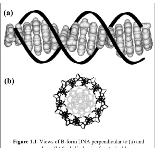
Double stranded deoxyribonucleic acid (DNA) represents a well-characterized system containing an extended π stack within its interior (Figure 1.1). A single DNA strand is composed of a polyanionic sugar-phosphate backbone linking together
combinations of the DNA bases adenine (A), guanine (G), cytosine (C), and thymine (T).
The DNA double helix is formed when two single strands combine to form an extended array of A-T and G-C base pairs. The helix is held together by hydrogen bonding between the complementary bases and stabilized by their stacking interactions.
The structurally well-defined DNA π stack may represent a unique medium for electron transfer. Numerous examples of solid-state π-stacked arrays have been
characterized, and these materials tend to exhibit semiconductor or conductor behavior, especially in the presence of dopants. Double helical DNA is perhaps the best
structurally characterized molecular π-stacked array. Hence, since the double helical structure was first elucidated, speculations that DNA, with its highly ordered stack of electronically coupled aromatic heterocycles, might be an ideal medium for charge transport have appeared.4–6
Molecular stacked assemblies share some characteristics with solid stacked materials, but there are also critical differences. Notably, molecular assemblies undergo greater dynamical motion. As will be described in this thesis, we have found that charge transport through the DNA helix depends sensitively upon these motions. Indeed, in characterizing charge transport through a molecular rather than a solid-state assembly, one tends to make different measurements and ask different questions. Specifically, one focuses not on molecular conductivity, but instead upon electron and/or electron hole transport or transfer properties. What is the efficiency, rate, and distance dependence of charge transfer between donors and acceptors within or bound to the DNA polymer? Is the system best represented by a single step transfer or a multistep transport process?
Over what molecular distance is transport feasible?
This introductory chapter details basic electron transfer theory and describes some of the previous and present efforts aimed at understanding the migration of charge
Figure 1.1 Views of B-form DNA perpendicular to (a) and down (b) the helical axis of π-stacked bases.
(a)
(b)
through the DNA double helix. This thesis focuses on experiments in which electron transfer probes are intimately intercalated or stacked within the DNA double helix. The stacking of these reactants and the intervening base pairs, both statically and dynamically, is the key parameter that ties together this body of work and the phenomenon of DNA- mediated ET as a whole.
1.2 Basic Electron Transfer Theory
Owing to the simplicity of electron transfer reactions, a theoretical formalism has been developed that describes electron transfer reactions in terms of a small number of experimentally accessible parameters.
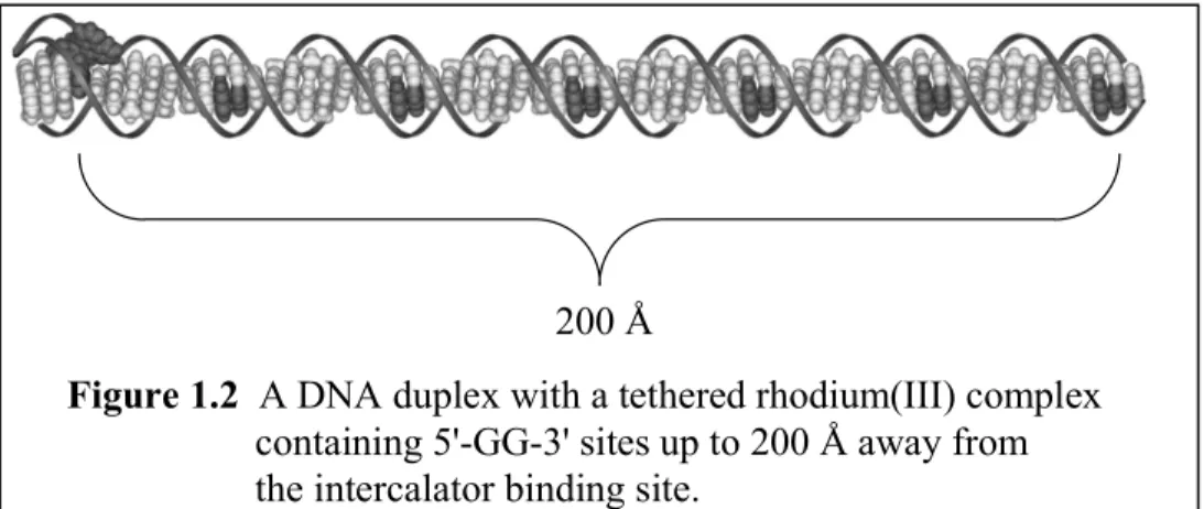
Long-range electron transfer through proteins and other σ-bonded frameworks is generally believed to proceed via superexchange interactions between the electron donor (D) and acceptor (A). Because the strength of this interaction decreases with the number of atoms separating the redox sites, the rate of electron transfer drops rapidly with the D- A distance. Charge transport through DNA, however, has been observed over distances as great as 200 Å (Figure 1.2), implying that the decay with distance is exceptionally shallow.7 Understanding the unique characteristics of DNA that allow such long-range events remains a major challenge of theory and experiment alike: as outlined below, recent studies suggest that energetics, inhomogeneities within the DNA π stack, and base dynamics all play important roles in distinguishing DNA-mediated charge transport.
200 Å
Figure 1.2 A DNA duplex with a tethered rhodium(III) complex containing 5'-GG-3' sites up to 200 Å away from the intercalator binding site.
According to semi-classical Marcus theory, the rate of electron transfer is given by the product of the frequency of nuclear motion along the transition state (ν), the probability of crossing from reactants to products at the transition state (κΕ), and the nuclear reorganization factor (κN).
kET = ν κΕ κN (1)
This third term involves the activation free energy for the reaction, and is defined by the thermodynamic free energy (∆Go) and the reorganizational parameter λ, which describes the extent of nuclear motion required to reach the transition state.
κN = exp(-(∆Go + λ)2/4λkbT) (2)
When the interaction between the donor and acceptor is weak, the transition state must be formed many times before the system crosses over from reactants to products (the non- adiabatic limit). The rate of the reaction is therefore limited by the rate of surface crossing at the transition state. Under these conditions, the activationless rate constant for the electron transfer can be expressed according to (3), where the electronic coupling matrix element (HAB) describes the interaction between the reactants (D + A) and
products (D+ + A-) at the transition-state configuration.
koET = 4π3
h2λkbTHAB2 (3)
The precise functional dependence of the electronic coupling element with distance therefore ultimately governs the limits of long-range electron transfer through a particular medium.
Several approaches have been developed to evaluate the factors that determine the magnitude of HAB. An early treatment by McConnell employs perturbation theory to describe the electronic coupling between a donor and acceptor separated by a bridge (b) comprised of identical repeat units.
HAB= hDbhbA
∆ε hb
∆ε
n−1
(4)
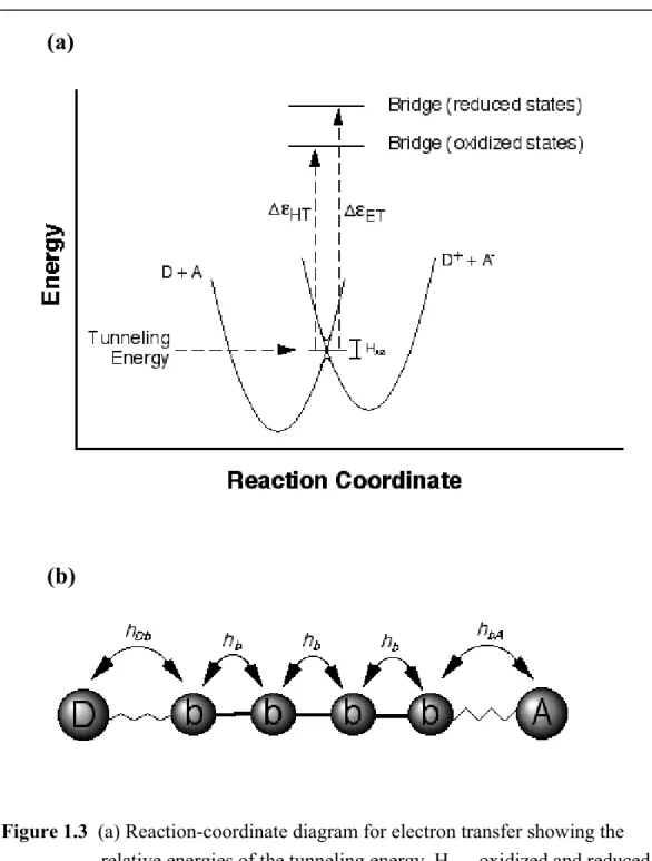
Here, the individual matrix elements describe the nearest-neighbor coupling between the donor and bridge (hDb), the acceptor and bridge (hbA) and adjacent bridge states (hb). The energy of the transition state is the tunneling energy, and the quantity ∆ε is equal to the difference between that energy and the one-electron reduced (for electron transfer) or one-electron oxidized (for hole transfer) states of the bridge (Figure 1.3).
As long as ∆ε is large, this simple model predicts that the rate of electron transfer will decay exponentially with distance, with a decay constant, β, equal to -2aln(hb/∆ε), where α is the length of the bridge unit. Indeed, exponential decay in the rates of σ- bond-mediated electron transfer reactions have now been observed through many proteins and straight-chain carbons, with β values typically ranging from ~0.85–1.2 Å-1.
Although this simple model assumes a uniform bridge, it nevertheless suggests that very small tunneling barriers (∆ε) may provide a mechanism for increased electronic coupling and longer-range electron transfer. For example, Siebbeles and associates have published calculations on DNA-mediated CT that indicate a smooth transition occurs between superexchange and “wire-like” conduction as the injection barrier drops and the transferring electron (or hole) becomes completely delocalized onto the bridge.8 Thus, as long as the redox potential of the excited state is above the electronic energy of the bridge, the reaction is expected to be quite rapid.
Figure 1.3 (a) Reaction-coordinate diagram for electron transfer showing the relative energies of the tunneling energy, HAB, oxidized and reduced bridge states, and tunneling energy barriers (∆ε). (b) Individual couplings relevant to bridge-mediated electron transfer.
(a)
(b)
Two additional factors must be taken into account when evaluating charge transport reactions within the double helix: (a) the non-homogeneity of the bridge repeat units (e.g., the inherent differences of the ionization potentials AT vs. GC base steps as a function of sequence context), and (b) dynamical processes within DNA that can affect both adjacent bridge couplings, as well as D- and/or A-bridge interactions. Again these are factors that distinguish DNA from π-stacked structures in the solid state.
The first factor has been invoked with the proposal that long-range charge transport in DNA involves hole hopping between localized sequences within the double helix. The overall charge transfer rate, then, will be proportional to the number of hops the charge takes along its journey. Most simply, kct ∼ n-η, where n is the number of hops and η is a constant. So how does this relate to specific sequences with differing redox potentials in DNA? The purine bases guanine and adenine are the most easily oxidized in DNA (G: E(+/0) = 1.3 V vs. NHE; A: E(+/0) = 1.4 V vs. NHE); the
pyrimidine bases cytosine and thymine are the most difficult to oxidize (C: E(+/0) = 1.7 V vs. NHE; T: E(+/0) = 1.8 V vs. NHE).9 Conversely, C and T are much easier to reduce (E(0/-) = -1.1 V vs. NHE)10 than are A and G (E ≤ -1.5 vs. NHE).11 Thus in the hopping mechanism, holes essentially jump from guanine to guanine along an extended duplex.12 Given the relatively large differences in reduction potentials between GC and AT base steps, these jumps can themselves be considered individual tunneling steps whose rates depend on the number of intervening AT base pairs.
Recently, it has become apparent, however, that this model needs modification.13 Assemblies of different lengths, sequence, and conformation may allow tunneling, hopping, or some mixture of the two mechanisms to actually dominate. Certainly, dynamical processes within DNA affect adjacent bridge couplings, base energetics, and therefore mechanism. Specifically, it was found that tunneling through A tracts is not required; hopping onto a bridge containing runs of A’s was demonstrated.14 Occupation of the bridge and its relative energetics therefore also depend upon the local DNA
conformation, i.e., the sequence-encoded domain. The redox potentials of individual bases within the DNA stack and the base-base couplings surely vary depending upon the local sequence dependent structure of DNA.
Considering energetics within the framework of a bridge-assisted hopping model also leads to other questions. It has been demonstrated that both ground state and excited state oxidants can induce oxidative damage at remote guanine sites along the DNA double helix in mixed-sequence DNA. Since a single step hop from the oxidant to the distant 5’ guanine is extremely unlikely, the injected hole must make several hops down the helix. In these hops within the bridge it will encounter various DNA bases, all with different redox potentials. While thermally induced hopping of the hole from one base or domain close in energy to another is quite possible, what happens when that energy gap is large (> 0.3 eV)? Does the originally injected hole equilibrate to the redox potential of the nearest, most easily oxidized base, or is the charge free to jump from strand to strand in its travel down the helix? This question is even more puzzling when trying to
understand the mechanism of the DNA-mediated oxidative repair of thymine dimers ( E(+/0) = 1.8 V ) by photoexcited intercalators up to 40 Å away.
Theory has been extremely helpful8,12,15–19 in explaining seemingly disparate observations from different laboratories, but many questions remain. Since this molecular π-stacked assembly adopts a variety of local conformations and undergoes a range of dynamical motions depending upon sequence context, the molecular π-stacked array clearly differs from π stacks in the solid state. These motions and conformations affect the coupling, energetics and even number of steps available in the transport process. In fact, if we could predict quantitatively the energetics and base-base couplings in DNA as a function of sequence, we might be able to utilize charge transport studies to probe the local conformations and motions of DNA.
1.3 Approaches to Study Long-Range Charge Transport Through DNA The introduction of a variety of techniques and of chemical assemblies was critical to the characterization of DNA charge transport. The synthesis of different DNA assemblies provided a powerful tool to explore a range of distances, timescales, and energetics. A variety of photophysical, biochemical, and electrochemical assays was also needed to examine charge migration through the DNA double helix in these different regimes. Below some of the different approaches used and the critical experiments carried out are described in more detail. What is remarkable in reviewing these results is the similarity in conclusion obtained, irrespective of the timescale, probe, or experiment.
1.3.1 Probes and assemblies
Since the preliminary photochemical experiments on Ru(phen)32+ excited-state quenching by Co(II) and Rh(III) complexes in the presence of DNA carried out more than a decade ago,20,21 a whole range of donors and acceptors bound to DNA have been employed to characterize charge transport through the intervening base pair stack. For example, the use of metallointercalators bound to DNA, as π-stacked donors and acceptors, provided a way of interrogating the π stack directly and highlighted the importance of stacking effects in long-range charge transport. Thus excited-state oxidative quenching of dipyridophenazine (dppz) complexes of ruthenium(II) by 9,10- phenanthrenequinone diimine (phi) complexes of rhodium(III) intercalated into DNA occurs on an extremely fast timescale (kET>3x10¹⁰ s-1).22 Subsequent transient-
absorption studies revealed long-lived osmium(III)23 and ruthenium(III)24 species after quenching of the corresponding *Os(II) or *Ru(II) excited states, establishing electron transfer as the mechanism of the quenching reaction.
To allow precise control of the location of the donors and acceptors along the double helix, synthetic techniques were developed to append the metallointercalators covalently to the DNA duplex, and these were proven to be critically important. The first experiment involving tethered metallointercalators was reported in 1993.25 In that study
a ruthenium(II) intercalator was tethered to one end of a single DNA strand, and a rhodium(III) intercalator was tethered to the complementary single strand. When the strands were annealed, ruthenium(II) steady-state luminescence was completely
quenched by the rhodium(III) intercalator positioned over 40 Å down the DNA π stack.
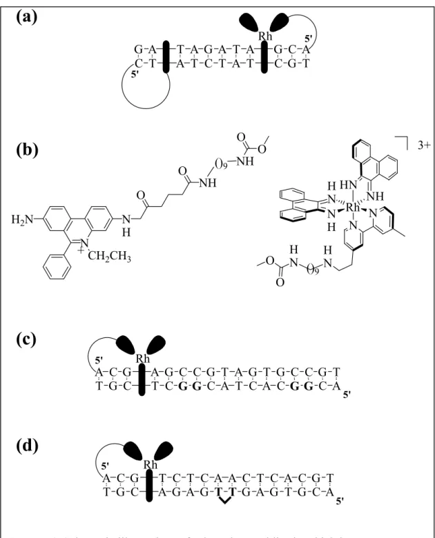
A similar system, this time with the organic intercalator ethidium as the photoexcited donor and rhodium(III) as the acceptor (Figures 1.4a and 1.4b), showed luminescence quenching over distances ranging from 20 to 30 Å.26 The importance of stacking was illustrated in an experiment reported by Meade and Kayyem in 1995.27 Non-intercalating ruthenium complexes were tethered to the ends of a DNA duplex but, because the ET reactants were not well coupled to the DNA π stack, efficient charge transfer was not observed. Experiments using these kinds of structurally well-characterized chemical assemblies became essential in characterizing DNA charge transport. Indeed, one of the real strengths in applying DNA assemblies was the ability to manipulate and vary synthetically accessible DNA oligomers of known structure.
Another important step in characterizing DNA-mediated charge transport was the application of the DNA bases (or modified bases) themselves as reactants, the donors and/or acceptors. Again the assemblies utilized a tethered rhodium intercalator as a photooxidant, but now it was used to oxidatively damage guanine doublet sites in DNA (Figure 1.4c).28 In these studies, the yield of oxidative damage was found to vary little with the distance separating the intercalator and guanine site of oxidation. Furthermore, these studies established for the first time “chemistry at a distance” on DNA and became useful in considering the biological consequences of DNA charge transport within the cell.29,30 DNA charge transport chemistry was also crucial in achieving another chemical reaction at a distance, the repair of the thymine dimer lesion in DNA (Figure 1.4d). Here too the reaction was mediated by the DNA helix, depended upon stacking of the
intercalating oxidant and bases, and the efficiency of repair showed little dependence on the distance separating the intercalator and thymine dimer.
G A T A G A T A G C A
C T A T C T A T C G T
Rh
T G C C G A G C A
A C G G C T C G
T A G T G C C G
T C A C G G C T A Rh
A A C T C T G C A
T T G A G A C G
T C T C A C G T
G A G T G C A Rh
+
HN NH
Rh NH HN
N N
HN HN O
O
3+
N H2N
CH2CH3 NH
O
O NH
NH
O O
()9
5'
5' 5'
5' 5'
5'
()9
(a)
(b)
(c)
(d)
Figure 1.4 Schematic illustrations of selected assemblies in which long-range DNA- mediated charge transport was demonstrated. (a) The fluorescence of a tethered ethidium intercalator is quenched by a tethered rhodium(III) intercalator located over 20 Å down the DNA helix. (b) Structures of the intercalators in Figure 1.4a. (c) A photoexcited rhodium intercalator is capable of oxidizing guanines at a distance down the DNA helix. (d) A tethered rhodium intercalator is also capable of oxidatively repairing at a distance a thymine dimer lesion in duplex DNA.
These studies above served as a foundation for a full range of experiments in many laboratories using a variety of photooxidants aimed at exploring the mechanism of DNA-mediated charge transport.7,13,14,31–43 Schuster and coworkers have studied guanine oxidation at a distance through the double helix in assemblies where various
anthraquinone photooxidants were attached to the end of the duplex.35,36 Saito et al. have looked at guanine oxidation triggered by derivatives of excited state benzophenone.41–43 Subsequently, photophysical studies were also carried out that involved direct reaction with the DNA bases, and these allowed measurements of reaction rates as well as yields to be performed. Luminescence quenching by the modified base 7-deazaguanine of excited-state ethidium tethered to a DNA duplex was characterized in our laboratory.44 Lewis and coworkers have extensively examined photoinduced charge separation between guanine and photoexcited stilbene in a series of synthetic DNA hairpins.45 In these structures, the stilbene caps the end of the double helix but remains stacked so that relatively efficient ET behavior is observed. Finally, direct base-base electron transfer, both intrastrand and interstrand, was probed by examining the quenching of the
fluorescent base 2-aminopurine by both guanine and 7-deazaguanine using ultrafast spectroscopies.46 The hallmark of these experiments demonstrating efficient DNA- mediated ET is the coupling of the reactants to the DNA base stack.
The latest tool to examine and also to exploit DNA charge transport has been electrochemistry experiments on DNA films.47–49 Here, too, the fabrication of DNA- modified electrodes and the application of a redox-active intercalator as a probe were critical elements in design. Electrochemistry has been used extensively to measure heterogeneous electron transfer dynamics at solid electrode surfaces. In a typical configuration, redox-active head groups are attached to thiol-terminated alkyl chains of variable length, and are subsequently self-assembled into well-ordered monolayers on a gold surface. Electrons (or holes) are then pushed through the linker to the head group, and the rates of charge transport are measured by evaluating the resulting electrochemical
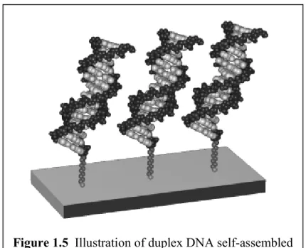
current. We developed analogous methods for preparing DNA-modified surfaces (Figure 1.5), in which the redox-active “head groups” are DNA intercalators (e.g., methylene blue or daunomycin) located at precisely defined positions along the base sequence.50 Extensive structural studies confirmed that the duplexes pack with high density in a
morphology in which the individual helices are oriented in an upright position with respect to the gold surface.50,51 These well-defined assemblies allowed the systematic evaluation of both distance and sequence on the dynamics of charge transport. Because tunneling through the aliphatic linker limits the rate of electron transfer; however, these studies report on events that occur on a much longer timescale than the photophysical experiments described above.
1.3.2 Biochemical measurements of oxidative damage
Several biochemical techniques have been developed to address long-range, DNA-mediated charge transport, the most common one being a probe for oxidative damage in DNA. Such damage is observed primarily at guanine (G), as predicted by theoretical and experimental studies which have determined that G is the most easily
Figure 1.5 Illustration of duplex DNA self-assembled monolayers on a gold surface.
oxidized base. The specific residues of damage, usually the 5’ G in a 5’-GG-3’ or 5’- GGG-3’ sequence, are correlated with the oxidation potential of G in different sequence contexts.52,53 This sequence context is important, as selective 5’ G damage is a hallmark of DNA-mediated charge transfer; non-specific guanine damage is often a sign of reaction with singlet oxygen.
Typical experiments involve tethering an intercalator at one end of the DNA duplex and placing guanine sites at various positions distant from the intercalative reactant. Photoexcitation (or in situ generation of a highly oxidizing ground state) of the intercalator creates a reactive species with enough driving force to oxidize guanine.
Oxidized guanine nucleotides are revealed as strand breaks upon subsequent piperidine treatment of the DNA; therefore, the various sites and yields of guanine damage may be visualized by polyacrylamide gel electrophoresis (PAGE)54 followed by autoradiographic imaging.
What are the advantages of this method? The most obvious is the ability to observe the final chemical results initiated by charge transfer: true chemistry at a
distance. Others include being able to analyze charge transport over great distances (tens of nanometers) and not having the analysis limited to a fast timescale. The major
drawbacks are not being able to observe directly charge-transfer intermediates or charge transfer events as they occur in real time.
1.3.3 Photophysical studies of DNA-mediated charge transfer
One of the most useful tools employed to study DNA charge transport has been spectroscopy, including time-resolved and steady-state absorption and emission. In our experiments, a hole donor is excited by near visible or far UV light. This creates an excited state which has enough energy to oxidize a hole acceptor located somewhere else along the DNA double helix.
Examination of excited-state lifetimes allows us to determine the timescale for charge transfer events. Faster ET will deplete the donor excited-state population more
16 rapidly than the same population in the absence of the acceptor, showing up in time- resolved emission spectra as a faster decay. Efficient steady-state emission quenching shows up as a lower quantum yield for the same reason. Time-resolved transient absorption gives us a chance to identify spectrally the charge transfer intermediates and search for back electron transfer events. However, in taking these approaches we are limited by the timescales on which our instruments are able to take data. We must always be cognizant of the fact that the data we are observing are in no way a complete picture of all the dynamics taking place in a system. Another drawback is the effort required to obtain ultrafast spectroscopy data; while gathering data on the milli- to
nanosecond timescale is fairly trivial, obtaining pico- to femtosecond data is not nearly as easy a chore as the other techniques described in this section.
1.3.4 Electrochemistry using DNA films
The construction and application of self-assembled monolayers of double- stranded DNA oligomers with redox-active probe molecules on gold electrode surfaces has enabled the systematic evaluation of charge transport as a function of distance, sequence, and base-stacking perturbations using electrochemistry. In these experiments DNA-modified gold electrodes are prepared containing well-packed DNA duplexes and a bound intercalator as redox probe.47–49 Reduction of the intercalator is monitored using cyclic voltammetry, chronocoulometry, and other common electrochemical methods.
This reaction involves no photoexcitation and instead measures the efficiency of
intercalator reduction through DNA-mediated charge transport. Like the photochemical and photophysical systems described above, these studies also point to the double helix as an exceptional medium for rapid, long-range transport events. Notably, the charge- transport dynamics in these studies appear also to be independent of both distance and DNA sequence, but extremely sensitive to even subtle defects in the π stack.55,56 Yet unlike the photochemical systems that involve ultrafast reactions between high-energy intermediates, the electrochemical processes occur on a much longer timescale
(milliseconds) and involve reactants in their ground electronic states. As a result, they provide complementary information concerning charge-transport phenomena through DNA, both in new kinetic and energetic regimes.
1.4 Parameters Explored in Characterizing DNA Charge Transport Chemistry Given the experimental approaches and assemblies described above, what parameters are important to consider in those frameworks when evaluating experiments aimed at understanding charge transport through DNA? Previous studies of electron and hole transfer through DNA have suggested the material is anything from a conducting wire to an insulating medium. The dichotomy of such results may be trying to tell us which variables lead to efficient charge transfer and which variables inhibit the flow of charge. It is therefore imperative to examine experimental results that may provide common clues to the factors governing the efficacy of long-range, DNA-mediated charge transport.
1.4.1 Perturbations to the π stack in studies of photoinduced charge transport One way to disrupt the flow of charge to distal sites down the helix is to introduce disorder into the π stack. In the solid state, such perturbations represent defects and these disrupt the flow of current. In the DNA duplex, there are many ways to introduce such defects and these defects sensitively affect charge transport (Figure 1.6).
A single-base mismatch in DNA, where bases opposing one another in the duplex do not represent Watson-Crick A-T or G-C pairs, provides such a perturbation in the base stack. The sensitivity of DNA charge transport to intervening mismatches was realized first in photophysical experiments. In an 11 base pair duplex containing ethidium and rhodium intercalators tethered to opposite ends, ethidium luminescence is quenched 21%
by the tethered rhodium.26 Introduction of a single, intervening C-A mismatch
diminishes the quenching to a negligible value. Thermal denaturation of the modified duplex achieves similar results. If a G-A mismatch, which is known to be well stacked
18 within a B-form DNA double helix, is introduced, the luminescence quenching (26%) is enhanced relative to the duplex containing normal Watson-Crick base pairs. These observations established two important points: (1) the path of the charge transfer is through the DNA double helix; (2) stacking of the bases is critical if DNA is to serve as a medium conducive to charge transport.
Rh
Rh
Rh Rh
Rh
Ru
(a) (b)
(d) (c)
e-
e- e-
X
e-
e-
X
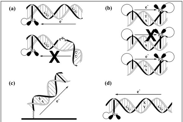
Figure 1.6 Schematics of various assemblies used to illustrate the importance of stacking perturbations in DNA-mediated charge transfer. (a) A 3-base bulge disrupts the π stack and decreases the yield of guanine damage at a distal site. (b) A C-A mismatch inhibits ethidium fluorescence quenching by a rhodium(III) intercalator; whereas, a well-stacked G-A mismatch facilitates efficient quenching. (c) Mismatches (X, Y = A, G, C, or T) prevent reduction of the intercalator methylene blue in DNA duplexes tethered to a gold electrode. (d) Poorly stacked mismatches (X, Y = A, G, C, or T) inhibit oxidative charge transport from a ruthenium intercalator to a distal guanine site.
Oxidative guanine damage at a distance down the DNA helix has also been studied as a function of different intervening, single-base DNA mismatches.57 A full range of distal/proximal damage ratios was observed depending upon the identity of the single base pair mismatch intervening between two guanine doublet sites positioned along a 22-mer duplex with a tethered ruthenium intercalator as the oxidant. The efficacy of charge transport through a given DNA mismatch was found to correlate simply with how well the bases in the mismatch are stacked within the helix.
Another way to disrupt the flow of charge involves placing an ATA bulge into the helix. NMR spectroscopy of a DNA duplex containing an ATA bulge shows substantial, but not complete, local disruption of the π stack, with the remainder of the ordered stack intact.58 In a normally stacked DNA duplex containing a rhodium intercalator tethered at one end and 5’-GG-3’ sites proximal (closest to) and distal (farthest from) the
intercalator, photooxidatively triggered DNA damage is spread evenly among the two sites. Introduction of an ATA bulge between the two guanine doublets leads to a 75%
diminution in oxidation at the distal site relative to the proximal site.59 Analogously, a decrease in the repair efficiency of a thymine dimer lesion by a tethered rhodium intercalator is also observed if an ATA bulge is introduced between the two reactants along the DNA double helix.60
Experiments have also been performed to examine the ability of DNA-bound proteins to facilitate or inhibit charge transport (as measured by guanine damage at remote sites from a tethered rhodium(III) intercalator) through the DNA double helix.61 Here too, protein binding serves to introduce a defect site-specifically into the helix, because the binding of some proteins to DNA causes a local perturbation in the base pair stack. The methyltransferase Hha I (M. Hha I) enzyme binds the sequence 5'-GCGC-3' and affects methylation at each of the internal cytosines.62,63 To gain access to each target cytosine for methylation, the enzyme extrudes the cytosine from the DNA helix. A key feature of the base-flipped complex is the insertion of a glutamine residue within the
20
Rh Rh Rh
(a)
(b)
(c)
e-
e-
X
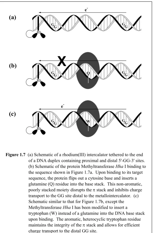
Figure 1.7 (a) Schematic of a rhodium(III) intercalator tethered to the end of a DNA duplex containing proximal and distal 5'-GG-3' sites.
(b) Schematic of the protein Methyltransferase Hha I binding to the sequence shown in Figure 1.7a. Upon binding to its target sequence, the protein flips out a cytosine base and inserts a glutamine (Q) residue into the base stack. This non-aromatic, poorly stacked moiety disrupts the π stack and inhibits charge transport to the GG site distal to the metallointercalator. (c) Schematic similar to that for Figure 1.7b, except the
Methyltransferase Hha I has been modified to insert a
tryptophan (W) instead of a glutamine into the DNA base stack upon binding. The aromatic, heterocyclic tryptophan residue maintains the integrity of the π stack and allows for efficient charge transport to the distal GG site.
DNA cavity created with extrusion of the cytosine.64,65 The discontinuity of the DNA base stack at the site of base flipping with the glutamine insertion then serves to inhibit long-range charge transport (Figure 1.7). Importantly, however, using a M. Hha I mutant which inserts an aromatic tryptophan residue instead of a glutamine restores long-range damage at distal 5’-GG-3’ sites. An intact, well-coupled π stack is therefore the key to restoring long-range charge transport.
In marrying biochemical and photochemical results from the same system, transient absorption spectroscopy of ruthenium-modified assemblies containing the bound M. Hha I tryptophan mutant established directly the formation of transient radical species having both tryptophan and guanine radical character.66 These studies exemplify how DNA-binding proteins might play a role both as inhibitor and as activator of DNA- mediated charge transport and underscore the importance of an intact aromatic π stack in facilitating long-range DNA charge transport.
1.4.2 The effects of stacking and dynamics on DNA charge transport
Our first hints of the sensitivity of DNA charge transport chemistry to stacking came simply from comparing reactions of left- and right-handed metallointercalator enantiomers with DNA. The ∆ enantiomers intercalate more deeply and tightly into the right-handed DNA helix, and not surprisingly, then, ∆-[Ru(phen)2dppz]2+ luminescence is quenched more efficiently by ∆-[Rh(phi)2bpy]3+ than by Λ−[Rh(phi)2bpy]3+.22,67 Clearly, to a first approximation the driving force, reorganizational energies, etc., for the two enantiomers should be the same. Nonetheless differences are observed, and these depend upon stacking or coupling of the different donors and acceptors into the helix. Since those first experiments, it became clear that stacking by our intercalators generally yielded more efficient DNA charge transport. Conversely, donors and acceptors that were poorly coupled into the base pair stack showed, at best, inefficient charge transport.
Our most detailed examination of the importance of stacking dynamics was obtained in studies of the oxidation of deazaguanine incorporated into a DNA duplex by
tethered, photoexcited ethidium (Figure 1.8); ethidium, one should note, is the classic organic DNA intercalator used in flourescence experiments over the past forty years.
Ultrafast charge transport between tethered ethidium and the modified base 7-
deazaguanine was observed with time constants of 5 ps and 75 ps, and these rates were found to be essentially independent of the donor-acceptor separation over the range of 10–17 Å.68 Notably, however, the yield of quenching decreased with increased separation between ethidium and deazaguanine. These two components of the decay corresponded to the charge transport rate within the helix, and these components were not evident with duplexes containing guanine (where oxidation is not favored) rather than deazaguanine. Both with deazaguanine and guanine were observed the 1.5 ps solvation
N H2N
CH2CH3 NH2
N N
NH NH2 O
N N
NH NH2 O
Guanine (G) 7-Deazaguanine (Z)
Ethidium (Et+)
{Et+*, Z}
{Et+, Z}
{Et, Z+} ket
krec
hν kd
(a)
(b) (c)
e-
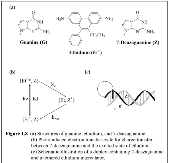
Figure 1.8 (a) Structures of guanine, ethidium, and 7-deazaguanine.
(b) Photoinduced electron transfer cycle for charge transfer between 7-deazaguanine and the excited state of ethidium.
(c) Schematic illustration of a duplex containing 7-deazaguanine and a tethered ethidium intercalator.
dynamics and the typical biexponential radiative decay of excited-state ethidium within the DNA duplex. The 5-ps component was therefore assigned as the inherent rate of charge injection into the DNA bridge that occurs without inhibition from structural dynamics. The 75-ps component was assigned to the reorientation of the ethidium intercalator within the duplex before charge transfer may proceed, presumably on a faster timescale. The assignment of the 75-ps component was also supported by fluorescence polarization measurements that identified this component as the motional timescale for the ethidium within the DNA intercalation site. Hence charge transport within the DNA duplex appeared to be gated by the dynamical motion of ethidium within the DNA. This conformational gating is not by any means a concept unique to DNA charge transport;
indeed, conformational gating of electron transfer processes has been observed in other molecular assemblies.69 These results are described in more detail in Chapter 2.
The lack of a distance dependence in the charge transfer rates in the ethidium / 7- deazaguanine system raised the question of what is responsible for the distance
dependence of the ethidium fluorescence quenching yield. Here too, the observations can be understood in the context of stacking dynamics. The dynamical nature of DNA, with base pair motions on the picosecond to millisecond timescales gives rise to a distribution of conformations, only a fraction of which are able to facilitate charge transfer.
Throughout the lifetime of the ethidium excited state, then, the motions of DNA are many. Charge transfer, with the rate constants given above, occurs if a favorable
conformation for donor-acceptor coupling is achieved during the excited state lifetime of ethidium. Introducing more bases between donor and acceptor gives a higher probability of stacking defects (on the timescale of the lifetime of excited ethidium) which leads to fewer, on average, distinct charge transfer events taking place. Thus the fluorescence quenching yield falls off with increasing donor-acceptor distance.
It is interesting that in these assemblies, as well, the introduction of a C-A
mismatch between donor and acceptor lowered the fluorescence quenching yield. In fact
we can really consider the mismatches simply as more dramatic examples of dynamical variations in stacking. Indeed the closest correlation for data for long-range oxidative damage in DNA as a function of intervening mismatches was found with NMR studies of mismatched base pairs motions.57
Dynamical effects within DNA clearly depend upon sequence. Often these effects are subtle and not as easy to discern. 5’-TATA-3’ regions are known to be flexible.70 The greater dynamical motions of such a region should inhibit charge transport. This prediction turns out to be true, as that specific intervening sequence
decreases the yield of oxidative damage caused by a photoexcited rhodium(III)
intercalator at a remote 5’-GG-3’ site.7,14 Earlier studies showed a similar diminution in charge transport and this was attributed to energetic factors. However, energetics can be ruled out as a contributing factor to the diminished yields because an intervening 5’- TATATATA-3’ sequence gives rise to increased oxidative damage yields relative to the 5’-TATA-3’ sequence. Clearly, the 5’-TATA-3’ sequence represents a unique domain, whether it be structural or dynamic, that must be considered when designing sequences to evaluate DNA-mediated charge transport.
The sensitivity of reactions to stacking, both static and dynamic, was also evident quite clearly in our photophysical studies of base-base charge transfer. Since all four DNA bases absorb in the same region and are very weak emitters, modified DNA bases were chosen to serve as donors, acceptors, or both (Figure 1.9). A few fluorescent bases (2-aminopurine (Ap) and 1-N6-ethenoadenine (εA)) are readily available, and both are capable of oxidizing guanine residues when excited with UV light (λ = 325 nm).51 The reactivities of Ap and εA are quite similar in solution. However, once incorporated into duplex DNA, there are striking differences. Fast charge transfer (k = 1010–1011 s-1) initiated by photoexcited Ap occurs over a 3.4–13.6 Å.71 Instead εA exhibits slower (by a factor of 100) charge transfer with a steep distance dependence. High-resolution
nuclear magnetic resonance (NMR) studies of duplexes containing εA72 (Figure 1.9c) and Ap73 (Figure 1.9d) provided insight into the clear difference between these modified bases: stacking within the DNA helix. εA is sterically bulky, does not pair with T, and adopts a nonrigid, poorly stacked conformation within the base stack. Ap undergoes normal Watson-Crick pairing with T and is stacked within the DNA helix quite similarly to the natural bases. The very different distance dependences for these reactions may indicate that different reaction pathways are accessed. It is quite remarkable nonetheless that these subtle differences in stacking can lead to such large changes in charge transfer rates and efficiencies.
The use of modified bases as donors and acceptors also allowed us to compare interstrand versus intrastrand charge transfer. Reaction of excited-state Ap with guanine on the same strand occurs approximately 100 times faster than reaction with guanine on the opposite strand. Thus, charge transfer proceeds preferentially down one strand in double-helical DNA. Within B-form DNA, stacking is essentially only intrastrand.74 Thus, when reactants are directly coupled through stacking along one strand, fast reaction kinetics result. If H-bonded base pairs must be traversed, the charge transfer kinetics slow considerably. Base-base charge transfer is further examined in Chapter 3.
1.4.3 Electrochemical measurements of stacking perturbations
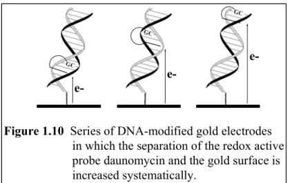
Electrochemical studies have been carried out on a collection of DNA films in which the separation of the redox-active intercalating probe molecule and the gold surface was varied systematically. Figure 1.10 shows a series of such sequences,
featuring daunomycin that has been covalently crosslinked to specific G residues
positioned at different sites in the duplex. Even though the daunomycin-gold separations span more than ~ 45 Å, there is no apparent variation in the heterogeneous rate of
electron transfer. Again, the use of an intercalator, such as daunomycin, is critical;
reduction of other redox probes, electrostatically bound near the top of the DNA film and distant from the gold electrode, is not accomplished.
e- e-
e-
Figure 1.10 Series of DNA-modified gold electrodes in which the separation of the redox active probe daunomycin and the gold surface is increased systematically.
The electrochemical results with daunomycin represent another example of gated electron transfer, where one step in the mechanism determines the overall reaction rate.
A likely candidate for gating in this system is tunneling through the aliphatic linker. The measured rate constant (k ~ 100 s-1) is very reasonable for electron tunneling through the 16-atom bridge that links the DNA to the gold, assuming a very low reorganizational energy for the charge transport event. A key element, however, is the electron acceptor, the well-stacked duanomycin intercalator. Once coupled into the π stack, the charge transfer is extremely fast.
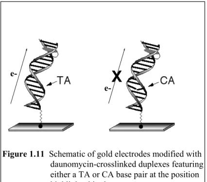
The electrochemical reaction is also exquisitely sensitive to π-stacking effects within the helix. To illustrate this, a CA mismatch was introduced into the monolayer between the probe molecule and the electrode; as illustrated in Figure 1.11, this single- base change turns off electron transfer to the daunomycin completely. It is noteworthy that doping the original films with increasing percentages of mismatched duplexes results in a linear decrease in the daunomycin electrochemical signals; lateral charge migration between adjacent duplexes is evidently quite slow.
X
e-
Figure 1.11 Schematic of gold electrodes modified with daunomycin-crosslinked duplexes featuring either a TA or CA base pair at the position highlighted in the sequence.
e-
CA mismatches are not the only lesions that attenuate electron transfer through these films. All of the possible mismatches, as well as the DNA products of several natural biological reactions (including oxidative damage (e.g., 8-oxo-A), depurination (an abasic site), and hydroxyl radical addition (e.g., 5,6-dihydrothymine)) diminish the efficiency of charge transport. Interestingly, the magnitude of the effect correlates quite closely with measurements of long-range oxidative damage as a function of intervening mismatches. This correlation becomes all the more remarkable when one considers the different timescales of the two experiments and the fact that one involves a
photooxidation and the other, a ground state reduction. A notable exception to the correlation between experiments is the GT wobble base pair. While thermodynamically stable, this mismatch disrupts electrochemically triggered charge transport across the DNA films, yet does not attenuate the efficiency of biochemical reaction. A likely explanation for this dichotomy can be found when considering the timescales of the various charge transport and base pair dynamics. NMR studies suggest that the GT wobble motion is many orders of magnitude slower than photoinduced charge transport.75,76 Therefore during any particular charge transport event, the bases are essentially frozen out in a relatively well-stacked configuration. Because the
electrochemical timescale is rendered orders of magnitude slower as a result of σ bond tunneling through the linker, the electrochemical reduction can now be additionally gated by GT base dynamics.
We have also recently undertaken electrochemical studies of protein binding to DNA.77 In this case as well, results parallel those seen in biochemical studies of
oxidative damage. Proteins that disrupt base pair stacking inhibit charge transport through the DNA films. Those proteins that introduce a π-stacking amino acid within the DNA duplex restore charge transport. This methodology represents a very sensitive electrical probe of protein-DNA interactions and may prove to be a useful new tool in biochemical assays.
Surprisingly, the efficiency of charge transport in these DNA films appears to be remarkably independent of both sequence and base content. Thus, while the yields of long-range charge migration are attenuated by the flexible 5’-TATA-3’ sequences, no measurable differences occur in the electrochemical responses of films whose base
contents vary from 100% AT to 100% GC. Perhaps this difference reflects a lower extent of flexibility generally for the oligomers that are well packed within the DNA films.
1.4.4 Energetics of donors and acceptors
Although the functional roles of distance and sequence on the efficiencies of long- range charge transport through the double helix appear to vary significantly depending on the particular assay employed, it is important to note that the different assays themselves employ reactants that span an unusually large range of redox energetics. For example, the distribution of GG-oxidation products following ultraviolet photoexcitation of intercalated rhodium complexes is markedly different from those found using activated sugar radicals. These latter experiments have been interpreted within the framework of a hopping mechanism, in which discrete G radicals are generated within the duplex and then diffuse down the π stack via a series of discrete single-step tunneling events.
Because these reactions occur near the “average” ionization potential of the DNA bridge, inhomogeneities within the bridge play important roles in determining the actual
outcomes of the reactions. Accordingly, charge migrates freely across DNA sequences rich in guanine, but exhibits a much steeper dependence through AT-rich regions. On the other hand, long-range oxidative repair of thymine dimers across extended regions of AT base steps may indicate that the initial excited state is sufficiently high in energy for direct hole injection into the bridge (Figure 1.12).
At the other energetic extreme, the apparent “wire-like” behavior of
electrochemically probed DNA films is difficult to understand given the very large energy gap between the reduction potential of daunomycin ( E(-/0) = -0.38 vs. NHE ) and the π-stacked bases of DNA. Nor is it obvious how hopping mechanisms that depend on
ionizing radiation to generate discrete radical charge carriers can be applied directly to these systems. Perhaps even more problematic is the apparent lack of sequence dependence observed for charge migration through the electrode-bound duplexes.
Indeed, hopping mechanisms by definition imply that DNA sequences are inhomogeneous with respect to charge transport phenomena.
Heller and associates have suggested that electrochemical conductivity through DNA-modified films can be understood in terms of solid-state semiconductor effects. A well-ordered array of DNA duplexes is particularly easy to polarize electrochemically due to the mobility of counter ions paired to the negatively charged phosphate backbone.
Owing to this property, the films are expected to exhibit an unusually large dielectric constant along the helical axis. Interestingly, a high internal dielectric for DNA was estimated based upon biochemical studies of DNA charge transport where the DNA charge distributions were varied.78 Because the mean free path of an electron in a semiconductor is inversely related to the magnitude of the dielectric constant, the
polarized film is predicted to be highly conductive along the longitudinal axis. The DNA bases (guanine or adenine) or redox-active intercalators may act as charge dopants, given that their ionization energies are also inversely related to the dielectric constant.79
Mismatches and other lesions may therefore disrupt the efficiencies of charge transport by acting as defects within the film.
In fact, determining the true energetics is an issue for all these studies. While redox potentials for some donors and acceptors, and even for the individual bases, can be measured in aqueous solution, how do these values correspond to those within the DNA helix? Moreover, as we consider radical migration through the DNA, is it the radical cation or deprotonated neutral radical that needs to be considered? In others do these represent proton-coupled charge transport processes which are gated also by proton transfer? A challenge for the future remains establishing these relevant values precisely.
D D-
∆ε >> 0 -
D
∆ε ~ 0 D*
+
D
∆ε < 0 D*
+
EnergyEnergyEnergy
(a) Direct Hole Injection
(b) Hole Hopping
(c) Electrochemical ET
Figure 1.12 Schematic illustration of the important energetics and electronic couplings involved in various DNA-mediated charge transfer reactions.
1.5 Current Focus
Biochemical, photophysical, and electrochemical experiments have now established that double helical DNA can mediate efficient charge transport. The
molecular stacked array of base pairs holds some similarities to solid-state materials. But there are also crucial differences. Coupling of donors and acceptors, the connections to the DNA stack, as well as the dynamics, the molecular motions, within the DNA base pair stack are important elements in determining the extent of charge transfer rates and efficiency. These are characteristics intrinsic to a molecular π-stacked array.
In the context of charge transport, the fragility of the DNA double helix is a unique feature, one that varies sensitively with sequence dependent structure and protein binding. Another critical feature is that the structures of DNA assemblies can be easily prepared and encoded by the base pair sequence. Thus, in taking advantage of these attributes, DNA might serve uniquely and powerfully in molecular electronic devices.
Experiments on DNA-modified electrodes already provide examples of how
characteristics of DNA charge transport may be exploited in the design of nanoscale sensors for mutations or protein binding. Indeed, one intriguing question remains
whether Nature has already exploited the unique charge transport characteristics of DNA within the cell.
Double helical DNA, containing a π-stacked array of base pairs within its interior, can be considered as a molecular analogue of solid-state π-stacked arrays. Like the solid- state materials, the DNA base pair stack provides a medium to facilitate charge transport.
However, owing to the dynamical motions of the base pairs within the molecular stack, as well as sequence dependent inhomogeneities in energetics and base-base couplings, DNA charge transport differs considerably from that in solid-state π-stacked materials.
This thesis aims to address several experimental techniques and chemical assemblies used to probe charge transport in DNA. The sensitivity of DNA-mediated charge transport to dynamical variations in base stacking and couplings is emphasized.
Chapter 2 describes the ultrafast spectroscopy examining the photoinduced ET reaction between an intercalator and a modified DNA base. Chapter 3 details similar experiments between modified DNA bases, while Chapter 4 discusses the results obtained from spectroscopy on metallointercalator/modified base DNA conjugates. Chapter 5 focuses on the creation and characterization of metallointercalators with high redox potentials and their interactions with DNA, while Chapter 6 summarizes the possible steric interactions (or lack thereof) of two metallointercalators bound to double helical DNA. Taken as a whole, these studies demonstrate how the structure, stacking, and dynamics of reactants and the intervening π stack affect the efficacy of DNA-mediated charge transport.
1.6 References
1. Marcus, R. A.; Sutin, N. Biochim. Biophys. Acta 1985, 811, 265.
2. Winkler, J. R.; Gray, H. B. Chem. Rev. 1992, 92, 369.
3. Isied, S. S.; Ogawa, M. Y.;Wishart, J. F. Chem. Rev. 1992, 92, 381.
4. Gyorgi, A. S. Proc. Natl. Acad. Sci., U.S.A. 1960, 46, 1444.
5. Eley, D. D.; Spivey, D. I. Trans. Faraday Soc. 1962, 58, 411.
6. Ladik, J J. Int. J. Quant. Chem. 2000, 78, 450.
7. Núñez, M E.; Hall, D. B.; Barton, J. K. Chem. Biol. 1999, 6, 85.
8. Grozema, F. C.; Berlin, Y. A.; Siebbeles, L. D. A. J. Am. Chem. Soc. 2000, 122, 10903.
9. Steenken, S.; Jovanovic, S. V. J. Am. Chem. Soc. 1997, 119, 617.
10. Steenken, S.; Telo, J. P.; Novais, H. M.; Candeias, L. P. J. Am. Chem. Soc. 1992, 114, 4701.
11. Kittler, L.; Löber, J.; Gollmick, F. A.; Berg, H. J. Electroanal. Chem. 1980, 116, 503.
12. Bixon, M.; Giese, B.; Wessely, S.; Langenbacher, T.; Michel-Beyerle, M. E.; Jortner, J. Proc. Natl. Acad. Sci., U.S.A. 1999, 96, 11713.
13. Giese, B.; Amaudrut, J.; Kohler, A. K.; Spormann, M.; Wessely, S. Nature 2001, 412, 318.
14. Williams, T. T.; Odom, D. T.; Barton, J. K. J. Am. Chem. Soc. 2000, 122, 9048.
15. Jortner, J.; Bixon, M.; Langenbacher, T.; Michel-Beyerle, M. E. Proc. Natl. Acad.
Sci., U.S.A. 1998, 95, 12759.
16. Bixon, M.; Jortner, J. J. Phys. Chem. B. 2000, 104, 3906.
17. Voityuk, A. A.; Rosch, N.; Bixon, M.; Jortner, J. J. Phys. Chem. B. 2000, 104, 9470.
18. Berlin, Y. A.; Burin, A. L.; Ratner, M. A. J. Phys. Chem. A. 2000, 104, 443.
19. Berlin, Y. A.; Burin, A. L.; Ratner, M. A. J. Am. Chem. Soc. 2001, 123, 260.
20. Barton, J. K.; Kumar, C. V.; Turro, N. J. J. Am. Chem. Soc. 1986, 108, 6391.
21. Purugganan, M. D.; Kumar, C. V.; Turro, N. J.; Barton, J. K. Science 1988, 241, 1645.
22. Arkin, M. A.; Stemp, E. D. A.; Holmlin, R. E.; Barton, J. K.; Hörmann, A.; Olson E. J.; Barbara, P. F. Science 1996, 273, 475.
23. Holmlin, R. E.; Stemp, E. D. A.; Barton, J. K. J. Am. Chem. Soc. 1996, 118, 5236.
24. Stemp, E. D. A.; Arkin, M. A.; Barton, J. K. J. Am. Chem. Soc. 1995, 117, 2375.
25. Murphy, C. J.; Arkin, M. A.; Jenkins, Y.; Ghatlia, N. D.; Bossmann, S. H.; Turro, N.
J.; Barton, J. K. Science 1993, 262, 1025.
26. Kelley, S. O.; Holmlin, R. E.; Stemp, E. D. A.; Barton, J. K. J. Am. Chem. Soc.
1997, 119, 9861.
27. Meade, T. J.; Kayyem, J. F. Angew. Chem. Int. Ed. Engl. 1995, 34, 352.
28. Hall, D. B.; Holmlin, R. E.; Barton, J. K. Nature 1996, 382, 731.
29. Rajski, S. R.; Jackson, B. A.; Barton, J. K. Mutation Res. 2000, 447, 49.
30. Núñez, M. E.; Holmquist, G. P.; Barton, J. K. Biochemistry 2001, 40, 12465.
31. Stemp, E. D. A.; Arkin, M. A.; Barton, J. K. J. Am. Chem. Soc. 1997, 119, 2921.
32. Arkin, M. A.; Stemp, E. D. A.; Coates Pulver, S.; Barton, J. K. Chem. Biol. 1997, 4, 389.
33. Hall, D. B.; Kelley, S. O.; Barton, J. K. Biochemistry 1998, 37, 15933.
34. Núñez, M. E.; Noyes, K. T.; Gianolio, D. A.; McLaughlin, L. W.; Barton, J. K.
Biochemistry 2000, 39, 6190.
35. Gasper, S. M.; Schuster, G. B. J. Am. Chem. Soc. 1997, 119, 12762.
36. Ly, D.; Sanii, L.; Schuster, G. B. J. Am. Chem. Soc. 1999, 121, 9400.
37. Sanii, L.; Schuster, G. B.; J. Am. Chem. Soc. 2000, 122, 11545.
38. Meggers, E.; Kusch, D.; Spichty, M.; Wille, U.; Giese, B. Angew. Chem. Int. Ed.
Engl. 1998, 37, 460.
39. Meggers, E.; Michel-Beyerle, M. E.; Giese, B. J. Am. Chem. Soc. 1998, 120, 12950.
40. Giese, B.; Wessely, S.; Spormann, M.; Lindemann, U.; Meggers, E.; Michel- Beyerly, M. E. Angew. Chem. Int. Ed. Engl. 1999, 38, 996.
41. Saito, I.; Nakamura, T; Nakatani, K; Yoshioka, Y.; Yamaguchi, K; Sugiyama, H.
J. Am. Chem. Soc. 1998, 120, 12686.
42. Nakatani, K.; Dohno, C.; Saito, I. J. Am. Chem. Soc. 1999, 121, 10854.
43. Nakatani, K.; Dohno, C.; Saito, I. J. Am. Chem. Soc. 2000, 122, 5893.
44. Kelley, S. O.; Barton, J. K. Chem. Biol. 1998, 5, 413.
45. Lewis, F. D.; Wu, T. F.; Zhang, Y. F.; Letsinger, R. L.; Greenfield, S. R.;
Wasielewski, M. R. Science 1997, 277, 673.
46. Kelley, S. O.; Barton, J. K. Science 1999, 283, 375.
47. Kelley, S. O.; Barton, J. K.; Jackson, N. M.; Hill, M. G. Bioconj. Chem. 1997, 8, 31.
48. Kelley, S. O.; Jackson, N. M.; Hill, M. G.; Barton, J. K. Angew Chem. Int. Ed. Engl.
1999, 38, 941.
49. Hartwich, G.; Caruana, D. J.; de Lumley-Woodyear, T.; Wu, Y. B.; Campbell, C. N.
Heller, A. J. Am. Chem. Soc. 1999, 121, 10803.
50. Kelley, S. O.; Barton, J. K.; Jackson, N. M.; McPherson, L. D.; Potter, A. B.; Spain, E. M.; Allen, M. J.; Hill, M. G. Langmuir 1998, 14, 6781.
51. Sam, M.; Boon, E. M.; Barton, J. K.; Hill, M. G.; Spain, E. M. Langmuir 2001, 19, 5727.
52. Saito, I.; Takayama, M.; Sugiyama, H.; Nakatani, K.; Tsuchida, A.; Yamamoto, M.
J. Am. Chem. Soc. 1995, 117, 6406.
53. Sugiyama, H.; Saito, I. J. Am. Chem. Soc. 1996, 118, 7063.
54. Chung, M.-H.; Kiyosawa, H.; Nishimura, S.; Kasai, H. Biochem. Biophys. Res.
Comm. 1992, 188, 1.
55. Kelley, S. O.; Boon, E. M.; Barton, J. K.; Jackson, N. M.; Hill, M. G. Nucl. Acids.
Res. 1999, 27, 4830.
56. Boon, E. M.; Ceres, D. M.; Drummond, T. G.; Hill, M. G.; Barton, J. K. Nature Biotech. 2000, 18, 1096.
57. Bhattacharya, P. K.; Barton, J. K. J. Am. Chem. Soc. 2001, 123, 8649.
58. Rosen, M. A.; Shapiro, L.; Patel, D. J. Biochemistry 1992, 31, 4015.
59. Hall, D. B.; Barton, J. K. J. Am. Chem. Soc. 1997, 119, 5045.
60. Dandliker, P. J.; Holmlin, R. E.; Barton, J. K. Science 1997, 275, 1465.
61. Rajski, S. R.; Kumar, S.; Roberts, R. J.; Barton, J. K. J. Am. Chem. Soc. 1999, 121, 5615.
62. Cheng, X.; Kumar, S.; Posfai, J.; Pflugrath, J. W.; Roberts, R. J. Cell 1993, 74, 299.
63. O’Gara, M.; Klimasauskas, S.; Roberts, R. J.; Cheng, X. Mol. Biol. 1996, 261, 634.
64. Mi, S.; Alonso, D.; Roberts, R. J. Nucl. Acids Res. 1995, 23, 620.
65. Garcia, R. A.; Bustamonte, C. J.; Reich, N. O. Proc. Natl. Acad. Sci., U.S.A. 1996, 93, 7618.
66. Wagenknecht, H.-A.; Rajski, S. R.; Pascaly, M.; Stemp, E. D. A.; Barton, J. K. J.
Am. Chem. Soc. 2001, 123, 4400.
67. Arkin, M. R.; Stemp, E. D. A.; Turro, C.; Turro, N. J.; Barton, J. K. J. Am. Chem.
Soc. 1996, 118, 2267.
68. Wan, C.; Fiebig, T.; Kelley, S. O.; Treadway, C. R.; Barton, J. K.; Zewail, A. H.
Proc. Natl. Acad. Sci., U.S.A. 1999, 96, 6014.
69. Davis, W. B.; Ratner, M. A.; Wasielewski, M. R. J. Am. Chem. Soc. 2001, 123, 7877.
70. Dickerson, R. E. Nucl. Acids Res. 1998, 26, 1906.
71. Wan, C.; Fiebig, T.; Schiemann, O.; Barton, J. K.; Zewail. A. H. Proc. Natl. Acad.
Sci. U.S.A. 2000, 97, 14052.
72. Kouchakdjian, M.; Eisenberg, G. M.; Yarema, K.; Basu, A.; Essigman, J.; Patel, D. J.
Biochemistry 1991, 30, 1820.
73. Nordlund, T. M.; Anderson, S.; Nilsson, L.; Rigler, R.; Graslund, A.; McLaughlin, L. W. Biochemistry 1989, 28, 9095.
74. Saenger, W. Principles of Nucleic Acid Structure; Springer-Verlag: New York, 1984.
75. Brown, T.; Kennard, O.; Kneale, G.; Rabinovich, D. Nature 1985, 315, 604.
76. Allawi, H. T.; SantaLucia, Jr., J. Nucl. Acids. Res. 1998, 26, 4925.
77. Boon, E. M.; Salas, J.; Barton, J. K. Nature Biotech. 2002, 20, 282.
78. Williams, T. T.; Barton, J. K. J. Am. Chem. Soc. 2002, 124, 1840.
79. Kittel, C. Introduction to Solid State Physics, 5th ed.; Wiley: New York, 1976.