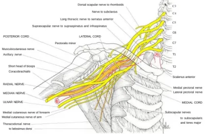
The photographs were completed by Dr McArdle at Gogarburn with assistance from the Department of Medical Illustration at the University of Edinburgh. Modifications have been made to some of the diagrams and a new diagram of the lumbosacral plexus has been included. A new set of color photographs has been prepared for this edition, the brachial and lumbosacral plexus diagrams have been retained, but all other diagrams have been redrawn.
Patricia Archer PhD for the drawings of the brachial plexus and lumbosacral plexus Ralph Hutchings for the photography. The nature and purpose of the tests should be explained to the patient, so that his interest and cooperation is secured. It should be noted that each of the methods used usually tests the action of the muscles in a single joint.
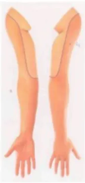
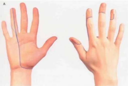
The patient presses the palms hard against a wall with the elbows fully extended. 4 The approximate area within which sensory changes can be found in complete lesions of the brachial plexus (C5, C6, C7, C8, T1). 5 The approximate area within which sensory changes can be found in lesions of the upper roots (C5.C6) of the brachial plexus.
6 The approximate area within which sensory changes can be found in lesions of the lower roots (C8, T1) of the brachial plexus.
MUSCULOCUTANEOUS NERVE
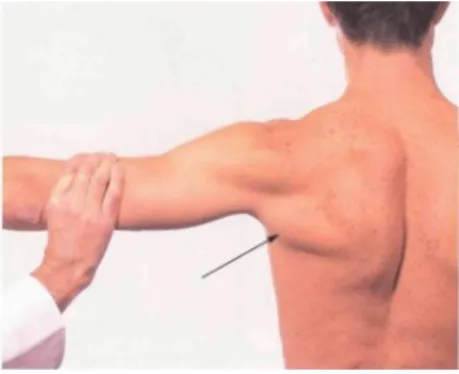
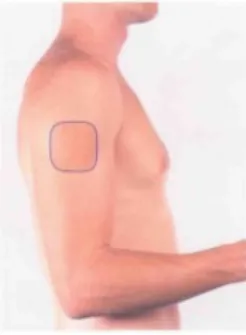
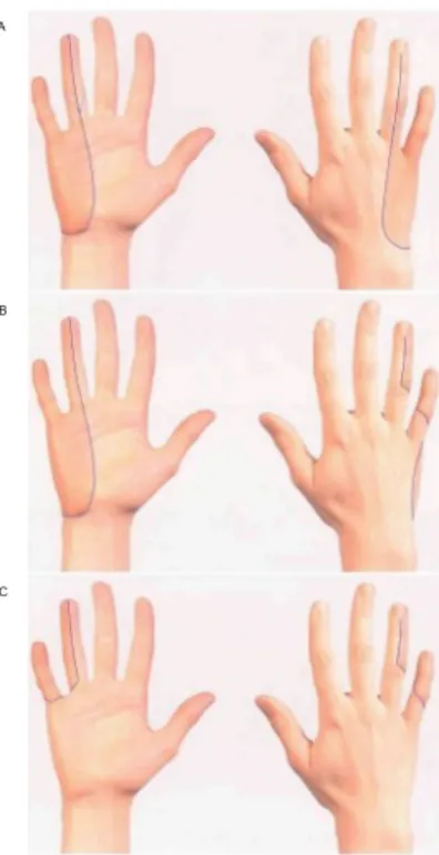
17 The approximate area within which sensory changes can be found in lesions of the musculocutaneous nerve. The distribution of the lateral cutaneous nerve of the forearm.). 19 Diagram of the axillary nerve, its main cutaneous branch and the muscles it supplies. 20 The approximate area within which sensory changes can be found in lesions of the axillary nerve.
AXILLARY NERVE
RADIAL NERVE
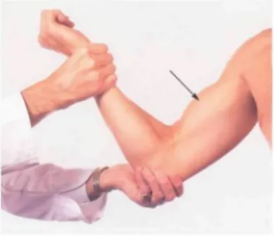
The mean area is usually significantly smaller and no sensory change is observed. 25 The approximate area within which sensory changes can be found in lesions of the radial nerve above the elbow joint and below the origin of the posterior cutaneous nerve of the forearm. The patient flexes the forearm against resistance, halfway between pronation and supination.
The patient supinates the forearm against resistance, with the forearm extended at the elbow. Extension of the metacarpophalangeal joints is maintained against the resistance of the fingers of the examiner's left hand. 32 Abductor Pollicis Longus (Posterior interosseous nerve; C7, C8) The patient abducts the thumb at the level of the carpo-metacarpal joint in a plane perpendicular to the palm.
Arrow: the tendon can be seen and felt anteriorly and just next to the tendon of extensor pollicis brevis (cf. 33 Extensor Pollicis Longus (posterior interosseous nerve; C7, C8). The patient extends the thumb at the interphalangeal joint against resistance.
MEDIAN NERVE
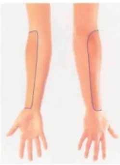
36 The approximate areas within which sensory changes can be found in lesions of the median nerve in: A the forearm, B the carpal tunnel. The patient flexes the finger at the proximal interphalangeal joint against resistance with the proximal phalanx. This test does not eliminate the possibility that flexion at the proximal interphalangeal joint is produced by flexor digitorum profundus.
40 Flexor Digitorum Profundus I and II (Anterior interosseous nerve; C7, C8) The patient flexes the distal phalanx of the index finger against resistance with the middle phalanx. The patient flexes the distal phalanx of the thumb against resistance while the proximal phalanx is fixed. The patient touches the base of the little finger with the thumb against resistance.
44 1st lumbric-interosseous muscle (median and ulnar nerves; C8, T1) The patient extends the finger at the proximal interphalangeal joint against resistance with the metacarpophalangeal joint hyperextended and fixed.
ULNAR NERVE
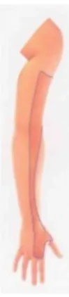
46 The approximate areas within which sensory changes can be found in lesions of the ulnar nerve: A above the origin of the dorsal cutaneous branch, B below the origin of the dorsal cutaneous branch and above the origin of the palmar branch, C below the origin of the palmar branch. 47 The approximate area within which sensory changes can be found in lesions of the medial cutaneous nerve of the forearm. The tendon of fiexor carpi ulnaris can be seen and felt (arrow) as the muscle acts to fixate the pisiform seen even when abductor digiti minimi is paralyzed (see also Fig. 49).
The patient flexes the distal interphalangeal joint against resistance while the middle phalange is fixed. The patient flexes the little finger at the metacarpophalangeal joint against resistance while extending the finger at both interphalangeal joints. 53 First dorsal interosseous muscle (ulnar nerve; C8, T1) The patient abducts the index finger against resistance.
54 Second Palmar Interosseous Muscle (Ulnar nerve; C8, T1) The patient adducts the index finger against resistance. The patient adducts the thumb perpendicular to the palm against the resistance of the examiner's finger.
LUMBOSACRAL PLEXUS
NERVES OF THE LOWER LIMB
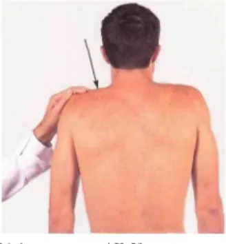
58 Diagram of the nerves on the back of the lower limb, their cutaneous branches and the muscles they supply. INFERIOR GLUTEAL NERVE Gluteus maximus POSTERIOR CUTANEOUS NERVE OF Thigh Biceps, long head Biceps, short head. 59 Approximate area within which sensory changes can be found in lesions of the lateral cutaneous nerve of the thigh.
60 Approximate area within which sensory changes can be found in femoral nerve lesions. Distribution of the intermediate and medial cutaneous nerves of the thigh and the saphenous nerve.). 61 Approximate area within which sensory changes can be found in obturator nerve lesions.
62 The approximate range within which sensory changes can be found in lesions of the posterior cutaneous nerve of the thigh. 63 The approximate range within which sensory changes can be found in lesions of the sciatic nerve trunk. 64 The approximate range within which sensory changes can be found in lesions of both the sciatic and posterior cutaneous nerves of the thigh.
65 The approximate area within which sensory changes can be found in lesions of the common peroneal nerve above the origin of the superficial peroneal nerve. 66 The approximate area within which sensory changes can be found in lesions of the deep peroneal nerve. 67 The approximate area within which sensory changes can be found in lesions of the sural nerve.
68 The approximate area within which sensory changes can be found in lesions of the tibial nerve. Arrows: the tendons of the biceps (lateral) and semitendinosus (medial) can be felt and usually seen. The patient lies on his back with the leg extended and flexes the foot against resistance.
82 Small muscles of the foot (medial and lateral plantar nerves; S1, S2) The patient cups the sole of the foot; the small muscles can be felt and sometimes seen. 85 Extensor Hallucis Longus (Deep peroneal nerve; L5, S1) The patient dorsiflexes the distal phalanx of the big toe against resistance.

DERMATOMES
NERVES AND MAIN ROOT SUPPLY OF MUSCLES
Flexor carpi ulnaris C7, C8, T1 Flexor digitorum profundus III & IV C7, C8 Hypotenarmuskler C8, T1 Adductor pollicis C8, T1 Flexor pollicis brevis C8, T1 Palmar interossei C8, T1 Dorsal interossei C8, T1 Lumbricals, T1 & IV Nedre ekstremitet Spinal Rødder Lårnerve.
COMMONLY TESTED MOVEMENTS