the Bacterial Pathogen Pseudomonas aeruginosa and the Role of Proteases
Thesis by David W. Basta
In Partial Fulfillment of the Requirements for the Degree of
Doctor of Philosophy
CALIFORNIA INSTITUTE OF TECHNOLOGY Pasadena, California
2019
(Defended May 13, 2019)
© 2019 David W. Basta
ORCID: 0000-0003-4176-6566
ACKNOWLEDGEMENTS
I have had the rare privilege of being mentored by brilliant women at every stage of my scientific career. It was in the lab of my undergraduate research advisor, Professor Helen J.
Wing, where I first discovered a passion for science, and where I began to thoughtfully and critically examine all aspects of my professional and personal life. My experiences in the Wing Lab radically changed my career goals and aspirations, and set me on an intellectual journey that I am still navigating to this day. Thank you, Dr. Wing.
In graduate school I was taken under the wing of the postdoc (now professor!) Megan Bergkessel. With her guidance, I developed the technical skills and experimental rigor befitting a good scientist. I credit Megan with destroying my idealism. She taught me that science, especially biology, can be difficult and messy, and that oftentimes the most important discoveries are hidden within the subtleties of an experimental result. Thank you, Megan.
Finally, I owe most of my current and future career success to my graduate research advisor, Dianne Newman. I have recognized so many of the qualities required to be a successful investigator by observing Dianne, and I admire her for vigorously promoting a culture of academic freedom in her lab. Dianne has always empowered me to take complete ownership of my research, and has instilled in me a strong sense of scientific independence—one of the most important qualities I believe a research advisor can impart to her students. Invariably, she enthusiastically encourages me whenever I approach her with a new idea, a new result, a new setback, or a new frustration, and she has pushed me to develop as a scientist and as an individual every step of the way. She has always had my back. She has never dropped the ball on a single responsibility that I entrusted to her, despite constantly having a mountain of reasonable excuses to do so. I cannot ask for a more supportive and responsible advisor.
Thank you, Dianne.
A big thank you to all of the lab members that have come and gone during my time in the Newman Lab: Will DePas, Scott Saunders, Lucas Meirelles, Daniel Dar, Darcy McRose, Brittany Belin, Elena Perry, John Ciemniecki, Kurt Dahlstrom, Melanie Spero, Lev Tsypin,
Chelsey Vandrisse, Elise Tookmanian, Gargi Kulkarni, Lisa Racki, Shannon Park, Flavia Costa, Naomi Kreamer, Nate Glasser, Kyle Costa, Ruth Lee, Elise Cowley, Sebastian Kopf, Nick Shikuma, Chia-Hung Wu, Brett Babin, Peter Jorth, and Jessica Ricci. I am so lucky to have been surrounded by such a fun, welcoming group of colleagues and friends. The Newman Lab is a wonderful place to be a scientist, and I am grateful for the timely support and encouragement I have received from each lab member along the way. I hope that I was as supportive and encouraging to some of you as you all were to me. Special thanks to my baymates Gargi Kulkarni and Elise Tookmanian for providing welcome respite from the inevitable frustrations that accompany scientific research.
Many thanks to my thesis committee members David Chan, Sarkis Mazmanian, and Alexander Varshavsky. Your gracious offerings of time, support, and thought-provoking discussion are truly appreciated.
Thank you to my co-authors and collaborators David Angeles-Albores and Tri Vu for participating in stimulating discussions and fun brain-storming sessions with me.
Thank you to the Proteome Exploration Laboratory members Annie Moradian and Michael Sweredoski. Your patience and kindness are just as appreciated as your technical expertise.
Thank you to the Caltech and USC staff Kristy Nguyen, Liz Ayala, Raina Beaven, and Roland Rapanot. Your support ensured that I could freely focus on my science throughout my Ph.D.
Thank you to the three most important women in my life: my mom Mervat Basta, my partner Kateryna Kozachenko, and my sister Lucy Basta. Your patience and support is the most convincing evidence for true love I will ever find. I love you all.
Thank you to my dad, Wagdy Basta, for your tireless support. You are the most reliable person I will ever know.
I have learned a whole lot about a whole lot over the course of my Ph.D. But some of the most significant lessons were on the topic of failure. I learned not only how to fail, but that I
should expect to fail, and fail repeatedly. Although it took longer than I would have liked, I finally embraced the fact that failure in science, and in life, is inevitable. I realized that my negative outlook on failure was arresting my growth, not just as a scientist, but as a human being. In the process of learning how bacteria respond to adversity, how they react when the future looks bleak, I was also learning important lessons about my own responses to hardship.
I learned that resilience is mandatory for success, and just like in microbiology, I learned that it is essential to not just focus on the good times, but to acknowledge the times in life when nothing seems to be going right, when it feels like what once nourished us has become depleted. It is only when we refuse to ignore our inevitable struggles and failures, and instead confront them head on, are we truly capable of growth. And with growth comes a surprising realization: “we’ve never failed at all, we’ve just found 10,000 ways it won’t work.”
This thesis is dedicated to the few people in my life who never once asked me when I was going to graduate.
ABSTRACT
Growth arrest is the dominant mode of microbial existence on the planet, yet the molecular mechanisms that underpin survival during growth arrest remain far less studied than other growth states. A better understanding of these mechanisms would provide valuable insight into the activity of microbial communities in both biogeochemical and clinical contexts, including the treatment of chronic infections. This thesis investigates the genetic requirements for survival of the bacterium Pseudomonas aeruginosa, a metabolically versatile opportunistic pathogen that thrives in diverse environments in which growth arrest is often caused by energy limitation. After reviewing our current knowledge of the strategies used by growth-arrested bacteria to adjust metabolism, regulate transcription and translation, and maintain the chromosome, I perform a functional genomic screen to identify genes that promote fitness of P. aeruginosa during growth arrest caused by carbon or oxygen starvation.
I find that P. aeruginosa can survive for days to weeks in these energy-starved conditions by maintaining a reduced steady-state level of ATP, and that many functional classes of genes are required for fitness. Intriguingly, a majority of genetic fitness determinants differ between carbon and oxygen starvation, despite the common endpoint of reduced ATP levels in these two conditions. Among the few genes generally required for fitness are the stress response sigma factor encoded by rpoS and the heat shock protease encoded by ftsH. Using independently-generated deletion strains, I show that mutants in distinct functional categories exhibit temporal fitness dynamics during oxygen starvation: regulatory genes generally manifest a phenotype early during growth arrest, whereas genes involved in cell wall metabolism are required later. Building on these findings, I investigate the functional role of FtsH during growth arrest more deeply and find a surprising negative genetic interaction between ftsH and rpoS, with mutations in rpoS alleviating the fitness defects of ΔftsH during growth arrest. I also find that FtsH functions coordinately with the other conserved heat shock proteases to maintain cellular integrity and delay aging of P.
aeruginosa during growth arrest. Finally, I investigate the role of FtsH and the other heat shock proteases in a novel N-terminal protein degradation pathway and find that the molecular details of this pathway likely differ between E. coli and P. aeruginosa. Together,
these findings uncover essential molecular processes that promote fitness of an important bacterial pathogen during growth and survival.
PUBLISHED CONTENT AND CONTRIBUTIONS
Basta D.W., Angeles-Albores D., Newman D.K. (2019) Heat shock proteases delay aging of Pseudomonas aeruginosa during energy-limited growth arrest. In preparation.
D.W.B. conceived the study and performed the experiments. D.W.B., D.A.A., and D.K.N analyzed the results and D.W.B. wrote the manuscript.
Basta D.W., Bergkessel M., Newman D.K. (2017) Identification of fitness determinants during energy-limited growth arrest in Pseudomonas aeruginosa. MBio 8(6).
doi:10.1128/mBio.01170-17.
D.W.B. and D.K.N. conceived the study. D.W.B. performed the experiments, analyzed the results, and wrote the manuscript.
Bergkessel M., Basta D.W., Newman D.K. (2016) The physiology of growth arrest: uniting molecular and environmental microbiology. Nat Rev Microbiol 14(9):549–562.
doi:10.1038/nrmicro.2016.107.
D.W.B. participated in collecting and reviewing the literature and wrote the section on bacterial metabolism.
TABLE OF CONTENTS
Acknowledgements ... iii
Abstract ... vi
Published Content and Contributions ... viii
Table of Contents ... ix
Chapter 1: Introduction ... 1
Overview ... 3
Chapter 2: The Physiology of Growth Arrest ... 5
Abstract ... 5
Introduction ... 5
Metabolism: maintaining energy supply ... 7
Alternative sources of substrates for energy and biosynthesis ... 7
Re-routing metabolic pathways ... 10
Further questions ... 11
Regulation of gene expression ... 12
Regulatory paradigms of different growth states ... 12
Tuning gene expression capacity and rate to match limited substrates ... 16
Further questions ... 17
Maintenance and replication of DNA ... 18
The nucleoid: condensation and protection from DNA damage ... 18
Further questions ... 21
Concluding remarks ... 23
Box 1: Non-growing states ... 24
Stationary phase ... 24
Long-term stationary phase (GASP) ... 25
Oxygen limitation ... 26
VBNC ... 26
Persisters ... 26
Box 2: Heterogeneity and growth arrest ... 27
Box 3: New tools for studying non-growing states ... 29
Sensitive and selective population measurements ... 30
Single-cell measurements ... 32
Key points ... 33
Glossary ... 34
Acknowledgements ... 36
Literature cited ... 37
Chapter 3: Fitness Determinants During Energy-Limited Growth Arrest in P. aeruginosa ... 43
Abstract ... 43
Importance ... 44
Introduction ... 44
Results ... 46
Viability and energy levels of P. aeruginosa during carbon or oxygen limitation ... 46
Tn-seq experimental approach ... 48
Identification of known fitness determinants during carbon or oxygen limitation ... 51
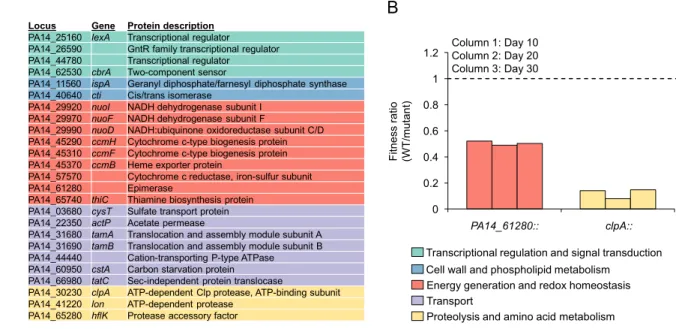
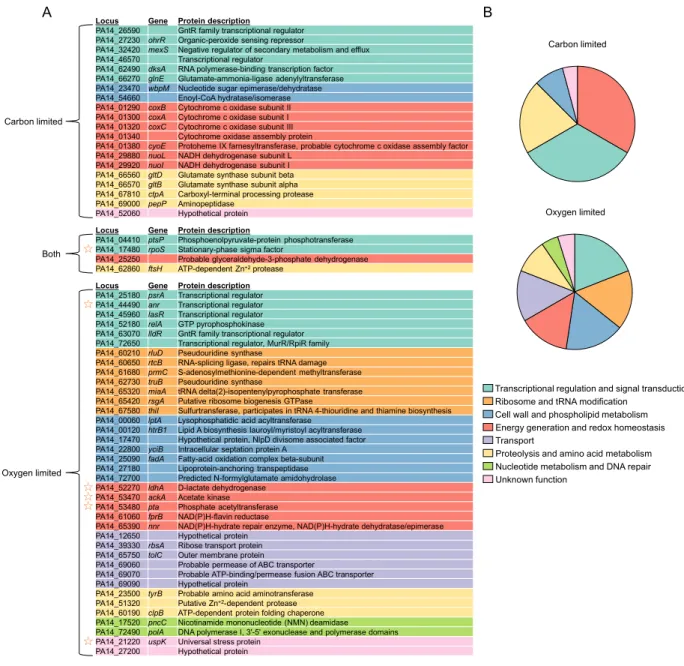
Functional classification of fitness determinants ... 53
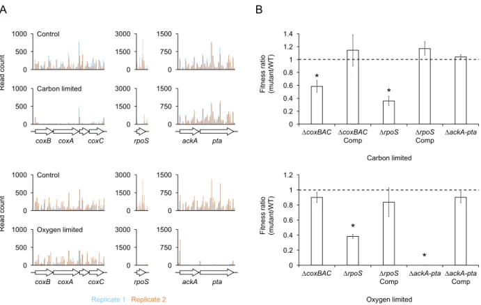
Experimental validation of Tn-seq results ... 54
Fitness dynamics of mutants during oxygen limitation ... 55
Discussion ... 59
Two methods to maintain the PMF and ATP synthesis during energy-limited growth arrest ... 60
Functional categories required specifically during oxygen limitation ... 61
Essentiality of proteolysis ... 62
A central role for RpoS ... 62
Open questions ... 63
Conclusion ... 64
Materials and Methods ... 65
Bacterial strains, plasmids, primers, and growth conditions ... 65
Generation of the transposon library ... 65
Tn-seq sample preparation ... 66
Sequencing and data analysis ... 66
Strain construction ... 67
Viability measurements ... 68
ATP measurements ... 68
Pyruvate measurements ... 68
Competition assays ... 69
Acknowledgements ... 69
Supplemental files accompanying Chapter 3 ... 70
Literature cited ... 71
Chapter 4: Heat Shock Proteases Delay Aging During Growth Arrest .... 77
Abstract ... 77
Significance statement ... 78
Introduction ... 78
Results ... 80
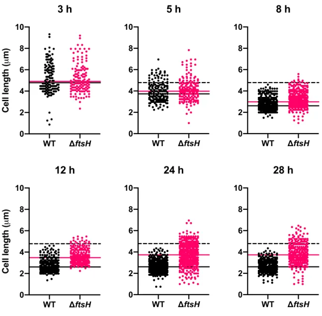
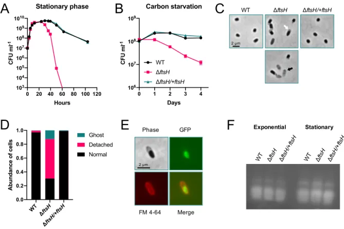
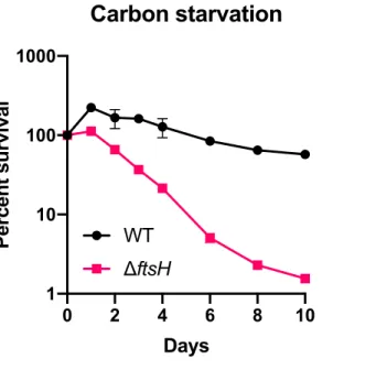
FtsH maintains cell integrity during growth arrest ... 80
Cell integrity of ΔftsH is not compromised by LPS overproduction during growth arrest ... 83
Identification of genetic interactions with ftsH during growth arrest .. 87
Identification of genetic interactions with ftsH during growth ... 90
Deletion of hslVU exacerbates survival of ΔftsH during growth arrest ... 93
ATP-dependent heat shock proteases cooperatively promote survival during growth arrest ... 94
Heat and alkaline pH exacerbate survival during growth arrest ... 97
Heat shock proteases delay protein aggregation during growth arrest ... 99
Discussion ... 100
Materials and Methods ... 103
Strains and growth conditions ... 103
Growth arrest survival assay ... 104
LPS measurement ... 104
Generation of the transposon library in ΔftsH ... 104
Tn-seq sample preparation and data analysis ... 105
Growth rate measurements ... 106
Microscopy ... 106
Supplemental files accompanying Chapter 4 ... 106
Literature cited ... 107
Chapter 5: Role of Heat Shock Proteases in a Novel N-degron Pathway ... 114
Abstract ... 114
Introduction ... 114
Results ... 116
Discussion ... 118
Materials and Methods ... 120
Acknowledgements ... 121
Literature cited ... 122
Chapter 6: Conclusions ... 124
Summary ... 124
Future directions ... 125
Functional genomics in multiple protease mutant backgrounds ... 125
Relevance of proteostasis in different ecological contexts ... 125
Relevance of proteostasis in growth arrest survival of different organisms ... 126
Role of ATP in mediating proteostasis during growth arrest ... 126
Appendix: A Protocol for Efficient Transposon Library Generation in P. aeruginosa Strain UCBPP-PA14 ... 128
Abstract ... 128
Protocol ... 128
Day 1 (Pregrowth) ... 128
Day 2 (Pregrowth) ... 128
Day 3 (Conjugation and plating) ... 128
Day 4 (Harvesting) ... 130
C h a p t e r 1 INTRODUCTION
“The mathematics of uncontrolled growth are frightening. A single cell of the bacterium E. coli would, under ideal circumstances, divide every twenty minutes. That is not particularly disturbing until you think about it, but the fact is that bacteria multiply geometrically: one becomes two, two become four, four become eight, and so on. In this way it can be shown that in a single day, one cell of E. coli could produce a super-colony equal in size and weight to the entire planet Earth.”
-Michael Crichton (1969) The Andromeda Strain.
Since its inception, the discipline of microbiology has been predicated on our ability to grow bacteria in the laboratory. In an effort to study their physiology, microbiologists have gone to great lengths to engineer pristine, ideal environments with just the right temperature, the right pH, the right amount of oxygen, and the right concoction of an amply nutritious food source in order to coax their bacterium to grow. For many bacteria, it doesn’t take much coaxing, because in contrast to multicellular organisms like ourselves, bacteria are opportunistic dividers. When conditions are favorable, a bacterium will invariably grow and divide to form two copies of itself. As Michael Crichton elegantly illustrates in the opening quote, the truth is that many species of bacteria have evolved to grow fast. In fact, they’ve evolved to grow so fast that the rate-limiting step is often the time it takes to fully replicate their genome so that it can be partitioned equally into each daughter cell. This is the state in which microbiologists generally prefer to study their bacterium of choice:
when it is happy, healthy, and growing so quickly that it might forget to include a copy of its genome in each new cell. We have derived much of our insight into the physiology of the bacterial cell, as well as that of our own cells, by studying these fast-growing bacteria in the prime of their lives and at the top of their game.
Pseudomonas aeruginosa is one well-studied example of these fast-growing bacteria. Because of its metabolic versatility, P. aeruginosa is happy to make a living in diverse environments, sometimes with a food source that could be considered meager at best. These environments vary widely: from
open ocean or fresh water streams, to the soil beneath our feet, to the biofilm growing in our hot tub.
Unfortunately, its versatility also allows P. aeruginosa to thrive in more sinister environments, like the lungs of a patient with pneumonia or cystic fibrosis, or a wound caused by a severe burn or surgical procedure.
But in all of these environments, from lake to lung or test tube to toilet bowl, we know that P.
aeruginosa can not grow indefinitely. At some point it will exhaust a key nutrient and cease its growth. What happens to P. aeruginosa after it stops growing? What happens to any bacterial community, for that matter, when the food or air it was happily consuming is all used up? Most of our study of bacterial physiology ends here, at the “stationary” or “growth arrest” phase of the bacterial life cycle. Microbiologists generally pay less attention to bacteria in this phase. For some, it is simply an “in between” phase, i.e., in between bouts of fast growth, and in between where their interests lie. This is an understandable sentiment. Studying growth arrest can be mechanistically tricky and highly variable from one experiment or organism to the next. In fact, when studying a growth-related process, it’s a good idea to minimize the emergence of growth-arrested cells altogether. Among the few valiant souls who devote their time and energy to try and better understand the physiology of growth arrest, I am certain all will readily admit that it’s HARD!
Studying growth arrest is just hard—a point that I hope will resonate with the reader of this thesis.
Because of the difficulty of its study, growth arrest is still very much a black box in our understanding of bacterial physiology. While we have developed a relatively detailed and exquisite picture of the many highly-conserved processes required for bacterial growth, an analogous framework does not exist for growth arrest. The question “what must cells do to grow?” is much more readily answered than “what must cells do to survive?” Most of the literature that has addressed the latter question has made it clear that bacteria do not simply “hang out” during periods of growth arrest, in what might be considered a “dormant” state. Rather, they must coordinately activate and maintain regulatory and metabolic responses to ensure their survival. These responses often differ greatly depending on the organism and the type of growth arrest—a concept that will be emphasized throughout this thesis.
Whether or not we care about bacterial growth arrest (although there are many reasons we should care, as will be made clear in this thesis) certainly the bacteria care. After all, they’re the ones who
are starving. And the fact is the real world is not like the laboratory, where a friendly scientist is always on standby to replenish nutrients. The real world is harsh. And more often than not, the time to feast is over and famine has befallen the land. How well bacteria can survive these periods of nutrient scarcity determines who will still be around when nutrients once again become available.
This thesis is about acknowledging the resilience of bacteria and appreciating what it takes for them not just to grow, but to survive. My hope is that by the end of this thesis the reader will also join me in acknowledgement and appreciation.
Overview
In Chapter 2, together with Megan Bergkessel, a postdoctoral scholar in the Newman Lab, I review much of our recent knowledge on how diverse organisms survive growth arrest, with emphasis on their metabolic, transcriptional, and translational responses, and the mechanisms they use to maintain genomic integrity. This chapter describes some of the recent technology that has facilitated the study of growth arrest, including techniques to measure the dynamics of protein synthesis, assess metabolic heterogeneity at the single-cell level, and screen for genetic fitness determinants on a genome-wide scale.
Chapter 3 utilizes one of these techniques, called Tn-seq, to identify genes required for fitness of P.
aeruginosa when it is growth arrested due to energy limitation. I show that P. aeruginosa is capable of extended survival when limited for organic carbon as an electron donor or oxygen as a terminal electron acceptor, and that the genes required for survival vary greatly depending on the type of energy limitation. I also show that many of these genes are temporally important for survival, suggesting that growth arrest encompasses a more dynamic state than was previously appreciated.
Building on the findings in Chapter 3, Chapter 4 uncovers a central role for heat shock proteases in promoting survival of P. aeruginosa during carbon starvation. Using Tn-seq and multiple knockout strains, I show that protein turnover is a fundamental problem for P. aeruginosa during growth arrest, and that a highly redundant genetic system is in place to delay what might be considered “aging” of growth-arrested cells.
Chapter 5 explores a possible connection between the heat shock proteases of P. aeruginosa and a novel degradation pathway that recognizes the N-terminal formyl group of bacterial proteins. This project resulted from a collaboration with the Alexander Varshavsky Lab at Caltech, who first described this degradation pathway in E. coli. Together with Tri Vu, a postdoctoral scholar in the Varshavsky Lab, we show that none of the major heat shock proteases in P. aeruginosa appear to play a role in degrading N-terminally formylated proteins in vivo. However, the possibility remains that the mechanistic details of this degradation pathway are not conserved between E. coli and P.
aeruginosa.
Finally, Chapter 6 provides some concluding remarks and a few suggested experiments to further build on the findings of this thesis.
An appendix is also included that details a protocol I developed to generate highly-saturated transposon libraries in P. aeruginosa strain UCBPP-PA14. This protocol could prove useful to those seeking to make their own transposon libraries in P. aeruginosa.
C h a p t e r 2
THE PHYSIOLOGY OF GROWTH ARREST
This chapter is adapted from:
Bergkessel M., Basta D.W., Newman D.K. (2016) The physiology of growth arrest: uniting molecular and environmental microbiology. Nat Rev Microbiol 14(9):549–562.
doi:10.1038/nrmicro.2016.107.
Abstract
Most bacteria spend the majority of their time in prolonged states of very low metabolic activity and little or no growth, in which electron donors, electron acceptors and/or nutrients are limited, but cells are poised to undergo rapid division cycles when resources become available. These non-growing states are far less studied than other growth states, which leaves many questions regarding basic bacterial physiology unanswered. In this Review, we discuss findings from a small but diverse set of systems that have been used to investigate how growth-arrested bacteria adjust metabolism, regulate transcription and translation, and maintain their chromosomes. We highlight major questions that remain to be addressed, and suggest that progress in answering them will be aided by recent methodological advances and by dialectic between environmental and molecular microbiology perspectives.
Introduction
Much of our knowledge of the molecular machinery that is responsible for energy generation, gene expression and DNA replication comes from studying fast-growing model organisms, such as Escherichia coli, during exponential phase in nutritionally complete medium. Under these conditions, a single E. coli cell would grow to a population with the mass of the Earth within 2 days.
Clearly, this does not occur, but the discrepancy between potential and actual growth underscores that the estimated 5x1030 bacteria that are on our planet spend the vast majority of their time in energy-limited states and not dividing. Although environmental microbiologists have long
appreciated the importance of extremely energy-limited states (1, 2), their focus has primarily been on exploring theoretical and empirical energetic limits of diverse metabolisms (3, 4). With a few notable exceptions (5-7), molecular microbiologists have largely avoided the study of growth- arrested cells. Consequently, relatively little is known about the mechanisms that underpin the dominant modes of bacterial existence; this gap in our knowledge hinders our understanding of the roles that bacterial communities have in driving global biogeochemical cycles and influencing plant and animal health.
The dearth of information is due, in part, to the challenges in defining, reproducing and measuring non-growing states of bacteria in the laboratory. Microbiologists have traditionally relied on population-level measurements of activity to draw conclusions about molecular mechanisms—an approach that benefits from high levels of biochemical activity and homogeneous populations. The few existing studies of non-growing states have focused on conditions that cause population growth arrest through the severe limitation of at least one basic resource (Box 1). These studies have provided most of our insights into these growth states (referred to as ‘long-term stationary phase’
(6), ‘continuous activity stationary phase’ (8)or ‘starvation–survival’ (2)) and have shown that growth-arrested cells synthesize proteins at rates that can be orders of magnitude slower than in exponential phase but stay viable for several days to years, and are usually able to rapidly resume growth when nutrients become available. These populations are neither highly active nor likely to be homogeneous (Box 2), potentially including subpopulations that undergo infrequent division cycles, but we assume, based on their static population numbers, that they represent the best available proxies for non-growing states. A major area for future research, as measurement sensitivity and selectivity continue to improve (Box 3), will be to better understand how these populations are composed of distinct cellular states.
Our focus on non-growing states of relatively fast-growing bacteria purposefully excludes some related states and other survival strategies. We view the responses to nutritional downshift that are part of the transition to stationary phase, which have been well studied and reviewed elsewhere (9, 10), as distinct from the strategies that are used later in stationary phase, and we point out these differences throughout the review (see also Box 1). Another state that we do not address is the endospore—a mode of survival that uses almost no energy for potentially thousands of years—which
has been well reviewed elsewhere (11). Finally, inherent slow growth in bacteria with low-energy core metabolisms is outside the scope of this review. These organisms, which have doubling times of months or much longer, might be viewed as ‘non-growing’ for substantial periods of time even when doubling at their maximum rates (reviewed in 4). How basic molecular mechanisms function in such contexts is fascinating, and may be related to the growth-arrested strategies of otherwise fast- growing bacteria, but the challenges of direct study remain extreme.
In this Review, we focus on general insights from current research and highlight remaining questions for three fundamental cellular challenges that are encountered during growth arrest. First, how are cellular energy stores maintained and managed during starvation? Second, how is gene expression regulated under extreme limitation for nucleotide and amino acid substrates? Third, how is chromosomal DNA protected in a way that allows occasional replication and low levels of transcription?
Metabolism: maintaining energy supply
The primary goal of growth-arrested organisms is to maintain the supply of the energy and biosynthetic precursors that are required to maintain essential macromolecular components of the cell, sustain active regulatory mechanisms for sensing and responding to the environment, and, perhaps most importantly, preserve the electrochemical gradient of the membrane (12, 13).
Preserving this gradient (commonly known as the proton motive force (PMF)) is, in turn, crucial for transporting energetic and biosynthetic substrates into the cell in all bacteria, for flagellar motility in flagellated bacteria and for ATP synthesis in bacteria that are unable to sustain substrate-level phosphorylation (14). Thus, the continual supply of energy and building blocks is highly interdependent on the preservation of the PMF. Many bacteria can re-route metabolism to respond to specific limitations in this supply with impressive flexibility, enabling them to shift to alternative sources of energy and building blocks while balancing flux through central metabolic pathways.
Alternative sources of substrates for energy and biosynthesis.
Severe substrate limitation is often the underlying cause of growth arrest, as it leads to reduced rates of both anabolicand catabolicmetabolism, and forces cells to rely on alternative sources of energy
and biosynthetic substrates, including internal stores. Indeed, cellular components themselves can be catabolized, which solves two problems that arise from severe substrate limitation: it provides nutrients and it removes the burden of maintaining cellular machinery that has become dispensable as overall cellular activity decreases. The result of this catabolism is a large reduction in cell size and volume (4), which increases the surface area-to-volume ratio of the cell and thus, in theory, could increase the efficiency of substrate transport (see below). Catabolism in growth-arrested bacteria has been shown most clearly to target ribosomes (see below) and membrane phospholipids. For example, genetic studies in E. coli found that derepression of genes that are involved in β-oxidation, and thus fatty acid degradation, is required for long-term survival in stationary phase (15). Vibrio cholerae also undergoes a substantial decrease (>99%) in total lipids within 7 days of starvation, despite an increase in viable cell count due to reductive divisions(see below), which led to the proposal that membrane phospholipids are an endogenous energy source to maintain viability in these cells (16).
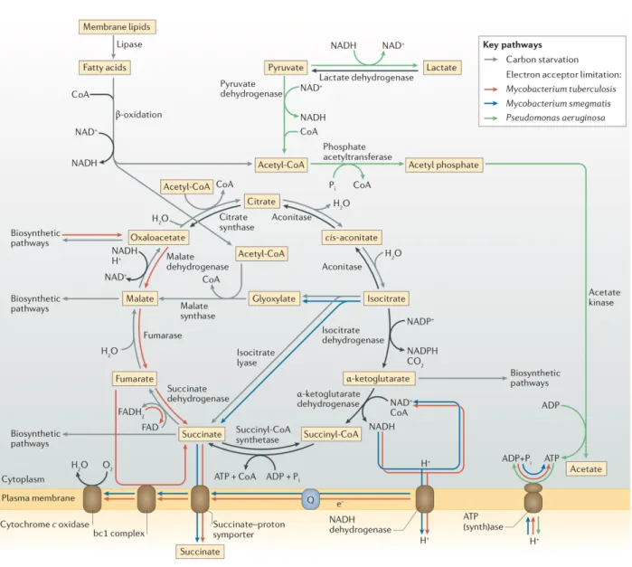
A recent microarray analysis of carbon-starved Vibrio harveyi cells supported this hypothesis, showing that genes that are involved in fatty acid β-oxidation were upregulated during starvation, concomitant with a decrease in cell size. Genes that have important roles in metabolic reactions that require acetyl-CoA, which is the major metabolite that is generated from the degradation of fatty acids, were also upregulated; many of these genes are part of the glyoxylate shunt, which is an anapleroticpathway that bypasses most of the tricarboxylic acid (TCA) cycle, which is substantially downregulated during starvation (17) (Fig. 1).
Figure 1. Metabolic rewiring during growth arrest. Different organisms use distinct metabolic strategies to meet cellular requirements under growth-limiting conditions. Both Mycobacterium spp. and Pseudomonas aeruginosa must adjust their strategies for maintaining their membrane electrochemical gradient under oxygen-limited conditions. Mycobacterium spp. continue to use the electron transport chain, using either low levels of oxygen or fumarate as the terminal electron acceptor, and also contribute to the membrane electrochemical gradient by secreting succinate, which is generated through reversal of the tricarboxylic acid (TCA) cycle in Mycobacterium tuberculosis and by the glyoxylate shunt in Mycobacterium smegmatis. In P.aeruginosa, fluxes through the TCA cycle and electron transport chain drop close to zero under anoxic conditions, and substrate-level phosphorylation generates ATP to run the ATP synthase in reverse, thus pumping protons outward across the membrane. Under carbon-limiting conditions, fatty acid β- oxidation generates acetyl-CoA, which can be fed into biosynthetic pathways through the glyoxylate shunt; this generates the TCA cycle intermediates that are most useful as precursors without overproducing other intermediates. Pi, inorganic phosphate; Q, quinone.
In addition to catabolism of internal stores, increased active acquisition of exogenous nutrients is an important survival strategy during nutrient limitation. In heterotrophs, limitation of organic carbon causes shortages of both energy and biosynthetic precursors. Many heterotrophs respond to this shortage by upregulating high-affinity transporters to scavenge carbon from the environment. For example, Vibrio sp. strain Ant-300 effectively depletes nanomolar concentrations of arginine from its environment and, interestingly, only exhibits chemotaxis toward arginine after starvation (18).
Both high-affinity and low-affinity transport depend on respiration rather than ATP hydrolysis for energy, which suggests that the maintenance of the PMF is crucial for uptake by these transporters.
Limitation of phosphate, nitrogen, sulfur and other nutrients also induces the upregulation of scavenging mechanisms in diverse organisms (19-21).
The evolutionary significance of competitive scavenging for resources is illustrated by the dynamics of populations of E. coli in batch culture. During long-term stationary phase in rich complex medium, in which the ‘growth advantage in stationary phase’ (GASP; Boxes 1,2) phenotype arises, E. coli mutants arise that take over the population. These mutants are able to outcompete ‘naive’ wild-type cells in stationary phase competition experiments (22). Strikingly, the majority of the GASP mutations that have been studied result in improved amino acid uptake and catabolism, by affecting genes such as those that encode leucine-responsive regulatory protein (Lrp; a nucleoid-associated protein) or the sigma factorRpoS (see below), or genes that are located in the amino acid transport locus ybeJ–gltJKL (6, 23, 24). Increased amino acid uptake ability is particularly important in the long-term stationary phase that is associated with the GASP phenotype, as the breakdown products of dead and dying cells are the major sources of nutrients.
Re-routing metabolic pathways.
A final strategy by which cells respond to nutrient limitation is to re-route metabolic fluxes to maintain acceptable levels of affected intermediates. For example, the preferentially aerobic pathogens Mycobacterium tuberculosis and Pseudomonas aeruginosa have very different responses to the hypoxic conditions that they experience during human infections (25). As oxygen is preferred by these bacteria as a terminal electron acceptor, hypoxia causes decreased flux through the electron transport chain, which, in turn, causes a decrease in the PMF and an accumulation of reducing
equivalents (primarily NAD(P)H). In M. tuberculosis, hypoxic conditions lead to a decreased but stable level of ATP, the maintenance of which requires the ATP synthase and some degree of PMF across the membrane (26). The PMF is maintained by the forward fluxes through the TCA cycle and the electron transport chain that can be sustained by the trace amounts of oxygen available to accept electrons, supplemented by electrogenic secretionof succinate. This succinate can be produced by one or both of two pathways: a reversal of the reductive branch of the TCA cycle through the upregulation of phosphoenolpyruvate carboxykinase, malic enzyme and fumarate reductase (which also re-oxidizes NADH); and the glyoxylate shunt, which bypasses steps of the TCA cycle that produce reducing equivalents (27-29) (Fig. 1). Interestingly, M. tuberculosis preferentially generates succinate by reversing the TCA cycle, whereas Mycobacterium smegmatis uses the glyoxylate shunt and Mycobacterium bovis uses both pathways (29), which shows that the re-routing of metabolic pathways in response to the limitation of terminal electron acceptors can vary, even between closely related bacterial species. M. tuberculosis can use metabolic reorganization as part of a strategy to survive hypoxic conditions for years without observable growth (30, 31); indeed, even when oxygen is completely absent, M. tuberculosis has been suggested to survive by using endogenously generated fumarate as an alternative terminal electron acceptor for oxidative phosphorylation (26) (Fig. 1). In contrast to mycobacteria, P. aeruginosa dispenses with oxidative phosphorylation altogether when oxygen is severely limited, and can instead maintain the PMF anaerobically by reversing the reaction that is catalysed by the ATP synthase (32), with ATP supplied by substrate- level phosphorylation using pyruvate or arginine as a substrate (33, 34) (Fig. 1). Under these conditions, fluxes through the TCA cycle and the electron transport chain are presumably close to zero. Together, these examples illustrate the flexibility of metabolic networks in maintaining crucial metabolic parameters during severe nutrient and energy limitation.
Further questions.
Although recent work has advanced our understanding of the metabolic strategies that are used to survive growth arrest, much is still unknown. At the level of single cells, questions remain about the absolute minimal energy requirements for viability, and how these limits vary according to the organism and environment, despite progress in investigating these boundary conditions (4). At an environmental level, complexities such as co-limitation for different substrates in a changing
environment and interaction with other microorganisms are likely contributors to energy dynamics in the natural world and significant forces in the evolution of strategies to regulate metabolism (35, 36). For example, in at least some cases, including low-energy environments, different species of microorganism can form cooperative metabolic interactions—an arrangement known as syntrophy (37). Indeed, mutually beneficial metabolic interactions may be more common than is currently appreciated; for example, redox-active phenazine pigments that are synthesized by Pseudomonas spp. were recently shown to support substrate-level phosphorylation by P. aeruginosa (32).
Furthermore, studies have suggested that other organisms may also benefit from the presence of phenazines produced by P. aeruginosa in some contexts (38). Understanding metabolic network connectivity, even within one organism, still presents a research challenge, and we are just beginning to probe the metabolic interactions of microbial communities. Identifying new energy yielding pathways within community contexts is a priority for future research.
Regulation of gene expression
Limitation for energy, nucleotides and amino acids are common features of non-growing states that probably impose general constraints on gene expression, although the precise identities of upregulated and downregulated genes vary depending on the organism and the underlying causes of growth arrest. The constraints on gene expression may differ in non-growing states from those imposed by the better understood challenges of exponential growth and the initial transition to stationary phase, and recent technical advances have made feasible the study of how gene expression might adapt to the constraints that are imposed by growth arrest at a molecular level.
Regulatory paradigms of different growth states.
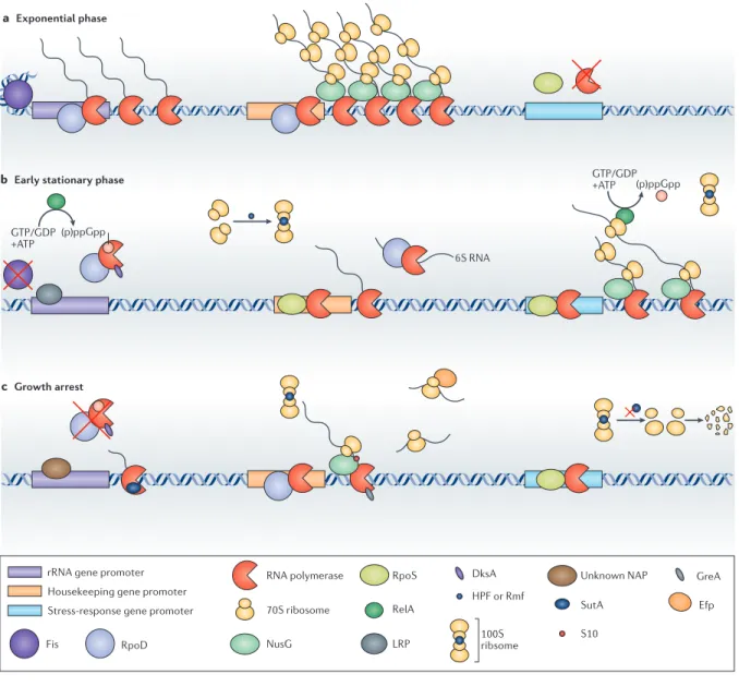
During exponential growth in nutritionally complete media, most gene expression is directed towards the biosynthesis of ribosomes, which are the principal mass constituent of each new cell being produced and are the drivers of the biosynthesis of all other proteins (reviewed in (39); Fig.
2a). Under these conditions, rates of translation elongation are limited by the intrinsic properties of the ribosome rather than the availability of amino acids (40). However, the unlimited availability of amino acids is short-lived even in rich medium batch culture. Thus, some resources will soon become limiting, at which time global regulatory systems will coordinate a transition away from maximum
growth. Two global regulators of this transition that are phylogenetically widely distributed but best characterized in E. coli are the sigma factor RpoS, which is induced by stresses such as heat and osmotic shock as well as nutrient downshift, and the stringent response alarmone guanosine pentaphosphate ((p)ppGpp), which is synthesized in response to signals of nutrient limitation (reviewed in (9)and (41), respectively). RpoS has relaxed sequence specificity compared with the housekeeping sigma factor RpoD, thus favoring the expression of a regulonof stress-adaptive genes with non-consensus promoters, but also drives lower levels of transcriptional activity (42). (p)ppGpp has pleiotropic effects on many cellular processes, and, together with the co-regulator DksA, strongly represses rRNA and ribosomal protein gene expression (43, 44). Together, RpoS and (p)ppGpp lead to a reduction in the total rates of gene expression, but also lead to a redirection of biosynthetic capacity away from ribosome biogenesis and towards more urgent concerns, such as preventing or repairing DNA damage (see next section), osmoprotection, refolding damaged pro- teins or increasing the synthesis of missing biosynthetic intermediates (Fig. 2b).
14
Figure 2. Transcription and translation during different growth phases. a | During exponential phase, rRNA genes are among the most highly transcribed in the cell, as ribosome biogenesis is a top biosynthetic priority. In addition, genes with promoters that have strong consensus sequences for RpoD binding are highly expressed and efficiently co-transcriptionally translated, aided by the transcription elongation factor NusG, which helps physically associate a ribosome with the RNA polymerase. The stationary phase sigma factor RpoS is synthesized to some extent but fails to compete with RpoD for RNA polymerase; consequently, stress-responsive genes with promoters that do not match the RpoD consensus sequence are not efficiently expressed. b | In the transition to stationary phase, limitation for amino acids activates RelA, which senses uncharged tRNAs and synthesizes the alarmone guanosine pentaphosphate ((p)ppGpp). (p)ppGpp, in conjunction with DksA, represses the transcription of rRNA by destabilizing the rRNA open promoter complex. The decrease in abundance of the nucleoid-associated protein Fis and the increase in abundance of leucine- responsive regulatory protein (Lrp) also contribute to rRNA repression. Hibernation promoting factor (HPF) and ribosome modulation factor (Rmf) are upregulated and lead to the dimerization of ribosomes to 100S complexes that are inactive for translation. RNA polymerase complexes with RpoD are selectively sequestered through several mechanisms, including binding to a small RNA (6S
Open promoter complexes The intermediate in transcription initiation in which RNA polymerase has bound to a promoter and unwound the double-stranded DNA, which allows the template strand of the DNA to pass through the active site of the polymerase.
possible dilution rates, which suggests that prolonged RpoS activity may be maladaptive7,49. Although gene products of the RpoS regulon have important roles in growth-arrested states, the many layers of regulation that affect levels of RpoS suggest a delicate balance between preparing for growth arrest and actually surviving a prolonged non-growing state50–52.
Nucleotide and energy levels continue to decrease after growth is arrested53, potentially affecting both the priorities and mechanisms of gene expression regulation.
As discussed, the priorities that are reflected by upregu- lated genes include using alternative energy and nutrient sources, and carrying out essential repair and replacement of cellular components. The mechanisms of expression regulation are less clear — the biochemical implications of severe nucleotide and amino acid shortages are of great interest. Studies of E. coli in exponential phase and
early stationary phase suggest that even moderate nutri- ent downshifts lead to profound changes in the stability of open promoter complexes and the tendencies of both polymerases and ribosomes towards pausing, perma- nently arresting or terminating40,43,44,54 (see REFS 55,56 for reviews). A successful regulatory strategy for non-growing states must favour the expression genes that are essential for survival, but must also ensure that the transcriptional and translational activities of the cell are matched to the available resources, and that relevant cellular machinery can handle slow elongation rates and long pauses (FIG. 2c). Tuning the capacity and rate of gene expression to match limited substrates. One strategy for tolerating shortages of nucleotides and amino acids is to limit the number of active polymerases and ribosomes so that fewer of these complexes are competing for the limited Nature Reviews | Microbiology a Exponential phase
b Early stationary phase
c Growth arrest
Fis RpoD
rRNA gene promoter Housekeeping gene promoter Stress-response gene promoter
NusG
RpoS
LRP 70S ribosome
HPF or Rmf
Unknown NAP 6S RNA
(p)ppGpp
(p)ppGpp GTP/GDP +ATP
RelA SutA
DksA GreA
S10
Efp RNA polymerase
100Sribsome GTP/GDP
+ATP
556 | SEPTEMBER 2016 | VOLUME 14 www.nature.com/nrmicro
RNA), and levels of RpoS are also increased, which leads to increased transcription of stress-responsive genes in the RpoS regulon.
RpoS can also drive transcription of housekeeping genes that have RpoD-consensus promoters, but does so at much lower levels than transcription of these genes that is mediated by RpoD. c | During growth arrest, overall gene expression activity is much lower than in exponential phase or the transition to stationary phase. Although much remains to be elucidated about how these very low levels of activity are regulated, the observations that several global regulators change in abundance suggest some possible mechanisms.
Regulators that are important during the transition to stationary phase, such as DksA, (p)ppGpp, RpoS and HPF, seem to be downregulated during growth arrest. Also, the complement of nucleoid-associated proteins (NAPs) changes substantially, which probably affects the expression of rRNA and other genes, although details remain to be explored. In Pseudomonas aeruginosa, some factors that are thought to contribute to transcription and translation elongation (GreA, S10 and elongation factor P (Efp)) were upregulated, possibly suggesting that they could help buffer against pausing and arrest in severely substrate-limited conditions.
Although some ribosomes are catabolized, with the dual benefit of decreasing the number of ribosomes that are competing for amino acid substrates and liberating nutrients to be used for energy and maintenance, the newly identified transcriptional regulator SutA, which is upregulated during growth arrest in P.aeruginosa, enhances rRNA and ribosomal protein gene expression, which suggests that some repair and replacement of ribosomes may also be important.
Decreased availability of nucleotides and amino acids probably begins to affect transcription and translation rates during this transition phase, but (p)ppGpp and DksA function, in part, by further sensitizing the initiation of transcription to the availability of nucleotides—a strategy that makes sense during a transition from high to low nucleotide availability, but perhaps not if limiting nucleotides become a long-term challenge. Indeed, several lines of evidence suggest that RpoS and (p)ppGpp govern a transient, active adjustment to dynamic conditions rather than survival of a long- term non-growing state. DksA and (p)ppGpp have been shown to decrease to low levels in late stationary phase in P. aeruginosa and E. coli, respectively (45, 46), and two studies have suggested that levels of RpoS in P. aeruginosa may actually be lower in the non-growing state than in mildly nutrient-limited states (47, 48). In addition, mutations in rpoS that decrease function cause a GASP phenotype and provide a selective advantage during continuous culture at the lowest possible dilution rates, which suggests that prolonged RpoS activity may be maladaptive (7, 49). Although gene products of the RpoS regulon have important roles in growth-arrested states, the many layers of regulation that affect levels of RpoS suggest a delicate balance between preparing for growth arrest and actually surviving a prolonged non-growing state (50-52).
Nucleotide and energy levels continue to decrease after growth is arrested (53), potentially affecting both the priorities and mechanisms of gene expression regulation. As discussed, the priorities that are reflected by upregulated genes include using alternative energy and nutrient sources, and carrying out essential repair and replacement of cellular components. The mechanisms of expression
regulation are less clear—the biochemical implications of severe nucleotide and amino acid shortages are of great interest. Studies of E. coli in exponential phase and early stationary phase suggest that even moderate nutrient downshifts lead to profound changes in the stability of open promoter complexes and the tendencies of both polymerases and ribosomes towards pausing, permanently arresting or terminating (40, 43, 44, 54)(see (55, 56) for reviews). A successful regulatory strategy for non-growing states must favor the expression genes that are essential for survival, but must also ensure that the transcriptional and translational activities of the cell are matched to the available resources, and that relevant cellular machinery can handle slow elongation rates and long pauses (Fig. 2c).
Tuning gene expression capacity and rate to match limited substrates.
One strategy for tolerating shortages of nucleotides and amino acids is to limit the number of active polymerases and ribosomes so that fewer of these complexes are competing for the limited supply of substrates. Indeed, both RNA polymerases and ribosomes are sequestered at the entry to stationary phase, which reduces the number of active complexes. RNA polymerases bound to RpoD are sequestered by binding to a small RNA, at least in E. coli (57), and ribosomes are sequestered by hibernation promoting factor or ribosome modulation factor (or both, depending on the organism;
reviewed in (56)), which mediate the formation of inactive ribosome dimers. In Listeria monocytogenes, the abundance of hibernation promoting factor peaks at the entry to stationary phase, and then decreases (58). Further into growth arrest, ribosome degradation begins to have an important role in the regulation of the number of ribosomes, and has the dual benefit of both limiting the number of active ribosomes and converting unused ribosomes to nutrients (58, 59). In E. coli, studies have shown that individual 30S and 50S subunits are preferred substrates for degradation during starvation; therefore, ribosome dimerization at the entry to stationary phase may help delay degradation until it is essential (60). Many bacteria also encode additional ribosome-binding proteins that are upregulated in stationary phase and that may have distinct roles in modulating the activity, sequestration or degradation of ribosomes during growth arrest. For example, RsfA, which is a highly conserved factor that has been shown to prevent subunit joining in E. coli (61), is upregulated during anaerobic survival in P. aeruginosa (48). Understanding how these factors coordinate to affect the fates of ribosomes during growth arrest will require further study.
Another strategy may be to tune the transcription and translation machineries to be less sensitive to pausing and substrate limitation, through changes in the levels of accessory factors. Detailed in vivo studies of such factors have not, for the most part, been conducted in non-growing bacteria, but some possible mechanisms are suggested by considering the proteins that are upregulated during anaerobic survival in P. aeruginosa in light of their functions in other growth states. One such factor is an RNA polymerase-binding protein, SutA, which enhances transcription of rRNA and ribosomal protein genes during slow growth. SutA may decrease the sensitivity of these genes to shortages of nucleotides and amino acids, and thus enable some repair and replacement of ribosomes during growth arrest (48). Although homologues of SutA are found only in a subset of Gammaproteobacteria (not including E. coli), another RNA polymerase accessory factor that may have a similar function is CarD, which is widely distributed outside the Gammaproteobacteria. CarD has been shown to be upregulated during nutrient limitation (62) and to enhance the transcription of rRNA genes (63) in M. smegmatis, which suggests that this type of regulatory response may be of broad importance. Two additional factors that are known to have roles in transcription elongation, at least in growing cells, and that are upregulated in P. aeruginosa during anaerobic survival are the ribosomal protein S10, which can ‘moonlight’ as a transcription elongation factor through its interaction with NusG (64), and GreA, which can help to rescue RNA polymerases that are arrested in ‘back-tracked’ states (known as backtracked RNA polymerases) (65). Finally, upregulation of the translation factor elongation factor P (Efp) may help buffer translation against stalling-induced arrest (66).
Further questions.
Although the observations described here imply that the non-growing state is actively regulated, the molecular details of the specific mechanisms that are responsible remain largely unknown, and there are hints that the solutions to fundamental problems of non-growing states may be quite diverse between phylogenetically divergent bacteria. This means that researchers must broaden the scope of inquiry to include more model organisms, including ones that are less adapted to laboratory growth conditions, and indicates that comparative and functional genomics techniques may become increasingly useful for identifying and understanding regulatory paradigms. Exploring molecular mechanisms in low-activity, heterogeneous states is challenging, but new applications for doing so
have shown promise, notably those that are based on next-generation sequencing or proteomics techniques (Box 3).
Maintenance and replication of DNA
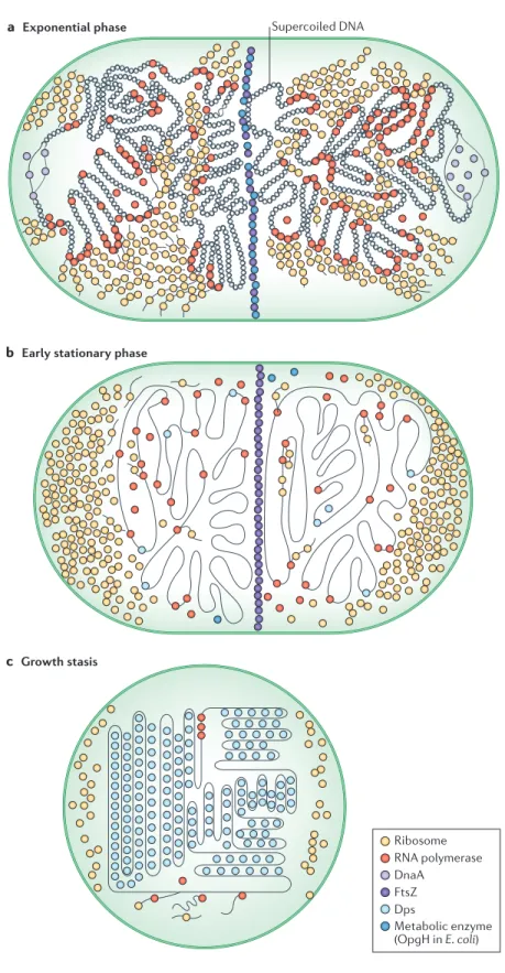
A defining feature of non-growing states is the repression of DNA replication and cell division, and accompanying changes to the nucleoid have mostly been studied in E. coli and other model organisms as they transition towards stationary phase. The most observable feature of the stationary- phase nucleoid is a condensed, even crystalline, morphology when viewed by electron microscopy (67), which is thought to protect the DNA from damage and confer a survival advantage.
The generation times of exponentially growing E. coli cells are shorter than the time that is required to replicate the chromosome, which means that a new round of replication must start before the previous one completes. However, this process must be regulated to ensure a yield of exactly one replicated chromosome per cell division (Fig. 3a). One of the main factors in the regulation of the initiation of chromosome replication is DnaA, which is the ATPase that binds to the origin of replication. To prevent new rounds of initiation as the cell transitions to a non-growing state, the level, availability and activity of DnaA are markedly downregulated by several mechanisms (reviewed in (68, 69)). During this transition to stationary phase, ongoing rounds of replication can still be completed, but the accompanying cell divisions result in small progeny because ribosome biosynthesis and other biosyntheses are suppressed. Recent work suggests that the cell division machinery in E. coli may be capable of directly sensing the nutritional status of the cell through interactions between the membrane glycosyltransferase osmoregulated periplasmic glucans H (OpgH; also known as MdoH) and the cytokinesis regulator FtsZ. Poor nutrient status leads to lower levels of OpgH, which removes a block to FtsZ assembly and cytokinesis, permitting reductive divisions during nutrient starvation (70) (Fig. 3b).
The nucleoid: condensation and protection from DNA damage.
As cells progress towards growth arrest, the nucleoid undergoes increasingly extreme morphological changes. In general, the nucleoid becomes more condensed, while clusters of bound, transcribing RNA polymerase dissipate and ribosomes move from the central region of the cell, which they had
shared with the nucleoid, to the poles (71). Some of these changes are driven by the global changes in gene expression that are discussed above; in particular, the substantial decrease in rRNA transcription can sufficiently alter the distribution of RNA polymerase to affect the gross morphology of the nucleoid (72) (Fig. 3b, c). However, a crucial factor that drives changes in the nucleoid is the more specific transcriptional changes that occur in the expression of individual nucleoid-associated proteins (NAPs), which are proteins that bind to DNA with low or no sequence specificity and can give rise to higher-order structures. Most organisms encode several NAPs, which range in specificity from nearly universal to species specific (reviewed in (73)). The most abundant NAP shifts from factor for inversion stimulation (FIS) (74) during nutrient upshift and early exponential phase (75)to DNA-binding protein from starved cells (Dps) in stationary phase—a change that is driven, in part, by the regulatory activity of RpoS (76, 77). Dps, which has homologs in many bacterial species, can condense DNA through ordered self-aggregation, both in vivo and in vitro (67, 78), and transmission electron micrographs and atomic force microscopy have shown that nucleoid condensation in stationary-phase E. coli is dependent on Dps (79, 80) (Fig. 3c).
Condensation of the nucleoid by DPS separates the DNA from potentially damaging reactants in the cytoplasm, holding it in a low-energy equilibrium state that is distinct from the dynamic disequilibrium of the nucleoid in exponential phase (79). In addition to protecting DNA by this spatial separation, Dps may protect DNA from oxidative damage (81). Dps has structural homology to ferritin (82) and can oxidize ferrous iron to its ferric, insoluble form (81). This reaction is proposed to compete with the Fenton reaction, thus protecting cellular DNA from oxidative damage by decreasing the production of oxidizing free radicals (76).
20
Figure 3. Overview of cellular morphology with emphasis on nucleoid. Cells undergo gross morphological changes in transitions between different growth states. a | In exponential phase, the chromosome has a high degree of negative supercoiling, owing to large
Origin of replication The site on the bacterial chromosome, determined by its sequence, where the two strands of DNA are unwound to enable replication of the chromosome to begin.
Maintenance and replication of DNA
A defining feature of non-growing states is the repres- sion of DNA replication and cell division, and accompa- nying changes to the nucleoid have mostly been studied in E. coli and other model organisms as they transition towards stationary phase. The most observable fea- ture of the stationary-phase nucleoid is a condensed,
even crystalline, morphology when viewed by electron microscopy67, which is thought to protect the DNA from damage and confer a survival advantage.
The generation times of exponentially growing E. coli cells are shorter than the time that is required to repli- cate the chromosome, which means that a new round of replication must start before the previous one completes.
However, this process must be regulated to ensure a yield of exactly one replicated chromosome per cell division
(FIG. 3a). One of the main factors in the regulation of the initiation of chromosome replication is DnaA, which is the ATPase that binds to the origin of replication. To pre- vent new rounds of initiation as the cell transitions to a non-growing state, the level, availability and activity of DnaA are markedly downregulated by several mecha- nisms (reviewed in REFS 68,69). During this transition to stationary phase, ongoing rounds of replication can still be completed, but the accompanying cell divisions result in small progeny because ribosome biosynthesis and other biosyntheses are suppressed. Recent work suggests that the cell division machinery in E. coli may be capable of directly sensing the nutritional status of the cell through interactions between the membrane glyco syltransferase osmoregulated periplasmic glu- cans H (OpgH; also known as MdoH) and the cytoki- nesis regulator FtsZ. Poor nutrient status leads to lower
Figure 3 | Overview of cellular morphology with emphasis on nucleoid. Cells undergo gross
morphological changes in transitions between different growth states. a | In exponential phase, the chromosome has a high degree of negative supercoiling, owing to large amounts of active transcription. RNA polymerase is mostly bound to DNA and is gathered in large clusters of highly transcriptionally active genes, which tend to migrate to the periphery of the nucleoid region. Ribosomes are observed in the central portion of the cell, sharing space with the nucleoid. New rounds of replication, initiated by DnaA, begin even before previous rounds have completed.
Cytokinesis is inhibited by interaction of an abundant metabolic enzyme (for example, OpgH in Escherichia coli) with FtsZ, thus maintaining a larger cell size. b | In the transition to stationary phase, decreases in total transcription, and rRNA transcription in particular, lead to less supercoiling of the chromosome but a more condensed overall morphology, with fewer RNA polymerases and ribosomes associated with the nucleoid region in the centre of the cell. New rounds of replication are inhibited by the decreased abundance and activity of DnaA, but cell division to segregate chromosomes that have already been replicated can still take place (facilitated, in part, by a decrease in OpgH, which releases FtsZ), leading to progeny with a small cell size. The abundance of the nucleoid-associated DNA-binding protein from starved cells (Dps) begins to increase, owing to transcriptional upregulation by the stationary phase sigma factor RpoS. c | During growth arrest, an extremely high abundance of Dps leads to a highly condensed, crystalline appearance of the nucleoid region. Reductive divisions in stationary phase, combined with catabolism of ribosomes and membranes, leads to much smaller cell sizes.
Transcription and translation still occur, despite reduced ribosome abundance, but at greatly decreased rates.
Nature Reviews | Microbiology
a Exponential phase Supercoiled DNA
b Early stationary phase
c Growth stasis
Ribosome RNA polymerase DnaA
FtsZ
Metabolic enzyme (OpgH in E. coli) Dps
◀
558 | SEPTEMBER 2016 | VOLUME 14 www.nature.com/nrmicro