BASIS FOR THE DISCRIMINATION OF SUPERCOIL HANDEDNESS DURING DNA CLEAVAGE BY HUMAN AND BACTERIAL TYPE II TOPOISOMERASES
By Jeffrey Jian Dissertation
Submitted to the Faculty of the Graduate School of Vanderbilt University
in partial fulfillment of the requirements for the degree of
DOCTOR OF PHILOSOPHY in
Biochemistry May 12, 2023 Nashville, Tennessee
Approved:
Neil Osheroff, Ph.D.
Charles R. Sanders, Ph.D.
James M. Dewar, Ph.D.
Katherine L. Friedman, Ph.D.
Martin Egli, Ph.D.
©2023 Jeffrey Yuchen Jian
“You don't raise heroes, you raise sons. And if you treat them like sons, they'll turn out to be heroes, even if it's just in your own eyes.”
– Walter Schirra
To Meredith, Salem, and Spooky
ACKNOWLEDGEMENTS
Well. Where to begin. First and foremost, my biggest thanks and gratitude go out to my Ph.D. advisor, Dr. Neil Osheroff, for giving me a chance in his research team. This doctoral experience was not easy by any means, not the least of which was a global pandemic that set everything back. Thank you, Neil, for being a fantastic mentor and guide as I navigated the ins and outs of my project. You’ve built your lab to be an outstandingly educational and motivating place for all of us, with the shoulders of topoisomerase giants to stand on. Your dedication and enthusiasm to your research has certainly not been unnoticed by me, with the consistent abstract/manuscript/poster/review/proposal draft edits at wee ungodly hours in the morning and Zooming in from who-knows-where-in-the-world-this-time to make sure I understand what I’m doing. I will attempt to draw inspiration from your unflagging work ethic, but you will have to forgive me if I can’t fully keep up. And of course, thank you for demonstrating to me the qualities to look for in a mentor: wit, patience, confidence, sarcasm-with-an-unreadable- expression, and an endless stream of personal life lessons and anecdotes. I’ll try to remember to bring/send you a bottle of Dr. Konstantin Frank white wine the next time I’m up at the Finger Lakes area.
Up next: my dissertation committee–Dr. Chuck Sanders, Dr. James Dewar, Dr. Kathy Friedman, and Dr. Martin Egli. Thank you all for your guidance, advice, and support throughout these years. While you have all held me to high standards that were tough to achieve, I am now a better scientist and researcher for it and I will always be grateful. James, thank you for the work that you do to assist the wellbeing of graduate students and postdocs; the increased awareness and activism of graduate students supporting each other is because of you. Chuck, thank you for
sticking it out with me through the toughest of times. You have seen it all, and I am relieved to have been able to show you a happy ending to my graduate school career.
My lab mates (including former ones)! To Jo Ann Byl: simply put, you are the backbone of our lab. You make sure that everything runs smoothly and you’ve been consistently reliable in pushing us to be the best scientists we can be. Thank you for your continued guidance and for your help as I navigated the toughest parts of my project. My levels of precision and detail towards experimental design and execution have substantially improved over the years because of you.
Esha Dalvie, Alexandria Oviatt, Jessica Collins, Jill Armenia, Samika Joshi, and Soziema Dauda: it has been a wild rollercoaster of surviving graduate school in the middle of a global pandemic. These were indeed interesting times that we’ve lived through. I will attempt to keep up the tradition of playing Christmas music early (starting in November). A special thanks to Esha and Alexandria for their training and guidance during my early transition months into the Osheroff Lab. I am grateful to have (had) you as (older) lab sisters, and you have created a welcoming environment that the rest of us will certainly pass down to newer grad students in the years to come. To Katie Rothamel, Sarah Arcos, and Byungil Kim: I am grateful to have had your support and training during the first few years of my graduate school career. Katie, I am certain that we will make time to get bubble tea, fried popcorn chicken, and long afternoon walks again.
To my research collaborators–Dr. Keir Neuman, Dr. Ian Morgan, Dr. Fred Guengerich, and Kevin McCarty: thank you all for your guidance, training, advice, and support during the final stretches of my Ph.D. I have learned a lot from each of you, and I am grateful to have had you as work companions as my project grew and evolved. To Dr. Beth Bowman: you have been
a never-ending source of inspiration and motivation through your unbridled enthusiasm towards and energy for looking after all students. Your demonstratively incredible resilience will be something that I aspire to emulate throughout the rest of my career. To Dr. Ashley Brady and Kate Stuart: thank you both (and the rest of the BRET ASPIRE team) for your encouragement and for making sure that I am on top of my career development. I promise that my LinkedIn profile will be complete at some point.
To my undergraduate research advisor, Dr. Kenneth Simpson, and my supervisors and colleagues: Dr. Belgin Dogan and Dr. Shiying Zhang. Thank you for granting me the training necessary to begin my graduate school journey. I look forward to returning to campus and catching up with you all.
And, of course, to my friends and support people in New York City area and Nashville.
David, Payam, Dana, Hannah, Greg, Iris, Esco, Jaclyn, Dysart, Natalie, Gabriel, Manuel, Mollie, Callan, Peter, George, Sabrina, Balthazar, and far, far too many others to be listed here. None of this would have been possible without any of you, and thank you for putting up with my graduate school journey in Nashville during this last half-decade. I look forward to being able to visit you all again soon; easing my ramen and dim sum withdrawals depend on it. To my Vanderbilt Magic group, my fellow D&D Doorknockers, my fellow giraffe competitive PTCG testers, and my Climb Nashville friends: thank you all for enriching my time here in Nashville and in Tennessee. Dr. Janice Aber: it’s been a long journey and you’ve seen it all at this point. I’m grateful to have badgered you over the last decade and then some. Two out of three and I’ll probably hold at this point.
Phew, final stretch! To my current family: 谢谢妈妈, 老爸, 和弟弟的帮助这几年. 我希 望我可以尽快帮助我们的家. 终于完成了我的博士研究. 更上一层楼.
To my family-to-be: Renee, Stephen, Sam(wise), Charles, and Jennifer. I am immensely grateful to have been welcomed so readily into your family, and I thank all of you for your warmth and support these last few years. I look forward to having all of our family together soon, and for many more years to come. A special thanks to Renee for physically bringing a specific person into my life, and speaking of which…
…to my fiancée Meredith: you cannot imagine how much love and kindness you have brought into my life. You have constantly demonstrated unparalleled and unwavering confidence and support in me and my work, and I would not be here today without you. Suffice to say that I have an inkling of suspicious that my lab prefers you over me, but I’ll take whatever brownie points I can get. Beyond my research, you have made me a better person and a better (assuming heteronormativity) man, and I am extremely fortunate that you took a chance on me to see what we could become together. I am incredibly proud of how much you have grown as a researcher and person, and I look forward to spending the rest of my life with you. And last but certainly not least, our kitties with arguably the biggest personalities of this whole Acknowledgements section: Salem the evil genius, and Spooky the adorable dumdum. The two of you are the sources of unaware curiosity, unbridled joy, insatiable appetites, and meowing chaos, but I wouldn’t have it any other way. We’ll work on building a social media presence for the two of you yet.
TABLE OF CONTENTS
Page
COPYRIGHT...ii
DEDICATION...iii
ACKNOWLEDGEMENTS...iv
LIST OF FIGURES...xi
LIST OF TABLES...xiv
LIST OF ABBREVIATIONS...xv
Chapter 1. INTRODUCTION...1
DNA...1
DNA Topology...2
Topoisomerases...9
Type I topoisomerases ...10
Type II topoisomerases...14
Type II topoisomerase structure overview...17
Human type II topoisomerase structure...19
Bacterial type II topoisomerase structure...20
Type II topoisomerase function overview...25
Human type II topoisomerase function...27
Gyrase and topoisomerase IV function...30
Type II topoisomerases in cellular environments: when good enzymes go bad...31
Type II topoisomerases as drug targets...36
Anticancer drugs...37
Antibacterial drugs...41
The effects of DNA topology on human and bacterial type II topoisomerases...43
Relaxation of supercoiled DNA...43
Catenation/decatenation of supercoiled DNA...45
Cleavage of supercoiled DNA...46
Scope of the Dissertation...48
2. BASIS FOR THE DISCRIMINATION OF SUPERCOIL HANDEDNESS DURING DNA CLEAVAGE BY TYPE II TOPOISOMERASES...50
Introduction...50
Methods...51
Enzymes...51
DNA substrates...52
Drugs...53
DNA cleavage...53
Persistence of topoisomerase-DNA cleavage complexes...56
DNA religation...57
Results...58
Effects of supercoil handedness on DNA cleavage mediated by type II topoisomerases and the persistence of cleavage complexes...58
Effects of supercoil handedness on DNA cleavage mediated by human type II topoisomerases and the persistence of total enzyme-DNA cleavage complexes in
the presence of anticancer drugs...69
Effects of supercoil handedness on DNA cleavage mediated by bacterial type II topoisomerases and the persistence of total enzyme-DNA cleavage complexes in the presence of antibacterial drugs...76
Effects of supercoil handedness on the ability of human and bacterial type II topoisomerases to religate DNA...77
Effects of supercoil handedness on the rate of DNA cleavage by human and bacterial type II topoisomerases...78
Discussion...100
3. CONCLUSIONS AND IMPLICATIONS...104
REFERENCES...108
LIST OF FIGURES
Figure Page
Chapter 1:
1.1. Topological states of DNA...5
1.2. Movement of DNA tracking machinery causes topological problems...8
1.3. Functions of type I and type II topoisomerases...12
1.4. Structures of type IIA topoisomerases...16
1.5. The catalytic cycle of type II topoisomerases...24
1.6. The critical balance between DNA cleavage and religation...34
1.7. Structures of antibacterial and anticancer drugs...35
Chapter 2: 2.1. Effects of pBR322 supercoil geometry on DNA cleavage mediated by type II topoisomerases in the absence of drugs...60
2.2. Effects of pUC18 supercoil geometry on DNA cleavage mediated by type II topoisomerases in the absence of drugs...61
2.3. Effects of supercoil geometry on the persistence of DNA cleavage complexes generated by human type II topoisomerases in the absence of drugs...64
2.4. Effects of supercoil geometry on the persistence of DNA cleavage complexes generated by E. coli type II topoisomerases in the absence of drugs...65
2.5. Effects of supercoil geometry on the persistence of DNA cleavage complexes generated by B. anthracis type II topoisomerases in the absence of drugs...66
2.6. Effects of supercoil geometry on the persistence of DNA cleavage complexes generated by M. tuberculosis type II topoisomerases in the absence of drugs...67
2.7. Effects of supercoil geometry on DNA cleavage and the persistence of cleavage complexes generated by human topoisomerase II⍺ in the presence of etoposide...71
2.8. Effects of supercoil geometry on DNA cleavage and the persistence of cleavage complexes generated by human topoisomerase IIβ in the presence of etoposide...72
2.9. Effects of supercoil geometry on DNA cleavage and the persistence of cleavage
complexes generated by human topoisomerase II⍺ in the presence of F14512...73 2.10. Effects of supercoil geometry on DNA cleavage and the persistence of cleavage
complexes generated by human topoisomerase IIβ in the presence of F14512...74 2.11. Effects of supercoil geometry on DNA cleavage and the persistence of cleavage
complexes generated by E. coli gyrase in the presence of ciprofloxacin...79 2.12. Effects of supercoil geometry on DNA cleavage and the persistence of cleavage
complexes generated by B. anthracis gyrase in the presence of ciprofloxacin...80 2.13. Effects of supercoil geometry on DNA cleavage and the persistence of cleavage
complexes generated by M. tuberculosis gyrase in the presence of ciprofloxacin...81 2.14. Effects of supercoil geometry on DNA cleavage and the persistence of cleavage
complexes generated by E. coli topoisomerase IV in the presence of ciprofloxacin...83 2.15. Effects of supercoil geometry on DNA cleavage and the persistence of cleavage
complexes generated by B. anthracis topoisomerase IV in the presence of ciprofloxacin...84 2.16. Effects of supercoil geometry on DNA religation by human type II topoisomerases in the presence or absence of drugs...86 2.17. Effects of supercoil geometry on DNA religation by E. coli type II topoisomerases in the presence or absence of drugs...87 2.18. Effects of supercoil geometry on DNA religation by B. anthracis type II
topoisomerases in the presence or absence of drugs...88 2.19. Effects of supercoil geometry on DNA religation by M. tuberculosis gyrase in the presence or absence of drugs...89 2.20. Effects of supercoil geometry on forward rates of DNA cleavage mediated by human type II topoisomerases in the presence of drugs...92 2.21. Effects of supercoil geometry on forward rates of DNA cleavage mediated by bacterial type II topoisomerases in the presence of drugs...93 2.22. Effects of supercoil geometry on initial rates of DNA cleavage mediated by human type II topoisomerases in the presence of drugs...94 2.23. Effects of supercoil geometry on initial rates of DNA cleavage mediated by bacterial type II topoisomerases in the presence of drugs...95 2.24. Effects of supercoil geometry on rates of DNA cleavage mediated by human type II topoisomerases in the absence of drugs...97
2.25. Effects of supercoil geometry on rates of DNA cleavage mediated by bacterial type II topoisomerases in the absence of drugs...98
LIST OF TABLES
Table Page
Chapter 2:
2.1. Effects of supercoil geometry on the lifetime of cleavage complexes generated by human and bacterial type II topoisomerases in the absence of stabilizing drugs...68 2.2. Effects of supercoil geometry on DNA cleavage and the lifetime of cleavage complexes generated by human type II topoisomerases in the presence of etoposide or F14512...75 2.3. Effects of supercoil geometry on DNA cleavage and the lifetime of cleavage complexes generated by bacterial gyrase in the presence of ciprofloxacin...82 2.4. Effects of supercoil geometry on DNA cleavage and the lifetime of cleavage complexes generated by bacterial topoisomerase IV in the presence of ciprofloxacin...85 2.5. Effects of supercoil geometry on forward rates of DNA cleavage generated by human and bacterial type II topoisomerases in the presence of stabilizing drugs...96 2.6. Effects of supercoil geometry onforward rates of DNA cleavage generated by human and bacterial type II topoisomerases in the absence of stabilizing drugs...99
LIST OF ABBREVIATIONS
(-)SC negatively supercoiled
(+)SC positively supercoiled
ATP adenosine triphosphate
bp base pair
DTT dithiothreitol
EDTA ethylenediaminetetraacetic acid
kb kilobase
L linear
Lk linking number
N nicked
R relaxed
SC supercoiled
SDS sodium dodecyl sulfate
Tw twist
Wr writhe
ΔLk change in linking number
CHAPTER ONE
INTRODUCTION
DNA
All living organisms, ranging from bacteria to plants to humans, have their genetic information encoded in the form of deoxyribonucleic acids (DNA) (Watson and Crick 1953, Watson and Crick 1953A, Voet and Voet 2004). As the hereditary material, DNA is composed of monomeric units known as nucleotides. Each nucleotide consists of one nitrogenous base (adenine [A], cytosine [C], thymine [T], and guanine [G]), a deoxyribose moiety, and a phosphate group (Watson and Crick 1953, Watson and Crick 1953A). The nucleotide bases on one strand of the DNA double helix are connected to each other by phosphodiester linkages and form hydrogen bonds with their complementary bases of the second strand, thus forming the rungs of a ladder with a sugar-phosphate backbone (A with T; C with G). This complementary duplex stores the biological information encoded by the sequences of nucleotide base pairs and can take the shape of two braided linear (free ends, like a ladder) or circular (like a circle) chains.
The sequence of nucleotide base pairs that form the DNA polynucleotide duplex contains the biological information necessary for the development, reproduction, growth, and function of all known bacteria, animals, and plants. The determination of the DNA double helix by Watson and Crick laid the groundwork for decades of additional research into the genetic material.
In their seminal publication in 1953, Watson and Crick put forth the structure of the DNA double helix, later known as B-form DNA (Watson and Crick 1953). Watson and Crick later
material for nucleic acid processes that required access and manipulation of DNA itself, such as replication, transcription, and chromosomal segregation (Watson and Crick 1953A). Subsequent studies demonstrated that the structural nature of the double helix itself, coupled with the necessary separation of the two strands of DNA, could led to foreseeable problems with the spatial relationships of the DNA duplex that needed to be resolved for proper biological function of occur. Although DNA can be visualized as a ladder, the way that the nucleotide base pairs stack upon each other introduces a slight twist in the structure, such that the ladder is converted into a double-stranded helix. This plectonemic coiling leads to a number of topological problems with DNA (Bates and Maxwell 2005, Deweese, Osheroff et al. 2008, Pommier, Sun et al. 2016, Ashley and Osheroff 2019). Consequently, questions about the topological, or three-dimensional, properties of the genetic material began to gain scientific attention in the 1970s.
DNA Topology
Topological properties of DNA are defined as those that cannot be changed without breaking one or both strands of the DNA double helix (Bates and Maxwell 2005, Deweese, Osheroff et al. 2008, Deweese and Osheroff 2009, Pommier, Sun et al. 2016, Ashley and Osheroff 2019). These properties become extremely important upon consideration of the sheer amount of DNA that exists in eukaryotic species, such as humans, and bacteria, such as Escherichia coli. In humans, the diploid genome is encoded in ~6 billion base pairs across 46 chromosomes. When the DNA from a single human cell is laid out end-to-end, there is ~2 m in length of genetic material that needs to be compressed and properly stored in a nucleus of ~10 µm in diameter (Kornberg and Baker 1992, Voet, Voet et al. 2002). Given that the human body is comprised of ~30 trillion cells, there are ~180 sextillion base pairs of DNA that would stretch
~60 billion km in length if laid end-to-end. Putting this into geographical context, it would be the equivalent of traveling from the Earth to the Sun and back more than 200 times! In addition, each molecule of DNA in human cells experiences extreme compaction and high friction associated with a two-braid of such length, as well as anchorage to scaffolding proteins that “fix” or prevent free rotation of the ends of DNA (Deweese, Osheroff et al. 2008, Deweese and Osheroff 2009).
In most bacterial cells, DNA exists as a circular molecule (and therefore has no ends) and is tethered to membrane structures. These properties, together with the interwound nature and restricted rotational movement of the double helix, create a number of topological issues that profoundly affect all functions of DNA (Deweese, Osheroff et al. 2008, Deweese and Osheroff 2009). As such, although the genetic information is organized in a linear sequence of nucleotide bases, it is the topological state of DNA that facilitates or restricts access to this information (Bates and Maxwell 2005, Deweese, Osheroff et al. 2008, Deweese and Osheroff 2009, Liu, Deibler et al. 2009, Finzi and Olson 2016).
DNA topology can be defined mathematically by three concepts: twist (Tw), writhe (Wr), and linking number (Lk; Figure 1.1) (Bauer, Crick et al. 1980, White and Cozzarelli 1984, Bates and Maxwell 2005, Deweese, Osheroff et al. 2008, Pommier, Sun et al. 2016, Ashley and Osheroff 2019). Twist is the total number of double helical turns in a defined DNA segment and represents the torsional or rotational stress that the double helix undergoes at any given point in time. By convention, positive twist (right-handed twist) is observed in the normal Watson-Crick DNA structure (Figure 1.1). Writhe is defined as the number of times the double helix crosses itself if the DNA segment is projected in two dimensions and represents axial stress in the molecule. Writhe is a spatial property of the DNA molecule and is defined as the number of times the double helix crosses itself. Writhe is assigned a value of -1 (negative) or +1 (positive),
depending on the handedness or directionality of the crossover to “reset” the DNA molecule. If the resetting directionality must be rotated clockwise, then the writhe is negative (i.e., right- handed). If the directionality must be rotated counterclockwise, then the writhe is positive (i.e., left-handed). Linking number represents the sum of twist and writhe, and is expressed as the sum thereof:
Lk = Tw + Wr
Assuming the ends of DNA strands are “fixed” in cellular environments and the double helix is not broken, the linking number is invariant. In biological systems, each of these properties of DNA is extremely relevant. In an environment absent of external stress, DNA duplexes that are not undergoing torsional strain, as observed in the canonical Watson-Crick structure, are known as “relaxed” molecules, with the two strands twisting around the helical axis once every ~10.5 base pairs (Bauer, Crick et al. 1980, Deweese, Osheroff et al. 2008). One important consideration is that the Lk for a right-handed plectonemically coiled structure is always positive; the Lk for a left-handed structure is negative. Having a Lk = 0 would mean that the DNA double helix is melted (i.e., paranemic) with no crossings between the two strands.
Thus, DNA topology is expressed as the change in linking number, ΔLk, which is defined as the difference between the actual Lk of the molecule and the Lk if the molecule was fully relaxed. It is the addition or removal of twist strain that imposes torsional stress upon the molecule.
C a t e n a t e d K n o t t e d Right-handed
(negative) crossovers
Left-handed (negative) crossovers Negatively
Supercoiled
Positively Supercoiled Relaxed
Catenated Knotted
Figure 1.1: Topological states of DNA. Top: DNA that contains no torsional stress is considered “relaxed” ( top middle). Underwinding or overwinding the DNA results in negatively supercoiled [(-)SC, top left] or positively supercoiled [(+)SC, top right] DNA. Here, DNA supercoiling is depicted as writhe (Wr) for visual clarity but twist (Tw) and Wr are interconvertible within these molecules. Bottom: Intermolecular catenanes (bottom left) and intramolecular knots (bottom right) can also form in DNA. In these cases, Tw and Wr are not
As a mathematical example, the Escherichia coli pBR322 circular plasmid, which is a plasmid commonly used in DNA topology assays, has a linking number of ~ +415. This value is determined based on the number of base pairs in a single pBR322 plasmid (4361) and the average number base pairs it takes to form one helical turn along the DNA backbone (10.5). If the pBR322 plasmid molecule is linearized and the ends are constrained, the linking number will not change (White and Bauer 1986, Deweese, Osheroff et al. 2008, Dorman and Dorman 2016, Ashley and Osheroff 2019). However, when one end of the linear molecule is rotated 360° in a right-handed direction (overwinding), the linking number will increase by +1, generating a positive twist or supercoil [(+)SC] in the segment (ΔLk = +1). Conversely, a rotation by 360° in a left-handed direction (underwinding) will decrease the linking number by -1, generating a negative twist or supercoil [(-)SC, ΔLk = -1]. If the linear plasmid were to be continuously rotated in a given direction and the ends were allowed to move closer to each other, eventually the twist (positive or negative) would be converted to writhe and the double helix would begin to wrap around itself, forming supercoils.
Nucleic acid processes such as recombination and replication also generate knots and tangles (i.e., catenanes; Figure 1.1) in the cell (Liu, Deibler et al. 2009). Knots are formed within a DNA molecule during processes such as DNA recombination (Bates and Maxwell 2005, Falaschi, Abdurashidova et al. 2007, McClendon and Osheroff 2007, Deweese, Osheroff et al.
2008, Ashley and Osheroff 2019). In contrast, catenanes form between multiple DNA molecules as a result of genome replication (Fortune and Osheroff 2000, Bates and Maxwell 2005, McClendon and Osheroff 2007, Deweese, Osheroff et al. 2008, Ashley and Osheroff 2019).
Catenanes must be removed to enable separation of sister chromatids during mitosis. If knots accumulate in the genome, DNA tracking systems will be unable to properly separate the two
strands of the double helix. If tangles are left unresolved prior to cell division, cells will die of mitotic failure (Holm, Goto et al. 1985, Uemura, Ohkura et al. 1987, Baxter and Diffley 2008, Baxter, Sen et al. 2011, Bauer, Marie et al. 2012, Sen, Leonard et al. 2016).
Taken together, there are biological implications of DNA supercoiling, as well as the formation of structures such as knots and tangles. DNA supercoiling is extremely relevant during essential nucleic acid processes that require DNA strand separation, such as DNA replication, and transcription (Espeli and Marians 2004, Bates and Maxwell 2005, Falaschi, Abdurashidova et al. 2007, Travers and Muskhelishvili 2007, Deweese, Osheroff et al. 2008, Ashley and Osheroff 2019). Globally, the DNA double helix exists in a slightly (~6%) underwound [i.e., (-)SC] state (Bates and Maxwell 2005, Deweese, Osheroff et al. 2008, Ashley and Osheroff 2019). This has important ramifications because the two strands of the DNA duplex must be separated to access the genetic information. The globally underwound nature of the genome imparts increased single-stranded character to the double helix and reduces the energy needed to melt (i.e., break) the hydrogen bonds between complementary bases, facilitating ease of strand separation (Liu and Wang 1987, Wang 1996, Wang 2002, Schvartzman and Stasiak 2004).
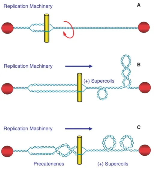
However, once movement of DNA tracking machinery begins, the deleterious effects of DNA topology manifest (Figure 1.2). Since helicases and polymerases separate, but do not unwind, the two strands of DNA, they do not remove any of the turns of the double helix. Consequently, there is an imparted increase of torsional stress that needs to be resolved. An acute overwinding [i.e., (+)SC] subsequently begins ahead of DNA tracking machinery (Postow, Crisona et al.
2001). If not resolved, the accumulation of (+)SCs will present a block to replication and transcription, and these processes will stall rapidly (Brill, DiNardo et al. 1987, Kim and Wang 1989, Wang 1996, Peter, Ullsperger et al. 1998, Wang 2002).
Figure 1.2: Movement of DNA tracking machinery causes topological problems. As DNA tracking systems move through the DNA, twists are pushed ahead of replication forks and transcription complexes, resulting in DNA overwinding that is converted into (+)SCs (A). In the case of replication, precatenanes form behind the fork (B); during transcription, (-)SCs form behind the moving DNA tracking machinery (C).
Topoisomerases
To maintain the appropriate levels of DNA supercoiling and remove knots and tangles in the genome, cells encode enzymes known as topoisomerases (Deweese, Osheroff et al. 2008, Deweese and Osheroff 2009, Vos, Tretter et al. 2011, Chen, Chan et al. 2013, Pommier, Sun et al. 2016, Austin, Lee et al. 2018, Ashley and Osheroff 2019). These enzymes are ubiquitous to all domains of life and are necessary for cellular survival. All topoisomerases modulate the levels of supercoiling in the genome through the creation of a transient break in the DNA helical backbone. Broadly, there are two types of topoisomerases, classified by the number of DNA strands cleaved per enzyme reaction cycle (Bates and Maxwell 2005, Deweese, Osheroff et al.
2008, Deweese and Osheroff 2009, Chen, Chan et al. 2013, Ashley and Osheroff 2019). Type I topoisomerases generate a single-stranded break, or “nick,” in the genetic material (Deweese, Osheroff et al. 2008, Chen, Chan et al. 2013, Ashley and Osheroff 2019, McKie, Neuman et al.
2021). In contrast, type II topoisomerases create a double-stranded break in the DNA double helix (Deweese, Osheroff et al. 2008, Deweese and Osheroff 2009, Vos, Tretter et al. 2011, Chen, Chan et al. 2013, Ashley and Osheroff 2019, McKie, Neuman et al. 2021). All topoisomerases require the use of active site tyrosine residues for catalysis and the process of cutting their DNA substrate results in the formation of covalent bonds between the tyrosine residues and the phosphate backbone of the cleaved DNA substrate. The transiently-cleaved, covalently-linked enzyme-DNA structure that is formed is known as the “cleavage complex”
(Deweese, Osheroff et al. 2008, Deweese and Osheroff 2009, Liu, Deibler et al. 2009, Vos, Tretter et al. 2011, Chen, Chan et al. 2013, Ashley and Osheroff 2019, McKie, Neuman et al.
2021, Vann, Oviatt et al. 2021). Formation of the cleavage complex during enzyme catalysis is thus tightly coordinated to prevent formation of permanent DNA breaks or disruption to genomic
integrity (Deweese, Osheroff et al. 2008, Deweese and Osheroff 2009, Forterre and Gadelle 2009, Vos, Tretter et al. 2011, Chen, Chan et al. 2013, Pommier, Sun et al. 2016, Austin, Lee et al. 2018, Ashley and Osheroff 2019).
Type I Topoisomerases
Type I topoisomerases are primarily monomeric enzymes (with the exception of reverse gyrase, which is tetrameric), most of which do not require a high-energy cofactor, such as ATP, to function (McClendon and Osheroff 2007, Chen, Chan et al. 2013, Seol and Neuman 2016, Ashley and Osheroff 2019). There are three subclasses of type I enzymes: IA, IB, and IC (Wang 1971, Champoux and Dulbecco 1972, Slesarev, Stetter et al. 1993, Deweese, Osheroff et al.
2008, Vos, Tretter et al. 2011).
Type IA topoisomerase was the first topoisomerase discovered and biochemically characterized (i.e., bacterial ω protein) (Wang 1971). Type IA topoisomerases function by creating a transient cut in one strand of the DNA double helix and then passing the opposite intact strand through the break, otherwise known as a “single-strand passage” event (Schvartzman and Stasiak 2004, Deweese, Osheroff et al. 2008, Vos, Tretter et al. 2011, Ashley and Osheroff 2019). All type IA topoisomerases require the coordination of divalent cations, such as Mg2+, for DNA scission. Upon catalytic cleavage of the sugar-phosphate DNA backbone, the bond energy is conserved via formation of a new covalent bond between a tyrosine residue of the enzyme active site and the newly generated 5’-terminal phosphate of the cleaved DNA. This single-stranded DNA passage mechanism changes value of the linking number by 1. As such, type IA enzymes are able to relax the supercoiled molecule but cannot remove knots and tangles.
Type IA topoisomerases mainly function on (-)SCs, preventing the accumulation of
hypernegatively supercoiled DNA and formation of R-loops (a DNA:RNA hybrid) during transcription (Drolet, Broccoli et al. 2003, Tan, Zhou et al. 2015). However, a few type IA enzymes, such as the reverse gyrase enzyme that is only found in thermophilic bacterial species and archaea, are able to introduce (+)SCs into DNA substrates, but only in the presence of ATP (Kikuchi and Asai 1984, Confalonieri, Elie et al. 1993).
Type IB topoisomerases, in contrast to type IA enzymes, do not utilize a strand passage mechanism to regulate DNA supercoiling. Instead, type IB enzymes cut one strand of the DNA substrate and then perform a controlled rotation of the cleaved end around the opposite intact strand (Champoux and Dulbecco 1972, Stivers, Harris et al. 1997, Deweese, Osheroff et al.
2008, Ashley and Osheroff 2019). This “swivel” mechanism occurs when the enzyme is covalently attached to the 3’-terminal phosphate of the cleaved end, allowing the 5’-DNA terminus to freely rotate. This rotation is controlled by the level of torque contained in the DNA molecule (Koster, Croquette et al. 2005). As such, the greater the level of supercoiling in the DNA substrate, the more supercoils are relaxed by the enzyme in one rotation cycle. Similar to the type IA enzymes, the linking number is changed by 1 for every 360º rotation. Type IB enzymes can function on both underwound and overwound DNA substrates but cannot function to decatenate substrates (Vos, Tretter et al. 2011).
Lastly, type IC topoisomerases have been identified only in a single hyperthermophilic methanogenic bacterial species. The only known member of this class is topoisomerase V (Slesarev, Stetter et al. 1993, Ashley and Osheroff 2019). Topoisomerase V activity is independent of ATP and divalent cations, and also behaves similarly to type IB enzymes in its controlled rotation mechanism (Taneja, Patel et al. 2006). The linking number is also changed by
1 for every rotation of the DNA double helix. There is little structural similarity between the type IB and type IC enzymes.
Relaxation
Unknotting
Decatenation Replication and
Transcription
Recombination and Repair
Replication/
Mitosis
Type I Type II
Type II
Type II
Figure 1.3: Functions of type I and type II topoisomerases. The different functions of type I and type II topoisomerases from human and bacterial species allow them to work on DNA substrates of different topological states. Because type I enzymes only cut one strand of DNA, they are only able to work on Tw. Because type II enzymes cut both strands of the DNA double helix, they are able to work on Wr.
Bacteria encode primarily type IA topoisomerases, topoisomerase I and topoisomerase III. Historically, bacterial topoisomerase I has been referred to as the ω protein (Wang 1971, Ashley and Osheroff 2019). Despite the similar numerical designation, bacterial topoisomerase I is unrelated to human topoisomerase I (i.e., bacterial topoisomerase I is a type IA enzyme, whereas human topoisomerase I is a type IB enzyme). ω protein functions in tandem with bacterial gyrase (a type II topoisomerase that will be discussed later) to regulate the overall level of DNA supercoiling in the bacterial genome (Nöllmann, Crisona et al. 2007). Bacterial topoisomerase III is related to human topoisomerase IIIα and IIIβ and is involved in maintaining genomic stability. Although the specific cellular activities of bacterial topoisomerase III are less clear, it has been shown to be more efficient at resolving knots and tangles over relaxing supercoils (Terekhova, Gunn et al. 2012). Bacterial species from genera such as Mycobacterium also encode type IB topoisomerases (Tse-Dinh 1998, Krogh and Shuman 2002, Forterre, Gribaldo et al. 2007, Forterre and Gadelle 2009, Sandhaus, Chapagain et al. 2018). These bacterial type IB enzymes do not structurally resemble those of archaeal or eukaryotic species, but instead are similar to those of poxviruses (Krogh and Shuman 2002, Forterre and Gadelle 2009).
Humans encode both type IA (topoisomerase III and III) and IB (topoisomerase I) enzymes (Deweese, Osheroff et al. 2008, Vos, Tretter et al. 2011, Ashley and Osheroff 2019).
Human topoisomerase III is believed to function to relax hypernegatively supercoiled DNA and prevent inappropriate recombination, as well as resolve recombination intermediates and stalled replication forks (Vos, Tretter et al. 2011). The III isoform can resolve single-stranded DNA tangles (i.e., hemicatenanes) that can arise during replication, repair, and recombination processes; deletion of the III isoform is lethal in mice (Hiasa, DiGate et al. 1994, Harmon,
DiGate et al. 1999). The human III isoform can act as a dual DNA and ribonucleic acid (RNA) topoisomerase, performing both cleavage and strand passage on RNA (Ahmad, Shen et al. 2017, Goto-Ito, Yamagata et al. 2017). In humans, deletion or mutation of topoisomerase III has been linked to schizophrenia and neurodevelopmental disorders (Stoll, Pietilainen et al. 2013, Ahmad, Shen et al. 2017). In mice, deletion of the III isoform is known to shorten life span and can negatively affect neurodevelopment (Stoll, Pietilainen et al. 2013, Xu, Shen et al. 2013). Human topoisomerase I mainly functions to relax (+)SCs and remove torsional stress ahead of replication and transcription machinery. Topoisomerase I has been implicated in maintaining genomic stability and gene-specific transcription. Topoisomerase I is dispensable at the cellular level but appears to be necessary for proper development in multicellular organisms (Lee, Brown et al. 1993, Morham, Kluckman et al. 1996, Nitiss 1998, Miao, Player et al. 2007).
Given the focus of my dissertation, the remainder of the Introduction will focus solely on type II topoisomerases and type I enzymes will not be further discussed.
Type II Topoisomerases
Type II topoisomerases can be classified into two subgroups, IIA and IIB, based on sequence and structural homology (Bates and Maxwell 2005, Deweese, Osheroff et al. 2009, Ashley and Osheroff 2019). The first type IIA topoisomerase (bacterial DNA gyrase) was discovered in 1972 (Gellert, Mizuuchi et al. 1976), and the first type IIB enzyme (topoisomerase VI) was identified in 1997 (Levine, Hiasa et al. 1998, Corbett and Berger 2004, Sissi and Palumbo 2010). All of these type II enzymes can relax DNA supercoils and resolve knots and tangles, and undergo similar catalytic cycles, but can differ in their sequences and structural domains. In contrast to the type I enzymes, type II topoisomerases function via a double-stranded
passage reaction, whereby the DNA double helix is cut on both strands (Deweese, Osheroff et al.
2008, Deweese, Osheroff et al. 2009, Ashley and Osheroff 2019). As such, during reactions, the linking number of their DNA substrates is changed in steps of 2. Lower eukaryotes, such as yeast, and invertebrates encode only one type IIA enzyme, topoisomerase II. Vertebrates such as humans express two closely related forms of the IIA enzyme, topoisomerase II and topoisomerase II (Deweese, Osheroff et al. 2008, Deweese and Osheroff 2009, Liu, Deibler et al. 2009, Nitiss 2009, Chen, Chan et al. 2013, Pommier, Sun et al. 2016, Austin, Lee et al. 2018, Ashley and Osheroff 2019, McKie, Neuman et al. 2021, Vann, Oviatt et al. 2021). Most bacterial species, such as the Gram-negative E. coli and the Gram-positive Bacillus anthracis, encode two type IIA enzymes, gyrase and topoisomerase IV. A few bacterial species, such as Mycobacterium tuberculosis, which is classified as neither Gram-positive nor Gram-negative, encode only a single type IIA topoisomerase, gyrase (Austin and Marsh 1998, Deweese, Osheroff et al. 2008, Deweese and Osheroff 2009, Forterre and Gadelle 2009, Vos, Tretter et al.
2011, Chen, Chan et al. 2013, Pommier, Sun et al. 2016, Austin, Lee et al. 2018, Ashley and Osheroff 2019). The only known type IIB topoisomerase, topoisomerase VI, exists only in plants and archaeal species (Bergerat, Gadelle et al. 1994, Sugimoto-Shirasu, Stacey et al. 2002). Type IIB enzymes are heterotetramers (similar to bacterial type IIA enzymes) but differ from type IIA enzymes in that the TOPRIM domain (to be discussed later) exists in the A subunit of gyrase (instead of in the B subunit, as with bacterial species) (Bergerat, de Massy et al. 1997, Gadelle, Krupovic et al. 2014). The A subunits of type IIA and type IIB topoisomerases do not share sequence or structural identity (Gadelle, Krupovic et al. 2014).
Given the focus of my dissertation, type IIA topoisomerases will often be collectively referred to as type II topoisomerases and type IIB enzymes will not be further discussed.
Figure 1.4: Structures of type IIA topoisomerases. The domain structures of three type IIA topoisomerases, bacterial (Escherichia coli) DNA gyrase and topoisomerase IV, and human topoisomerase IIα are shown. Regions of homology among the enzymes are indicated by colors.
The N-terminal (i.e., GyrB) homology domains contain the regions responsible for ATP binding and hydrolysis (GHKL). The vertical white stripes represent the three conserved motifs that define the ATP-binding domain. The N-terminal domain also contains the binding site for divalent metal ions (TOPRIM). The central (i.e., GyrA) region (WHD) contains the active site tyrosyl residue that forms the covalent bond with DNA during scission. For bacterial gyrase, the variable C-terminal domain contains the “GyrA box” that is necessary for the wrapping mechanism. For human topoisomerase IIα, the CTD contains nuclear localization sequences (NLS) and phosphorylation sites (PO4). The active site tyrosine residue is indicated for each
Type II Topoisomerase Structure Overview
Whereas most bacterial type II topoisomerases are comprised of two heterotetramer units (A2B2), the human type II topoisomerases are formed by two fused heterotetramer units, forming homodimers (A2). All known type IIA topoisomerases in humans and bacteria share a number of common structural features across three regions: the N-terminus, the catalytic core, and the C- terminus (Figure 1.4) (Deweese, Osheroff et al. 2008, Deweese and Osheroff 2009, Vos, Tretter et al. 2011, Chen, Chan et al. 2013, Ashley and Osheroff 2019, Dalvie and Osheroff 2021, McKie, Neuman et al. 2021). The N-terminal region, where the DNA double helical segment enters the enzyme (i.e., the DNA-gate), includes the ATPase, also known as the GHKL (DNA gyrase, Hsp90, bacterial CheA-family histidine kinases, and MutL), domain, and the transducer domain that relays hydrolysis information to the catalytic core (Nitiss 2009, Wendorff, Schmidt et al. 2012). The catalytic core contains the topoisomerase/primase (TOPRIM) domain that coordinates the active site divalent cations, the winged-helix domain (WHD) that contains the active site tyrosine residue, and the tower domain that maintains polar and electrostatic interactions with the DNA substrate (Wendorff, Schmidt et al. 2012, Chang, Wang et al. 2013).
The two domains fundamental to the Mg2+-dependent double-stranded DNA cleavage reaction are the WHD domain and the TOPRIM domain. The C-terminal domain is an intrinsically disordered region (IDR) that contains nuclear localization signals, sites for posttranscriptional modification, and is critical to the recognition of DNA topology (McClendon and Osheroff 2007, Deweese and Osheroff 2009, Nitiss 2009, Lindsey, Pendleton et al. 2014).
First, the WHD contains a helix-turn-helix fold, which a characteristic of all WHD proteins but is commonly found in proteins with DNA-binding function (Harrison and Aggarwal 1990, McKie, Neuman et al. 2021). In addition to its ability to bind DNA, the WHD also
contains the active site tyrosine residue, which is necessary for the nucleophilic attack on the scissile phosphate of the DNA double helical backbone and the formation of the transient reversible topoisomerase-DNA covalent bind (Nitiss 2009, Wendorff, Schmidt et al. 2012, Lindsey, Pendleton et al. 2014).
Second, the TOPRIM domain is formed from an // Rossman-like fold (an extended beta sheet that is sandwiched by alpha helices) (Chang, Wang et al. 2013, McKie, Neuman et al.
2021). The TOPRIM domain is necessary for the transesterification reaction between the scissile phosphate of the DNA backbone and active site tyrosine residue. The active site divalent cation is held by an aspartate-any residue-aspartate (DxD) motif and a glutamate residue that can act as a general acid-base moiety (Aravind, Leipe et al. 1998, Sissi and Palumbo 2009). Collectively, the DxD motif and its coordinate divalent cation in the TOPRIM domain, with the active site tyrosine of the WHD, enable the formation of the two transient cuts of the DNA backbone.
Third, the tower domain functions in DNA bending. The tower domain contains a beta sheet that can interact with one of the captured DNA double helices (the gate or G-segment, to be discussed later), bending the DNA segment to promote cleavage (Dong and Berger 2007, Lee, Jung et al. 2012, Jang, Son et al. 2019, McKie, Neuman et al. 2021). The presence of a conserved, invariant isoleucine residue (across eukaryotic and bacterial species) has been found to intercalate between two base pairs of the G-segment, inducing a ~150º bend. Deletion or mutation of this isoleucine has been found to interfere with proper DNA bending and subsequent relaxation of (+)SC or (-)SC substrates (Dong and Berger 2007, Lee, Dong et al. 2013).
Fourth, the GHKL domain contains an ATP-binding region that is formed from an 8- stranded antiparallel beta sheet surrounded by alpha helices (Corbett and Berger 2004, McKie, Neuman et al. 2021). Binding of ATP induces dimerization and shifting the N-gate into a closed
conformation. The bound ATP interacts with a lysine residue in the transducer domain, and subsequently facilitates rotation (11–18º) between the GHKL and transducer domains (Corbett and Berger 2003, Corbett and Berger 2005, McKie, Neuman et al. 2021). Despite these similarities in enzyme structure among human and bacterial type II topoisomerases, there are substantial differences, which will be discussed below.
Human Type II Topoisomerase Structure
Overall, eukaryotic type II topoisomerases are homologous to bacterial enzymes (described above) (Deweese, Osheroff et al. 2008, Deweese and Osheroff 2009, Vos, Tretter et al. 2011, Chen, Chan et al. 2013, Ashley and Osheroff 2019, Dalvie and Osheroff 2021, McKie, Neuman et al. 2021). There is an N-terminus, followed by a catalytic core region, and then a C- terminus. However, in contrast to the bacterial enzymes, eukaryotic type II topoisomerases consist of a fusion of the two subunits into a single polypeptide sequence with protomer masses
~160-180 kDa (Deweese, Osheroff et al. 2008, Deweese and Osheroff 2009, Chen, Chan et al.
2013, Ashley and Osheroff 2019). In humans, there are two isoforms of type II topoisomerases:
topoisomerase II and topoisomerase II (Austin and Marsh 1998, Deweese, Osheroff et al.
2008, Deweese and Osheroff 2009, Forterre and Gadelle 2009, Vos, Tretter et al. 2011, Chen, Chan et al. 2013, Gentry and Osheroff 2013, Ashley and Osheroff 2019). These isoforms are related in amino acid sequence (~70%) and enzyme structure, but they are encoded by different genes (TOP2A and TOP2B, located at chromosomal bands 17q21–22 and 3p24, respectively) and differ in molecular mass. Whereas human topoisomerase II is 170 kDa in protomer mass, topoisomerase II is 180 kDa (Deweese, Osheroff et al. 2008, Deweese and Osheroff 2009, Chen, Chan et al. 2013, Ashley and Osheroff 2019). On the basis of amino acid sequence
comparisons with E. coli gyrase, the N-terminus of eukaryotic type II enzymes is homologous to GyrB and the central domain is homologous to GyrA (McClendon and Osheroff 2007, Vos, Tretter et al. 2011, Ashley and Osheroff 2019, McKie, Neuman et al. 2021). Crystal structures of the N-terminus and catalytic core in humans have been solved (Schmidt, Osheroff et al. 2012, Wendorff, Schmidt et al. 2012). The C-termini of eukaryotic topoisomerase II and II do not share homology with the corresponding C-terminal domain of gyrase or topoisomerase IV.
Eukaryotic C-terminus contains nuclear localization sequences and sites for posttranslational modifications such as phosphorylation and SUMOylation (McClendon and Osheroff 2007, Ashley and Osheroff 2019, McKie, Neuman et al. 2021). For the II isoform, these modifications can allow the enzyme to be concentrated at centromeres during mitosis (Linka, Porter et al. 2007, Antoniou-Kourounioti, Mimmack et al. 2019). Like E. coli gyrase, it is the C- terminus of human type II topoisomerases that allow it to recognize supercoil handedness during relaxation, preferentially relaxing (+)SC faster than (-)SC DNA (McClendon and Osheroff 2006, McClendon, Gentry et al. 2008, Ashley and Osheroff 2019). To date, the structure of the eukaryotic C-terminal region has not yet been solved (McClendon, Gentry et al. 2008).
Bacterial Type II Topoisomerase Structure
The founding type II enzyme, gyrase, is comprised of two distinct subunits, GyrA (~96 kDa) and GyrB (~88 kDa). Gyrase was first discovered in 1972 during E. coli sedimentation analyses of DNA that showed the presence of negative supercoiling (Worcel and Burgi 1972, Gellert, Mizuuchi et al. 1976). The enzyme was later purified in 1976 (Gellert, Mizuuchi et al.
1976). Structurally, the GyrA portion of the enzyme (WHD, Tower, CTD; Figure 1.4) contains the active site tyrosine residue that forms the covalent bond with DNA during the cleavage
reaction, as well as the C-terminal domain (Corbett and Berger 2004, Vos, Tretter et al. 2011, Gentry and Osheroff 2013, McKie, Neuman et al. 2021). The GyrB portion contains motifs that allow ATP and divalent cation binding as part of the N-terminal domain (GHKL, Transducer, TOPRIM; Figure 1.4) (Corbett and Berger 2004, Vos, Tretter et al. 2011, Gentry and Osheroff 2013, McKie, Neuman et al. 2021). As such, one can envision that GyrB “comes before” GyrA when visualizing the linear polypeptide sequences from left to right. The main function of gyrase in bacteria is to maintain the proper supercoil density of the bacterial genome; gyrase can relax (+)SCs that accumulate ahead of DNA tracking machinery during processes such as replication and transcription, but it is also able to, to a lesser efficiency, decatenate (i.e., unlink) DNA in the presence of divalent cations (Marians 1987, Ullsperger and Cozzarelli 1996, Deweese, Osheroff et al. 2008, Deweese and Osheroff 2009, Vos, Tretter et al. 2011, Chen, Chan et al. 2013, Ashley and Osheroff 2019, Dalvie and Osheroff 2021).
One major distinguishing feature of gyrase is a seven-amino acid motif in the C-terminal domain of the GyrA subunit (Figure 1.4) (Kramlinger and Hiasa 2006, Sissi and Palumbo 2010, Vos, Tretter et al. 2011, Lanz and Klostermeier 2012, McKie, Neuman et al. 2021). This structure is known as the GyrA box, and it uniquely allows for the wrapping of the DNA substrate to introduce (-)SCs and rapidly relax (+)SC DNA in the presence of ATP (Deweese, Osheroff et al. 2008, Sissi and Palumbo 2010, Aldred, Kerns et al. 2014, Gibson, Ashley et al.
2018, Ashley and Osheroff 2019, McKie, Neuman et al. 2021). The GyrA box is found within a loop between strands 1 and 6 of a six-stranded beta pinwheel structure in the C-terminal region (Corbett, Shultzaberger et al. 2004, McKie, Neuman et al. 2021). Previous studies have demonstrated that the GyrA box (sequence QRRGGKG), when mutated via deletions or alanine substitutions, abrogate the ability of the enzyme to introduce (-)SCs (Kampranis and Maxwell
1996, Kramlinger and Hiasa 2006, Ashley, Dittmore et al. 2017, Ashley and Osheroff 2019, McKie, Neuman et al. 2021). Mutations within the GyrA box affect the ability of gyrase to rapidly relax (+)SC DNA and relax (-)SC in the presence of ATP (Kramlinger and Hiasa 2006, Ashley, Dittmore et al. 2017, Ashley and Osheroff 2019, McKie, Neuman et al. 2021). It thus appears that the GyrA box is the defining feature of gyrase that enables it to freely supercoil DNA. Negative supercoils are introduced into the DNA substrate when the G-segment, being bound to the DNA-gate of the N-terminus, is chirally wrapped around one of the GyrA C- terminal domains to form a constrained (+)SC (Nöllmann, Crisona et al. 2007, Deweese, Osheroff et al. 2008, Ashley and Osheroff 2019, McKie, Neuman et al. 2021). This positive supercoil is then converted into a negative supercoil following strand passage, where the G- segment is translocated at a 60° angle, introducing negative writhe to the molecule (Deweese, Osheroff et al. 2008, Ashley and Osheroff 2019, McKie, Neuman et al. 2021).
Like gyrase, topoisomerase IV is also a heterotetramer that contains two subunits. The nomenclature of topoisomerase IV stemmed from their first identification in 1990 as gyrase homologs required for chromosomal segregation and cellular partitioning (Kato, Nishimura et al.
1990, Deweese, Osheroff et al. 2008, Deweese and Osheroff 2009, Forterre and Gadelle 2009, Vos, Tretter et al. 2011, Chen, Chan et al. 2013, Ashley and Osheroff 2019). In Gram-negative species such as E. coli, the topoisomerase IV subunits are designated as ParC (~88 kDa, homologous to GyrA) and ParE (~70 kDa, homologous to GyrB) (Kato, Nishimura et al. 1990, Kato, Suzuki et al. 1992). In Gram-positive species such as B. anthracis, the subunits of topoisomerase IV are named GrlA (Gyrase-like gene A) and GrlB (Gyrase-like gene B) (Levine, Hiasa et al. 1998, Gentry and Osheroff 2013, Ashley, Dittmore et al. 2017). “Reading” from left to right, the enzyme “order” would thus be ParE/GrlB and ParC/GrlA. Like gyrase,
topoisomerase IV is also able to relax (+)SC DNA. However, topoisomerase IV differs from gyrase because of the inability to supercoil DNA (Hiasa and Marians 1994, Crisona, Strick et al.
2000, Zechiedrich, Khodursky et al. 2000). In comparison to gyrase, the ParC C-terminal domain does not contain the necessary structure to supercoil DNA, instead having a “broken” five (not six) beta pinwheel, and the absence of a GyrA box (Corbett, Shultzaberger et al. 2004, Corbett, Schoeffler et al. 2005, Tretter, Lerman et al. 2010, Vos, Lee et al. 2013). Remnants of the canonical GyrA motif have been found in each of its pinwheel “blades” (Tretter, Lerman et al.
2010, Vos, Lee et al. 2013). Nonetheless, the ParC C-terminal domain contains positively charged moieties on its outer surface, suggesting a role in binding DNA (Corbett and Berger 2004). The loss of the ParC C-terminal domain has been found to impede the ability of E. coli topoisomerase IV to distinguish between topologically distinct substrates when relaxing and decatenating DNA substrates of different supercoil handedness (Corbett, Schoeffler et al. 2005, Vos, Lee et al. 2013).
+ M g2 +
+ 2 A T P A T P è A D P
A D P çA T P 1
2 3
4
5
6 7
Gate (G) segment Transfer (T) segment
Topoisomerase
Figure 1.5: The catalytic cycle of type II topoisomerases. The double-stranded DNA passage reaction of topoisomerase II can be separated into seven discrete steps. 1) Type II enzyme (blue) binding to two segments of DNA: the gate segment (green) and transport segment (yellow). 2) Bending of the gate segment, which requires the presence of Mg2+ or other divalent metal ions. These metal ions are required for all subsequent steps. 3) Double-stranded DNA cleavage of the gate segment (i.e., formation of the cleavage complex). 4) Passage of the transport segment through the DNA gate generated by cleavage. This reaction requires the binding of 2 ATP molecules, and strand passage proceeds more rapidly if one of the two ATP molecules is hydrolyzed. 5) Ligation of the cleaved DNA gate segment. 6) Hydrolysis of the second ATP molecule, which allows release of the gate segment through a C-terminal gate in the protein. 7) Enzyme turnover and closing of the protein gate, which regenerates the enzyme to initiate a new round of catalysis. Artwork from Ashley et al., 2019.
Type II Topoisomerase Function Overview
All type II topoisomerases function by forming transient double-stranded DNA breaks and modulate the topological state of DNA by a double-stranded passage reaction (Figure 1.5) (Deweese, Osheroff et al. 2008, Deweese and Osheroff 2009, Vos, Tretter et al. 2011, Ashley and Osheroff 2019, McKie, Neuman et al. 2021, Vann, Oviatt et al. 2021). The enzyme begins its catalytic cycle by first capturing a segment of DNA through the opening of the N-terminal region of the enzyme (Figure 5, step 1). This first segment will be cut by the enzyme and is known as the “gate” or G-segment. In contrast, the segment that will be secondly captured and eventually transported through the transiently cleaved G-segment is known as the “transport” or T-segment. In the presence of a divalent cation such as Mg2+ and in coordination with the TOPRIM domain, the G-segment is assessed for bendability (Jang, Son et al. 2019). DNA sequences that can be bent are distorted to an angle of ~150° and can be used as the site for scission (Dong and Berger 2007, Lee, Dong et al. 2013).
The bent G-segment is then cleaved via a nucleophilic attack by the two active site tyrosine residues on the phosphate backbone of the double helix (Figure 1.5, step 2). Cleavage is initiated when a general base, which is believed to be a conserved histidine residue, deprotonates the hydroxyl group of the active site tyrosine, allowing the oxyanion to attack the scissile phosphate (Figure 1.5, step 3). Two cofactors are needed by the enzyme to carry out this and the subsequent double-stranded DNA passage reactions (Deweese, Osheroff et al. 2008, Deweese and Osheroff 2009, Vos, Tretter et al. 2011, Chen, Chan et al. 2013, Ashley and Osheroff 2019).
The first cofactor is a divalent cation, such as magnesium (i.e., Mg2+), for all steps beyond enzyme-DNA binding. The enzyme uses a non-canonical two-metal ion mechanism at each cut site (Noble and Maxwell 2002, Deweese and Osheroff 2009, Schmidt, Burgin et al. 2010,
Pommier, Sun et al. 2016). The presence of one divalent cation enables interaction with the bridging 5’-oxygen molecule of the scissile bond and speeds up rates of enzyme-mediated cleavage at the first cut site. A second divalent cation is believed to make critical contacts with and help deprotonate the active site tyrosine, thereby stabilizing the DNA transition state. Once the first DNA strand is cut, the second strand is cleaved ~20-fold faster (Deweese, Guengerich et al. 2009). The resulting transiently-cleaved cleavage complex has the enzyme covalently bound to the scissile 5’-phosphate of the double helical backbone. The second cofactor is ATP, which drives the strand passage reaction. ATP is not necessary for either the DNA cleavage or religation of the DNA substrate (Deweese, Osheroff et al. 2008, Deweese and Osheroff 2009, Vos, Tretter et al. 2011, Chen, Chan et al. 2013, Ashley and Osheroff 2019, McKie, Neuman et al. 2021, Vann, Oviatt et al. 2021).
In greater detail, the cleavage complex is a transient enzyme-DNA structure connected by two staggered 4-base single-stranded cohesive overhangs in the 5’-end of one DNA sequence, a 3’-hydroxyl moiety on the opposite terminus of the cleaved strand, and a gap in the double helix (Deweese and Osheroff 2009, Nitiss 2009, Pommier, Leo et al. 2010, Vos, Tretter et al. 2011, Ashley and Osheroff 2019, McKie, Neuman et al. 2021, Vann, Oviatt et al. 2021). To maintain the bond energy of the sugar-phosphate backbone as well as genomic integrity during the cleavage process, the type II enzyme forms covalent bonds between the active site tyrosine and the 5’-phosphate group of the DNA backbone, generating a phosphotyrosyl linkage (Deweese and Osheroff 2009, Nitiss 2009, Pommier, Leo et al. 2010, Vos, Tretter et al. 2011, Ashley and Osheroff 2019, McKie, Neuman et al. 2021, Vann, Oviatt et al. 2021). Upon the binding of two ATP molecules, the N-terminal gate is closed. Closing of the N-terminal gate triggers a conformational change in the enzyme that helps translocate the T-segment through the transient
opening in the enzyme active site, performing strand passage (Figure 1.5, step 4). Although hydrolysis of the high-energy cofactor is not necessarily a prerequisite for strand passage to occur, it appears that this step occurs faster if one of the two bound ATP molecules is hydrolyzed (Lindsley and Wang 1993).
After strand passage, a second, post-strand passage, cleavage complex is formed (Figure 1.5, step 5). The type II enzyme then religates the cleaved DNA to regenerate the intact DNA double helix. DNA religation is initiated when a general acid removes the hydrogen from the 3’- terminal hydroxyl group (Deweese and Osheroff 2009, Wendorff, Schmidt et al. 2012). Another nucleophilic attack is then initiated on the phosphotyrosyl bond, regenerating the intact DNA double helical backbone and the enzyme active site. The T-segment is then released from the protein (Figure 1.5, step 6). Hydrolysis of a second ATP molecule then occurs, and the enzyme releases the G-segment. Lastly, the type II enzyme conformation is reset, allowing for the next cycle of catalysis (Figure 1.5, step 7) (Osheroff 1986, Roca and Wang 1992, Wang 1998, Wilstermann and Osheroff 2001, Deweese and Osheroff 2009, Vos, Tretter et al. 2011, Vann, Oviatt et al. 2021).
Human Type II Topoisomerase Function
In humans, type II topoisomerases play a role in virtually every major nucleic acid process (Deweese, Osheroff et al. 2008, Deweese and Osheroff 2009, Forterre and Gadelle 2009, Vos, Tretter et al. 2011, Chen, Chan et al. 2013, Pommier, Sun et al. 2016, Ashley and Osheroff 2019). These functions include untangling daughter chromosomes that form during replication, resolving knots formed during recombination, removing (+)SC DNA generated ahead of replication forks and transcription complexes, and maintaining proper chromosome organization
and structure as the major non-histone protein of the mitotic chromosome scaffold and the interphase nuclear matrix (Earnshaw, Halligan et al. 1985, Gasser, Laroche et al. 1986, Deweese and Osheroff 2009, Vos, Tretter et al. 2011). Lower eukaryotes and non-vertebrate species such as yeast encode only a single type II enzyme, topoisomerase II (Wyckoff and Hsieh 1988, McClendon, Rodriguez et al. 2005). In vertebrates and humans, however, both topoisomerase II
and topoisomerase II are expressed; it remains unclear why two distinct isoforms are encoded (Deweese, Osheroff et al. 2008, Deweese and Osheroff 2009, Forterre and Gadelle 2009, Vos, Tretter et al. 2011, Chen, Chan et al. 2013, Pommier, Sun et al. 2016, Ashley and Osheroff 2019). Despite their broad similarities, there are several crucial differences in expression and function between these two isoforms that will be discussed.
Human topoisomerase II and topoisomerase II are distinct in their expression patterns (Heck and Earnshaw 1986, Heck, Hittelman et al. 1988, Woessner, Mattern et al. 1991, McClendon and Osheroff 2007, Vos, Tretter et al. 2011). Topoisomerase II is required for the survival of proliferating cells (Heck and Earnshaw 1986, Heck, Hittelman et al. 1988, Hsiang, Wu et al. 1988, McClendon and Osheroff 2007, Ketron and Osheroff 2014). Levels of topoisomerase II expression increase throughout S-phase of the cell cycle, beginning at lower levels during G1 and rising through S, eventually peaking at the G2/M phase boundary (Heck, Hittelman et al. 1988, Woessner, Mattern et al. 1991, Kimura, Saijo et al. 1994). Topoisomerase II is found almost exclusively in actively proliferating tissues, localizes predominantly in the nucleus, is associated with replication forks and transcription machinery, and has been found to be tightly bound to chromosomes and sister chromatids throughout mitosis (Uemura, Ohkura et al. 1987, Woessner, Mattern et al. 1991, Kimura, Saijo et al. 1994, Mirski, Gerlach et al. 1997, Grue, Grasser et al. 1998, Mirski, Gerlach et al. 1999, McClendon and Osheroff 2007, Lee and
Berger 2019). As such, the type II enzyme is believed to be the main isoform that functions in growth-related processes such as replication and chromosomal segregation (Grue, Grasser et al.
1998, Nitiss 2009, Pommier, Sun et al. 2016, McKie, Neuman et al. 2021, Vann, Oviatt et al.
2021). While there is evidence that topoisomerase II can act ahead of replication forks, it is believed to primarily act behind them (Heintzman, Campos et al. 2019).
In contrast to topoisomerase II, the II isoform is not required for survival at the cellular level, and II activity cannot replace that of II (Dereuddre, Delaporte et al. 1997, Grue, Grasser et al. 1998, Bakshi, Galande et al. 2001, Nitiss 2009, Ketron and Osheroff 2014, Austin, Lee et al. 2018). The concentration of topoisomerase II expressed in cells is independent of the stage of the cell cycle and this isoform is found at generally consistent levels in most cell types regardless of cell proliferation status (Austin and Marsh 1998, Christensen, Larsen et al. 2002, Cowell, Sondka et al. 2012, McKie, Neuman et al. 2021). Unlike its II counterpart, the II
enzyme dissociates from chromosomes during mitosis (Austin and Marsh 1998, Isaacs, Davies et al. 1998, Linka, Porter et al. 2007). Although the precise cellular functions of topoisomerase II
have yet to be fully understood, it has been shown to be involved in the regulation of hormonally-related genes at the transcriptional level (Yang, Li et al. 2000, Ju, Lunyak et al.
2006, Deweese and Osheroff 2009, Ketron and Osheroff 2014, Austin, Lee et al. 2018, McKie, Neuman et al. 2021, Vann, Oviatt et al. 2021). Topoisomerase II is necessary for proper neural development in mouse embryos (Yang, Li et al. 2000, Lyu and Wang 2003). Conditional knockouts have implicated topoisomerase II activity in proper retinal development and ovulation (Zhang, Yu et al. 2013, Li, Hao et al. 2014). More recently, NGS studies using chromatin immunoprecipitation sequencing (ChIP-seq) have mapped II activity at borders of
Hou et al. 2016, Martínez-Garcia, García-Torres et al. 2021, McKie, Neuman et al. 2021). Taken together, there may be functions of topoisomerase II in proper hormonal development and cellular differentiation via interactions with transcription machinery (Yang, Li et al. 2000, Ju, Lunyak et al. 2006, McClendon and Osheroff 2007, Liu, Deibler et al. 2009, Nitiss 2009, Pommier, Sun et al. 2016, Bollimpelli, Dholaniya et al. 2017, Austin, Lee et al. 2018).
Gyrase and Topoisomerase IV Function
Despite their similarities in structure, gyrase and topoisomerase IV play different roles in cellular environments. In addition to its unique ability to introduce (-)SCs into DNA, the strand wrapping mechanism of gyrase allows it to remove (+)SCs substantially faster than it introduces (-)SCs into DNA (Ashley, Blower et al. 2017, Ashley, Dittmore et al. 2017). Because of its ability to wrap DNA during catalysis, gyrase functions primarily to relax or generate supercoils (Levine, Hiasa et al. 1998, Khodursky, Peter et al. 2000, Hsu, Chung et al. 2006, Tadesse and Graumann 2006, Deweese, Osheroff et al. 2008, Aldred, Kerns et al. 2014, Gibson, Ashley et al.
2018, Ashley and Osheroff 2019). To put this into the context of biological function, DNA gyrase plays an important role in removing the (+)SCs that can form ahead of DNA tracking machinery (i.e., polymerases and helicases) during essential nucleic acid processes such as replication and transcription (Levine, Hiasa et al. 1998, Khodursky, Peter et al. 2000, Hsu, Chung et al. 2006, Tadesse and Graumann 2006, Deweese, Osheroff et al. 2008, Aldred, Kerns et al. 2014, Gibson, Ashley et al. 2018). Its strong ability to rapidly and preferentially relax (+)SCs versus introduce (-)SCs would make it a suitable enzyme to function ahead of replication forks and transcription complexes to alleviate torsional stress induced by DNA overwinding.
Additionally, gyrase works in conjunction with protein (a type I topoisomerase) to maintain
the global negative superhelicity of DNA (Mirkin, Zaitsev et al. 1984, Tse-Dinh 1998, Ashley and Osheroff 2019).
In contrast to gyrase, topoisomerase IV is the enzyme primarily responsible for resolving knots and tangles that form during nucleic acid processes (Levine, Hiasa et al. 1998, Tadesse and Graumann 2006, Deweese, Osheroff et al. 2008, Wang, Reyes-Lamothe et al. 2008, Liu, Deibler et al. 2009, Sissi and Palumbo 2010, Aldred, Kerns et al. 2014, Zawadzki, Stracy et al. 2015, Gibson, Ashley et al. 2018, Ashley and Osheroff 2019). As such, topoisomerase IV mainly resolves precatenanes that form behind replication forks and removes DNA knots that form during recombination. Nonetheless, topoisomerase IV has been found to remove (+)SCs from DNA substrates more efficiently than it does (-)SCs (Ashley, Dittmore et al. 2017).
Topoisomerase IV may also play a role ahead of DNA tracking systems, but the precise nature of this process is less understood (Vos, Lee et al. 2013, Ashley, Dittmore et al. 2017).
Type II Topoisomerases in Cellular Environments: When Good Enzymes Go Bad
All type II topoisomerases generate a transiently-cleaved, covalently-linked enzyme- DNA complex during the strand passage reaction, which is necessary for maintaining the proper topological state of DNA in cellular systems. However, the formation of the cleavage complex itself poses a potential danger to the cell (Figure 1.6) (Anderson and Osheroff 2001, Deweese and Osheroff 2009, Nitiss 2009, Aldred, Kerns et al. 2014, Ketron and Osheroff 2014, Pendleton, Lindsey et al. 2014, Pommier, Sun et al. 2016, Vann, Oviatt et al. 2021). Given that a