Lynn S. Bickley, MD, FACP
Clinical Professor of Internal Medicine School of Medicine
University of New Mexico Albuquerque, New Mexico
Peter G. Szilagyi, MD, MPH
Professor of Pediatrics
Chief, Division of General Pediatrics
University of Rochester School of Medicine and Dentistry Rochester, New York
Product Manager: Annette Ferran Editorial Assistant: Ashley Fischer Design Coordinator: Joan Wendt Art Director, Illustration: Brett MacNaughton Manufacturing Coordinator: Karin Duffield Indexer: Angie Allen
Prepress Vendor: Aptara, Inc.
7th Edition
Copyright © 2013 Wolters Kluwer Health | Lippincott Williams & Wilkins.
Copyright © 2009 by Wolters Kluwer Health | Lippincott Williams & Wilkins. Copyright © 2007, 2004, 2000 by Lippincott Williams & Wilkins. Copyright © 1995, 1991 by J. B. Lippincott Company.
All rights reserved. This book is protected by copyright. No part of this book may be reproduced or transmitted in any form or by any means, including as photocopies or scanned-in or other electronic copies, or utilized by any information storage and retrieval system without written permission from the copyright owner, except for brief quotations embodied in critical articles and reviews. Materials appear- ing in this book prepared by individuals as part of their official duties as U.S. government employees are not covered by the above-mentioned copyright. To request permission, please contact Lippincott Williams & Wilkins at Two Commerce Square, 2001 Market Street, Philadelphia PA 19103, via email at [email protected] or via website at lww.com (products and services).
9 8 7 6 5 4 3 2 1 Printed in China
Library of Congress Cataloging-in-Publication Data Bickley, Lynn S.
Bates’ pocket guide to physical examination and history taking / Lynn S. Bickley, Peter G. Szilagyi. — 7th ed.
p. ; cm.
Pocket guide to physical examination and history taking
Abridgement of: Bates’ guide to physical examination and history-taking. 11th ed. / Lynn S.
Bickley, Peter G. Szilagyi. c2013.
Includes bibliographical references and index.
Summary: “This concise pocket-sized guide presents the classic Bates approach to physical exami- nation and history taking in a quick-reference outline format. It contains all the critical information needed to obtain a clinically meaningful health history and to conduct a thorough physical assessment.
Fully revised and updated, the Seventh Edition will help health professionals elicit relevant facts from the patient’s history, review examination procedures, highlight common findings, learn special assess- ment techniques, and sharpen interpretive skills.The book features a vibrant full-color art program and an easy-to-follow two-column format with step-by-step examination techniques on the left and abnormalities with differential diagnoses on the right.”—Provided by publisher.
ISBN 978-1-4511-7322-2 (pbk. : alk. paper)
I. Bates, Barbara, 1928-2002. II. Szilagyi, Peter G. III. Bickley, Lynn S. Bates’ guide to physical examination and history-taking. IV. Title. V. Title: Pocket guide to physical examination and history taking.
[DNLM: 1. Physical Examination—methods—Handbooks. 2. Medical History Taking—
methods—Handbooks. WB 39]
616.07′51—dc23 2012030529
Care has been taken to confirm the accuracy of the information presented and to describe gener- ally accepted practices. However, the authors, editors, and publisher are not responsible for errors or omissions or for any consequences from application of the information in this book and make no warranty, expressed or implied, with respect to the currency, completeness, or accuracy of the contents of the publication. Application of this information in a particular situation remains the professional responsibility of the practitioner; the clinical treatments described and recommended may not be con- sidered absolute and universal recommendations.
The authors, editors, and publisher have exerted every effort to ensure that drug selection and dosage set forth in this text are in accordance with the current recommendations and practice at the time of publication. However, in view of ongoing research, changes in government regulations, and the constant flow of information relating to drug therapy and drug reactions, the reader is urged to check the package insert for each drug for any change in indications and dosage and for added warn- ings and precautions. This is particularly important when the recommended agent is a new or infre- quently employed drug.
Some drugs and medical devices presented in this publication have Food and
Drug Administration (FDA) clearance for limited use in restricted research settings. It is the responsi- bility of the health care provider to ascertain the FDA status of each drug or device planned for use in his or her clinical practice.
LWW.COM
and to students world-wide committed to clinical excellence.
The Pocket Guide to Physical Examination and History Taking, 7th edition is a concise, portable text that:
● Describes how to interview the patient and take the health history.
● Provides an illustrated review of the physical examination.
● Reminds students of common, normal, and abnormal physical findings.
● Describes special techniques of assessment that students may need in specific instances.
● Provides succinct aids to interpretation of selected findings.
There are several ways to use the Pocket Guide:
● To review and remember the content of a health history.
● To review and rehearse the techniques of examination. This can be done while learning a single section and again while combining the approaches to several body systems or regions into an integrated examination (see Chap. 1).
● To review common variations of normal and selected abnormalities.
Observations are keener and more precise when the examiner knows what to look, listen, and feel for.
● To look up special techniques as the need arises. Maneuvers such as The Timed Get Up and Go test are included in the Special Techniques sections in each chapter.
● To look up additional information about possible findings, including abnormalities and standards of normal.
The Pocket Guide is not intended to serve as a primary text for learn- ing the skills of history taking or physical examination. Its detail is too brief for these purposes. It is intended instead as an aid for student review and recall and as a convenient, brief, and portable reference.
vii
+0)8<-:
1
Overview: Physical Examination and History Taking 1+0)8<-:
2
Clinical Reasoning, Assessment, and Recording Your Findings 15+0)8<-:
3
Interviewing and the Health History 31+0)8<-:
4
Beginning the Physical Examination: General Survey, Vital Signs, and Pain 49+0)8<-:
5
Behavior and Mental Status 67+0)8<-:
6
The Skin, Hair, and Nails 83+0)8<-:
7
The Head and Neck 99+0)8<-:
8
The Thorax and Lungs 127+0)8<-:
9
The Cardiovascular System 147+0)8<-:
10
The Breasts and Axillae 167+0)8<-:
11
The Abdomen 179+0)8<-:
12
The Peripheral Vascular System 199+0)8<-:
13
Male Genitalia and Hernias 211+0)8<-:
14
Female Genitalia 225+0)8<-:
15
The Anus, Rectum, and Prostate 241+0)8<-:
16
The Musculoskeletal System 251+0)8<-:
17
The Nervous System 285+0)8<-:
18
Assessing Children: Infancy Through Adolescence 323+0)8<-:
19
The Pregnant Woman 359+0)8<-:
20
The Older Adult 373Index 395
ix
1
Overview: Physical 1
Examination and History Taking
This chapter provides a road map to clinical proficiency in two critical areas: the health history and the physical examination.
For adults, the comprehensive history includes Identifying Data and Source of the History, Chief Complaint(s), Present Illness, Past History, Family History, Personal and Social History, and Review of Systems. New patients in the office or hospital merit a comprehensive health history; however, in many situations, a more flexible focused, or problem-oriented, interview is appropriate. The components of the comprehensive health history structure the patient’s story and the format of your written record, but the order shown below should not dictate the sequence of the interview. The interview is more fluid and should follow the patient’s leads and cues, as described in Chapter 3.
Overview: Components of the Adult Health History
Identifying Data ◗Identifying data—such as age, gender, occupation, marital status
◗Source of the history—usually the patient, but can be a family member or friend, letter of referral, or the medical record
◗If appropriate, establish source of referral because a written report may be needed
Reliability ◗Varies according to the patient’s memory, trust, and mood
Chief Complaint(s) ◗The one or more symptoms or concerns causing the patient to seek care
(continued)
Be sure to distinguish subjective from objective data. Decide if your assessment will be comprehensive or focused.
Overview: Components of the Adult Health History (continued)
Present Illness ◗Amplifies the Chief Complaint; describes how each symptom developed
◗Includes patient’s thoughts and feelings about the illness
◗Pulls in relevant portions of the Review of Systems, called “pertinent positives and negatives” (see p. 3)
◗May include medications, allergies, habits of smoking and alcohol, which frequently are pertinent to the present illness
Past History ◗Lists childhood illnesses
◗Lists adult illnesses with dates for at least four categories: medical, surgical, obstetric/gynecologic, and psychiatric
◗Includes health maintenance practices such as immunizations, screening tests, lifestyle issues, and home safety
Family History ◗Outlines or diagrams age and health, or age and cause of death, of siblings, parents, and grandparents
◗Documents presence or absence of specific illnesses in family, such as hypertension, coronary artery disease, etc.
Personal and Social History
◗Describes educational level, family of origin, current household, personal interests, and lifestyle Review of Systems ◗Documents presence or absence of common symp-
toms related to each major body system
Subjective Data Objective Data
What the patient tells you What you detect during the examination The history, from Chief Complaint
through Review of Systems
All physical examination findings
The Comprehensive Adult Health History
As you elicit the adult health history, be sure to include the following:
date and time of history; identifying data, which include age, gender, marital status, and occupation; and reliability, which reflects the quality of information the patient provides.
T T
The e C C Co om mp p pre ehe en nsiv ve e A Adu u ult He e ealth Hi is story y
CHIEF COMPLAINT(S)
Quote the patient’s own words. “My stomach hurts and I feel awful”;
or “I have come for my regular check-up.”
PRESENT ILLNESS
This section is a complete, clear, and chronologic account of the prob- lems prompting the patient to seek care. It should include the prob- lem’s onset, the setting in which it has developed, its manifestations, and any treatments.
Every principal symptom should be well characterized, with descrip- tions of the seven features listed below and pertinent positives and negatives from relevant areas of the Review of Systems that help clarify the differential diagnosis.
The Seven Attributes of Every Symptom
◗Location
◗Quality
◗Quantity or severity
◗Timing, including onset, duration, and frequency
◗Setting in which it occurs
◗Aggravating and relieving factors
◗Associated manifestations
In addition, list medications, including name, dose, route, and frequency of use; allergies, including specific reactions to each medication; tobacco use; and alcohol and drug use.
HISTORY
List childhood illnesses, then list adult illnesses in each of four areas:
●Medical (e.g., diabetes, hypertension, hepatitis, asthma, HIV), with dates of onset; also information about hospitalizations with dates; number and gender of sexual partners; risky sexual practices
●Surgical (dates, indications, and types of operations)
●Obstetric/gynecologic (obstetric history, menstrual history, birth control, and sexual function)
●Psychiatric (illness and time frame, diagnoses, hospitalizations, and treatments)
Also discuss Health Maintenance, including immunizations, such as tetanus, pertussis, diphtheria, polio, measles, rubella, mumps, influenza, varicella, hepatitis B, Haemophilus influenzae type b, pneumococcal vaccine, and herpes zoster vaccine; and screening tests, such as tuber- culin tests, Pap smears, mammograms, stool tests, for occult blood colonoscopy, and cholesterol tests, together with the results and the dates they were last performed.
FAMILY HISTORY
Outline or diagram the age and health, or age and cause of death, of each immediate relative, including grandparents, parents, siblings, children, and grandchildren. Record the following conditions as either present or absent in the family: hypertension, coronary artery disease, ele- vated cholesterol levels, stroke, diabetes, thyroid or renal disease, cancer (specify type), arthritis, tuberculosis, asthma or lung disease, headache, seizure disorder, mental illness, suicide, alcohol or drug addiction, and allergies, as well as conditions that the patient reports.
PERSONAL AND SOCIAL HISTORY
Include occupation and the last year of schooling; home situation and significant others; sources of stress, both recent and long term; impor- tant life experiences, such as military service; leisure activities; religious affiliation and spiritual beliefs; and activities of daily living (ADLs).
Also include lifestyle habits such as exercise and diet, safety measures, and alternative health care practices.
REVIEW OF SYSTEMS (ROS)
These “yes/no” questions go from “head to toe” and conclude the inter- view. Selected sections can also clarify the Chief Complaint; for example, the respiratory ROS helps characterize the symptom of cough. Start with a fairly general question. This allows you to shift to more specific ques- tions about systems that may be of concern. For example, “How are your ears and hearing?” “How about your lungs and breathing?” “Any trouble
with your heart?” “How is your digestion?” The Review of Systems ques- tions may uncover problems that the patient overlooked. Remember to move major health events to the Present Illness or Past History in your write-up.
Some clinicians do the Review of Systems during the physical examination.
If the patient has only a few symptoms, this combination can be efficient but may disrupt the flow of both the history and the examination.
General. Usual weight, recent weight change, clothing that fits more tightly or loosely than before; weakness, fatigue, fever.
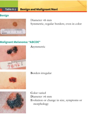
Skin. Rashes, lumps, sores, itching, dryness, color change; changes in hair or nails; changes in size or color of moles.
Head, Eyes, Ears, Nose, Throat (HEENT). Head: Headache, head injury, dizziness, lightheadedness. Eyes: Vision, glasses or contact lenses, last examination, pain, redness, excessive tearing, double or blurred vision, spots, specks, flashing lights, glaucoma, cataracts.
Ears: Hearing, tinnitus, vertigo, earache, infection, discharge. If hear- ing is decreased, use or nonuse of hearing aid. Nose and sinuses: Fre- quent colds, nasal stuffiness, discharge or itching, hay fever, nosebleeds, sinus trouble. Throat (or mouth and pharynx): Condition of teeth and gums; bleeding gums; dentures, if any, and how they fit; last dental examination; sore tongue; dry mouth; frequent sore throats; hoarseness.
Neck. Lumps, “swollen glands,” goiter, pain, stiffness.
Breasts. Lumps, pain or discomfort, nipple discharge, self-examination practices.
Respiratory. Cough, sputum (color, quantity), hemoptysis, dyspnea, wheezing, pleurisy, last chest x-ray. You may wish to include asthma, bronchitis, emphysema, pneumonia, and tuberculosis.
Cardiovascular. “Heart trouble,” hypertension, rheumatic fever, heart murmurs, chest pain or discomfort, palpitations, dyspnea, orthopnea, paroxysmal nocturnal dyspnea, edema, past electrocardio- graphic or other cardiovascular tests.
Gastrointestinal. Trouble swallowing, heartburn, appetite, nausea.
Bowel movements, color and size of stools, change in bowel habits, rectal bleeding or black or tarry stools, hemorrhoids, constipation, diarrhea. Abdominal pain, food intolerance, excessive belching or passing of gas. Jaundice, liver or gallbladder trouble, hepatitis.
Peripheral Vascular. Intermittent claudication; leg cramps; varicose veins; past clots in veins; swelling in calves, legs, or feet; color change in fingertips or toes during cold weather; swelling with redness or tenderness.
Urinary. Frequency of urination, polyuria, nocturia, urgency, burn- ing or pain on urination, hematuria, urinary infections, kidney stones, incontinence; in males, reduced caliber or force of urinary stream, hesitancy, dribbling.
Genital. Male: Hernias, discharge from or sores on penis, testicu- lar pain or masses, history of sexually transmitted infections (STIs) or diseases (STDs) and treatments, testicular self-examination practices.
Sexual habits, interest, function, satisfaction, birth control methods, condom use, problems. Concerns about HIV infection. Female: Age at menarche; regularity, frequency, and duration of periods; amount of bleeding, bleeding between periods or after intercourse, last menstrual period; dysmenorrhea, premenstrual tension. Age at menopause, meno- pausal symptoms, postmenopausal bleeding. In patients born before 1971, exposure to diethylstilbestrol (DES) from maternal use during pregnancy. Vaginal discharge, itching, sores, lumps, STIs and treat- ments. Number of pregnancies, number and type of deliveries, number of abortions (spontaneous and induced), complications of pregnancy, birth control methods. Sexual preference, interest, function, satisfaction, problems (including dyspareunia). Concerns about HIV infection.
Musculoskeletal. Muscle or joint pain, stiffness, arthritis, gout, backache. If present, describe location of affected joints or muscles, any swelling, redness, pain, tenderness, stiffness, weakness, or limita- tion of motion or activity; include timing of symptoms (e.g., morn- ing or evening), duration, and any history of trauma. Neck or low back pain. Joint pain with systemic features such as fever, chills, rash, anorexia, weight loss, or weakness.
Psychiatric. Nervousness; tension; mood, including depression, memory change, suicide attempts, if relevant.
Neurologic. Changes in mood, attention, or speech; changes in ori- entation, memory, insight, or judgment; headache, dizziness, vertigo;
fainting, blackouts, seizures, weakness, paralysis, numbness or loss of sensation, tingling or “pins and needles,” tremors or other involuntary movements, seizures.
Hematologic. Anemia, easy bruising or bleeding, past transfusions, transfusion reactions.
Endocrine. “Thyroid trouble,” heat or cold intolerance, excessive sweating, excessive thirst or hunger, polyuria, change in glove or shoe size.
The Physical Examination:
Approach and Overview
Conduct a comprehensive physical examination on most new patients or patients being admitted to the hospital. For more problem-oriented, or focused, assessments, the presenting complaints will dictate which segments you elect to perform.
●The key to a thorough and accurate physical examination is a sys- tematic sequence of examination. With effort and practice, you will acquire your own routine sequence. This book recommends exam- ining from the patient’s right side.
●Apply the techniques of inspection, palpation, auscultation, and per- cussion to each body region, but be sensitive to the whole patient.
●Minimize the number of times you ask the patient to change position from supine to sitting, or standing to lying supine.
●For an overview of the physical examination, study the sequence that follows. Note that clinicians vary in where they place different segments, especially for the musculoskeletal and nervous systems.
BEGINNING THE EXAMINATION:
SETTING THE STAGE
Take the following steps to prepare for the physical examination.
Preparing for the Physical Examination
◗Reflect on your approach to the patient.
◗Adjust the lighting and the environment.
◗Make the patient comfortable.
◗Determine the scope of the examination.
◗Choose the sequence of the examination.
◗Observe the correct examining position (the patient’s right side) and handedness.
T T
The e P Ph hy ysi iccal E E Exa am min nat tio o on:
A A
Ap ppr rro oacch h h an nd d d O Ove ervie ew w w
Think through your approach, your professional demeanor, and how to make the patient comfortable and relaxed. Always wash your hands in the patient’s presence before beginning the examination.
The Physical Examination: Suggested Sequence and Positioning
◗General survey
◗Vital signs
◗Skin: upper torso, anterior and posterior
◗Head and neck, including thyroid and lymph nodes
◗Optional: Nervous system (mental status, cranial nerves, upper extremity motor strength, bulk, tone, cerebellar function)
◗Thorax and lungs
◗Breasts
◗Musculoskeletal as indicated:
upper extremities
◗Cardiovascular, including JVP, carotid upstrokes and bruits, PMI, etc.
◗Cardiovascular, for S3 and murmur of mitral stenosis
◗Nervous system: lower extremity motor strength, bulk, tone, sensation;
reflexes; Babinskis
◗Musculoskeletal, as indicated
◗Optional: Skin, anterior and posterior
◗Optional: Nervous system, including gait
◗Optional: Musculoskeletal, comprehensive
◗Women: Pelvic and rectal examination
◗Men: Prostate and rectal examination
◗Cardiovascular, for murmur of aortic insufficiency
◗Optional: Thorax and lungs—
anterior
◗Breasts and axillae
◗Abdomen
◗Peripheral vascular; Optional:
Skin—lower torso and extremities
Key to the Symbols for the Patient’s Position
Sitting
Lying supine, with head of bed raised 30 degrees Same, turned partly to left side
Standing
Lying supine, with hips flexed, abducted, and externally rotated, and knees flexed (lithotomy position)
Lying on the left side (left lateral decubitus)
Sitting, leaning forward Lying supine
Each symbol pertains until a new one appears. Two symbols separated by a slash indicate either or both positions.
Reflect on Your Approach to the Patient. Identify yourself as a student. Try to appear calm, organized, and competent, even if you feel differently. If you forget to do part of the examination, this is not uncommon, especially at first! Simply examine that area out of sequence, but smoothly.
Adjust Lighting and the Environment. Adjust the bed to a convenient height (be sure to lower it when finished!). Ask the patient to move toward you if this makes it easier to do your physical examination. Good lighting and a quiet environment are important. Tangential lighting is optimal for structures such as the jugular venous pulse, the thyroid gland, and the apical impulse of the heart. It throws contours, elevations, and depressions, whether moving or stationary, into sharper relief.
Make the Patient Comfortable. Show concern for privacy and modesty.
●Close nearby doors and draw curtains before beginning.
●Acquire the art of draping the patient with the gown or draw sheet as you learn each examination segment in future chapters. Your goal is to visualize one body area at a time.
●As you proceed, keep the patient informed, especially when you antic- ipate embarrassment or discomfort, as when checking for the femoral pulse. Also try to gauge how much the patient wants to know.
●Make sure your instructions to the patient at each step are courteous and clear.
●Watch the patient’s facial expression and even ask “Is it okay?” as you move through the examination.
When you have finished, tell the patient your general impressions and what to expect next. Lower the bed to avoid risk of falls and raise the bedrails if needed. As you leave, clean your equipment, dispose of waste materials, and wash your hands.
Determine the Scope of the Examination. Comprehensive or Focused? Choose whether to do a comprehensive or focused examination.
Choose the Sequence of the Examination. The sequence of the examination should
●maximize the patient’s comfort
●avoid unnecessary changes in position, and
●enhance the clinician’s efficiency.
In general, move from “head to toe.” An important goal as a student is to develop your own sequence with these principles in mind. See Chapter 1 of the textbook for a suggested examination sequence.
Observe the Correct Examining Position and Handedness. Examine the patient from the patient’s right side. Note that it is more reliable to estimate jugular venous pressure from the right, the palpating hand rests more comfortably on the apical impulse, the right kidney is more frequently palpable than the left, and examining tables are frequently positioned to accommodate a right-handed approach. To examine the supine patient, you can examine the head, neck, and anterior chest.
Then roll the patient onto each side to listen to the lungs, examine the back, and inspect the skin. Roll the patient back and finish the rest of the examination with the patient again supine.
The Comprehensive Adult Physical Examination
General Survey. Continue this survey throughout the patient visit.
Observe general state of health, height, build, and sexual develop- ment. Note posture, motor activity, and gait; dress, grooming, and personal hygiene; and any odors of the body or breath. Watch facial expressions and note manner, affect, and reactions to persons and things in the environment. Listen to the patient’s manner of speaking and note the state of awareness or level of consciousness.
Vital Signs. Ask the patient to sit on the edge of the bed or exam- ining table, unless this position is contraindicated. Stand in front of the patient, moving to either side as needed. Measure the blood pressure.
Count pulse and respiratory rate. If indicated, measure body temperature.
Skin. Observe the face. Identify any lesions, noting their location, distribution, arrangement, type, and color. Inspect and palpate the hair and nails. Study the patient’s hands. Continue to assess the skin as you examine the other body regions.
T T
The e C C Co om mp p pre ehe en nsiv ve e A Adu u ult Ph hys sica al E
E
Exa am m mina at tiion n
HEENT. Darken the room to promote pupillary dilation and vis- ibility of the fundi. Head: Examine the hair, scalp, skull, and face.
Eyes: Check visual acuity and screen the visual fields. Note position and alignment of the eyes. Observe the eyelids. Inspect the sclera and conjunctiva of each eye. With oblique lighting, inspect each cornea, iris, and lens. Compare the pupils, and test their reactions to light.
Assess extraocular movements. With an ophthalmoscope, inspect the ocular fundi. Ears: Inspect the auricles, canals, and drums. Check auditory acuity. If acuity is diminished, check lateralization (Weber test) and compare air and bone conduction (Rinne test). Nose and sinuses: Examine the external nose; using a light and nasal speculum, inspect nasal mucosa, septum, and turbinates. Palpate for tenderness of the frontal and maxillary sinuses. Throat (or mouth and pharynx):
Inspect the lips, oral mucosa, gums, teeth, tongue, palate, tonsils, and pharynx. (You may wish to assess the Cranial Nerves at this point in the examination.)
Neck. Move behind the sitting patient to feel the thyroid gland and to examine the back, posterior thorax, and lungs. Inspect and palpate the cervical lymph nodes. Note any masses or unusual pulsations in the neck. Feel for any deviation of the trachea. Observe sound and effort of the patient’s breathing. Inspect and palpate the thyroid gland.
Back. Inspect and palpate the spine and muscles.
Posterior Thorax and Lungs. Inspect and palpate the spine and muscles of the upper back. Inspect, palpate, and percuss the chest.
Identify the level of diaphragmatic dullness on each side. Listen to the breath sounds; identify any adventitious (or added) sounds, and, if indicated, listen to transmitted voice sounds (see p. 133).
Breasts, Axillae, and Epitrochlear Nodes. The patient is still sit- ting. Move to the front again. In a woman, inspect the breasts with patient’s arms relaxed, then elevated, and then with her hands pressed on her hips. In either sex, inspect the axillae and feel for the axillary nodes; feel for the epitrochlear nodes.
A Note on the Musculoskeletal System. By now, you have made pre- liminary observations of the musculoskeletal system, including the hands, the upper back, and, in women, the shoulders’ range of motion (ROM). Use these observations to decide whether a full musculoskeletal examination is warranted: With the patient still sitting, examine the hands, arms, shoulders, neck, and temporomandibular joints. Inspect and palpate the joints and check their ROM.
(You may choose to examine upper extremity muscle bulk, tone, strength, and reflexes at this time, or you may decide to wait until later.)
Palpate the breasts, while continuing your inspection.
Anterior Thorax and Lungs. The patient position is supine.
Ask the patient to lie down. Stand at the right side of the patient’s bed.
Inspect, palpate, and percuss the chest. Listen to the breath sounds, any adventitious sounds, and, if indicated, transmitted voice sounds.
Cardiovascular System. Elevate head of bed to about 30 degrees, adjusting as necessary to see the jugular venous pulsa- tions. Observe the jugular venous pulsations, and measure the jugular venous pressure in relation to the sternal angle. Inspect and palpate the carotid pulsations. Listen for carotid bruits.
/ Ask the patient to roll partly onto the left side while you listen at the apex. Then have the patient roll back to supine while you listen to the rest of the heart. Ask the patient to sit, lean forward, and exhale while you listen for the murmur of aortic regurgitation. Inspect and palpate the precordium. Note the location, diameter, amplitude, and duration of the apical impulse. Listen at the apex and the lower sternal border with the bell of a stethoscope. Listen at each ausculta- tory area with the diaphragm. Listen for S1 and S2 and for physiologic splitting of S2. Listen for any abnormal heart sounds or murmurs.
Abdomen. Lower the head of the bed to the flat position. The patient should be supine. Inspect, auscultate, and percuss. Palpate lightly, then deeply. Assess the liver and spleen by percussion and then palpation.
Try to feel the kidneys; palpate the aorta and its pulsations. If you suspect kidney infection, percuss posteriorly over the costovertebral angles.
/ Peripheral Vascular System. With the patient supine, palpate the femoral pulses and, if indicated, popliteal pulses. Palpate the inguinal lymph nodes. Inspect for edema, discoloration, or ulcers in the lower extremities. Palpate for pitting edema. With the patient standing, inspect for varicose veins.
/ Lower Extremities. Examine the legs, assessing the three systems (see next page) while the patient is still supine. Each of these systems can be further assessed when the patient stands.
/ Nervous System. The patient is sitting or supine. The exami- nation of the nervous system can also be divided into the upper extremity
examination (when the patient is still sitting) and the lower extremity examination (when the patient is supine) after examination of the peripheral nervous system.
Mental Status. If indicated and not done during the interview, assess orientation, mood, thought process, thought content, abnormal per- ceptions, insight and judgment, memory and attention, information and vocabulary, calculating abilities, abstract thinking, and construc- tional ability.
Cranial Nerves. If not already examined, check sense of smell, fun- duscopic examination, strength of the temporal and masseter muscles, corneal reflexes, facial movements, gag reflex, strength of the trapezia and sternomastoid muscles, and protrusion of tongue.
Motor System. Muscle bulk, tone, and strength of major muscle groups. Cerebellar function: rapid alternating movements (RAMs), point-to-point movements such as finger to nose (F → N) and heel to shin (H → S); gait. Observe patient’s gait and ability to walk heel to toe, on toes, and on heels; to hop in place; and to do shallow knee bends. Do a Romberg test; check for pronator drift.
Sensory System. Pain, temperature, light touch, vibrations, and discrimination. Compare right and left sides and distal with proximal areas on the limbs.
Reflexes. Include biceps, triceps, brachioradialis, patellar, Achilles deep tendon reflexes; also plantar reflexes or Babinski reflex (see pp. 301–303).
Additional Examinations. The rectal and genital examinations are often performed at the end of the physical examination.
/ Male Genitalia and Hernias. Examine the penis and scrotal contents. Check for hernias.
Rectal Examination in Men. The patient is lying on his left side for the rectal examination. Inspect the sacrococcygeal and perianal areas. Palpate the anal canal, rectum, and prostate. (If the patient can- not stand, examine the genitalia before doing the rectal examination.)
Genital and Rectal Examination in Women. The patient is supine in the lithotomy position. Sit during the examination with the speculum, then stand during bimanual examination of uterus,
adnexa, and rectum. Examine the external genitalia, vagina, and cervix.
Obtain a Pap smear. Palpate the uterus and adnexa. Do a bimanual and rectal examination.
Standard and Universal Precautions
The Centers for Disease Control and Prevention (CDC) have issued several guidelines to protect patients and examiners from the spread of infectious disease. All clinicians examining patients are well advised to study and observe these precautions at the CDC Web sites. Advi- sories for standard and methicillin-resistant Staphylococcus aureus (MRSA) precautions and for universal precautions are briefly sum- marized below.
●Standard and MRSA precautions: Standard precautions are based on the principle that all blood, body fluids, secretions, excretions except sweat, nonintact skin, and mucous membranes may contain trans- missible infectious agents. These practices apply to all patients in any setting. They include hand hygiene; when to use gloves, gowns, and mouth, nose, and eye protection; respiratory hygiene and cough eti- quette; patient isolation criteria; precautions relating to equipment, toys and solid surfaces, and handling of laundry; and safe needle- injection practices.
Be sure to wash your hands before and after examining the patient.
This will show your concern for the patient’s welfare and display your awareness of a critical component of patient safety. Antimicro- bial fast-drying soaps are often within easy reach. Change your white coat frequently, because cuffs can become damp and smudged and transmit bacteria.
●Universal precautions: Universal precautions are a set of precautions designed to prevent transmission of HIV, hepatitis B virus (HBV), and other blood-borne pathogens when providing first aid or health care.
The following fluids are considered potentially infectious: all blood and other body fluids containing visible blood, semen, and vaginal secretions; and cerebrospinal, synovial, pleural, peritoneal, pericardial, and amniotic fluids. Protective barriers include gloves, gowns, aprons, masks, and protective eyewear. All health care workers should observe the important precautions for safe injections and prevention of injury from needlesticks, scalpels, and other sharp instruments and devices.
Report to your health service immediately if such injury occurs.
S S
Sta an nd darrd an nd d U Univ ve ers sal Pr reca autio ons s
15
Clinical Reasoning, 2
Assessment, and Recording Your Findings
Assessment and Plan: the Process of Clinical Reasoning
Because assessment takes place in the clinician’s mind, the process of clinical reasoning often seems inaccessible to beginning students.
As an active learner, ask your teachers and clinicians to elaborate on the fine points of their clinical reasoning and decision making.
As you gain experience, your clinical reasoning will begin at the outset of the patient encounter, not at the end. Listed below are principles underlying the process of clinical reasoning and certain explicit steps to help guide your thinking.
Identifying Problems and Making Diagnoses:
Steps in Clinical Reasoning
◗Identify abnormal findings. Make a list of the patient’s symptoms, the signs you observed during the physical examination, and available laboratory reports.
◗Localize these findings anatomically. The symptom of a scratchy throat and the sign of an erythematous inflamed pharynx, for example, clearly localize the problem to the pharynx. Some symptoms and signs, such as fatigue or fever, cannot be localized but are useful in the next steps.
◗Interpret the findings in terms of the probable process. There are a number of pathologic processes, including congenital, inflammatory or infectious, immunologic, neoplastic, metabolic, nutritional, degenerative, vascular, traumatic, and toxic. Other problems are pathophysiologic, reflect- ing derangements of biologic functions, such as heart failure. Still other problems are psychopathologic, such as headache as an expression of a somatization disorder.
(continued)
A A
Assse essssm me e ent t a an nd d P Pl la an: : th h he Pro o oce ess of f C
C
Clini icca al Re e eas so oni ing g g
The Case of Mrs. N
Now study the case of Mrs. N. Scrutinize the findings recorded, apply your clinical reasoning, and analyze the assessment and plan.
◗Make hypotheses about the nature of the patient’s problems. Draw on your knowledge, experience, and reading about patterns of abnormali- ties and diseases. By consulting the clinical literature, you embark on the lifelong goal of evidence-based decision making. The following steps should help:
1. Select the most specific and critical findings to support your hypothesis.
2. Match your findings against all the conditions you know that can produce them.
3. Eliminate the diagnostic possibilities that fail to explain the findings.
4. Weigh the competing possibilities and select the most likely diagnosis.
5. Give special attention to potentially life-threatening and treatable conditions. One rule of thumb is always to include “the worst-case scenario” in your list of differential diagnoses and make sure you have ruled out that possibility based on your findings and patient assessment.
◗Test your hypotheses. You may need further history, additional maneuvers on physical examination, or laboratory studies or x-rays to confirm or to rule out your tentative diagnosis or to clarify which possible diagnosis is most likely.
◗Establish a working diagnosis. Make this at the highest level of explicitness and certainty that the data allow. You may be limited to a symptom, such as
“tension headache, cause unknown.” At other times, you can define a prob- lem explicitly in terms of its structure, process, and cause, such as “bacterial meningitis, pneumococcal.” Routinely listing Health Maintenance helps you track several important health concerns more effectively: immunizations, screening measures (e.g., mammograms, prostate examinations), instruc- tions regarding nutrition and breast or testicular self-examinations, recom- mendations about exercise or use of seat belts, and responses to important life events.
◗Develop a plan agreeable to the patient. Identify and record a Plan for each patient problem, ranging from tests to confirm or further evaluate a diagno- sis; to consultations for subspecialty evaluation; to additions, deletions, or changes in medication; or to arranging a family meeting.
Identifying Problems and Making Diagnoses:
Steps in Clinical Reasoning (continued)
T T
The e C C Ca ase e o of M Mrs s. N N
Health History 8/25/12 11:00 am
Mrs. N is a pleasant, 54-year-old widowed saleswoman residing in Espanola, New Mexico.
Referral. None
Source and Reliability. Self-referred; seems reliable.
Chief Complaint: “My head aches.”
Present Illness: For about 3 months, Mrs. N has had increasing problems with frontal headaches. These are usually bifrontal, throbbing, and mild to moder- ately severe. She has missed work on several occasions because of associated nausea and vomiting. Headaches now average once a week, usually are related to stress, and last 4 to 6 hours. They are relieved by sleep and putting a damp towel over the forehead. There is little relief from aspirin. No associated visual changes, motor-sensory deficits, or paresthesias.
“Sick headaches” with nausea and vomiting began at age 15, recurred throughout her mid-20s, then decreased to one every 2 or 3 months and almost disappeared.
The patient reports increased pressure at work from a new and demanding boss; she is also worried about her daughter (see Personal and Social History).
She thinks her headaches may be like those in the past but wants to be sure, because her mother died following a stroke. She is concerned that they inter- fere with her work and make her irritable with her family. She eats three meals a day and drinks three cups of coffee a day and tea at night.
Medications. Aspirin, 1 to 2 tablets every 4 to 6 hours as needed. “Water pill” in the past for ankle swelling, none recently.
*Allergies. Ampicillin causes rash.
Tobacco. About 1 pack of cigarettes per day since age 18 (36 pack-years).
Alcohol/drugs. Wine on rare occasions. No illicit drugs.
Past History
Childhood Illnesses. Measles, chickenpox. No scarlet fever or rheumatic fever.
Adult Illnesses. Medical: Pyelonephritis, 1998, with fever and right flank pain; treated with ampicillin; developed generalized rash with itching several days later. Reports x-rays were normal; no recurrence of infection.
Surgical: Tonsillectomy, age 6; appendectomy, age 13. Sutures for laceration, 2001, after stepping on glass. Ob/Gyn: 3-3-0-3, with normal vaginal deliver- ies. Three living children. Menarche age 12. Last menses 6 months ago. Little interest in sex, and not sexually active. No concerns about HIV infection.
Psychiatric: None.
Health Maintenance. Immunizations: Oral polio vaccine, year uncertain;
tetanus shots × 2, 1991, followed with booster 1 year later; flu vaccine, 2000, no reaction. Screening tests: Last Pap smear, 2008, normal. No mammograms to date.
*You may wish to add an asterisk or underline important points.
(continued)
Family History
Train accident Stroke, varicose veins, headaches 43 67
High blood pressure
Heart attack
Infancy 67 58 54
33 31 27 Headaches
Migraine headaches Indicates patient Deceased male Deceased female Living male Living female OR
Father died at age 43 in train accident. Mother died at age 67 from stroke; had varicose veins, headaches.
One brother, 61, with hypertension, otherwise well; second brother, 58, well except for mild arthritis; one sister, died in infancy of unknown cause.
Husband died at age 54 of heart attack.
Daughter, 33, with migraine headaches, otherwise well; son, 31, with head- aches; son, 27, well.
No family history of diabetes, tuberculosis, heart or kidney disease, cancer, anemia, epilepsy, or mental illness.
Personal and Social History: Born and raised in Las Cruces, finished high school, married at age 19. Worked as sales clerk for 2 years, then moved with husband to Amarillo, had 3 children. Returned to work 15 years ago because of financial pressures. Children all married. Four years ago, Mr. N died suddenly of a heart attack, leaving little savings. Mrs. N has moved to small apartment to be near her daughter, Isabel. Isabel’s husband, John, has an alcohol problem.
Mrs. N’s apartment now a haven for Isabel and her 2 children, Kevin, 6 years, and Lucia, 3 years. Mrs. N feels responsible for helping them; feels tense and nervous but denies depression. She has friends but rarely discusses family problems: “I’d rather keep them to myself. I don’t like gossip.” No church or other organizational support. She is typically up at 7:00 a.m., works 9:00 to 5:30, eats dinner alone.
Exercise and diet. Gets little exercise. Diet high in carbohydrates.
Safety measures. Uses seat belt regularly. Uses sunblock. Medications kept in an unlocked medicine cabinet. Cleaning solutions in unlocked cabinet below sink. Mr. N’s shotgun and box of shells in unlocked closet upstairs.
(continued)
Review of Systems
General. *Has gained about 10 lbs in the past 4 years.
Skin. No rashes or other changes.
Head, Eyes, Ears, Nose, Throat (HEENT). See Present Illness. No history of head injury. Eyes: Reading glasses for 5 years, last checked 1 year ago. No symptoms.
Ears: Hearing good. No tinnitus, vertigo, infections. Nose, sinuses: Occasional mild cold. No hay fever, sinus trouble. *Throat (or mouth and pharynx): Some bleeding of gums recently. Last dental visit 2 years ago. Occasional canker sore.
Neck. No lumps, goiter, pain. No swollen glands.
Breasts. No lumps, pain, discharge. Does breast self-exam sporadically.
Respiratory. No cough, wheezing, shortness of breath. Last chest x-ray, 1986, St. Vincent’s Hospital; unremarkable.
Cardiovascular. No known heart disease or high blood pressure; last blood pressure taken in 2006. No dyspnea, orthopnea, chest pain, palpitations. Has never had an electrocardiogram (ECG).
Gastrointestinal. Appetite good; no nausea, vomiting, indigestion. Bowel movement about once daily, *though sometimes has hard stools for 2 to 3 days when especially tense; no diarrhea or bleeding. No pain, jaundice, gallbladder or liver problems.
Urinary. No frequency, dysuria, hematuria, or recent flank pain; nocturia × 1, large volume. *Occasionally loses some urine when coughs hard.
Genital. No vaginal or pelvic infections. No dyspareunia.
Peripheral Vascular. Varicose veins appeared in both legs during first preg- nancy. For 10 years, has had swollen ankles after prolonged standing; wears light elastic pantyhose; tried “water pill” 5 months ago, but it didn’t help much;
no history of phlebitis or leg pain.
Musculoskeletal. Mild, aching, low back pain, often after a long day’s work;
no radiation down the legs; used to do back exercises but not now. No other joint pain.
Psychiatric. No history of depression or treatment for psychiatric disorders.
See also Present Illness and Personal and Social History.
Neurologic. No fainting, seizures, motor or sensory loss. Memory good.
Hematologic. Except for bleeding gums, no easy bleeding. No anemia.
Endocrine. No known thyroid trouble, temperature intolerance. Sweating average. No symptoms or history of diabetes.
Physical Examination
Mrs. N is a short, overweight, middle-aged woman, who is animated and responds quickly to questions. She is somewhat tense, with moist, cold hands. Her hair is well-groomed. Her color is good, and she lies flat without discomfort.
Vital Signs. Ht (without shoes) 157 cm (5′2″). Wt (dressed) 65 kg (143 lb).
BMI 26. BP 164/98 right arm, supine; 160/96 left arm, supine; 152/88 right arm, supine with wide cuff. Heart rate (HR) 88 and regular. Respiratory rate (RR) 18.
Temperature (oral) 98.6°F.
(continued)
Skin. Palms cold and moist, but color good. Scattered cherry angiomas over upper trunk. Nails without clubbing, cyanosis.
Head, Eyes, Ears, Nose, Throat (HEENT). Head: Hair of average texture.
Scalp without lesions, normocephalic/atraumatic (NC/AT). Eyes: Vision 20/30 in each eye. Visual fields full by confrontation. Conjunctiva pink; sclera white.
Pupils 4 mm constricting to 2 mm, round, regular, equally reactive to light.
Extraocular movements intact. Disc margins sharp, without hemorrhages, exudates. No arteriolar narrowing or A-V nicking. Ears: Wax partially obscures right tympanic membrane (TM); left canal clear, TM with good cone of light. Acu- ity good to whispered voice. Weber midline. AC > BC. Nose: Mucosa pink, septum midline. No sinus tenderness. Mouth: Oral mucosa pink. Several interdental papillae red, slightly swollen. Dentition good. Tongue midline, with 3 × 4 mm shallow white ulcer on red base on undersurface near tip; tender but not indu- rated. Tonsils absent. Pharynx without exudates.
Neck. Neck supple. Trachea midline. Thyroid isthmus barely palpable, lobes not felt.
Lymph Nodes. Small (<1 cm), soft, nontender, and mobile tonsillar and poste- rior cervical nodes bilaterally. No axillary or epitrochlear nodes. Several small inguinal nodes bilaterally, soft and nontender.
Thorax and Lungs. Thorax symmetric with good excursion. Lungs resonant. Breath sounds vesicular with no added sounds. Diaphragms descend 4 cm bilaterally.
Cardiovascular. Jugular venous pressure 1 cm above the sternal angle, with head of examining table raised to 30°. Carotid upstrokes brisk, without bruits.
Apical impulse discrete and tapping, barely palpable in the 5th left interspace, 8 cm lateral to the midsternal line. Good S1, S2; no S3 or S4. A II/VI medium- pitched midsystolic murmur at the 2nd right interspace; does not radiate to the neck. No diastolic murmurs.
Breasts. Pendulous, symmetric. No masses; nipples without discharge.
Abdomen. Protuberant. Well-healed scar, right lower quadrant. Bowel sounds active. No tenderness or masses. Liver span 7 cm in right midclavicular line; edge smooth, palpable 1 cm below right costal margin (RCM). Spleen and kidneys not felt. No costovertebral angle tenderness (CVAT).
Genitalia. External genitalia without lesions. Mild cystocele at introitus on straining. Vaginal mucosa pink. Cervix pink, parous, and without discharge.
Uterus anterior, midline, smooth, not enlarged. Adnexa not palpated due to obesity and poor relaxation. No cervical or adnexal tenderness. Pap smear taken. Rectovaginal wall intact.
Rectal. Rectal vault without masses. Stool brown, negative for occult blood.
Extremities. Warm and without edema. Calves supple, nontender.
Peripheral Vascular. Trace edema at both ankles. Moderate varicosities of saphenous veins in both lower extremities. No stasis pigmentation or ulcers.
Pulses (2+ = brisk, or normal):
Radial Femoral Popliteal Dorsalis Pedis Posterior Tibial
RT 2+ 2+ 2+ 2+ 2+
LT 2+ 2+ 2+ 2+ 2+
(continued)
Musculoskeletal. No joint deformities. Good range of motion in hands, wrists, elbows, shoulders, spine, hips, knees, ankles.
Neurologic. Mental Status: Tense but alert and cooperative. Thought coher- ent. Oriented to person, place, and time. Cranial Nerves: II–XII intact.
Motor: Good muscle bulk and tone. Strength 5/5 throughout (see p. 295 for grading system). Cerebellar: Rapid alternating movements (RAMs), point- to-point movements intact. Gait stable, fluid. Sensory: Pinprick, light touch, position sense, vibration, and stereognosis intact. Romberg negative.
Reflexes:
Biceps Triceps Brachioradialis Patellar Achilles Plantar
RT 2+ 2+ 2+ 2+ 1+ ↓
LT 2+ 2+ 2+ 2+/2+ 1+ ↓
OR
Laboratory Data None Currently. See Plan.
Assessment and Plan
1. Migraine headaches. A 54-year-old woman with migraine headaches since childhood, with a throbbing vascular pattern and frequent nausea and vomiting. Headaches are associated with stress and relieved by sleep and cold compresses. There is no papilledema, and there are no motor or sensory deficits on the neurologic examination. The differential diagnosis includes tension headache, also associated with stress, but there is no relief with massage, and the pain is more throbbing than aching. There are no fever, stiff neck, or focal findings to suggest meningitis, and the lifelong recurrent pattern makes subarachnoid hemorrhage unlikely (usually described as “the worst headache of my life”).
(continued) +
+++ + +
+ + ++ + + ++++
+ +
+ + + +
+ +
+ +
_ _
Assessment and Plan (continued)
Plan:
◗Discuss features of migraine vs. tension headaches.
◗Discuss biofeedback and stress management.
◗Advise patient to avoid caffeine, including coffee, colas, and other caf- feinated beverages.
◗Start NSAIDs for headache, as needed.
◗If needed next visit, begin prophylactic medication, because patient is having more than three migraines per month.
2. Elevated blood pressure. Systolic hypertension is present. May be related to anxiety from first visit. No evidence of end-organ damage to retina or heart.
Plan:
◗Discuss standards for assessing blood pressure.
◗Recheck blood pressure in 1 month.
◗Check basic metabolic panel; review urinalysis.
◗Introduce weight reduction and/or exercise programs (see #4).
◗Reduce salt intake.
3. Cystocele with occasional stress incontinence. Cystocele on pelvic exami- nation, probably related to bladder relaxation. Patient is perimenopausal.
Incontinence reported with coughing, suggesting alteration in bladder neck anatomy. No dysuria, fever, flank pain. Not taking any contributing medica- tions. Usually involves small amounts of urine, no dribbling, so doubt urge or overflow incontinence.
Plan:
◗Explain cause of stress incontinence.
◗Review urinalysis.
◗Recommend Kegel exercises.
◗Consider topical estrogen cream to vagina next visit if no improvement.
4. Overweight. Patient 5′2″, weighs 143 lb. BMI is ∼26.
Plan:
◗Explore diet history; ask patient to keep food intake diary.
◗Explore motivation to lose weight; set target for weight loss by next visit.
◗Schedule visit with dietitian.
◗Discuss exercise program, specifically, walking 30 minutes most days each week.
5. Family stress. Son-in-law with alcohol problem; daughter and grand- children seeking refuge in patient’s apartment, leading to tensions in these relationships. Patient also has financial constraints. Stress currently situational. No evidence of major depression at present.
Plan:
◗Explore patient’s views on strategies to cope with stress.
◗Explore sources of support, including Al-Anon for daughter and financial counseling for patient.
◗Continue to monitor for depression.
(continued)
Assessment and Plan (continued)
6. Occasional musculoskeletal low back pain. Usually with prolonged standing. No history of trauma or motor vehicle accident. Pain does not radiate; no tenderness or motor-sensory deficits on examination. Doubt disc or nerve root compression, trochanteric bursitis, sacroiliitis.
Plan:
◗Review benefits of weight loss and exercises to strengthen low back muscles.
7. Tobacco abuse. 1 pack per day for 36 years.
Plan:
◗Check peak flow or FEV1/FVC on office spirometry.
◗Give strong warning to stop smoking.
◗Offer referral to tobacco cessation program.
◗Offer patch, current treatment to enhance abstinence.
8. Varicose veins, lower extremities. No complaints currently.
9. History of right pyelonephritis, 1998.
10. Ampicillin allergy. Developed rash but no other allergic reaction.
11. Health maintenance. Last Pap smear 2004; has never had a mammogram.
Plan:
◗Teach patient breast self-examination; schedule mammogram.
◗Schedule Pap smear next visit.
◗Provide three stool guaiac cards; next visit discuss screening colonoscopy.
◗Suggest dental care for mild gingivitis.
◗Advise patient to move medications, caustic cleaning agents, gun and ammunition to locked cabinet—if possible, above shoulder height.
Approaching the Challenges of Clinical Data
As you can see from the case of Mrs. N, organizing the patient’s clini- cal data poses several challenges. The following guidelines will help you address these challenges.
●Clustering data into single vs. multiple problems. The patient’s age may help. Young people are more likely to have a single disease, while older people tend to have multiple diseases. The timing of symptoms is often useful. For example, an episode of pharyngitis 6 weeks ago probably is unrelated to fever, chills, pleuritic chest pain, and cough that prompt an office visit today.
If symptoms and signs are in a single system, one disease may explain them. Problems in different, apparently unrelated systems often require more than one explanation. Again, knowledge of dis- ease patterns is necessary.
A A