Biomolecular Tools for Noninvasive Imaging and
Manipulation of Engineered Cells
Thesis by Di Wu
In Partial Fulfillment of the Requirements for the Degree of Doctor of Philosophy
in Medical Engineering
CALIFORNIA INSTITUTE OF TECHNOLOGY Pasadena, California
2021
(Defended September 18th, 2020)
ã 2020 Di Wu
ORCID: 0000-0002-6848-668X
ACKNOWLEDGEMENTS
First and foremost, I wish to acknowledge my PhD advisor Mikhail Shapiro, who is hands down the best mentor and role model I could have asked for. I am extremely grateful for the opportunities he provided that challenged me to grow both scientifically and personally.
Mikhail gave me interesting problems to tackle and room to explore, which allowed me to discover the pleasure of finding things out. He provided valuable feedback and words of encouragement when I needed them the most. In addition, Mikhail has fostered a fun, stimulating, and caring lab culture that I am grateful to be a part of, day in and day out.
I would like to thank the scientific mentors that I had before coming to Caltech: Guosong Hong, Hongjie Dai, and Warren Chan. They have initiated me to life in a lab, helped to shape my research interests, and continued to offer helpful advice on life and career.
I would like to thank YC Tai, Mory Gharib, and Rob Phillips for being members of my PhD committee. I would also like to thank YC for creating the Medical Engineering program at Caltech of which I am proud to be a member of its inaugural class. I am grateful to Rob for his penetrating questions on science and life that I often return to and gain from.
I am grateful to the many collaborators and colleagues with whom I had the good fortune of working alongside in the Shapiro Lab over the years: Mohamad Abedi, Maria Paulene Abundo, Avinoam Bar-Zion, Raymond Bourdeau, Marjorie Buss, Hunter Davis, Xiaozhe Ding, Mengtong Duan, Arash Farhadi, Abdullah Farooq, Rob Hurt, Gabrielle Ho, Zhiyang Jin, Jeong Hoon Ko, Jérôme Lacroix, Anupama Lakshmanan, Justin Lee, Audrey Lee- Gosselin, Bill Ling, George Lu, David Mittelstein, Dina Malounda, David Maresca, Arnab Mukherjee, Dan Piraner, Claire Rabut, Pradeep Ramesh, Danny Sawyer, Shirin Shivaei, Jerzy Szablowski, Teresa Tran, Yuxing Yao, and Sangjin Yoo. These are incredibly talented and motivated scientists, and I have gained so much from interacting with each of them.
I am also extremely fortunate to have worked with amazing collaborators outside the lab:
Diego Baresch, Colin Cook, Zhichao Ma, Tian Qiu, Peer Fischer, James Friend. They have been extremely generous to share their time and expertise, which have been instrumental to the work presented in this thesis.
I am grateful to the Caltech Medical Engineering Department and the Amgen-Charles Lee Powell Foundation for their generous support during my initial years at Caltech.
Thank you to Christine Garske for going above and beyond in making my life effortless in Medical Engineering.
On a more personal note, I would like to extend my profound appreciation for the people in the Shapiro Lab who, in addition to being incredible colleagues, have also become my family and lifelong friends. The past 7 years have been the most memorable years of my life because of them. Thank you, Mo, for being like a brother to me, and being on this PhD journey with me from start to finish. Thank you, Pradeep, for welcoming me with open arms and making me feel at home from the very beginning. Thank you, Dan, for taking me on as his rotation student and initiating me into the lab. Thank you, Zhiyang and Teresa for their caring friendship and all the heartfelt conversations. Thank you, Mittelstein, Danny, and Abdullah for always being up for a wild discussion, and for being wonderful travel companions. Thank you, Maresca, Jerzy, George, and Avinoam for sharing their unique perspectives on life and academia. Thank you to Bill, Shirin, Anu, Justin, Rob, and Arash for all the laughs and shenanigans over the years.
Thank you to Cathy Miao, for sharing part of this journey with me, and for teaching me to have balance in my life.
Finally, my deepest gratitude goes to my family for their unparalleled love and support. I am grateful to my mom, Hong, for being my mentor in life and a constant source of inspiration.
I am grateful to my grandparents, BangJian and Fuhe, for raising me during my early years and for their continued love and support. I am grateful to my dad, Zhuxin, for teaching me to live authentically. I am also grateful to my family for supporting my moving away from home towards better opportunities.
It takes a village, and I am extremely fortunate to have these people in my life. This thesis is dedicated to them.
ABSTRACT
Today’s most advanced tools for imaging and controlling cellular function are based on fluorescent or light-controlled proteins, which have limited utility in large organisms or engineered living materials due to the scattering of photons. Deeply penetrant forms of energy such as magnetic fields and sound waves, while routinely used to monitor and treat diseases on the tissue and organism level, do not process the equivalent set of biomolecular tools for interfacing with biology at the molecular and cellular scale. Emerging technologies discussed in this thesis aim to bridge this gap by harnessing biomolecules that have the appropriate physical properties to interact with sound waves or magnetic fields in such a way that enables the visualization and control of specific cells (Chapter 1). We describe two additions to the expanding toolkit for noninvasive imaging and control. In the first case, we show that gas vesicles, a class of hollow protein nanostructures naturally found in aquatic single-cell organisms, can be used as acoustic actuators to enable the control of cellular forces, movement, and patterning using ultrasound (Chapter 2). In the second case, we show that aquaporins, a class of membrane water channels, can be used to alter cellular permeability and serve as genetic reporters for magnetic resonance imaging (Chapter 3).
These tools provide critical capabilities for interfacing with cellular function noninvasively and could open the door to applications in various research, biomedical, and industrial settings.
PUBLISHED CONTENT AND CONTRIBUTIONS
Wu D, Baresch D, Cook C, Malounda D, Maresca D, Abundo MP, Mittelstein DR, Shapiro MG. (2019) “Genetically Encoded Nanostructures Enable Acoustic Manipulation of
Engineered Cells”. In: BioRxiv 691105. doi:10.1101/691105.
DW conceived the study with MGS, DB, and DMaresca, designed and fabricated the devices with CC and DRM, prepared the samples with DMalounda, designed and conducted the experiments with DB, analyzed the data, and wrote the manuscript with MGS.
Mukherjee A†, Wu D†, Davis HC, Shapiro MG (2016) “Non-Invasive Imaging Using Reporter Genes Altering Cellular Water Permeability”. In: Nature Communications 7, 13891. doi:10.1038/ncomms13891.
(†Equal Contribution)
DW designed, performed, and analyzed data from in vivo experiments with AM, and participated in the writing of the manuscript.
Piraner DI, Farhadi A, Davis HC, Wu D, Maresca D, Szablowski JO, Shapiro MG (2017)
“Going Deeper: Biomolecular Tools for Acoustic and Magnetic Imaging and Control of Cellular Function”. In: Biochemistry. doi:10.1021/acs.biochem.7b00443.
DW participated in the writing of the manuscript.
Maresca D†, Lakshmanan A†, Abedi M, Bar-Zion A, Farhadi A, Lu GJ, Szablowski JO, Wu D, Yoo S, Shapiro MG (2018) “Biomolecular Ultrasound and Sonogenetics”. In:
Annual Review of Chemical and Biomolecular Engineering. 9, 229–52.
doi:10.1146/annurev-chembioeng-060817-084034.
DW participated in the writing of the manuscript.
TABLE OF CONTENTS
Acknowledgements ... iii
Abstract ... iv
Published Content and Contributions ... v
Table of Contents ... vi
List of Illustrations and/or Tables ... vii
Chapter I: Biomolecular Tools for Imaging and Controlling of Cellular Function ... 1
Length scales for studying cellular function in vivo ... 2
Forms of energy for biological imaging and control ... 3
Biomolecular Tools for Ultrasound Imaging ... 4
Biomolecular Tools for Ultrasonic Actuation ... 7
Biomolecular Tools for Magnetic Resonance Imaging ... 11
Biomolecular Tools for Magnetic Control ... 14
Outlook ... 16
Chapter II: Genetically Encoded Actuators for Acoustic Inversion, Manipulation and Patterning of Engineered Cells ... 24
Abstract ... 24
Introduction ... 25
Results ... 27
Discussion ... 40
Methods ... 44
Supplementary Information ... 57
Chapter III: Non-invasive Imaging using Reporter Genes Altering Cellular Water Permeability ... 60
Abstract ... 60
Introduction ... 60
Results ... 62
Discussion ... 69
Methods ... 71
Supplementary Information ... 87
LIST OF ILLUSTRATIONS AND/OR TABLES
Number Page
1-1 Modalities for in vivo imaging and control of cellular function ... 2
1-2 Biomolecular tools for ultrasound imaging ... 5
1-3 Biomolecular tools for acoustic control ... 8
1-4 Biomolecular tools for magnetic resonance imaging ... 12
1-5 Biomolecular tools for magnetic control ... 15
2-1 Gas vesicles as biomolecular transducers of acoustic radiation force ... 26
2-2 Gas vesicles experience direct acoustic radiation force ... 29
2-3 Gas vesicle expression in bacteria inverts and magnifies their response to ARF ... 31
2-4 Dynamic patterning and one-step bioprinting with acoustic bacteria ... 34
2-5 Gas vesicles invert cellular response to ARF in mammalian cells ... 37
2-6 ARF-silencing of GVs allows in situ patterning, pressure sensing and multiplexed acoustic manipulation ... 39
2-S1 Control particles do not experience substantial ARF ... 57
2-S2 Calibration of the acoustic energy inside the acoustofluidic channel ... 57
2-S3 Cell patterns can be reconfigured on the timescale of seconds ... 58
2-S4 Bacteria cluster formation requires intact intracellular GVs ... 58
2-S5 Hologram phase mask ... 59
3-1 AQP1 functions as a genetically encoded reporter for diffusion weighted MRI ... 64
3-2 AQP1 reports gene expression over a large dynamic range ... 65
3-3 AQP1 expression is observable in mixed cell populations ... 67
3-4 AQP1 enables the imaging of gene expression in intracranial tumor xenografts ... 68
3-S1 Monte Carlo simulations of water diffusion in AQP1 and GFP cells as a function of cell membrane permeability, effective diffusion time, and percentage of AQP1-labeled cells ... 93
3-S2 Apparent diffusion coefficient of water in AQP1 and GFP-expressing CHO cells measured at short diffusion times ... 93
3-S3 AQP4 is a genetically encoded reporter for diffusion weighted MRI ... 94
3-S4 Western blotting of AQP1 expressed on the membrane of CHO cells ... 94
3-S5 Diffusion weighted images of horizontal slices of the mouse brains with bilateral tumor xenografts, acquired 48 hours following
intraperitoneal injection of doxycycline ... 95
C h a p t e r 1
BIOMOLECULAR TOOLS FOR ACOUSTIC AND MAGNETIC IMAGING AND CONTROL OF CELLULAR FUNCTION
This chapter is in large part a reformatted version of the manuscript entitled “ Going Deeper: Biomolecular Tools for Acoustic and Magnetic Imaging and Control of Cellular Function” published by Piraner D.I., Farhadi A, Davis H.C., Wu D., Maresca D., Szablowski J.O., and Shapiro M.G. in Biochemistry1. Under the supervision of Mikhail Shapiro, I contributed to the writing of the manuscript.
Abstract
Most cellular phenomena of interest to mammalian biology occur within the context of living tissues and organisms. However, today’s most advanced tools for observing and manipulating cellular function – based on fluorescent or light-controlled proteins – work best in cultured cells, transparent model species or small, surgically accessed anatomical regions.
Their reach into deep tissues and larger animals is limited by photon scattering. To overcome this limitation, we must design biochemical tools that interface with more penetrant forms of energy. For example, sound waves and magnetic fields easily permeate most biological tissues, allowing the formation of images and delivery of energy for actuation. These capabilities are widely used in clinical techniques such as diagnostic ultrasound, magnetic resonance imaging, focused ultrasound ablation, and magnetic particle hyperthermia. Each of these modalities offers spatial and temporal precision that could be used to study a multitude of cellular processes in vivo. However, connecting these techniques to cellular functions such as gene expression, proliferation, migration, and signaling requires the development of new biochemical tools that can interact with sound waves and magnetic fields as optogenetic tools interact with photons. Here, we discuss the exciting challenges this poses for biomolecular engineering, and provide examples of recent advances pointing the way to greater depth in in vivo cell biology.
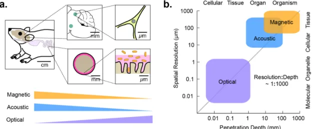
Length scales for studying cellular function in vivo
Before discussing technologies for cellular imaging and control, it is useful to think about the length scales on which these techniques must operate. Consider three representative
biological systems: the mammalian microbiome, the adaptive immune system, and the brain (Figure 1a). A microbe’s life in the mammalian gastrointestinal tract is intricately linked to its location along the length of the tract, its radial position within the lumen, and its place within a microenvironment such as the colonic crypt2. These locations are associated with length scales of centimeters, millimeters, and microns, respectively. Likewise, adaptive immunity is a multi-scale phenomenon. Antigen presentation and recognition occur at sub- micron immunological synapses, while immune cell recruitment from blood and lymphoid organs, proliferation, and regulatory signaling occur on the scale of millimeters to centimeters. Similarly, neural signaling is organized at length scales ranging from sub- micron synapses to millimeter-sized brain regions and centimeter-scale axonal projections.
In all three systems, key biological questions involve the function of particular cell types within a certain spatially-defined anatomical context. For example, which microbes can successfully colonize the small intestine? Which genes do T-cells express after migrating into a tumor and recognizing a neoantigen? How does the activity of excitatory neurons in a certain part of the hippocampus relate to the development of seizures? Each of these questions involves ensemble cellular behaviors occurring on the millimeter scale, which are difficult to recapitulate in in vitro models. Studying biology at this scale complements the understanding gained by examining cells at the single-cell and sub-cellular level, and requires a dedicated set of experimental tools.
Figure 1 | Modalities for in vivo imaging and control of cellular function. (a) Diagram of the length scales of several biological processes of interest in vivo, and the degree to which these length scales are accessible by imaging technologies. (b) Approximate length scales and maximal tissue penetration depths accessible by optical, acoustic, or magnetic imaging.
Forms of energy for biological imaging and control
The key elements of any technology for cellular imaging and control are the form of energy applied to or measured from the sample and the molecular mechanisms connecting this energy to a biological process of interest (Figure 1b). Since the work of van Leeuwenhoek, the dominant energy type used to study biological phenomena has been visible light, with modern microscopy taking advantage of an impressive array of molecular tools to optically visualize and perturb cellular processes. Unfortunately, visible light gets scattered within approximately one millimeter in most tissues, limiting its use to in vitro specimens and shallow or surgically accessed anatomical regions.
On the other hand, sound waves and magnetic fields are capable of penetrating deep into tissues. Ultrasound at MHz frequencies permeates through several centimeters, enabling imaging or focused energy deposition with a wavelength-dependent resolution down to approximately 100 µm3. This is further improved to below 10 µm with recently developed super-resolution techniques4. Due to this excellent performance, ultrasound imaging is widely used in the clinic and in pre-clinical research. In addition, ultrasound can be focused at depth to deliver mechanical forces or localized heating5. These capabilities are used clinically for non-invasive ablation of diseased tissues.
Likewise, magnetic fields experience minimal tissue attenuation. They can be used to produce high-contrast images of many organs by exploiting the context-dependent magnetic resonance behavior of nuclear spins, with a spatial resolution on the order of 100 µm. In addition, static or time-varying magnetic fields can produce mechanical forces or heat in tissues containing magnetic nanomaterials6, which can be localized in to the millimeter scale using field-free point scanning techniques7.
Based on their tissue penetration and spatiotemporal resolution, sound waves and magnetic fields are well-suited to imaging and controlling the function of cells in vivo (Figure 1b). All that is needed is a set of biomolecular tools that can link these forms of energy to specific cellular functions such as gene expression and signaling. Developing such tools presents an exciting challenge to biomolecular engineers. Just as the discovery of the green fluorescent protein stimulated the development of hundreds of reporters, sensors, and actuators through creative protein engineering, recent developments in acoustically and magnetically active
proteins may allow us to engineer a similar variety of biological tools for ultrasound and magnetic resonance. Initial inroads towards this goal are described in the following sections.
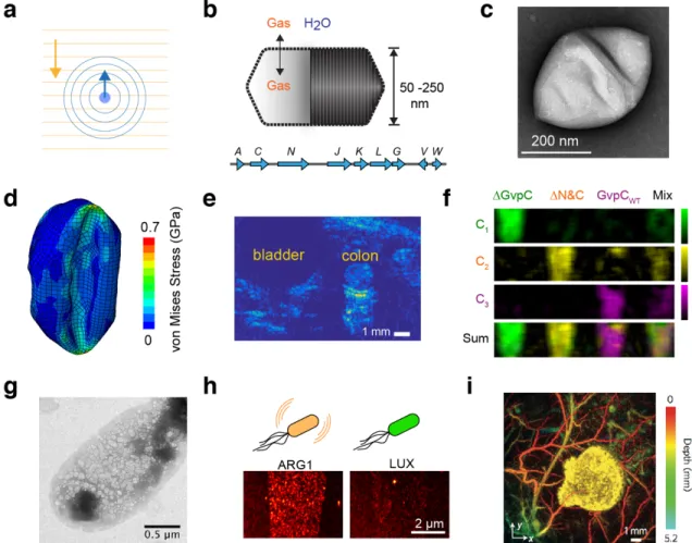
Biomolecular tools for ultrasound imaging
Diagnostic ultrasound uses the scattering of sound waves to delineate tissue boundaries, monitor the motion of organs such as the heart and quantify the velocity of blood flow (Figure 2a). Until recently, the prospect of using ultrasound to image the function of specific cells was remote due to the lack of suitable molecular reporters. Conventional ultrasound contrast agents are micron-sized synthetic bubbles that resonantly scatter sound waves. Although these microbubbles can be targeted to specific endovascular targets for molecular imaging in the bloodstream, their size and long-term instability makes it difficult to use them in labeling and monitoring the function of specific cells8. Alternatively, scattering synthetic nanoparticles have been explored as ultrasound contrast agents with the potential for cell labeling and extravascular interrogation9,10.
To connect ultrasound more closely with molecular and cellular biology, we recently adapted a unique class of gas-filled proteins, called gas vesicles or GVs, as the first biomolecular acoustic reporters. GVs evolved in aquatic photosynthetic microbes as a means to regulate buoyancy for optimal access to sunlight and other nutrients11. Despite their name, gas vesicles contain no lipids; they comprise a 2 nm-thick protein shell enclosing a hollow interior with dimensions on the order of 250 nm (Figure 2, b-c). Their shell allows gases dissolved in the surrounding media to freely permeate in and out of their interior, while their hydrophobic inner surface prevents the formation of a liquid aqueous phase. GVs are encoded by clusters of 8-14 genes, including two primary structural proteins and several minor constituents, chaperones, and regulators.
In 2014, we showed that GVs can produce ultrasound contrast in purified form, inside cells and in vivo, establishing them as the first acoustic biomolecules12. Since this initial discovery, considerable advances have been made in understanding the acoustic properties of GVs and improving the ability of ultrasound to detect them with greater sensitivity and specificity.
One key finding was that GVs undergo nanoscale buckling deformations under ultrasound (Figure 2d), resulting in non-linear scattering and allowing amplitude-modulated pulse sequences to detect GVs with greater specificity against background tissues (Figure 2e) in a
process analogous to two-photon microscopy13,14. Another key finding was that the acoustic properties of GVs can be engineered at the genetic level. In particular, a key component of the GV shell called GvpC influences the response of GVs to pressure, setting thresholds for buckling and irreversible collapse15. Tuning GvpC at the genetic level enables modulation of
Figure 2 | Biomolecular tools for ultrasound imaging. (a) Illustration of sound propagation in the imaging medium and received echo used to form the ultrasound image. (b) GVs are hollow protein nanostructures that freely allow diffusion of dissolved gas through their shell but exclude water12. GVs are encoded by operons consisting of 8-14 genes. (c) Representative transmission electron micrograph of purified GV from Halobacterium12. (d) Simulation illustrating nanoscale deformation of GVs under ultrasound leading to nonlinear backscattered echo13. (e) Amplitude-modulation pulse sequence reveals GVs in mouse colon14 (Reprinted from Maresca, D et al (2017). Nonlinear ultrasound imaging of nanoscale acoustic biomolecules. Applied Physics Letters, 110(7), 73704, with the permission of AIP Publishing). (f) Multiplexed imaging of genetically engineered GVs15. (g) Heterologous expression of GVs in E. coli using an optimized GV gene cluster16. (h) Ultrasound image of E. coli expressing GVs or the non-echogenic luminescence reporter, luciferase16. (i) Photoacoustic imaging of tumor expressing tyrosinase, and surrounding blood vessels19.
GVs’ nonlinear signals, as well as multiplexed imaging of GV variants with differential pressure sensitivity15 (Figure 2f). Additionally, fusions of GvpC with other polypeptides enable the tailoring of GV surface properties such as charge or affinity for molecular imaging targets15.
A major effort is also underway to express GVs heterologously as genetically encoded reporters. As an initial target, we have developed genetic constructs to express GVs in model commensal and pathogenic microbes such as E. coli and S. typhimurium (Figure 2g)16. Imaging these and other microbes in mammalian hosts could enable new studies of the microbiome and the tracking of engineered probiotic therapies. Cells expressing the current generation of acoustic reporter genes can be visualized at densities below 108 cells/ml, corresponding to a volume fraction of 0.005% – a level compatible with imaging microbes in the GI tract or tumors (Figure 2h)16. Another key milestone was our recent introduction of mammalian acoustic reporter genes, which enabled the expression of GVs in mammalian cells.17
An alternative mechanism by which ultrasound can facilitate the visualization of cells in vivo is photoacoustic imaging, a technique wherein optical excitation is absorbed and converted into thermoelastic pressure waves, which are detected by ultrasound transducers18. This enables the use of light to image deeper structures because photons are allowed to scatter en route to their target, with spatial information provided by ultrasound. The major advantage of photoacoustic imaging compared to pure ultrasound is its ability to leverage existing molecular tools developed for optical imaging, including fluorescent proteins or light- absorbing pigments such as melanin (Figure 2i)19,20. However, this technique is still difficult to employ at depths beyond one to two centimeters without causing tissue phototoxicity.
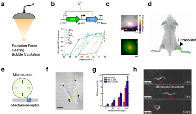
Biomolecular tools for ultrasonic actuation
In addition to imaging, ultrasound can be used to deliver energy to focused regions of tissue, with targeting on the scale of a single millimeter. Depending on beam intensity and pulse duration, this energy can be used to apply mechanical forces, drive resonant cavitation of bubbles, or deposit heat (Figure 3a)5. These capabilities are used clinically for non-invasive surgery21. If they could instead be harnessed, at lower intensities, to modulate the activity of
specific cells in vivo, this would facilitate the study of cellular function within relevant anatomical contexts.
Several nascent approaches have been proposed to enable this possibility. For example, the ability of ultrasound to controllably heat tissue within the well tolerated range of 37–42ºC can be coupled to natural or engineered temperature-dependent signaling pathways. This approach has been used to remotely activate transcription driven by the heat shock promoter, pHSP70, in cultured mammalian cells and live mice22. While this approach is highly effective in certain contexts, the thermal set-point and activity level of heat shock promoters varies
Figure 3 | Biomolecular tools for acoustic control. (a) Ultrasound can be focused at depth in tissue and apply several forms of energy to interface with cells. (b) Schematic of the gene circuit utilized to gate a GFP reporter gene with a temperature-sensitive repressor (TSR), and a panel of tuned variants of temperature-sensitive repressors24. (c) MRI thermometry imaging demonstrates a spatial temperature gradient induced by FUS on a plate of bacterial cells, resulting in spatially targeted gene expression24. (d) E. coli were injected into both hindlimbs of a nude mouse; after FUS application to the right hindlimb, reporter gene expression is significantly enriched at the site of heating24. (e) Diagram of mechanism by which microbubble cavitation can result in membrane deformation leading to mechanoreceptor activation. (f) Microbubbles attached to cultured retinal pigment epithelium cells27. (g) Uptake of membrane-impermeable dye into retinal pigment epithelium cells expressing MscL and functionalized with microbubbles27. (h) C. elegans worm motor response to ultrasound in a bath of microbubbles28.
between cell types, is not easily tunable, and responds to other stimuli in addition to temperature23.
In bacteria, endogenous heat shock promoters have only modest activation in response to this range of temperatures, necessitating the development of engineered thermal bioswitches. To address this need, we recently introduced two families of orthogonal, tunable temperature- dependent transcriptional repressors for remote control of bacterial function24. These bioswitches are based on the TlpA transcriptional repressor from S. typhimurium and a variant of the cI repressor from the Lambda bacteriophage (TcI). Unlike the ~ 10-fold thermal induction of heat shock promoters, the expression of genes downstream of TlpA and TcI operators is turned on by more than 100-fold in response to mild heating. We showed that TlpA and TcI can be engineered through directed evolution to actuate at different desired temperatures, as required by a given application (Figure 3b). In addition, they can be used in combination to build thermal logic circuits, for example to turn on two different functions at two different temperatures. We have demonstrated that these switches can be used to spatially pattern gene expression in plated bacterial cells (Figure 3c) and also in bacteria implanted in vivo (Figure 3d). In addition to controlling transcription, these bioswitches can also be used to control protein-protein interactions and protein localization. We showed that TlpA monomers can be engineered through rational design to heterodimerize at a tunable transition temperature.25 When fused to other proteins, engineered TlpA can reversibly control their association and membrane localization in mammalian cells.25
The modest fold change of heat shock promoters (HSPs) can also be improved through the engineering of genetic circuits. By allowing HSPs to drive feed-forward or recombinase- based circuits, we showed that a large or sustained transcriptional response can be achieved.26 Using these architectures in T-cells, we demonstrated the heat-inducible expression of chimeric antigen receptors and cytokines, and the killing of target tumor cells.26
Besides heating, ultrasound is also able to apply mechanical forces to tissues. These forces, which are amplified by acoustically active structures such as microbubbles, could be coupled to signaling elements such as mechanosensitive ion channels, allowing non-invasive control of cellular signaling. This concept was recently demonstrated in vitro by combining synthetic microbubbles with mammalian cells heterologously expressing the E. coli mechanosensitive ion channel MscL27. Microbubbles were similarly used to stimulate the endogenous
mechanosensor Trp4 in C. elegans28. Unlike thermal stimuli, which are typically associated with timescales on the order of seconds, mechanical effects can be produced on the order of milliseconds, potentially allowing more rapid control of cellular signaling. However, techniques requiring microbubbles are limited in their application to mammals due to the difficulty of delivering bubbles to relevant tissues.
In addition to directly controlling cellular function, ultrasound can be used to spatially target the delivery of genetically encoded tools or treatments. In the brain, such delivery can be targeted non-invasively by opening the blood-brain barrier reversibly at a specific location using focused ultrasound and intravascular microbbubles29. This technique allows the delivery of adeno-associated virus (AAV) vectors to targeted regions with millimeter precision30. This method has recently been used to deliver AAV vectors encoding for the chemogenetic receptors hM3D(Gq) and hM4D(Gi) into ultrasound-targeted brain regions, which can then be activated or inhibited upon the systemic administration of a small- molecule drug.31
Biomolecular tools for magnetic resonance imaging
Like ultrasound, MRI derives contrast from both, endogenous variation in the properties of tissue and molecular contrast agents. Taking advantage of the rich behavior of nuclear spins under various physical and chemical conditions has enabled the development of several classes of biomolecular MRI reporters32,33. One major class comprises proteins that contain paramagnetic metals, such as iron or manganese, or lead to the accumulation of these ions in tissue. Proteins in this class include ferritin, bacterial cytochromes, the transferrin receptor and other transporters (Figure 4a-b). Paramagnetic species in these proteins produce T1
contrast through spin exchange of coordinated water protons and T2 contrast by distorting the magnetic field near the protein. Another class of reporters includes proteins with large numbers of exchangeable protons – the nuclear spin most commonly imaged with MRI.
These protein-bound protons resonate at a distinct frequency (chemical shift) relative to water-bound protons, and can be selectively saturated with radiofrequency pulses, quenching their MRI signal. By applying such saturation while these protons exchange rapidly with the aqueous pool, the signal of the entire pool can be substantially reduced. This “catalytic”
contrast scheme is called chemical exchange saturation transfer, or CEST. Proteins detectable with this method include a synthetic lysine-rich protein34 and human protamine35. While these pioneering reporter types have been used to demonstrate the imaging of genetically defined cells using MRI, they are generally limited by their low molecular sensitivity (requiring protein concentrations on the order of µM) or the requirement of metal cofactors, which may not always be bioavailable. Recent efforts have therefore been focused on developing alternative classes of reporters that are more sensitive and do not require metals. For example, we recently introduced aquaporin 1 as a biomolecular reporter for MRI based on its ability to enhance the diffusion of water across cell membranes (Figure 4c)36. Aquaporins are transmembrane channels that passively conduct water with exquisite selectivity at rates of up to one billion water molecules per channel per second. We showed that the overexpression of this autologous, non-toxic, metal-free molecule produces contrast Figure 4 | Biomolecular tools for magnetic resonance imaging. (a) Metalloproteins interact magnetically with aqueous 1H nuclear spins, leading to T1 or T2 MRI contrast. (b) Migrating neuroblasts expressing ferritin produce a hypointense track (arrow) in T2 weighted MRI59. Asterisks denote adenovirus injection sites. (c) Overexpression of aquaporin enhances passive diffusion of water across the cell membrane, resulting in contrast on diffusion weighted MRI. (d) AQP1 expression in mouse xenograft shows significant contrast compared to contralateral GFP expressing xenograft after expression is induced with doxycycline36.(e) GVs interact with hyperpolarized xenon dissolved in biological media, producing contrast in 129Xe MRI.(f) Genetically distinct GVs produce different chemical shifts in 129Xe MRI, enabling multiplexed imaging39.
in diffusion-weighted MRI at concentrations below 500 nM, allowing non-invasive imaging of gene expression in vitro and in vivo (Figure 4d). In addition to aquaporin 1, other water-permeable channels such as the urea transporter can produce diffusion-based contrast, albeit with lower channel selectivity37.
To push the molecular and cellular sensitivity of MRI even further, recent work has focused on directly addressing a fundamental physical limitation of conventional magnetic resonance: the weak magnetic alignment of nuclear spins under thermal equilibrium. This low polarization results in overall MRI signals approximately 105 times weaker than they could be if all the available spins aligned with the applied magnetic field. This limitation can be overcome with hyperpolarization – an advanced technique in which nuclei are prepared via physical methods in a state of non-equilibrium polarization that is up to 10,000-fold stronger than baseline38. Hyperpolarized nuclei such as the noble gas 129Xe can then be delivered to the body by inhalation to be imaged during their polarization half-life of a few seconds. Because each hyperpolarized atom carries a much stronger signal than thermally polarized molecules, MRI reporters acting on these nuclei are detectable at much lower concentrations than their conventional counterparts. The first biomolecular reporters for hyperpolarized xenon MRI were GVs, the aforementioned gas-filled protein nanostructures.
GVs allow xenon dissolved in surrounding media to exchange in and out of their gaseous compartment, producing MRI contrast through CEST at picomolar concentrations (Figure 4, e-f)39. Other proteins active as contrast agents for 129Xe MRI and other hyperpolarized nuclei have also been reported40,41. GVs can also be imaged directly by MRI without 129Xe, owing to the difference in the magnetic susceptibility of air and water, allowing GVs to be visualized using susceptibility-weighted imaging.42
In addition to reporters connected to gene expression, biomolecules have also been engineered as MRI sensors – allowing dynamic tracking of processes such as neurotransmission and kinase signaling. One class of such sensors, inspired by pioneering synthetic approaches43, comprises iron-containing metalloproteins in which the accessibility of an open iron coordination site to water is modulated by the binding of small molecules, thereby altering T1 contrast44. Directed evolution allows the tuning of this small molecule binding site for selective interactions with neurotransmitters, such as dopamine and serotonin. The resulting reporters have been used to image the dynamics of neurotransmitter release and reuptake in rodent brains45. Other biomolecular sensor constructs, based on T2
and CEST mechanisms, have been developed to image signals such as kinase and protease activity46.
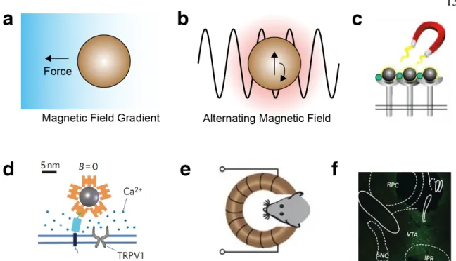
Biomolecular tools for magnetic control
Magnetic fields exert forces on magnetically active materials such as superparamagnetic and ferromagnetic particles6. Depending on the particle type, these forces can be sufficient to guide the movement of materials or cells in the body and actuate receptor signaling (Figure 5a). In addition, rapidly alternating magnetic fields can generate heat in particles whose magnetization oscillates with the applied field, which in turn can be used to control temperature-dependent processes (Figure 5b).
Most strategies for magnetic control of cell function have relied on synthetic magnetic nanoparticles as transducers of the magnetic field. For example, superparamagnetic iron oxide nanoparticles have been used to cluster cell surface receptors or apply directly actuating forces on integrin and notch47 (Figure 5c). In addition, superparamagnetic particles have been used in combination with alternating fields to activate temperature-sensitive ion channels such as TRPV148 (Figure 5d). This approach enabled remote control of neural signaling in vivo in mice surgically implanted with such particles49 (Figure 5e-f).
Additionally, cells containing iron oxide particles have been concentrated at certain locations in vivo50,51.
Translating these approaches into more versatile, fully-genetic constructs is challenging due to the unsolved problem of heterologous biosynthesis of strongly magnetic nanomaterials.
Although superparamagnetic and ferromagnetic iron oxide nanocrystals are made by magnetotactic bacteria52, the genes encoding their specialized organelle machinery for such synthesis have so far been transferred only to their close genetic relatives. The magnetic nanostructures formed in commensal microbes and mammalian cells, such as ferritin, tend to be paramagnetic or weakly superparamagnetic. Efforts to increase the magnetic strength of ferritin through genetic engineering have so far come up short of qualitatively altering its magnetic character.
Despite this physical limitation, some groups have reported that fusions of ferritin to temperature- and mechanically-sensitive ion channels allow neurons expressing these channels to be activated remotely using both alternating and static magnetic fields53,54. These reports are somewhat surprising based on classical theoretical estimates of the forces and temperatures that could be produced by ferritin55. Experimental quantification of the temperature generated by ferritin also revealed no temperature increase in alternating magnetic fields.56 However, it is possible that as-yet unknown alternative mechanisms are at play.
Outlook
Figure 5 | Biomolecular tools for magnetic control. (a) Magnetic field gradients exert a force on magnetic particles. (b) Alternating magnetic fields heat magnetic particles by inducing oscillations in the magnetic moment of the nanoparticle. (c) Magnetic fields can induce the clustering of magnetic particles, and receptors to which they are bound47. (d) Coupling magnetic nanoparticles to the heat- sensitive ion channel TRPV1 enables magnetic control of calcium influx to the cell48. (e) Remote deep brain stimulation using alternating magnetic fields applied to mice with implanted magnetic nanoparticles and virally transduced TRPV149. (f) Expression of the neural activity marker cFos in the ventral tegmental area (VTA) of the mouse brain after injection with TRPV1-encoding virus, magnetic nanoparticles and the application of alternating field stimulation49.
The development of biomolecular tools for non-invasive cellular imaging and control is in its infancy. Inspired by the history and impact of fluorescent and optogenetic proteins, many opportunities exist to develop acoustic and magnetic technologies connected to a variety of cellular processes. For example, biomolecular ultrasound imaging is a new field animated 6 years ago with the development of GVs as its first biomolecular reporter. Much remains to be learned about the acoustic properties of these molecules and how they can be tuned at the genetic level to increase imaging sensitivity, or engineered to respond dynamically as sensors of cellular signaling. In addition, more work is needed to transfer the machinery encoding GVs to a greater number of species. This work takes place against a backdrop of other exciting developments in ultrasound, exemplified by super-resolution imaging4 and the recent invention of functional ultrasound (fUS), a technique for imaging neural activity non- invasively with improved spatiotemporal resolution compared to functional MRI (< 100 µm and < 10 ms) using transducers that can be mounted on freely moving animals57.
Biomolecular MRI is only slightly more mature, with an expanding variety of contrast mechanisms but no clear frontrunner to become the go-to molecule for in vivo cellular imaging. Recently developed aquaporin-based reporter genes offer unique advantages in terms of their simplicity and biocompatibility, while GVs have the potential to bring the advantages of hyperpolarization to boosting the in vivo sensitivity of cellular MRI.
Engineering both of these molecules and accompanying in vivo imaging methods for maximum sensitivity and potential use as dynamic sensors are major avenues for future research. In addition, an outstanding grand challenge is the engineering of heterologous magnetite biosynthesis, which would provide powerful MRI contrast, as well as opportunities for actuation. In parallel with these molecular efforts, progress is being made on improving the information content of MRI images. For example, we recently used nitrogen vacancy diamond magnetometry, an optical technique for imaging magnetic fields, to map the nanoscale magnetic field in cells containing iron oxide nanoparticles and connect these maps to the T2 contrast seen by MRI58. This study demonstrated experimentally that the spatial arrangement of magnetic materials inside cells strongly influences contrast, guiding the development of magnetic cellular reporters and sensors and imaging parameters for their specific identification in vivo.
Complementing these evolving imaging technologies, much additional work on genetically encodable agents is needed to control cellular responses with acoustic or magnetic energy.
For example, there is a lack of mammalian thermal bioswitches that are orthogonal to pleotropic heat shock pathways and tunable to different temperature thresholds analogously to the system we developed for microbial remote control. In addition to regulating gene expression, tools are needed to connect thermal inputs directly to signaling pathways.
Similarly, for ultrasound actuation based on mechanical forces, use in mammals will require eliminating the need for synthetic microbubbles to produce constructs that can be fully genetically encoded. Likewise, the synthetic magnetic particles used in well-accepted magnetic control techniques using thermal, mechanical, or clustering mechanisms must be replaced with genetically encodable materials. The potential use of ferritin for this purpose requires further study to reconcile its encouraging empirical performance and predicted lack of efficacy based on previously studied physical properties.
In summary, going deeper into the body to study cellular function within its native in vivo context requires engineering interactions between deeply penetrant forms of energy and biomolecules to enable non-invasive imaging and control. Several recent advances have provided exciting proofs or concept for this approach, and inform the development of new classes of biomolecular tools. Many depths remain to be plumbed.
References
1. Piraner, D. I. et al. Going Deeper: Biomolecular Tools for Acoustic and Magnetic Imaging and Control of Cellular Function. Biochemistry vol. 56 5202–5209 (2017).
2. Donaldson, G. P., Lee, S. M. & Mazmanian, S. K. Gut biogeography of the bacterial microbiota. Nat. Rev. Microbiol.14, 20–32 (2015).
3. Foster, F. S., Pavlin, C. J., Harasiewicz, K. A., Christopher, D. A. & Turnbull, D. H.
Advances in ultrasound biomicroscopy. Ultrasound Med. Biol.26, 1–27 (2000).
4. Errico, C. et al. Ultrafast ultrasound localization microscopy for deep super-resolution vascular imaging. Nature527, 499–502 (2015).
5. Humphrey, V. F. Ultrasound and matter-Physical interactions. Progress in Biophysics and Molecular Biology (2007) doi:10.1016/j.pbiomolbio.2006.07.024.
6. Pankhurst, Q. A., Connolly, J., Jones, S. K. & Dobson, J. Applications of magnetic nanoparticles in biomedicine. J. Phys. D. Appl. Phys.36, R167–R181 (2003).
7. Tasci, T. O. et al. Focused RF hyperthermia using magnetic fluids. Med. Phys.36, 1906–1912 (2009).
8. Abou-Elkacem, L., Bachawal, S. V & Willmann, J. K. Ultrasound molecular imaging:
Moving toward clinical translation. Eur. J. Radiol.84, 1685–93 (2015).
9. Sheeran, P. S., Luois, S. H., Mullin, L. B., Matsunaga, T. O. & Dayton, P. A. Design of ultrasonically-activatable nanoparticles using low boiling point perfluorocarbons.
Biomaterials33, 3262–3269 (2012).
10. Chen, F. et al. Exosome-like silica nanoparticles: a novel ultrasound contrast agent for stem cell imaging. Nanoscale9, 402–411 (2017).
11. Pfeifer, F. Distribution, formation and regulation of gas vesicles. Nature Reviews Microbiology (2012) doi:10.1038/nrmicro2834.
12. Shapiro, M. G. et al. Biogenic gas nanostructures as ultrasonic molecular reporters.
Nat. Nanotechnol.9, 311–316 (2014).
13. Cherin, E. et al. Acoustic behavior of Halobacterium salinarum gas vesicles in the high frequency range: experiments and modeling. Ultrasound Med. Biol. 1–15 (2017) doi:10.1016/j.ultrasmedbio.2016.12.020.
14. Maresca, D. et al. Nonlinear ultrasound imaging of nanoscale acoustic biomolecules.
Appl. Phys. Lett.110, 073704 (2017).
15. Lakshmanan, A. et al. Molecular Engineering of Acoustic Protein Nanostructures.
ACS Nano10, 7314–7322 (2016).
16. Bourdeau, R. et al. Acoustic reporter genes for non-invasive imaging of microbes in mammalian hosts.
17. Farhadi, A., Ho, G. H., Sawyer, D. P., Bourdeau, R. W. & Shapiro, M. G. Ultrasound imaging of gene expression in mammalian cells. Science (80-. ). 365, 1469–1475 (2019).
18. Wang, L. V & Yao, J. A practical guide to photoacoustic tomography in the life
sciences. Nat. Methods13, 627–638 (2016).
19. Jathoul, A. P. et al. Deep in vivo photoacoustic imaging of mammalian tissues using a tyrosinase-based genetic reporter. Nat. Photonics9, 239–246 (2015).
20. Yao, J. et al. Multiscale photoacoustic tomography using reversibly switchable bacterial phytochrome as a near-infrared photochromic probe. Nat. Methods13, 1–9 (2015).
21. Hynynen, K. MRIgHIFU: A tool for image-guided therapeutics. J. Magn. Reson.
Imaging34, 482–493 (2011).
22. Kruse, D. E., Mackanos, M. a, O’Connell-Rodwell, C. E., Contag, C. H. & Ferrara, K. W. Short-duration-focused ultrasound stimulation of Hsp70 expression in vivo.
Phys. Med. Biol.53, 3641–60 (2008).
23. Aït-Aïssa, S., Porcher, J. M., Arrigo, A. P. & Lambré, C. Activation of the hsp70 promoter by environmental inorganic and organic chemicals: Relationships with cytotoxicity and lipophilicity. Toxicology145, 147–157 (2000).
24. Piraner, D. I., Abedi, M. H., Moser, B. A., Lee-Gosselin, A. & Shapiro, M. G. Tunable thermal bioswitches for in vivo control of microbial therapeutics. Nat. Chem. Biol.
13, 75–80 (2016).
25. Piraner, D. I., Wu, Y. & Shapiro, M. G. Modular Thermal Control of Protein Dimerization. ACS Synth. Biol.8, 2256–2262 (2019).
26. Abedi, M. H., Lee, J., Piraner, D. I. & Shapiro, M. G. Thermal Control of Engineered T-cells. ACS Synth. Biol.9, 1941–1950 (2020).
27. Heureaux, J., Chen, D., Murray, V. L., Deng, C. X. & Liu, A. P. Activation of a bacterial mechanosensitive channel in mammalian cells by cytoskeletal stress. Cell.
Mol. Bioeng.7, 307–319 (2014).
28. Ibsen, S., Tong, A., Schutt, C., Esener, S. & Chalasani, S. H. Sonogenetics is a non- invasive approach to activating neurons in Caenorhabditis elegans. Nat. Commun.6, 1–12 (2015).
29. Hynynen, K., McDannold, N., Vykhodtseva, N. & Jolesz, F. a. Noninvasive MR imaging-guided focal opening of the blood-brain barrier in rabbits. Radiology 220, 640–646 (2001).
30. Poon, C., McMahon, D. & Hynynen, K. Noninvasive and targeted delivery of therapeutics to the brain using focused ultrasound. Neuropharmacology4, 519–526 (2016).
31. Szablowski, J. O., Lee-Gosselin, A., Lue, B., Malounda, D. & Shapiro, M. G.
Acoustically targeted chemogenetics for the non-invasive control of neural circuits.
Nat. Biomed. Eng.2, 475–484 (2018).
32. Gilad, A. A. et al. MRI Reporter Genes. J Nucl Med49, 1905–1908 (2008).
33. Mukherjee, A., Davis, H. C., Ramesh, P., Lu, G. J. & Shapiro, M. G. Biomolecular MRI Reporters: evolution of new mechanisms. Progress in Nuclear Magnetic Resonance Spectroscopy (2017). doi:10.1016/j.pnmrs.2017.05.002.
34. Farrar, C. T. et al. Establishing the Lysine-rich Protein CEST Reporter Gene as a
CEST MR Imaging Detector for Oncolytic Virotherapy. Radiology275, 746–754 (2015).
35. Oskolkov, N. et al. Biophysical characterization of human protamine-1 as a responsive CEST MR contrast agent. ACS Macro Lett.4, 34–38 (2015).
36. Mukherjee, A., Wu, D., Davis, H. C. & Shapiro, M. G. Non-invasive imaging using reporter genes altering cellular water permeability. Nat. Commun.7, 13891 (2016).
37. Schilling, F. et al. MRI measurements of reporter-mediated increases in transmembrane water exchange enable detection of a gene reporter. Nat. Biotechnol.
1–6 (2016) doi:10.1038/nbt.3714.
38. Barskiy, D. A. et al. NMR Hyperpolarization Techniques of Gases. Chem. - A Eur. J.
725–751 (2016) doi:10.1002/chem.201603884.
39. Shapiro, M. G. et al. Genetically encoded reporters for hyperpolarized xenon magnetic resonance imaging. Nat. Chem.6, 629–34 (2014).
40. Wang, Y., Roose, B. W., Palovcak, E. J., Carnevale, V. & Dmochowski, I. J. A Genetically Encoded ??-Lactamase Reporter for Ultrasensitive 129Xe NMR in Mammalian Cells. Angew. Chemie - Int. Ed.55, 8984–8987 (2016).
41. Patrick, P. S. et al. Detection of transgene expression using hyperpolarized 13 C urea and diffusion-weighted magnetic resonance spectroscopy. Magn. Reson. Med. 73, 1401–1406 (2015).
42. Lu, G. J. et al. Acoustically modulated magnetic resonance imaging of gas-filled protein nanostructures. Nat. Mater.17, 456–463 (2018).
43. Li, W. H., Fraser, S. E. & Meade, T. J. A calcium-sensitive magnetic resonance imaging contrast agent. J. Am. Chem. Soc.121, 1413–1414 (1999).
44. Shapiro, M. G. et al. Directed evolution of a magnetic resonance imaging contrast agent for noninvasive imaging of dopamine. Nat. Biotechnol.28, 264–270 (2010).
45. Lee, T., Cai, L. X., Lelyveld, V. S., Hai, A. & Jasanoff, A. Molecular-level functional magnetic resonance imaging of dopaminergic signaling. Science 344, 533–535 (2014).
46. Airan, R. D. et al. MRI biosensor for protein kinase A encoded by a single synthetic gene. Magn. Reson. Med.68, 1919–1923 (2012).
47. Mannix, R. J. et al. Nanomagnetic actuation of receptor-mediated signal transduction.
Nat. Nanotechnol.3, 36–40 (2008).
48. Huang, H., Delikanli, S., Zeng, H., Ferkey, D. M. & Pralle, A. Remote control of ion channels and neurons through magnetic-field heating of nanoparticles. Nat.
Nanotechnol.5, 602–606 (2010).
49. Chen, R., Romero, G., Christiansen, M. G., Mohr, A. & Anikeeva, P. Wireless magnetothermal deep brain stimulation. Science347, 1477–80 (2015).
50. Muthana, M. et al. Directing cell therapy to anatomic target sites in vivo with magnetic resonance targeting. Nat. Commun.6, 8009 (2015).
51. Felfoul, O. et al. Magneto-aerotactic bacteria deliver drug-containing nanoliposomes
to tumour hypoxic regions. Nat. Nanotechnol.11, 1–5 (2016).
52. Faivre, D. & Schüler, D. Magnetotactic Bacteria and Magnetosomes. Chem. Rev.108, 4875–4898 (2008).
53. Stanley, S. A., Sauer, J., Kane, R. S., Dordick, J. S. & Friedman, J. M. Remote regulation of glucose homeostasis in mice using genetically encoded nanoparticles.
Nat Med21, 92–98 (2015).
54. Wheeler, M. A. et al. Genetically targeted magnetic control of the nervous system.
Nat Neurosci19, 756–761 (2016).
55. Meister, M. Physical limits to magnetogenetics. Elife5, 1–14 (2016).
56. Davis, H. C. et al. Nanoscale Heat Transfer from Magnetic Nanoparticles and Ferritin in an Alternating Magnetic Field. Biophys. J.118, 1502–1510 (2020).
57. Sieu, L.-A. et al. EEG and functional ultrasound imaging in mobile rats. Nat Methods 12, 831–834 (2015).
58. Davis, H. C. et al. Mapping the Microscale Origins of MRI Contrast with Subcellular NV Diamond Magnetometry. (2016).
59. Iordanova, B. & Ahrens, E. T. In vivo magnetic resonance imaging of ferritin-based reporter visualizes native neuroblast migration. Neuroimage59, 1004–1012 (2012).
C h a p t e r 2
GENETICALLY ENCODED ACTUATORS FOR ACOUSTIC INVERSION, MANIPULATION, AND PATTERNING OF ENGINEERED CELLS
This chapter is in large part a reformatted version of the manuscript entitled “Genetically encoded actuators for acoustic inversion, manipulation and patterning of engineered cells”
by Wu D, Baresch D, Cook C, Ma Z, Malounda D, Maresca D, Abundo M.P., Lee J, Shivaei S, Mittelstein D.R., Qiu T, Fischer P and Shapiro M.G. currently in revision. A pre-print version can be found on bioRxiv1. Under the supervision of Mikhail Shapiro, my contributions to the work were to conceive, design, and conduct the experiments, analyze and interpret the data, and write the manuscript.
Abstract
The ability to selectively manipulate and control the spatial arrangement of genetically defined cells is critical for the fields of living materials, biomedicine, and synthetic biology.
Ultrasound has the ability to manipulate a variety of objects remotely and en masse with high spatial and temporal precision via acoustic radiation force (ARF). However, this capability is currently disconnected from intracellular genetic programs. Here, we show that gas vesicles (GVs) – a unique class of genetically encoded gas-filled protein nanostructures – can serve as genetically encodable actuators for ARF, enabling the selective acoustic inversion and manipulation of engineered cells. Due to their differential density and compressibility relative to water, GVs are effectively moved with acoustic standing waves despite their nanometer dimensions. When expressed inside genetically engineered cells, GVs amplify the ARF experienced by the cells by a factor of ten and invert their acoustic contrast, allowing the cells to be selectively manipulated with sound waves based on their genotype. This enables dynamic patterning, focal trapping, translation, and holographic bioprinting of specific cells with acoustic fields. In addition, the unique material properties of purified GVs enable new modes of acoustic interaction, including force inactivation, multiplexed manipulation, and endosomal labeling. Unlike fluorescent proteins, which have no intrinsic ability to serve as actuators for cellular manipulation, GVs provide a direct link between gene expression and mechanical
actuation, creating a new paradigm for molecular and cellular control in a broad range of contexts.
Introduction
The ability to remotely pattern, actuate, and apply force to genetically specified cells would have many applications in biomedicine and synthetic biology, ranging from the fabrication of biological living materials2 to drug delivery3 and noninvasive control of cellular function4–6. Ultrasound offers unique advantages in such contexts over optical, magnetic, and printing-based approaches due to its functionality in opaque media, non-invasiveness, relatively high spatial precision on the µm scale, and rapid, reconfigurable field formation.
Acoustic radiation force (ARF) allows ultrasound to manipulate materials whose density or compressibility differ from their surrounding medium. This capability has been used to manipulate, pattern, and sort synthetic particles and cells, for example by using acoustic standing waves to create stable attractors for such objects or to separate them in microfluidic devices7. However, due to the similarity of acoustic contrast factor among endogenous cellular materials, it is challenging to connect ARF-based actuation directly to intracellular gene expression. Doing so would require a genetically encodable agent capable of drastically altering the acoustic properties of a cell.
To address this need, we hypothesized that gas vesicles (GVs) – a unique class of biologically assembled air-filled protein nanostructures – could experience strong ARF and enable the selective acoustic manipulation of GV-expressing cells. GVs are genetically encoded protein-shelled nanostructures with hydrodynamic diameters on the order of 250 nm (Figure 1, a-b) which evolved in aquatic photosynthetic microbes as a means to achieve buoyancy for improved access to sunlight8. GVs consist of a physically stable, hollow compartment enclosed by a 2 nm-thick protein shell that is permeable to gas but excludes
liquid water. Based on their unique physical properties, GVs were recently developed as genetically encodable and engineerable contrast agents for non-invasive imaging9–14. However, the ability of GVs to serve as actuators of ARF has not been tested.
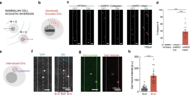
We hypothesized that GVs’ differential density and compressibility relative to aqueous media would allow these nanostructures to experience significant ARF (Figure 1c), and that cells genetically engineered to express GVs would experience a drastically different radiation force due to changes in their acoustic properties (Figure 1, d-e). We further hypothesized that the resulting forces would act in the opposite direction from other biomaterials, which are generally denser than water, allowing selective acoustic
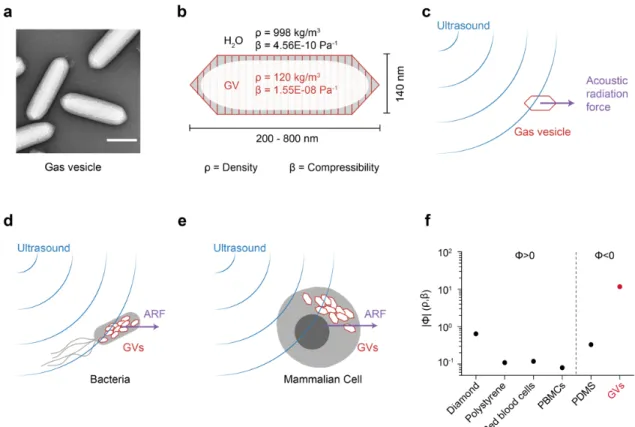
Figure 1 | Gas vesicles as biomolecular transducers of acoustic radiation force. (a) Transmission electron microscopy image of representative GVs from Anabaena flos-aquae. (b) Schematic drawing of a GV, showing its effective density (ρ) and compressibility (β) relative to that of the surrounding water. (c) Illustration of a GV experiencing acoustic radiation force due to applied ultrasound. (d) Illustration of a bacterium experiencing enhanced acoustic radiation force due to GVs inside the cell. (e) Illustration of a mammalian cell experiencing a unique acoustic radiation force due to the intracellular GVs. (f) Estimated magnitude of the acoustic contrast factor, |Φ|, of GVs and several common materials used in acoustic manipulation. Materials to the left and right of the vertical dashed line exhibit positive and negative acoustic contrast in water, respectively. PBMCs, peripheral blood mononuclear cell. PDMS, polydimethylsiloxane.
manipulation. This would connect mechanical actuation directly to the expression of a specific gene – a capability not provided by other genetic labels such as fluorescent proteins. In this study, we test these fundamental hypotheses and demonstrate the use of GVs in the selective acoustic trapping, translation, patterning and holographic bioprinting of genetically engineered cells. In addition, we show that the physical properties of purified GVs provide new capabilities for acoustic multiplexing, pressure measurement, and endocytic cell labeling.
Results
Gas vesicles experience direct acoustic radiation force
To estimate the expected ARF acting on GVs, we modeled them as spherical particles with an effective density of 120 kg/m3 (ref. 15) and compressibility of 1.55E-8 Pa-1 (ref. 16).
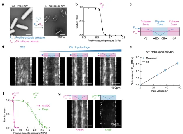
Because both of these values are radically different from water (Figure 1b), we predicted that GVs would have a strongly negative acoustic contrast in aqueous media, with a contrast factor of –11.7 (Figure 1f, Eq. 1 in Methods). While cells and most biological components exhibit positive acoustic contrast in aqueous solution, a few materials – such as microbubbles, polydimethylsiloxane (PDMS) elastomer microparticles and lipids – exhibit a negative contrast factor, allowing them to migrate up pressure gradients and efficiently separate from positive-contrast materials, as demonstrated in several important applications17–23. We hypothesized that GVs could be manipulated in a similar manner by responding directly to ARF at typical frequencies and energy densities of several MHz and
~10-100 J/m3 (ref. 24). Despite their nanometer dimensions, we anticipated that GVs’
exceptionally large contrast factor would allow them to overcome the challenges of sub- micron particle actuation caused by the volumetric scaling of ARF and the competing process of acoustic streaming25.
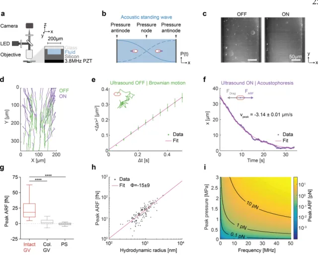
To test the ability of GV nanostructures to be manipulated with ARF, we purified GVs from the cyanobacterium Anabaena flos-aquae (Ana), chemically labeled them with a fluorescent dye, and imaged them in suspension inside a microfluidic channel coupled to a bulk piezoelectric resonator operating at 3.8 MHz (Figure 2a). The channel width of 200 µm represents a half-wavelength at this frequency, resulting in a pressure node at its center and antinodes (areas of highest pressure) at each wall (Figure 2b). As expected based on
their negative acoustic contrast, GVs readily migrated to the pressure anti-nodes upon ultrasound application (Figure 2, c-d). As a control, we imaged GVs that were collapsed before the experiment with hydrostatic pressure (Supplementary Figure 1). Neither collapsed GVs nor similarly-sized polystyrene tracer nanoparticles – included as an additional control and indicator of fluid motion – migrated in the acoustic field, confirming the absence of streaming.
Next, we quantified the ARF acting on GV particles in solution using single-particle tracking (Figure 2d). The Brownian motion of each particle before ultrasound application was used to determine its mobility and hydrodynamic size (Figure 2e, Eq. 2 & 5 in Methods). For the same particle, its motion within the acoustic field during ultrasound application was fitted to an equation accounting for the spatial field profile (Eq. 4 in Methods), allowing us to determine the peak particle velocity (Figure 2f). The maximum ARF acting on GV particles was then determined by a balance with hydrodynamic drag, and measured to be 24.5 ± 1.7 fN under the acoustic parameters used in this measurement (Figure 2g). In contrast, control particles showed no substantial ARF.
Colloidal association of individual GVs within the microfluidic channel resulted in tracked particles having a range of hydrodynamic radii larger than expected from a single GV.
Therefore, to estimate the ARF acting on a single GV, we plotted the dependence of the ARF on the hydrodynamic radius of the clusters and fitted it with a power law function accounting for fractal clustering26,27 (Figure 2h, Eq. 6 in Methods, force-mobility exponent
= 1.39 ± 0.06; R2 = 0.744). Given the acoustic energy applied in this experiment (0.25 ± 0.02 J/m3, Supplementary Figure 2), this single-particle force corresponds to an acoustic contrast factor of –15 ± 9, consistent with our theoretical estimate of –11.7 (Figure 1e).
Using this contrast factor, we can predict the ARF on a single GV across a range of typical acoustic parameters24 (Figure 2i), with the expected force spanning from 0.01 to 10 pN.
Forces of this magnitude are more than sufficient to overcome Brownian motion, as shown in our experiments, and are relevant to many biomolecular and cellular interactions28. Overall, these results establish GVs as a genetically encodable biomolecular nanomaterial that can be manipulated with acoustic fields.