THE POLYCH/E fOUS ANNELIDS OF PORK) RICO.
BY
AARON L. TREADWELL,
ProfessorofBiology, VassarCollege.
181
THE POLYCHAETOUS ANNELIDS OF PORTO RICO.
By AARON
L.TREADWELL,
Professorof Biology, VassarCollege.
The material here described was
collectedby the expedition
sent toPorto Rico
in
the winter
of1898-99, by Hon. George M. Bowers, United
StatesCommissioner
ofFish and
Fisheries.The
collectionincluded 85
speciesof
Polychsetes, ofwhich
32, so far as 1could determine, are new. All observations were made on preserved
material.Where no other preserving
fluid isindicated
indescriptions
of color, etc., it willbe understood
thatthe specimen was
in alcohol. Ifformalin was
used, that factisnoted
in the description.For
assistance inprocuring
literatureIam indebted
toDr. H. M. Smith,
ofthe United
StatesCommission
ofFish and
Fisheries,Prof. H.
C.Bumpus,
ofBrown
University, and Prof. H.
P.Johnson,
ofthe University
of California.All
the figures in thetext were drawn by the author.
Family SYLLIDfit.
SYLLISSav.
Syllis
spongiphila
Verrill.SyllisspongiphilaVerrill,Trans.Conn.Acad.,vol.4,pi.24, figs. 10,10a, 1881:Kept.U.S.F. C.for1888, pi. 42,figs.183,183a;
Proc.IT. S.Nat.Mns.1885, p. 435.
ProfessorVerrilldescribesthecolor as yellowish white.
He
does not sayifthatisthecase inthe livinganimal.Most
ofthese agreewithhisdescription,butinsome
the anterior portion ofthebody
wascoloredadarkbrown by two
ratherbroadbrown
bandsineachseg-ment. In the intersegmental constrictions is a narrower band,
more
sharply definedand
denserin color.The
eyes are fartherremoved
from the baseof themiddle antennain these than inthose figuredby
Verrill andthe terminaljoint ofthesetahasmore numerous
teeth.Collectedfrom
Boqueron
Bay, station 6065, Arroyo, Puerto Real, on corals atMayaguez.Syllis
eomplanata,
n.sp.Body
verymuch
flattened,withrow
ofdark-brownspotsaroundpos- terioredgeofheadandacrossposteriorportion ofeach segment. Similar spots scatteredirregularlyoverrest ofbody. Tentaclesand
all cirriarticu- lated,withrow
of pigment granules around eachannulus.Median
ten- taclelongerthanlateral,aboutfourtimesaslongashead.Two
tentacular cirri, dorsalone rather longerthanmedian
tentacle,ventral oneshorter.Palps thick at base, tapering torounded apex. Eyes four,anterior pair thelarger.
Arrangement
ofpigment suchas togive theappearanceof a deepcleftonposteriormarginofhead. (Fig.1.)Parapodium
uniramous,withseveralstoutaciculae. Setae few,
compound,
withlongterminal articles; latterwithstout subter- minaltoothand row
of smaller teethbehind it.No
tooth inpharynx.The
specimen, from Ponce, wasnot complete; about150segmentspresent. Length, 44mm.;
width, 2mm.
Fig.1. HeadofS. complcmata x3G. Right pairedantenna removedtoshowthepalp.
183
184 BULLETIN OE THE UNITED STATES FISH COMMISSION.
Family
HES10NID£.
HESIONE
Sav.Hesione proctochona Schm.
IlesioneproctochonaSchmarda, NeueWirbelloseThiere,1861, p.79,pi. 28,fig.22G.
Fallaciaproctochona Webster, Annelids fromBermuda,Bull. U.S.F. C.188-1,p. 311, pi.S,fig.21.
HesionevittigeraEhlers,Annelidsofthe Blake, Stem. Mus.Comp.Zool.,Cambridge,1887, p. 143, pi.41, figs.1-4.
HesioneprsetextaEhlers,ibid, p. 147, pi.41, figs. 5, 6.
AccordingtoSchmarda’s originaldescription, the anterior portionofH.proctochonaischaracter- ized
by
theabsenceofantennae,the presenceof eight pairs of tentacularcirri,and
foureyes. There aresixteen setigerous segments,and the anus is surroundedby
a funnel-likeexpansion,whose
edges areprolongedintoten conicalprojections.He
also describes,but does notfigure,two
long analcirri.Dorsal surfacebrown, eachsegmentdividedintotenbands
by
transversewhitelines.Between
everytwo
oftheselinesisabroaderwhite band.A
smallknob
oneitherside,infront ofeach parapodium.Webster(loc. cit.)describes from
Bermuda
specimenswhich
he identitiesas this species.To
Sehmarda’sdescriptionhe
adds the following points: There are twovery minuteantennae, so small as toescape detectionwithahand
lens; eachparapodium
bears on itsouter, upper angle a slender, lip-like projection, andthe ante-analsegment hasno
setae, butbearstwo
verylongcirri.Ehlers(loc.cit.
)compareshis
new
species,H.vittigera,
with11.proctochona.
As
betweenII.vittigeraand
II.proctochona., as describedby
Schmarda, theagreementsand
differences areas follows: Both have16 tentacularcirriand4eyes; neitherhas tentacles. (NoteWebster’s discovery of tentacles in II.proctochona.) II.vittigera has 15setigeroussegments, H.proctochonahas16; the ante-anal segment- in the former haslong cirri, whilein thelatterit is setigerous. (Note, again, Webster’s description of this segment inH.
proctochona,which
removes this distinction.) Ehlers’s pi. 41, fig. 1, shows unmistakably16 bundles of setae,which
leadstothe suggestion that possibly there might have been anerrorinthedescription. It seems probable thatthetwo may
agree in this respect. H.vittigerahas
two
unequal, lip-like projections on the dorsal surface of the parapodium,which
are absentin II.proctochona. (Note, again, Webster’s description of one such lip in the latter.) There areno
conicalprojectionssurroundingthe analopeninginII.vittigera,asdescribedforII.proctochona.The
PortoRicocollectioncontained alargenumber
of specimens of this genus,which show
somany
resemblancestoboththeabovespeciesthatitisverydoubtfulifthetwoaredistinct.Number
of setigerous segments, 16. There are eightpairs of tentacular cirri, four eyes,
and
two veryrudi-mentary
antennae, visibleonly on verycareful examination. Dorsal surfacemarked
withtransversebrown
lines, leavinga broader whiteband
atanterior end of each segment.The
outer angleof theparapodium
bearstwo
shortlips. Thesemay
benearly equal in size, oronemay
be verysmall and easilyoverlooked.The
ante-analsegmentbearsnoset®, buttwo
longcirri. In favorablespecimens theedgeof the anal funnelisseentobedrawn
outintoconical processes,though the structureswerelostin
most
of the specimens.The
PortoRico specimens agreewith II.proctochonain thenumber
of setigerous segments, in the possessionof rudimentaryantennae,and
inhaving lobesonthe anal funnel.They
agreewithII.vittigera inhavingtwolobestothe dorsal parapodial lip, the smallerbeingfrequentlyverysmall. In
allother respects they agree with both species. Since, except in the first of the above-described features,thedifferences are points which might easily escape detection, I
am
convinced that the sjpeciesareidentical,and
haveincludedallofthePortoRicospecimensof IlesioneunderthespeciesII.proctochona.
Under
thename
H.prsetexta,Ehlersdescribesanotherspecies differingfromII.vittigera,inhaving longitudinalinsteadof transversebrown
marking,and
in having but a single dorsal lipto the para- podium.Two
specimens from Porto Ricoshow
these longitudinalmarkings,butagreeinthe structure of theirparapodia withII. vittigera. Elderssuggeststhatthe differencesbetweenII.vittigeraand
II.prsetextaaremerelysexual. Thissuggestionisprobablycorrect.
Collected from Arroyo, Mayaguez, Hucares,
Boqueron
Bay, Playa de Ponce reef,Ensenada Honda
(Culebra), Guanica Bay, Fajardo, Puerto Real, Porto Rico, Ponce, stations 6072,6080, 6092, 6096, 6098.The
lastspecimenhadfifteensetigeroussegmentsand
themarkingsof II.prsetexta.THE POLY CHA3TOUS ANNELIDS OF PORTO RICO. 185
PODARKE
Ehlers.Podarke
agilis Ehlers.PodarkeagilisEhlers,DieBorstenwiirmer,p. 197, pi.8, figs.9-11.
A
single specimen I have referred, rather doubtfully, to this species.The
anteriorend was
mutilated,so that it was impossible tocompare
the cirri with Ehlers’s description.The median
antenna is proportionatelylonger than in Ehlers’s-description, and thenumber
ofbody
segments greater. Collectedfrom PuertoReal.CASTALIA
Sars.Castalia longicirrata, n. sp.
Head
roughly shield-shaped, its posterior end prolonged intotwo
diverging processes. (Seefig.2.) Antennaedelicate,atriflelongerthanpalps. Palps2-jointed,terminaljoint conical,narrower thanbasal.
When
proboscisisprotrudedtheheadabutsonaconicalprocessborne ondorsal surfaceofproboscis,
which
looks, in surface view, like a verythick
median
antenna.(Fig. 2,pr.).
Four
eyes, the anterior nearly twice as large as posterior,body
of19segments. Anteriorly, dorsalramus
of parapodium not
more
than one-third the length of ventral andmuch
narrower.Toward
the posterior end the ventral rami increase verymuch
in thicknessand
the differencebetween thetwo ismore
pronounced.Each
has astout,black aciculum. Setae ofventralbundlecompound.
(Fig.3.) Thoseofdorsalbundlelong, acicular,transversely striated, minutely serrated near the end.
The
eighteenth segment without parapodia, but, I think, with cirri.The
posteriorend
was ^oo badly mutilated to determine this point with certainty.Two
anal cirri. Ventral cirrus reachingbeyond
tip of parapodium. Dorsal cirrus very long, inlengthequaling fourtimes thediameterofthe body.Color pale yellow,with
marked
iridescence.An
indicationoftransversemarkingscouldbeseen onmostofthesegments, lookingasifthecolor originallypresenthad
beenremoved by
thealcohol.Collectedfromstation 6079.
Castalia mutilata, n. sp.
Head much
broader than long, with anteriorly amedian “tongue” marked
offby two
very indistinctlines. (See fig. 4.)With
highpower
two very delicatepro-cesses
may
be seen at anterior edge (fig. 4, ant?); these I interpret as rudimentary antenna*. Palps2-jointed, terminal jointmuch
longerthan basal. Eyes four,the anterior considerably larger than posterior pair.Six pairsof tentacular cirri.
Body
of 53 segments, broadest anteriorly, and narrowinggraduallytowardposterior end. Length, 17mm.
Greatest width, 1.5mm.
Parapodium
uniramous, with long conical anterior fipand
shorterand more
rounded posterior one. Seta- intwo
bundles,allcompound,
terminal article ofmost
ventrally-placed seta- considerably shorterthan thoseof dorsalones.A
delicateventralcirrusissituatedabout a quarter of the lengthof parapodium from its endand
reachestotheendof the posteriorlip.The
dorsal cirrus isverymuch
stouter, placed nearer thebody.
The
terminalarticles ofallthe dorsalcirriand
anal cirrihad
been broken away.CollectedfromSan Antoniobridge, San Juan.
I have identified these last
two
specimens as belonging to the genus Castalia from Ehler’s diagnosis, DieBorstenwiirmer,p. 187.Fig.4.
—
Headol Castalia muti- lata, x 26, ant?, antennae;pip,palps.
1
86 BULLETIN OF THE UNITED STATES FISH COMMISSION.
Family
POLYNOIBjE.
Inthe following descriptionsI have followed Johnson’s (Proe. Calif. Acad. Science, vol. 1, No.
5, )>. 165) diagnosisofthegeneraPolynoe
and
Harmothoe.HARMOTHOE
Kinberg.Harmothoe
polytricha Sehmarda.Polynoepolytricha Sclimarda,NeueWirbelloseThiere,i,xr,p. 156. Ehlers,Annelidsofthe Blake,p. 19; pi.10, figs. 9,1th pi. 11, fig. 1.
A number
ofmuch
mutilated specimens, comprising onlyafewof the most anterior segmentsand
destitute ofelytraand
dorsalcirri.From
theformof the head, parapodia,and
tentacles, I have identifiedthem
withthis species. Ehlersfiguresonlytwo
eyes. Thesehave,in addition,two
lateral eyesmuch
largerthantheothers,and ontheside ofthehead,where
theymighteasilybeoverlooked.Collectedfromstations 6079, 609], 6070,
and Mayaguez
Harbor.POLYNOE
Sav.Polynoe
brevisetosa Kinberg.PolynoebrevisetosaJohnson,Proc.Calif.Acad.Sci.,vol.1,No.5,p. 1C7;pi.6, fig.21; pi.7,figs. SI, 40,40a;pi.8,figs.46,40a.
Forreferencesto earlier literatureseep. 167ofJohnson’spaper.
Collectedfrom PuertoReal, Porto Rico, CaballoBlancoReef,
and
Cuanica Bay.Polynoe
branchiala, n. sp.Head
roughlyhexagonal (seefig.5) withanterior eyes at outer angles. Basaljoint of antenna extendingalittlebeyond
thatoftentacles. Terminaljoint of antennae reaching considerablybeyond
tentacles. Antennas
and
tentaclesbrown
foroverhalf theirlength,thenawhite band, thenasecondbrown
band, immediatelyunder the white, swollen end.A
delicate acute tipterminates antennas, tentacles,and
allcirri. Peristomialcirrishapedlike tentacles, withtwo brown
bands, oneabouthalfway
along their terminal joint,the other just beneath the swollen tip. Dorsaland
analcirri like peristomial,butwithonlyonebrown
band. Palpslong, conical, covered withfine,hair-likepapillae.Twelve
pairs of elytra. Surfaceof elytra.coveredwith minutetubercles,withanumber
ofsofter, larger, papillaenearoutermargin. Lateral andposteriormargin denselyfringed, withalongertufta littleto one side ofmedian
planeofbody
onmany
of elytra. Elytra completely cover the body, and the tuberclesand
filaments give it an appearance of being covered with finegraysand.Parapodium
withdorsalbundle of rather longsetae, toothedon
both edges,and
a ventralbundle of very stout, dark-brown setae (see fig. 6).Each
of the latter ends in a blunt pointand
carries at alittle distance fromitsenda transverserow
ofsharp teeth.One
ortwo
of these aremuch
stouter than the rest (fig. 7)..Dorsally eitheran elytrophore or a verylong dorsalcirrus (fig.6). Ven-
trallya long
narrow
cirrus.Between
eachpairof parapodia, fastenedtoanterior wallofparapodium, tobody
wall,and
a few to posterior wall of anterior parapodium are anumber
(ten or more) of finger-shaped processes (gills). (See fig. 6, hr.) These appear first between the thirdand
fourth setigerous segmentsand
are found throughout the greater part of body. Proboscissmooth with a row of dorsaland
ventralpapillsearounddistalopening.Length, 25
mm.
; width, 8mm.
Another specimen: Length,20mm.;
width, 6mm.
CollectedfromBoqueron Bay, Ponce, station 6065.
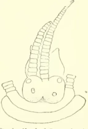
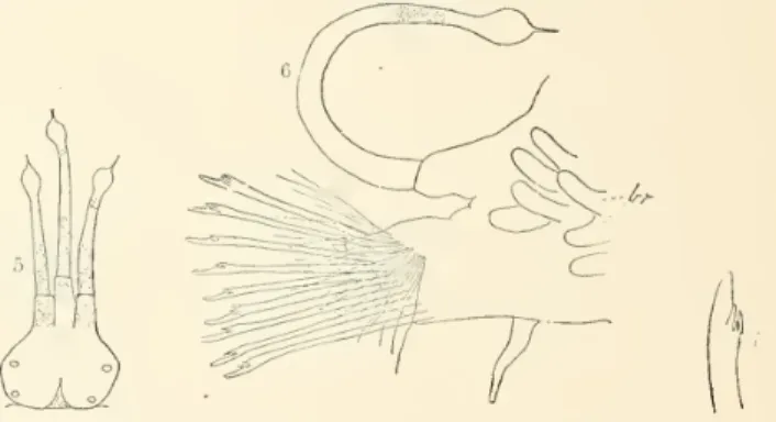
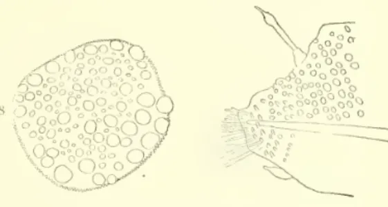
Figs. 5-7.
—
Polynoebranchiala. Fig.5,Head, x9. Fig.6,Parapodium, x16;br,branchial. Fig.7, Ventralseta,x90.
THE PQLYCH2ET0US ANNELIDS OF PORTO RIOO. 187 Polynoe nodosa,
n. sp.Body
plump, with short parapodia.The
specimens with elytraremoved
arecoiled,and
look not unlike the larvaof a coleopterous insect. Generalbody
color gray, with dark longitudinal dorsal band. Inoneof thetwo specimensatmy
disposal thisisduetocolorofbody
wall; intheother, to the color of minute tubercleswhich
cover it.Whole
dorsal surface, evenof parapodium, studded with small round tubercles.No
tubercles ventrally, but surface is studded with fine papillae, giving itavillous appearance.Head
withlateral edges rounded. Breadth about equal todistance from posterior margin to baseof antennae. Anterior eyes larger than posteriorand
situatedmore
towardside of head. Antennaelonger thanhead, withterminal swellingand
acute tip. Tip of ter-minalswelling
and
subterminalband
white; therest brown. Tentaclesnothalf aslong asantennae, likethelatterinformand
color, but lackingsubterminalband
of white. Tentacularand
dorsalcirri like antennae. Palps nearly twice as long a;
tentacles,tapering slowly to near apex,ending abruptly inasharppoint. Basal halfcolorless, terminalhalfbrown. Surfacestudded with very minute papillae,visible onlyunder high power.
Ina smaller specimen,about half the lengthof the above, the palps
were
uniformlybrown
and 8 onlyalittlelongerthanthe antennae.Elytraon segments2, 4, 5, 7,etc.,23.
Whole number
of pairs, 12.Body
segments27, includ- ing anal segment.Only
the anterior pair of elytrapresent in eitherspecimen. These werenearly square, theedge with a
row
of finepapil- Fig.9, Par lae,surfacestuddedwithtubercles. (Seefig.8.)
Parapodium
uniramous, very thick, its dorsal surface covered with tubercles. (See fig. 9.)
Toward
the end,and
ventrally, the tubercles are replacedby
fine villous-like papillae; dorsally, an elytrophore orcirrus. Short,"stout, ventralcirrus.A
few(ten) verystrong setae, withblunt-pointed aipex;asingle largetoothsome
distancefrom apex. Basal part striatedlongitudinally.A
singlelarge aciculum.Length25
mm;
width5mm.
Ofanother, length15mm.,
width3.5mm.
Collectedfrom Fajardoandstation 6079.
Polynoe,
sp.From Mayaguez
was collected afragment, probablyaPolynoe,butowing-to loss of the anterior segmentsthis could not be determined with certainty.An
elevated dorsal ridgemarks
off three distinctareasofthebody —
amedian and two
lateral. Surfaceirregularlymarked
with lightbrown and
gray. Elytra transparent, not coveringentiredorsalsurface.STHENELAIS
Kinberg.Sthenelais
simplex
Ehlers.Sthenelais simplex Ehlers,Annelidsofthe Blake,p.00, pi. 13, ligs.2and3; pi. 14,figs.1to0.
Ehlers says there arenoeyes. These,
which
agreeinallotherrespectswithhis diagnosis,show
a pairofverysmalldarkeyes, oneoneitherside ofthebaseofthe antenna.Collectedfromstation 6066.
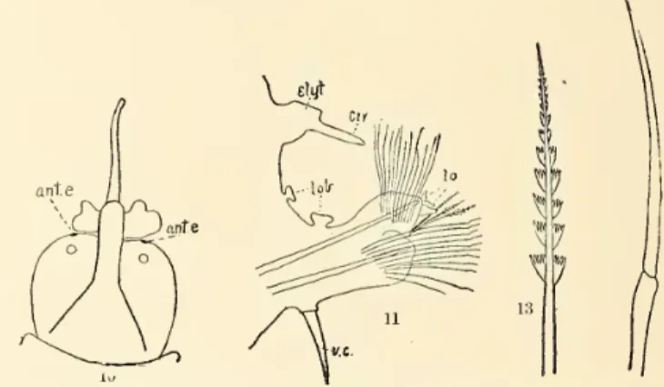
Sthenelais grubei, n. sp.
Grube, inhisdiagnosisofthisgenus (Annulata Semperiana,p.54),saysthat theelytraareborne on segments2,4,5,7,etc.,alternatelytosegment23,
and on
everysegmentposteriorto that. Schmarcla (NeueWirbellose Thiere,p.146) states that the alternation ceaseson
the twenty-seventh segment.The
specimens heredescribedagreewith Schmarda’sdescription.The
head(fig.10)isrounded,with a ratherbroadmedian
fissureintowhich
theantennafits. Base of antenna with broad lateralflap,narrowerat base. Palpsaslongasfirstninesegments.
Elytrawhite,,semitransparent. Firstpairbroad kidney-shaped; othersapproximatelyoval, the outer posterior border fringed with a fewdelicate papillae.
As
far assegment27thereisanarrow dorsalareanotcoveredby
theelytra.losa. Fig.8, Elytron,x20.
apodimn, x15.
188 BULLETIN OF THE
ITNT
TEL)STATES FISH COMMISSION.
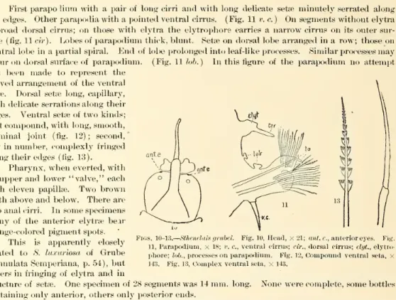
Firstparapolium with a pairof longcirri
and
withlong delicate setse minutelyserrated along theedges. Other parapodia with apointed ventral cirrus. (Fig.Hr.
c.)On
segmentswithoutelytra a broad dorsal cirrus; on those with elytra the elytrophore carries anarrowcirrus on itsoutersur- face (fig. 11cir). Lobesofparapodiumthick,blunt. Setseondorsal lobearrangedina row; thoseon ventral lobeinapartialspiral.End
oflobeprolongedinto leaf-like processes. Similar processesmay
occurondorsal surfaceof parapodium. (Fig. 11tub.) Inthisfigure of the
parapodium
no attempt has beenmade
to represent thecurved arrangementof the ventral setae. Dorsal setae long, capillary, withdelicate serrationsalongtheir edges. Ventralsetaeoftwokinds;
first
compound,
withlong,smooth, terminal joint (fig. 12); second, fewinnumber, complexly fringed alongtheiredges (fig. 13).Pharynx,
when
everted,with anupperand
lower“valve,” each with eleven papillae.Two brown
teeth aboveand
below. Therearetwo
analcirri. Insome
specimensmany
of the anterior elytrae bear orange-colored pigmentspots.This is apparently closely related to S. luxuriosa of
Grube
(Annulata Semperiana, p.54),but differs infringingof elytra and instructureof setae.
One
specimenof28segmentswas14mm.
long.None
werecomplete,some
bottles containingonlyanterior, othersonlyposteriorends.Collected from stations 6057, 6059, 6061, 6062, 6063, 6073; Puerto Rico,
Boqueron
Bay,and
San Antonio Bridge, San Juan.PSAMMOLYCE
Kinberg.Psammolyce
rigida Grube.Psammolycerigida Grube,Verhand. d. Zool.-Botan. Gesellschaft in Wien, 1868, p. 631, pi. 7, fig.1. Quoted from Grube, AnnulataSemperiana,p.55,1878.
Collectedfromstation 6062.
PANTHALIS
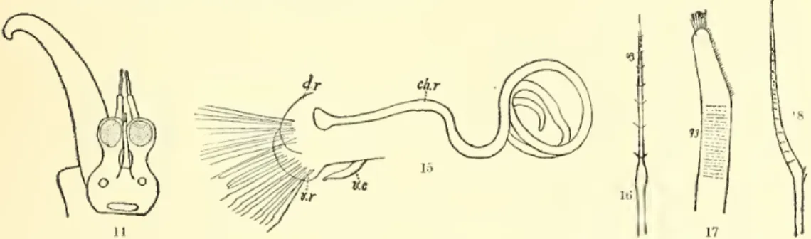
Kinberg.Panthalis oculea,n. sp.
Head
globular,prolongedanteriorlyintotwo
eye-stalks, whichcarrytheenormous
eyes (fig.14).Threetentacles,the
median
onanteriormarginofhead, the pairedbeneaththe eye-stalksand
project- ingbeyond'their ends.Apex
abruptlynarrowed,giving rise tomoderatelylongfilament,longerthan terminal filamentof unpaired. Palpslong, tapering,surfacecovered withminute filiform processes.First
parapodium
withtwo long cirri, ending likethe unpaired antenna,and nearlyaslong as thepalps, butmore
slender,and smooth. Palpsand
cirriwithnumerous brown
dots.A
tuft of setaeondorsal surfaceof firstparapodium.
A brown marking
atbaseofunpairedantennaand
a transversebrown band
atposterioredgeof head. Smallerpair of eyesnearbaseof antenna.Elytraon segments2,4, 5, 7,etc., through as
much
of thebody
aswas
preserved. Elytra nearly round, small,leavingthe greater partof thebody
uncovered, their surface dividedby
fine lines into nearlyequal, rectangular “cells.”A
brownish pigmentinmany
ofthese spaces,with a tendencyto accumulatein greateramount
toward dorsaland
posterior edges.Two
specimens, otherwise indis- tinguishable from the others,showed
no pigment on the dorsal surface of elytra and the posterior edgesofthelatterwereblack.Pharynx,
when
extruded, as long as first 20 segments, with asmooth
surface; atend with a dorsaland
ventral“valve,” eachfringedwithpapilhe, ofwhich
the dorsaland median
ventral aremuch
thelargest.Two
powerfulteethineachjaw,arow
ofsmaller teethrunninglaterallyoneither side ofeach.Figs. 10-13.
—
Sthenelaisgmbei. Fig.10.Head,x21; ant.e.,anterior eyes. Fig.11,Parapodium, x18; v.c.,ventralcirrus;cir.,dorsalcirrus;elyt.,elytro- phore;lob.,processeson parapodium. Fig.12,Compoundventral seta, x
143. Fig.13,Complexventralseta,x143.
THE POL YCH.ETOUS ANNELIDS OF PORTO RICO. 189 On
ventralview oftheentireanimalaseries of blackcoiled structuresmay
beseen lyingoneon either sidethemedian
linein eithersegment. Iftheparapodium becutoff, thesestructures pullout of the body, remaining attached to tip of parapodiumby
a flatexpansion. (Fig. 15, ch.r.)Each
is a chitinousrod,
which
easilysplitsup
into anumber
of fine threads. Dissectionshowsthat thefirstseptumappearsbetween segments21and22,thecoelominfront ofthatbeing a continuouscavity.
Thiscavityisnearlyfilled
by
theserods, whichareshorterandmuch
lesscoiledthan theyare farther back.They
lie just above the nephridia, which can be seen belowthem
as slender, short, white organs.Rami
ofparapodium almostfused. Dorsalramus
(fig.15, d.r.) rounded,thin, withatuft oflong setae; afewlarger thantheothers,lanceolateatend, with anumber
of pairs of lateral spines (fig. 16).Ventralsetaeoftwokinds; dorsal onesthick, brown, withendobliquely truncated,
and
coveredwith minute spines. (Fig.17.) Ventral ones colorless, notmore
thanone-fourthasthickasthedorsal, bentatsome
distancefrom end, withthe transversediametersomewhat
greateratpointof bending;fromthe
bend
toapex covered withtransverserowsofminutespines. (Fig.18.)
About
55 of anterior segments preserved inone specimenmeasured17mm.
in length, 2mm.
broad withoutparapodia,4
mm.
withparapodia.Collectedfromstations 6059, 6063,
and
Porto Rico.Figs. 14-18.
—
Panthalisocalea. Fig.14, Head, x14. OnlytheleftpalpIsfigured. Fig.15,Parapodium, x14; d.r.,v.r.dorsalandventralrami; v. c., ventralcirrus; c/t. r., chitinous rod. Fig.16, Setaofdorsalramus, x 143. Figs. 17 and18, Setsof ventralramus, x143.
EULEPIS
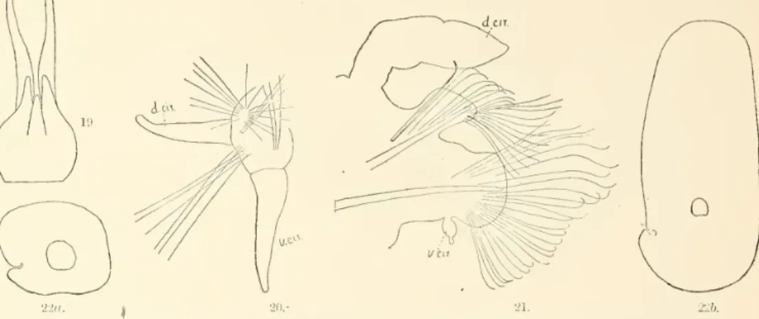
Grube.Eulepis
splendida,n.sp.Head
rounded, incised in front, unpaired tentacle small, rising from dorsal surface of head, reachingscarcely to halfthe lengthofthe paired; the latterarisingfromanterior lobe of head, about two-thirds as long as head (fig. 19). All antennae conical, with distal two-thirds dark brown,the very tip white. Palps long, smooth, white, tapering gradually to the end (fig. 19).No
eyes could be seen.First parapodium with twocirri and two tufts of delicate setae (fig. 20) arisingfromitssurface.
Parapodium
twistedso thatthetwo
cirricome
to lievery nearlyin a horizontal plane. Setae long, thread-like, afew with veryminuteserrationsalong oneborder. Other parapodia with verydistinct rami. Dorsalramus with about 15 stout, brown, chitinous setae, curved at apex, thecurvedportion pointing backward.Below
this is a tuft of fine thread-like setae,some
with fine serrations along their edges. Theseareverynumerous and
of a golden-redcolor. Ventralramus
broader thandorsal, with about 25 long setae. Setae about half the diameter of coarse dorsal setae, curved atapex, the curvedportionpointingbackward. Generalcolor of thesesetaeyellowish brown, withtips,asseenin reflectedlight, noticeablylighter. Ventral cirrus ovatewithbaseslightly narrowed, its apexdrawn
outintoaterminaljoint havingmuch
the form of the basal, but verymuch
smaller. Dorsalramus
witheitheracirrus oran
elytrophore (fig. 21).Elytra borne on segments2, 4, 5, 7,etc., 21, 24. Grube, in his diagnosis of thisgenus (Annu- lataSemperiana,p.51), says thatelytraalternate anteriorly, afterthe
manner
of the Polynoidee, but posteriorlyareborneon
allsegments. InhisdescriptionofE.hsemifera (loc.cit.,p.52),henotesthat theelytraarefound on segments2,4,5, 7,etc.,up
to 21; that then theyskip first two, then three,190 BULLETIN OF THE UNITED STATES FISH COMMISSION.
and from the twenty-eighth segment are found on all segments.
He
notes further that theelytra increase in sizeup
to the twelfth pair;and thenbecome
smaller. InE. splendida there aretwelve pairsofelytra,thelastmuch
the largest,borne on segment 24, but extending backso as tocoveras farasgreater partofsegment31, and theabovegeneric description—
thatallofthe posteriorsegments bear elytra—
appliesonlyifwe
regardthe broad, flatexpansionof the dorsalcirrus asanelytron (fig.Figs. 19-22/).
—
Eulepis splendida. Fig.19, Head, /17. Fig.20, Firstparapodium, x23; d.dr. 'andr.cir.,dorsal andventralcirri. Fig.21, Posteriorparapodium,x17. Figs.22a, 226, Seventh,andtwelfthelytra,x8.21). Thiscanhardlybe thecase, sinceit isfoundonallthe cirrus-bearing segments,exceptthemost anteriorones. Inthisrespectthesespecimensdo notagreewith Grube’sdiagnosis.
They
agree insomany
otherrespects, however, that I have no hesitation in assigningthem
tothisgenus. Probably theloss ofposteriorelytraiscorrelatedwiththeenormous
developmentofthe twelfthpair.Parapodia around
head
verymuch crowded
together; the secondand
third segments fused dorsally, sothatthesecondelytrophore apparentlyarises from anterior end of third segment.The
firstelytracompletelycoverthe head.
The
firstelytrawereremoved
inordertodraw
the head,and
wereunfortunatelylostbeforethey could be drawn.A
drawing of the seventh isgiven in fig. 22«
and
of the twelfth in fig. 22b.These are
drawn
tothesame
scale,toshow
the increase insizefrombeforebackward. Excepta small notchon
outer border, the edgeisentire.Theircolor is white,
and
they show, under the microscope, a finelygranular texture.At
ante^- rior edge of ventralramus
ofparapodium
is a darkspot.Body
of37 segments. Length,37mm. Width
withoutparapodia,3mm.
;withparapodia,5mm.
The
single entirespecimenhad
oneanalcirrus.Collectedfromstations6062
and
6065.Eulepis
fimbriata, n. sp.Figs.23,24.
—
Eulepis fimbriata. Fig. 23, Head, x18;at, line along which processfrom anterior elytrophore fuseswith head. Fig.24, Elytron,x22.Head rounded
(fig. 23).Antenna
small, globular, onashortstalk. Tentaclesreaching a littlebeyond
tip of antenna, rising from undersurface of head. Palps long,smooth, closelyappressed in middle line.
Head
thicklymarked
with yellowishbrown.Two
darkspots(eyes?)nearbaseand
onetoward apexjustbehindbaseofantenna.Allappendages around thehead very
much crowded
together.The
anteriorelytracompleted
cover the head, theirelytrophorestouching onmedian
line.An
anterior process frombaseof elytrophoreTHE POLY CHAETOUS ANNELIDS OF PORTO RICO. 191
fuseswithdorsalsurfaceofhead. (Seeitsline ofattachmentat, infig. 23.) Parapodia
and
set* like those of E. splendida,except that set* of dorsalramus
are possiblynot sonumerous and
lack the brilliant color characteristic of the latter. Secondand
third segmentsmore
or less fused above.Arrangement
of elytra as inE. splendida, the twelfth pairmuch
thelargest, borneon segment21 and covering nearlyalltherest ofbody.Elytra white, granular, like those of E. splendida,,but prolonged on lateral border into broad leaf-likeprocesses(fig. 24).
One
analcirrus.Number
ofbody
segments, 37.Length, 24
mm.;
width, withoutparapodia, 4mm.
Collectedfromstation60(51.
Family
PHYLLODOCID^.
PHYLLODOCE
Sav.Phyllodoce
oculata Ehlers.Phyllodocc oculata Ehlers,Annelidsofthe Blake,p. 135, pi.40, figs. 4,5,G.
AccordingtoEhlers, the ventral cirrus in each segment is fused along itswholedorsal edgeto ventralface of parapodium. Inthespecimenfrom PortoRico, althoughthecirrusiscloselyapposed totheparapodium, itisactuallyfusedonlyatitsbase. Ehlers describes, further,the parapodiumas uniramous, withananteriorand aposteriorlip,thelatterbeingthelarger
and
bifid atend. Intheseit isthe anteriorlip
which
islargerand
bifid.Through
the courtesyof Dr.W. M. Woodworth,
I havehad
an opportunityofexaminingthe type specimen fromtheMuseum
ofComparative Zoologyat Cambridge, Mass., and Ifind thatEhlers was certainlywrong
inboth theabovepoints.The
ventral cirrus in the type specimen is attached onlyat its base,and
the anterior lip is largerand
bifid.InallotherrespectsthePorto Ricospecimensagree,with Ehlers’s diagnosisofthespecies.
Collectedfromstation 6065.
Phyllodoce magma-oculata,
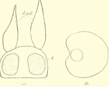
n. sp.Head
rounded, broader than long (fig. 25). Eyes verylarge (e, fig.25). Dorsal antennae-lanceolate,nearly twiceas longas head (d.ant, fig. 25). Ventral antennae on lowerface ofhead, equalin size to dorsal. Fourpairs of tentacular cirri, the largest 3.5 times as long as the antennae, thick, with acute termination.The
othercirri smaller, equal.The
gillshad
all beenremoved
from the anterior segmentsof thebody. Thosewhich
remained werecov- ered with a slimy deposit, containingnumerous
foreign particles.The
gills are especially liable to be brokenaway
inattempting toremove
thisdeposit. Gills broadly reniform (fig.26), withentire marginand
withpointofattachment nearthe baseofthehilus. Coloratpointofattachment,lightbrown.Each
gill contains
numerous
anastomosing bloodvesselsand numerous
small, round, lightand
darkbrown
pigmentgranules.Parapodium
asingleconical lobe, slightly bifid atthe end, withalarge aciculum.About
ninecompound
set* on either side of the aciculum. Basal joint of set* long, most extendingnearly or quite the length ofparapodium beyond
tip of latter.At
end the basal portion hasaclub-shaped enlargement,marked by
veryfinelongitudinallines. Terminalportion rathermore
thanhalf aslong as basal, atbaseasbroadasbasalportion, taperinggraduallytoafinepoint.Collectedfromstation 6067.
Phyllodoce, sp.
From
BoqueronBay
yvas obtained afragmentofaspecimenof thisgenustoomuch
injured for identification,head and
tail lacking.The
fragment was 25mm.
long, 5mm.
wide, and contained over 50 segments.Body
black, with adorsal longitudinalband,and
edgesof gills and cirrifringed withwhite.Pigs.25, 26.
—
Phyllodoce magna-oculata. Fig.25, Head, x 70; d. ant., dorsal antennre;e, eye.Fig.26, Gill,X70.
192 BULLETIN OF THE UNITED STATES FISH COMMISSION.
EULALIA
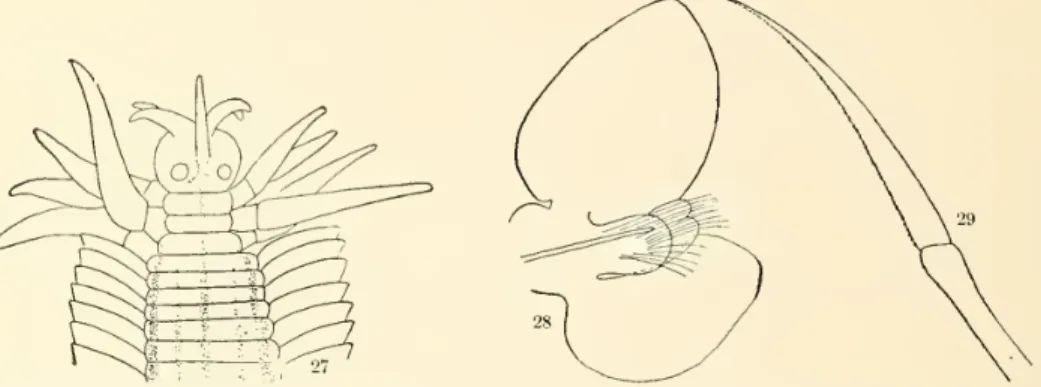
(Sav.)Malmgren.Eulalia quinquelineata, n. sp.
Head
oval, broadest just infrontof eyes, withaslightconstriction atbaseof tentacles (fig.27).Tentaclesfour,equal,three-fourthsaslongashead,stout,withacutepoints.
Median
unpairedtentacle arisingabouthalfway
from eyes tothe anteriormargin of head,much more
slenderthanpairedand reachingbeyond
their ends.Two
rows of small pigment spots begin atbase of unpaired tentacle and extend back betweeneyes, forming an )( shaped marking.On
firstsegment, onetentacularcirrus; onsecond, two (adorsalanda ventral); onthird,a long dorsal, tentacularcirrus,and
athick, fiat, ventral cirrus, thelatterlike the ventralcirriofsucceeding segments. Tentacularcirricomposed
ofa basal portion anda stout terminal portionpointed atend.Dorsalcirrus ofsecondandthatofthirdsegments aboutequal in size
and
slightly largerthanother two.First
and
second segments abouttwo-thirds thediameterofthe fourthand
succeeding segments.Body
450mm.
long; without the parapodia2mm.
wideat anterior end. It retains thiswidth untilnearposteriorend, where it narrows gradually.No
analcirrus presentinthe singlespecimen in this collection. Segments at anterior end six times wider than long; toward posterior end this proportion is diminished and the parapodiaof successive segments aremore
widelyseparated than anteriorly. Inthepreserved specimen,which
isdoubtlessmore
or less contracted, thegills of each segmentoverlap thoseofsegmentin front.On
accountofthe greatlength ofthebody and
theextent towhich
itwascoiledI was unabletodeterminetheprecisenumber
ofthesegments. Since,the length ofthe anteriorsegmentsisonlyabout0.33mm.
,increasingslightlytoward posteriorend,itfollows that theremust
beat least 1,300segmentsin thewholebody.Figs. 27-29.
—
Eulalia quinquelineata. Fig.27,Head,x7. Fig.28,Parapodium,x72. Fig.29,Compoundseta,x257.Parapodium
asingleramus
withanteriorbilobedlipand posteriorshorter,rounded,one. Dorsal cirrusnearly a regularovate,attachedby
ventraljoint (fig. 28). Ventral cirrus comparativelylarge, ovate,hollowedoutonsidenext parapodium. Both cirrishow numerous
ramificationsofblood vessels in their interior.Toward
posteriorend
the cirribecome
atriflemore
acuteat endsand
the whole appendage ismuch
smaller, buttheir relativeproportions are aboutthe same. Setfecompound,
the basaljoint long,swollenatend. Terminaljointbroadat base,bentslightlyand
tapering gradually to anacutepoint, withrow
ofminuteteeth on concave edge(fig. 29^.Colorof
body
yellowishbrown, with afaintgreenishtinge. Dorsal surfacewithfivelongitudinal blackbands—
amedian,two
admedian,and
twolateral, thelatter justat baseof parapodia.Median
narrow onfirst rive segments,becoming
broaderfarther back. Lateral narrower than median,begin- ning on thirdsegment.Admedian
abouthalfway
betweenthe other two, alittlenearer thelateral;narrowerand lighter colored than either.
They
begin on posterior edgeof fourthsegmentas small spots,which
are repeatedonfifthand
sixthsegments,becoming
continuouslinesonseventh (fig.27).Toward
posteriorend
this linebecomesmuch
less conspicuous.On
ventral surface amedian and
two lateralbands, similarin sizeand
position to correspondingdorsalones, butwith noadmedian
bands.CollectedfromHueares.
THE POLYCHiETOUS ANNELIDS OF PORTO RICO. 193
Family
NEREIDS.
NEREIS
Cuvier.Nereis
bairdii Webster.NereisbairdiiWebster, Annelids fromBermuda,Bull.No.25,U.S.Nat.Mus.,(>.312, pi.8,figs.22-28.
Thereareapparently two-well
marked
varieties in this species, differing incolorand
inform of parapodia.One
with outerportionofheadand
dorsal surface of anteriorsegmentsbrown.A row
of cclorlessspotsin thisband
onheadand
acrossanteriorendoffirstsix orsevensegments. Parapodia, especiallythe posteriorones,withexcessivedevelopmentofdorsalramus,asdescribedbyW
ebster.A
rectangularwhite patchondorsal surfaceof eachpairof segments, overlapping the linebetweenthe two,thoughlyingmainlyin posteriorone.
The
secondvarietyhas aband
ofbrown
aroundthehead,much
narrower thanthefirstand
withno whitespots.A
transversebrown band
on each segment,much
darker nearposterioredge. Lobesofparapodiummuch
blunterandmore
rounded thaninfirst varietyandposteriorparapodianotwith excessive developmentof dorsal ramus. Dorsal cirrimuch
longerthaninfirstvariety. Websterfigureslong
compound
setaewithsmoothterminaljoint. Inthe PortoRicospecimensthisterminaljointisfinelytoothed. These specimensagreeso closelywithone another,and
with Webster’s descriptionof the species,in somany
anatomical features, that I have thoughtitbesttoregardthem
ascolor varieties ofthesame
species.Collectedfrom PuertoReal, Arroyo, Boqueron Bay, Mayaguez, Porto Rico, stations 6065, 6091, 6092, 6062,
and
6063;Ensenada Honda,
Culebra.Nereis mirabilis Kinberg.
Nereis mirabilis Kinberg,AnnulataNova, Oefvers. af. K.Vet.Akad.Forh.1864,No.16, page 571. Quoted fromEhlers, Annelidsofthe Blake,p. 117, pi. 37,figs.1-6.
Nereisgracilis
W
ebster,Annelids fromBermuda,p. 313.From
a careful comparison of Ehlers’s with Webster’s description I conclude that these are identical species. Ehlersfiguresthe antennseas entire, whileWebsterfiguresthem
withrather long basaljoints.The
singlespecimenatmy
disposal (fromBoqueron
Bay) has a short basal joint, easily overlooked inpreservedmaterial.Nereis arroyensis,n. sp.
Head
broader thanlong, the dorsal surfaceshading off graduallyinto thepalps, withno
sharp line between the two. Anterior edge rounded, with two thick antennae. Eyes four, anterior pair semilunar in form (with transparent “lens”?) (fig. 30).The
posterior dorsal tentacular cirrihad
been lost; the other cirri short (fig. 30). Inother specimens than theonefigured thesecirri were longer.Body
colorless,broadestinfront,tapering graduallytoposterior end.
Body
of 73 segments.Two
verylong analcirri. 45
mm.
long, 2.5mm.
wide.Parapodium
withdorsalcirrus longerthanramus
(fig.31).Ramus
of
two
lingulae, the dorsal a little longer than the ventral. Setae of dorsalramus compound,
with long basal joint with prominent trans- verse striations. Terminal joint long,nearlystraight,finelytoothed.Dorsal setae of ventral
ramus
likesetaeofdorsalramus. Ventralsetaewithbasal joint like dorsal, terminal joint short,
hooked
at endand
with arow
of long delicate spines on side. Dorsal lingulaof ventralramus
with anterior and posterior lobe (“lip”), the anterior a little the larger. Ventral cirrus not quite so long as dorsal.Toward
posteriorend, parapodiamuch
as anteriorly, except that lobesbecome
alittlemore
pointed,and
thesetsewith long terminal jointbecome
relativelymuch more numerous
inthe ventral ramus.The number
ofsetseinthe dorsalramus becomes
verysmall.CollectedfromArroyo andstation6052.
2d—F. C.B.1900—13
Figs. 30, 31.
—
Nereis arroyensis. Fig. 30,Head,x17. Fig. 31, Parapo- dium,x18.194 BULLETIN OF THE UNITED STATES FISH COMMISSION.
A
singlemuch
mutilated specimen fromMayaguez
I have placed in this species, though it is possiblydistinct.The
formof the headand
palpswas
likethatof iV. arroyensis, as alsowasthatef the posterior parapodia.The
anterior8 to 10 parapodiahad
verythickrounded lobes, showing only a divisionintothetworami.Family
NEPHTHYDID^.
NEPTHYS
Cuv.Nepthys squamosa
Elders.NepthyssquamosaEhlers,Annelidsofthe Blake,p. 128,pi. 37, figs.7-10.
Collectedfromstations 6084, 6085, 6091, 6092, 6093.
Family
AMPHINOMIDAE.
HERMODICE
Kinberg.Hermodice carunculata
(Pall.) Kinberg.HermodicecarunculataWebster,Annelidsfrom Bermuda,Bull. U.S.Nat.Mus.1884, p.307 (See thispaperforreference to earlier literature.) Ehlers,Annelidsofthe Blake,p.27.
The
colorvariesfrombrown
toadecidedblue. Ehlers says theyoung
arealightbrown
witha blackmark
acrossthe back.Only
onespecimenofthis collection(18cm.long)showed
thismarking.Collected from Guanica Bay, Fajardo, Arroyo, Ponce, SanAntonio Bridge, SanJuan, Boqueron Bay, Mayaguez, Playa de PonceReef,
Ensenada Honda
(Culebra), stations 6092, 6088.N0T0PYG0S
Kinberg.Notopygos
crinitaGrube.JSTotopygoscrinitaGrube, Beschreibung neuer oderwenig bekannterAnneliden, Arehiv. f.Natur.Jhr,21, Bd. 1, 1885.
Grube,AnnulataSemperiana,1878, p.7. Ehlers,Annelidsofthe Blake,p. 24, pi.1, fig.3;pi.3, figs. 5,•,7.
A
very full description is givenby
Ehlers; he does not figure nor describe arow
of small, bead-like elevationson the dorsal surfaceof themedian
fold of the caruncle; these areabout 15 in number, very prominent in front,and
gradually fading out behind; theyare relativelymore
promi- nentinthelargethaninthe smallspecimens. Inaspecimen42mm.
longthefirsttenofthesebeads were dark brown. Insmallerspecimens onlyoneortwo show any
color.Collected fromstation 6079.
From
asecond specimenthe locality labelwas unfortunately lost in transferring.EURYTHOE
Kinberg.Eurythoe complanata
Pall.EurythoecomplanataPallas,MiscellaneaZoologica,Hagae-Comitum,p. 109, pi.8, figs.19-26. QuotedfromEhlers,Annelids ofthe Blake,p.29.
Body
light gray, withmarked
iridescence. Setae white. Ehlers describes the eyes as black;thesewerealightreddishbrown.
Collectedfrom Arroyo,Hucares,PuertoReal,
Ensenada Honda
(Culebra). Inoneotherspecimen thelocalitylabelwas
lost.EUPHROSYNE
Sav.Euphrosyne
trilobaEhlers.EuphrosynetrilobaEhlers,Annelidsofthe Blake,p.31,pi.4.
Collectedfromstation 6098.
CHLOEIA
Sav.Chloeia euglochisEhlers.
Chlocia euglochis Ehlers,Annelidsofthe Blake,p.18,pi.1, figs.1-8; pi.3,figs.1-4.
Two
specimensarein this collection.Eyes
notsonearly fusedas inEhlers’sdescription.Median
andpairedantennaeand
mostofthe dorsalcirriareofabrilliantviolet color.AMPHINOME
Brug.Amphinome
microcarunculata, n. sp.Body
of singlespecimenincomplete,onlyanterior 36segmentspreserved. Lengthofthese,38mm.
Breadthofhead,0.75
mm. Body
rapidlywidenstotwentieth segment,where
itsbreadthis10mm.;
THE POLYOHfiETOUS ANNELIDS OF PORTO RICO. 195
from here it narrows again rapidly; thirty-second segment, 5
mm.
wide. Color above, seal-brown, shading into ashy gray anteriorly; ventrally, ashy gray. Setae long, very fine, white. Caruncle small, smooth, notextendingbeyond
limits ofhead lobe. .Dorsal setae shorterthanventral. Dorsal cirrus arising atbaseof tuft ofsetae,a little behindand
ventral tothem. Cirrusaboutthree-fourths aslongas setae. Ventralramus witha thick fleshylip,fromthe dorsaledgeofwhich
the setae arise.Ventral cirrus slender, shorter than lip of ramus.
Two
tentaclesand two
subtentaclespresent, themedian
tentaclehaving beenlost. (Seefig. 32.)No
eyes couldbeseen.Mouth
surroundedby two
segments, the posterior lip lyinginthe interruption ofmedian
line of third segment. Gillsappear firstoneighthsegment,as a single filament, attaining their full size about segment 12.
They
are very inconspicuous, lying behind the dorsal cirrus,and
inpreserved materialalmost completely hiddeninthe con- striction between the segments. In its fully developed form each gilliscomposed
ofatuftof thick,shortfilaments.In the generic descriptionof
Amphinome
(Kinberg; Sven- ska. Vetensk. Akad. Ofversigt, vol. 14, pp- 11 to14, 1858) it is statedthat the gillsbegin on segment3. In the absenceof amedian
tentacleand
inthe factthatthegillsappear firstontheninthsegment, this specimendiffers fromthe genericdiagnosis. I have regardedtheformerasan accident,and
thelatteras notofsuffi- cientimportanceonwhich
toformanew
genus.Collectedfromstation 6070.
Family
CHRYSOPETALID./E.
BHAWANIA
Schmarda.Bhawania
goodei Webster.BhawaniagoodeiWebster, AnnelidafromBermuda,p. 308.
A number
offragments, lackingboth headand
tail,seem
undoubtedlytobelongto this species, thoughtheremainingparts differsomewhat
from Webster’sdescription. Dorsaland
ventralramiof parapodia separated rathermore
widely than inWebster’sdescription. Terminal portionofventralramus expanded
at base and narrowing rapidly toward apex.Webster
figures it as slenderand
conical. Dorsal
ramus
as inWebster’sdescription. Dorsalpalese inarow
extendingacrossback, themedian
onesbending inward,sothat those of opposite sidesoverlap. Set*of two kinds. Thoseof dorsal ramus constrictedat base, likedorsalpale*,and
for inner two-thirdsof their lengthmarked
withthe longitudinalandtransverse striationscharacteristic of latter. Theseare figuredby Webster
assmooth, with sharppoint
and
broadbase.The
set* of the dorsalramus
are regardedby Johnson
(Proc. Calif. Acad. Sci., vol. 1, No. 5, p. 162) asasecond formofpale*,and
areoneofthe charactersofhisnew
genusHeteropale.The
data inmy
possession are few,but fromwhat
I haveIam
inclined tobelieve thatJohnson’sHeteropale, shouldbediscarded infavorof Schmarda’s Bhawania. Intheshapeand
position of the otherset*, thesespecimensagreewithB. goodei.Ehlers (Annelidsofthe Blake,p. 34)describesfragmentsofa Chrysapetallidin
which
thepale*cover the back.
He
does not describeany
otherdetails.CollectedfromArroyo.
Family
EUNICIDAi.
EUNICE
Cuvier.Eunice ornata
Andrews.Eunice ornataAndrews, Annelida PolychsetaofBeaufort,N.C.,Proc. U.S.Nat. Mus.,vol.14,p. 284, 1891.
Collected from stations 6080, 6079, 6073, 6092; Mayaguez; Ponce; stations 6086, 6091; and afemalewith eggs fromArroyo. Inthespecimenfromstation6092 thegillsbeganonthe sixthseg- ment, insteadofthefifth, which, accordingtoAndrews, isthenormal.
Fig.32.
—
HeadofAmphinomemicrocarun- culata,x8;car.,caruncle.196 BULLETIN OF THE UNITED STATES FISH COMMISSION.
Eunice
denticulata Webster.EunicedenticulataWebster,AnnelidafromBermuda,Bull.U.S.Nat.Mus.1884.
Webster’sspecimens,preserved in alcohol, wereof ayellowishwhitecolor.
One
inthis collec- tion,preserved in formalin,was
adirtywhitethrough most of the body,dorsal surfaceof headand
anteriorsegments irregularlymarked
with dark green blotches. Gillsappearas singlefilamentson the twenty-eighthsetigeroussegment.Head
deeplybilobed. Tentaclessmooth, equal, about twice aslongasthe head.The
dorsalramus
of theparapodium
containscomb-shapedsetae,which
arenot describedby
Webster.The
headofonelargespecimenwas
mottled with brown.Collectedfromstations 6065, 6079,
on
corals atMayaguez,and
fromEnsenada Honda
(Culebra).Eunice violacea-maculata
Fillers.Euniceviolacea-maculata Ehlers,Annelidsofthe Blake,p. 86,pi.24,I,figs. 11,12; pi. 25,figs.1-7.
Two
longtransverselybanded
analcirri. Collected fromEnsenada Honda
(Culebra),and
from station 6079.Eunice
articulataEhlers.Eunicearticula Ehlers,Annelidsofthe Blake,p.83,pi.24, figs. 8, 9, 10.
Gillsbeginonthe third setigerous segment. Ehlers says there aretwoanalcirri. These have four,twolong, articulated,
and two
veryshortones.From
PlayadePoncereefwas
collectedaspecimensuperficiallyveryunlike Ehlers’sdescriptionof this species,but agreeing so closely inmost
charactersof importance that I have included it here.The
differences are possibly sexual.The body
ismuch
largerand
broader. Generalcolor, light brown. Segments3, 7, 8,and
9 white.A
narrowbrown band
atthe baseofeachsegmentofantennaeand
cirri.A
smallerspecimenfromstation137shows
thesesame
colormarkings.Collectedfromstations 6065, 6098, 6096,
and
Playa de Poncereef.Eunice
siciliensisGrube.EunicesiciliensisEhlers,Die Borstemviirmer,p. 353, pi.16. (See Ehlers’spaperforreferences topreviousliterature.)
Inan animalof350segmentsthegillsappearfirstasa simplefoldon segment145. Ehlerssays that the distinction in length between the
two
pairs of analcirriis notgreat. Inthesingleperfect specimenin this collectiontwo
of these aremuch
longerthanthe other two.Collectedfromstation 6064, CaballoBlancoreef,
and
Arroyo.Eunice
fucataEhlers.Eunice fucataEhlers,Annelidsofthe Blake,p.91,pi.25, figs.8-20.
Ehlers does not mentionthepresenceof analcirri. Inoneof these there are
two
long, rather fleshycirri. CollectedfromArroyo, CaballoBlancoreefs,and Boqueron
Bay.Eunice
auriculata, n. sp.Prostomium
bilobed,eachlobetriangular,withapex
pointing forward. (Seefig.33.) Thislobingis
much more
prominent onthe ventralface. Tentaclescomposed
ofa short articulated basal portionand
along,smooth, terminal portion, thelattertaperinggraduallytothe end.Median
antenna very long, reachingbackto the eighteenth segment.Median
paired antennae half as long as unpaired.Outerpaired antennaeabouthalf aslongasmedian. Peristomium long at sides, deeplyhollowed in front, sothat
median
length isscarcely two-thirds thatof side.Median
lengthaboutequaltothree succeeding segments. Third segment about equal in width to succeeding. Nuchal cirri smooth, tapering gradually'from a rather thick base to a sharply-pointed apex.Apex
reaching a littlebeyond
front borderof peristomium.The body
graduallynarrowstothe fourth segment,and
from heregradually increasesinwidthtoaboutthe tenth.From
there a gradual decrease againas far as thirtieth. Thirtieth segment a trifle narrower than the first. Dorsal cirri very large. Branchiae beginon
nineteenthsegmentasasinglefilament.On
thetwenty-first thishasdividedintotwo,and
ataboutthetwenty-seventhitdividesagain.
They
neverbecome
verycomplex.Anteriorparapodiablunt,withanterior