The Role of Cardiac Fibroblast Talins on Regulating Fibrosis and Hypertrophy Following Pressure Overload of the Heart
By Natalie A. Noll
Dissertation
Submitted to the Faculty of the Graduate School of Vanderbilt University
in partial fulfillment of the requirements for the degree of
DOCTOR OF PHILOSOPHY In
Biomedical Engineering March 31, 2022
Approved by:
W. David Merryman, Ph.D.
Cynthia Reinhart-King, Ph.D.
Franz Baudenbacher, Ph.D.
Roy Zent, M.D, Ph.D.
Hind Lal, Ph.D.
ii
Copyright © 2022 by Natalie A. Noll All Rights Reserved
iii
ACKNOWLEDGEMENTS
This work was only made possible with the support and help of many mentors, coworkers, friends, and family. I want to acknowledge my mentor Dave Merryman, and my colleagues, dissertation committee, and co-authors on the two manuscripts that have been adapted for this dissertation, as well as my funding sources: the NIH and Foundation Leducq.
I want to especially acknowledge my lab mates who I have had the privilege of overlapping with. There are many highs in science, but also many lows, and all of you were there to experience mine firsthand. Thank you for brainstorming with me, editing multiple manuscripts, helping me see the bright side of failures, celebrating my accomplishments, and helping me to adapt to working two years in lab during a pandemic. You made every day in lab better: David Armstrong, Matt Bersi, Nathen Bloodworth, Erin Booton, Meghan Bowler, Cyndi Clark, Tessa Huffstater, Cami Johnson, Ethan Joll, Olu Ogungbesan, Caleb Snider, Chrisi Scott, Michael Raddatz, Lance Riley, Mark Vander Roest, Allison Schroer, and Michael Valentine.
Through this journey beach volleyball has provided with me a place for mental recharge and rejuvenation, and the people that I have met through volleyball have become my second family. Thank you all for supporting me and becoming forever friends.
Chris, your support these last two years has been incredible. My stress disappears when we are together. Thank you for being my volleyball partner, but also my partner in life.
Lastly, but most importantly I want to thank my parents and sister – Nicola, Tom, and Stephanie, as well as my entire extended family. Thank you for always being there for me and supporting me from near and afar. I know you are always there for me, and your support means the world.
iv
TABLE OF CONTENTS
ACKNOWLEDGEMENTS ... iii
LIST OF TABLES ... vii
LIST OF FIGURES ... viii
LIST OF ABBREVIATIONS ... x
Dissertation Overview ... 1
Chapter 1 Cardiovascular Disease and Tissue Response to Hypertension ... 2
1.1 Heart failure disease burden ... 2
1.2 Heart failure classifications ... 3
1.3 Hypertension ... 6
1.3.1 Systolic blood pressure ... 6
1.3.2 Cardiac fibroblasts ... 7
1.3.3 Cardiac myocytes ... 10
1.4 Therapeutic shortcomings of hypertension ... 12
Chapter 2 Integrin Adhesions & Talin ... 14
2.1 Mechanotransduction between cells and the extracellular matrix ... 14
2.1.1 Integrins ... 15
2.1.2 Talin ... 16
2.2 Talin in the body ... 20
2.3 Talin in the heart... 21
Chapter 3 Mouse Models of Pressure Overload Injury to the Heart ... 23
3.1 Introduction ... 23
3.2 Mouse models of HFpEF ... 24
3.2.1 Hypertension ... 24
3.2.2 Pulmonary hypertension ... 25
3.2.3 Type 2 diabetes ... 25
3.2.4 Type 1 diabetes ... 26
3.2.5 Obesity ... 27
3.2.6 Aging ... 28
3.3 Advantages and disadvantages of HFpEF models ... 29
v
3.4 Mouse models of HFrEF ... 32
3.4.1 Left ventricular pressure overload ... 32
3.4.2 Ischemic injury ... 33
3.4.3 Other surgical models ... 34
3.4.4 Pharmacological models of HFrEF ... 34
3.4.5 Genetic models ... 36
3.5 Advantages and disadvantages of HFrEF models ... 38
3.6 Discussion ... 40
Chapter 4 Loss of Tln1 in Myofibroblasts During Pressure-Overload Induced HFpEF Results in Augmented Cardiac Hypertrophy ... 44
4.1 Introduction ... 44
4.2 Methods ... 46
4.3 Results ... 55
4.3.1 TAC injury results in pressure overload of the heart that leads to both HFpEF and HFrEF in WT and mice with myofibroblast deletion of Tln1 ... 55
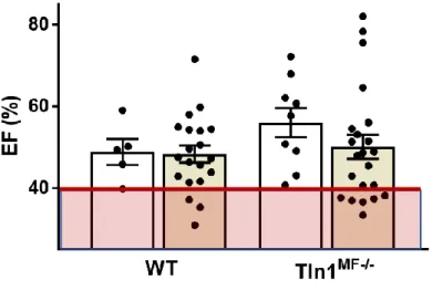
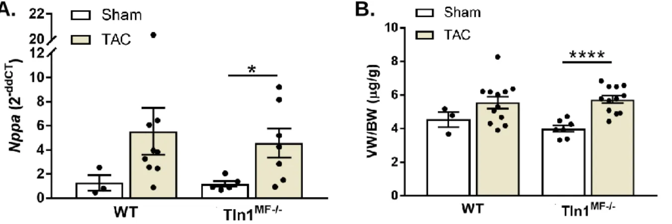
4.3.2 Myofibroblast deletion of Tln1 results in myocardial injury and cardiomyocyte hypertrophy in response to TAC injury with HFpEF ... 57
4.3.3 Myofibroblast deletion of Tln1 results in no change in cardiac fibrosis burden following TAC with HFpEF ... 60
4.3.4 siRNA knockdown of Tln1 in myofibroblasts alters cellular proliferation, migration, and contraction ... 61
4.4 Discussion ... 66
Chapter 5 Creating and Validating Models of Heart Failure Injury and Creation of the Tln2-Null; Cardiac Fibroblast-Specific Tln1 Knockout Mouse ... 69
5.1 Introduction ... 69
5.2 Methods ... 71
5.3 Validation of heart failure mouse models ... 76
5.3.1 Isoproterenol injections ... 76
5.3.2 Angiotensin II & Phenylephrine osmotic pumps ... 77
5.4 Creation and validation of a Tln2 null, CF-specific deletion of Tln1 ... 80
vi
5.4.1 Mice with a global deletion of Tln2 and CF-specific deletion of Tln1 does not
affect the ability of mice to survive myocardial infarction injury. ... 83
5.4.2 AngII-injury in Tln2-null mice results in cardiac hypertrophy ... 85
5.5 Discussion ... 89
Chapter 6 Loss of Talin in Cardiac Fibroblasts Results in Augmented Ventricular Cardiomyocyte Hypertrophy in Response to Pressure Overload ... 91
6.1 Abstract ... 91
6.2 Introduction ... 92
6.3 Methods ... 94
6.4 Results ... 100
6.4.1 Global deletion of Tln2 and CF-specific deletion of Tln1 causes a mild stress response in adult mice ... 100
6.4.2 Mice with a global deletion of Tln2 and CF-specific deletion of Tln1 develop exaggerated systolic hypertension in response to AngII infusion ... 101
6.4.3 CF deletion of Tln1 and Tln2 does not affect heart hemodynamics during AngII infusion ... 101
6.4.4 CF deletion of Tln1 and Tln2 results in cardiomyocyte hypertrophy in response to AngII infusion ... 103
6.4.5 CF deletion of Tln1 and Tln2 results in no change in cardiac fibrosis burden following AngII infusion ... 105
6.4.6 Global deletion of Tln2 and CF-specific deletion of Tln1 causes a change in genes associated with fibrosis and cardiac hypertrophy ... 107
6.5 Discussion ... 109
Chapter 7 Discussion and Future Directions ... 112
7.1 Summary and broader impact ... 112
7.2 Future directions... 118
REFERENCES ... 122
vii
LIST OF TABLES
Table Page
Table 1: Stages of Heart Failure and Treatment Options ... 3
Table 2: Mouse models used to induce HFpEF or HFrEF ... 41
Table 3: Primers used for genotyping. ... 47
Table 4: Primers used for qPCR. ... 49
Table 5: Primers used for genotyping mice. ... 72
Table 6: RIN numbers for RNAseq. ... 98
Table 7: PubMed results for top 10 enriched genes from RNAseq. ... 108
viii
LIST OF FIGURES
Figures Page
Figure 1: Disease states and presenting phenotypes of HFpEF and HFrEF ... 5
Figure 2: Cardiac fibroblasts to myofibroblast activation ... 9
Figure 3: The talin protein ... 16
Figure 4: Talin activation and the formation of focal adhesion complexes under applied force. ... 18
Figure 5: Loss of Tln1 and Tln2 in cardiomyocytes leads to dilated cardiomyopathy. ... 22
Figure 6: Postn-Cre activation after TAC ... 47
Figure 7: Experimental approach of TAC injury ... 48
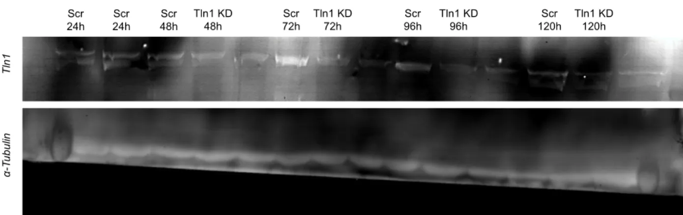
Figure 8: Western blot of Tln1 siRNA knockdown ... 51
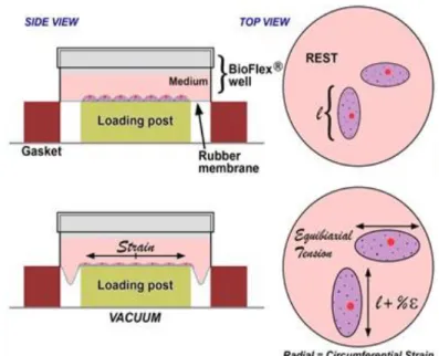
Figure 9: Flexcell diagram. ... 52
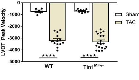
Figure 10: LVOT Peak V after TAC ... 55
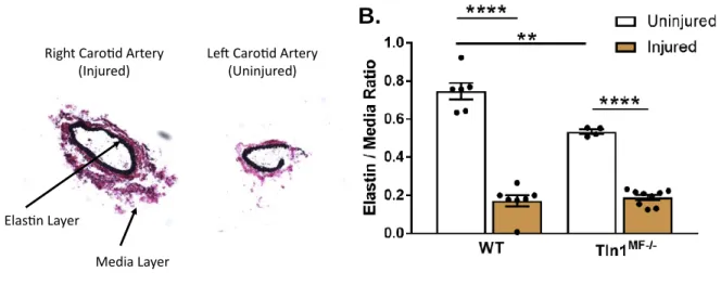
Figure 11: Elastin staining of carotid arteries after TAC. ... 56
Figure 12: EF of mice after TAC injury. ... 57
Figure 13: qPCR of Nppa and ventricle weight after TAC injury. ... 58
Figure 14: Echocardiographic assessment of LV thickness after TAC injury. ... 59
Figure 15: DL/BW ratio in TAC injured mice. ... 60
Figure 16: Measurement of interstitial fibrosis in TAC injured mice. ... 61
Figure 17: siRNA knockdown of Tln1 in CFs. ... 62
Figure 18: qPCR of α-SMA and Fn1 in Tln1 KD and Scr cells aft 10% strain. ... 63
Figure 20: Scratch wound and gel contraction assays of Tln1 KD and Scr CFs. ... 65
Figure 21: Experimental approach for ISO injection injury.. ... 76
Figure 22: EF and ventricular weight of ISO injured mice. ... 77
Figure 23: Experimental approach of AngII & PE injury. ... 78
Figure 24: EF of AngII & PE injured mice. ... 79
Figure 25: Measurements of ventricle weights in AngII & PE injured mice. ... 79
Figure 26: Measurement of interstitial fibrosis in AngII & PE injured mice.. ... 80
ix
Figure 27: Echocadiographic analysis of Tln2-/-; Tln1CF-/- and Tln2-/- mice at 12 weeks of age. .... 82
Figure 28: Experimental approach for MI injury. ... 83
Figure 29: Tcf21-Cre expression in mice after MI injury. ... 84
Figure 30: Survival curves pos-MI injury. ... 85
Figure 31: Experimental approach for AngII injury. ... 85
Figure 32: SBP of mice after AngII injury. ... 86
Figure 33: EF in mice after AngII inury. ... 87
Figure 34: Measurements of ventricle weight after AngII injury ... 88
Figure 35: Measurement of intersitial fibrosis after AngII injury. ... 88
Figure 36: Echocardiographic measurements at 12 weeks of age under basal conditions. ... 95
Figure 37: Experimental approach of 8-week AngII injury. ... 96
Figure 38: Measurements of SBP and mRNA Nppa expression in 8 week AngII injured mice. . 101
Figure 39: Echocardiographic measurements of heart function in 8-week AngII injured mice. ... 102
Figure 40: DL/BW ratio in 8-week AngII injured mice. ... 103
Figure 41: Ventricle / BW ratio in 8-week AngII injured mice. ... 104
Figure 42: WGA staining of cardiomyocyte area in 8-week AngII injured mice. ... 105
Figure 43: Measurements of interstitial fibrosis in 8-week AngII injured mice. ... 106
Figure 44: qPCR expression of α-SMA in 8-week AngII injured mice. ... 107
Figure 45: Volcano plot showing enriched genes from RNAseq. ... 108
x
LIST OF ABBREVIATIONS
Abbreviations
and Key Terms Definition
ACC American Heart Association
AFM Atomic force microscopy
AHA American Heart Association
AngII Angiotensin II
CF Cardiac fibroblast
CKD Chronic kidney disease
CO Cardiac output
DCM Dilated cardiomyopathy
DL/BW Dry lung / body weight
DOCA Deoxycorticosterone acetate
DOX Doxorubicin
ECM Extracellular matrix
EF Ejection fraction
F Blood flow
Gal-3 Galectin-3
GEO Gene Expression Omnibus
HF Heart failure
HFpEF Heart failure with preserved ejection fraction HFrEF Heart failure with reduced ejection fraction
HT Hypertension
IL-1β Interleukin-1β
IL-6 Interleukin -6
IR Ischemia reperfusion
ISO Isoproterenol
IVS Interventricular septum wall thickness LAD Ligation of the left anterior descending artery
LV American College of Cardiology
LVAW Left ventricular anterior wall thickness LVOT VTI Left ventricular outflow track time integral LVPW Left ventricle posterior wall thickness
MI Myocardial infarction
MMP Matrix metalloproteinase
MTJ Myotendinous junction
NPPA Natriuretic peptide A
NPPB Brain natriuretic peptide
PAB Pulmonary aortic banding
PE Phenylephrine
PH Pulmonary Hypertension
xi
POSTN Periostin
PSR Picrosirius red
qPCR Quantitative polymerase chain reaction
R Resistance of the vasculature
ROS Reactive oxygen species
SBP Systolic blood pressure
Scr Scramble
STZ Streptozotocin
T1D Type 1 Diabetes
T2D Type 2 Diabetes
TAC Transverse aortic constriction
TCF21 Transcription factor 21
TGF-β Transforming growth factor-β TIMP Tissue inhibitor of metalloproteinase
Tln1 Talin 1
Tln2 Talin 2
TLR2 DOX-receptor 2
TNF-α Tumor necrosis factor-α
TNF-β Tumor necrosis factor-β
TPR Total peripheral resistance
VEGEF Vascular endothelial growth factor A
VW/BW Ventricle / body weight
WGA Wheat germ agglutinin
αSMA α-smooth muscle actin
1
Dissertation Overview
My doctoral work has investigated the cell-specific contributions of the two talin proteins - Tln1 and Tln2 - in cardiac fibroblasts and myofibroblasts during pressure overload injury in the heart. The first focus of this research was to explore the effect that myofibroblast Tln1 had during transverse aortic constriction. The second aim of my work was to generate a Tln2 null, Tln1- cardiac fibroblast specific genetically modified mouse and then find and validate a dosing strategy that reproducibly resulted in interstitial fibrosis during pressure overload injury to the heart. Lastly, my work applied the model to study cardiac fibroblast remodeling post hypertension injury in the absence of Tln2 and loss of Tln1 in cardiac fibroblasts and the resulting cardiac remodeling that occurs.
In this dissertation, I provide a thorough background on heart failure, hypertension, and the roles of cardiac fibroblasts and cardiomyocytes to response to prolonged pressure overload of the heart. Next, I justify targeting Tln1 and Tln2 in cardiac fibroblasts in the context of hypertension disease. Following this, I summarize the known mouse models for inducing hypertension and pressure overload experimentally. Subsequentially, I present my research into the effects of cardiomyocyte Tln1 using the experimental model of transverse aortic constriction in vivo and siRNA knockdown in vitro. Following this, I describe the generation and creation of a novel genetic mouse to explore cardiac fibroblasts specific Tln1 and Tln2 contribution to fibrotic remodeling and cardiac hypertrophy in response to pressure overload injury. I then use this novel mouse to research the effect of cardiac fibroblast Tln1 and Tln2 loss following angiotensin II induced pressure overload of the heart. Echocardiography, RNA sequencing and in vitro assays were implemented to characterize the phenotypic alterations due to loss of Tln1 and Tln2 in cardiac fibroblasts. Finally, I discuss the impact of this work and potential future directions the research could be taken.
2
Chap ter 1
Chapter 1
Cardiovascular Disease and Tissue Response to Hypertension
Text for Chapter 1 was adapted from Noll NA, Lal H, Merryman WD. Mouse Models of Heart Failure with Preserved or Reduced Ejection Fraction. The American Journal of Pathology, Vol. 190, No. 8, August 2020.
1.1 Heart failure disease burden
Heart failure (HF) is the leading cause of death worldwide. There are approximately 6.5 million Americans living with HF with an incidence in 10 in 10,000 people over the age of 65.1 Recently, morbidity attributed to HF has dropped to one in nine deaths due to improvements in strategies focused on treating the conditions proceeding and leading to HF, including hypertension, myocardial infarction (MI) and atherosclerosis.2 However, even with improvements in treatment, mortality associated with HF is still high, with 50% of patients diagnosed with HF dying within five years of diagnosis.3 Current predictions show that by 2030, 8 million American adults will be diagnosed with HF.3
Two-thirds of all HF cases can be attributed to one of four underlying conditions: ischemic heart disease, chronic obstructive pulmonary disease, hypertensive heart disease, or rhematic heart disease.4 Of these, hypertension remains the major preventable cause of cardiovascular disease through pharmacological intervention and life style changes.5 This is done by treating the underlying symptoms, namely systolic blood pressure (SBP). However, these treatments do not address the chemical and cellular changes that are occurring in the heart leading to HF.
Therefore, there is a need to identify the cellular processes underlying the conditions leading to HF so that new therapeutic strategies can be developed.
3 1.2 Heart failure classifications
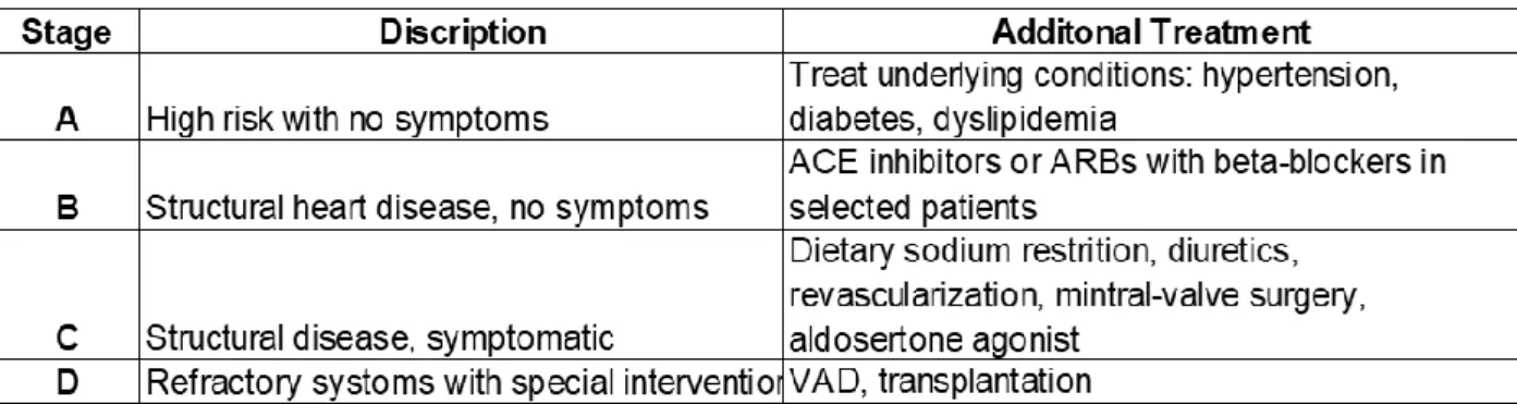
The American Heart Association (AHA) defines HF as a complex clinical syndrome that results from any structural or functional impairment of ventricular filling or ejection of blood.6 HF is classified based on ejection fraction (EF), and the progression of the disease based on cardiac deterioration. The American College of Cardiology (ACC)/AHA defines HF in four stages. These stages range from stage A, where there is a high risk of HF but no symptoms or structural damage to the heart, to stage D, where patients with refractory HF require advanced intervention (Table 1).7–9 Left-sided HF is associated with an increased risk of sudden death,10 and is subdivided based on left ventricular (LV) EF: HF with preserved ejection fraction (HFpEF; LVEF ≥ 50%), HF with mid-range ejection fraction (HFmrEF; LVEF 40-49%), and HF with reduced ejection fraction (HFrEF: LVEF <40).11 Medical advances have developed specific treatments for HFrEF by acting on the neuro-hormonal axis, but effective drugs for treatment of HFpEF are absent.12 This has led to an increase in HFpEF prevalence, and account for more than 50% of all HF cases in the United States.13
Table 1: Stages of Heart Failure and Treatment Options for Systolic Heart Failure. Adapted from Jessup et al.7,8
4
HFpEF is clinically defined as HF with normal EF and diastolic dysfunction, the inability of the ventricles to relax properly.14 HFpEF is usually the result of chronic diseases such as hypertension, diabetes mellitus, atrial fibrillation, aging, obesity, and/or renal dysfunction (Figure 1).15 Of these, hypertension is the most predominant underlying condition with a prevalence in 60-80% of all HFpEF cases.16 These chronic diseases gradually diminish the normal relaxation ability of the LV as the ventricular walls become stiffer from increasing interstitial fibrosis. As a result, the heart can no longer fill properly with blood during the resting period between each beat, which eventually leads to diastolic failure. Cardiomyocytes increase their thickness, by adding sarcomeres, the contractile unit of the cell, in parallel, resulting in cardiomyocyte hypertrophy and concentric hypertrophy of the heart. HFpEF occurs more often in women (79% vs 49% of all HFpEF cases) and is more prominent in older populations.14 HFpEF manifests clinically as exercise intolerance, dyspnea, edema, pulmonary hypertension and pulmonary edema, all of which are symptoms associated with cardiac hypertrophy, increased fibrosis, and decreased capillary content. Additional acute insults to the heart, or chronic high blood pressure can cause a transition from HFpEF to HFrEF during increased cardiomyocyte injury.
In contrast with the reduced relaxation capacity of HFpEF, HFrEF occurs when the ventricles lose their ability to contract normally. A wide range of cardiac conditions can cause HFrEF, including coronary artery disease, MI, and cardiomyopathies (Figure 1).15 These diseases result in apoptosis of cardiomyocytes which causes an imbalance in heart wall structure, causing eccentric remodeling with left ventricular dilation, but normal wall thickness.17–21 These adaptations initially allows the heart to normalize left ventricle (LV) wall stress and maintain cardiac output and EF. However, as remodeling continues, stiffening of the ventricular walls diminishes the cardiomyocyte’s ability to contact with enough force to adequately eject blood into the systemic circulation. This eventually leads to systolic failure. Patients with HFrEF have higher
5
levels of circulating brain natriuretic peptide (NPPB), a common biomarker for HF, and a higher mortality rate than those with HFpEF.22
Figure 1: Disease states and presenting phenotypes of HFpEF and HFrEF. Disease states and their resulting left ventricular remodeling leading to the development of heart failure with preserved ejection fraction (HFpEF) and heart failure with reduced ejection fraction (HFrEF).
Created with BioRender.com.
6 1.3 Hypertension
Hypertension (HT) is one of the main underlying conditions that leads to HFpEF and is the most important risk factor for the development of HFrEF in the United States.4,23,24 In the Framingham Heart Study, 91% of all patients developed hypertension that predated their newly diagnosed HF.16 HT is characterized by an increase in SBP which causes increased ventricular pressure in the heart. 2017 guidelines by the ACC/AHA define HT as blood pressure greater than 130/180 mmHg.25 Currently, there are 70 million adults in America with HT, only 52% of which have their blood pressure properly managed.26 Studies show that that a SBP reduction as low as 5mmHg can reduce the risk of HF by 24% in early onset HT, underscoring the importance of the development of therapies that target hypertension and SBP.27
1.3.1 Systolic blood pressure
Blood pressure is the pressure or tension that is exerted by the blood as it circulates the arterial vessels and is the result of cardiac output (CO) of the heart and the total peripheral resistance (TPR) of the systemic circulation (Equation 1).28 Blood flow (F) through the heart is maintained by the change in perfusion pressure (atrial – venous pressure) and the amount of resistance (R) in the vasculature (Equation 2).29 Under normal conditions, perfusion pressure and vascular resistance do not change. HT is caused by, but not limited to, malfunction of the humoral system, neuronal and autoregulatory systems.30 Under pathological conditions such as HT and during aging, narrowing of the vasculature causes an increase in vascular resistance. The heart initially responds through autoregulation. Autoregulation is the ability of the heart to maintain blood flow despite a change in perfusion pressure (Pa-Pv).29 The heart does this by increasing the flow of blood by dilating the vasculature to decrease resistance. This allows for the maintaining of homeostatic SBP. Under continual pathological insults, such as plaque buildup in the arteries, and a reduced coronary endothelium-dependent dilation capacity in HFpEF, vascular dilation alone cannot adequately reduce the vascular resistance, resulting in an increase in blood
7
pressure.31–37 This leads to a sequential increase in ventricular heart pressure, and ventricular wall stress.
𝑩𝑷 = 𝑪𝑶 𝒙 𝑻𝑹𝑷 Equation (1)
𝑭 = (𝑷𝒂−𝑷𝒗)
𝑹 Equation (2)
1.3.2 Cardiac fibroblasts
The adult heart is comprised of approximately 30% cardiomyocytes with the remaining 70% non-myocyte cells being primarily cardiac fibroblast (CFs).38 CFs are arranged in sheets that run in parallel with muscle fibers and they help maintain continuity of cell signaling between cardiomyocytes.39 Genetic lineage tracing has shown that Tcf21 is the best marker for CFs as it is expressed most widely expressed marker of all fibroblast-like cells in the heart, and is expressed by all activated cardiomyocytes derived from CFs.40
CFs are recognized chiefly as regulators of the extracellular matrix (ECM) and are involved in general maintenance of myocardial structure.41 Collagen is the major stress-bearing element within the ECM and forms a 3D network around bundles of myocytes to generate a stress-tolerant network.42 In the healthy heart, ~85% of the ECM is composed of thicker collagen I fibers, witch conifer tensile strength, and ~11% of collagen type III fibers that maintain the elasticity of ECM.43 Additionally, the ECM acts as an insulator for myocardial signaling. Electrical signals are passed between CFs and cardiomyocytes through gap junctions. In vitro analysis of single fibroblasts
8
have shown them to be capable of synchronizing contractions between myocytes, illustrating the role that CFs play in maintaining total heart contractility.44
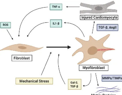
Pathological conditions such as systemic inflammation, hypoxia, cardiomyocyte death, mechanical stress, and activation by pro-fibrotic cytokines can lead to the phenotypic shift of quiescent CFs to active myofibroblasts. Myofibroblasts proliferate and migrate to the site of injury, where they secrete and compact ECM components.45,46 This migration is cytokine-induced and requires the co-coordinated activity of matrix metalloproteinases (MMPs) and tissue inhibitor of metalloproteinase (TIMPs) to move through the ECM network in the heart (Figure 2).47 CFs express a limited subset of MMPs; collagenases (MMP-1, MMP-13), gelatinases (MMP-2, MMP- 9), and stromelysin (MMP-3).42 Myofibroblasts also differ from inactivated CFs by having a more contractile phenotype, marked by the expression of alpha-smooth muscle actin (α-SMA) (Figure 2). This allows myofibroblasts to compact and arrange ECM components such as collagen types I and III, and fibronectin. The secretion and compaction of ECM components leads to interstitial and perivascular fibrosis of the ventricular walls, allowing for short-term adaptation to tissue injury.48–52
9
Figure 2: Cardiac fibroblasts to myofibroblast activation. Fibroblast to myofibroblast transition in the heart in response to cardiomyocyte injury, proinflammatory cytokines, and systemic inflammation through reactive oxygen species (ROS). Ang II, angiotensin II; Gal-3, galectin-3; MMP, matrix metalloproteinase;
TGF-β, transforming growth factor- β; TIMP, tissue inhibitor of metalloproteinase; TNF-α, tumor necrosis factor-α. Created with Bio-Render.com.
While necessary for initial survival, continued CF remodeling of the heart can lead to negative effects. Chronic pressure overload of the heart increases ventricular wall stiffness, and secretion of pro-fibrotic signaling factors resulting in a positive feedback loop of further myofibroblast activation (Figure 2). Unlike myofibroblasts in other regions which undergo apoptosis after healing is complete, cardiac myofibroblasts can persist in fibrotic areas, such as the scar post-MI.53 Clinical studies have shown that in the failing heart TIMP activity is decreased, while MMP activity is increased, pointing to an imbalance in the hearts ability to degrade unnecessary collagens.54–56 Angiotensin II (AngII), which is secreted by CFs, has been shown to induce collagen synthesis, as well as decrease TIMP-1 and TIMP-2 in humans.57 AngII stimulation causes the increased expression of TGF-β, resulting in collagen I and III secretion, and further
10
induction of the CF to myofibroblast transition.58 Additionally, myofibroblasts produce cytokines (TNF-α, IL-1B, IL-6, TNF-β), vasoactive peptides, and growth factors (AngII, TN-1, ANP, BNP, VEGEF), which can increase collagen synthesis in CFs, while also inducing cardiomyocyte hypertrophy (Figure 2). The pathological responses of myofibroblasts results in a stiffer, non- compliant myocardium that can lead to impaired cardiomyocyte contraction and hypertrophy (Figure 1).
During HFrEF, cardiac fibrosis occurs primarily due to the loss of cardiomyocytes, where myofibroblasts lay down ECM to fill the empty gaps left by cardiomyocytes death. This fibrosis leads to the impairment of cardiomyocyte contraction transduction resulting in uncoordinated contraction of cardiomyocyte bundles.59 Further disruption of the interactions between laminin, which connect cardiomyocytes and capillaries, causes a further reduction in cardiomyocyte mass.60 During HFpEF, excessive collagen deposition and a reduction in collagen III results in a stiffer, less compliant ventricular wall.61,62
1.3.3 Cardiac myocytes
Cardiomyocyte remodeling in HFpEF and HFrEF are driven by the amount of damage that cardiomyocytes endure during the initial injury of the heart. During HFrEF, remodeling is driven by cardiomyocyte damage and death, leading to an imbalance in the heart wall structure.17–21 This death can be driven by ischemia, an inappropriate inflammatory response, and pressure overload.63–66 Increasing levels of circulating Troponin-T leads to a reduction in cardiomyocyte mass, causing cardiomyocytes to become thinner and more elongated.17–21 This results in eccentric remodeling of the heart, with left ventricular dilation, but normal wall thickness (Figure 1). Increased stiffness of the heart walls due to interstitial fibrosis results in impaired cardiomyocyte contraction.59 This results in a decrease in SV and a decrease in EF as cardiomyocytes are unable to push blood out of the ventricles and into the systemic circulation as the same rate.
11
Conversely, under chronic injury to the heart, such as increase systolic blood pressure during hypertension, the heart undergoes HFpEF first. As the LV walls become stiffer from increasing interstitial fibrosis, cardiomyocytes lose their ability to relax properly. To normalize their ability to contract cardiomyocytes add sarcomeres in parallel to increase their contractility, which also results in increased cardiomyocyte thickness.67,68 This results in concentric hypertrophy, where the heart wall thickens and ventricular chamber volume decreases (Figure 1). As concentric remodeling continues, the heart can no longer fill properly due to the decrease ventricular chamber volume and altered cardiomyocyte relaxation, resulting in diastolic heart failure.
12
1.4 Therapeutic shortcomings of hypertension
Two-thirds of all HF cases can be attributed to one of four underlying conditions: ischemic heart disease, chronic obstructive pulmonary disease, hypertensive heart disease, or rhematic heart disease.4 Of these, HT remains the major preventable cause of cardiovascular disease.5 Treatment of adults with HT has centered around lowering blood pressure to less than 140/90 mmHg. However, new evidence from the Systolic Blood Pressure Intervention Trail suggests that lowering SBP to less than 130 mmHg may be vital for patients who are high-risk, including those with a history of cardiovascular disease and chronic kidney disease.69 Life style changes, in combination with pharmacotherapy are the most commonly used method to treat hypertension.
The most modifiable lifestyle changes are obesity, high sodium intake, insufficient physical activity, and excessive alcohol consumption. For patients eating a typical American diet, reduction to intake of 2400mg of sodium per day shows benefits in reduction of blood pressure across a wide range of patients.70 This benefit is increased with a reduction of sodium intake to 1500mg and 1000mg per day.
Therapeutics for high blood pressure fall into 3 categories: 1) Thiazide-type diuretics 2) calcium channel blockers (CCBs) 3) angiotensin converting enzyme (ACE) inhibitors / angiotensin II receptor blockers (ARBs).71 For stage 1 hypertension (SBP between 140-159 mmHg), each of the first 3 classes are similarly effective in lowering BP in 30-50% of the general adult population.72,73 However, initial monotherapy is unlikely to lower BP to the goal in patients whose BPs are greater than 20/10 mmHg of their goal BP. When this occurs, in stage 2 hypertension (SBP ≥ 160 mmHg), and in many cases of stage 1 hypertension, combination therapy using 2 drugs is needed.72,73
While the right combination of pharmacotherapy and lifestyle modifications can decrease blood pressure, there are currently no treatments for the underlying interstitial fibrosis,
13
cardiomyocyte hypertrophy, and additionally cellular changes that occurred during injury.
Additionally, lifestyle modifications may not be able to be made due to economic status, underlying risk factors, and living environment. Therefore, there is a need to identify the cellular process underlying the conditions leading to HT so that new therapeutic strategies can be developed to treat the cause of HT, and not the underlying symptom of increased blood pressure.
14
Chap ter 2
Chapter 2
Integrin Adhesions & Talin
2.1 Mechanotransduction between cells and the extracellular matrix
Mechanotransduction is the ability of cells to sense and transduce physical forces into biomechanical signals and a cellular response.74 In the heart, resident cardiomyocytes and cardiac fibroblasts (CFs) are subjected to physical forces during normal cardiac function such as membrane stretch, gain and loss of adhesion, and compression75. Mechanotransduction is vitally important in the heart, as this process drives cardiomyocyte hypertrophy, CF migration, and deposition of collagen in response to increased stiffness and stress on ventricular and atrial walls during acute and prolonged injury.
Integrin adhesion complexes allow cells to transduce forces between the extracellular environment and their cellular body. Integrin adhesion complexes require 4 components for their formation: an extracellular matrix (ECM) ligand, a transmembrane integrin heterodimer, a mechanosensitive protein (e.g., talin), and filamentous actin.76 Integrin adhesion complexes form a variety of adhesions from small, transient nascent adhesions, to larger, more stable focal adhesions that develop under high mechanical loads. Integrin adhesions can also form structures such as protostomes and invadopodium that mediate matrix degradation and remodeling, as well as fibrillar adhesions that mediate ECM assembly.77–79
Integrin adhesions function in a bidirectional manner, resulting in ‘outside-in’ and ‘inside- out’ signaling.80–82 Outside-in signaling is a result of integrins binding the ECM leading to intracellular signaling events that can influence a wide-range of cellular activities including
15
migration, proliferation, gene expression, survival, and alterations in cellular morphology. Inside- out signaling occurs when non-integrin cellular receptors modify integrin activation. This signaling results in a change in integrin binding affinity and clustering on the cellular membrane, which has been associated with chemical and mechanical signaling in cardiomyocytes and CFs.
2.1.1 Integrins
Integrins are heterodimeric transmembrane receptors that are comprised of an α and β subunit.83,84 Integrins binding to the ECM occurs in the extracellular domain of the integrin and is modulated by binding of proteins to its intracellular portion.85 Integrin activation occurs when talin, a cytoskeletal linker molecule, binds the intracellular β subunit of the integrin, resulting in a change in integrin affinity for the ligand.86,87 While integrins can be activated by numerous proteins, talin is the cytoskeletal link common to all integrin adhesions.76
Integrin expression is unique to each cell type and can change based on developmental stage and pathological state. In adult cardiomyocytes, α1β1, α5β1, and α7β1 are the most highly expressed integrin heterodimers, and are the binding receptors for collagen, fibronectin, and laminin, respectively.88 The β1 integrin subunit is unique as it has two differently spliced isoform, β1A and β1D.88–90 The β1A isoform is expressed embryonically, while the β1D isoform is expressed in adult cells, providing them with distinct, isoform-specific interactive properties with the ECM and signaling molecules.91 Knockout studies have shown that integrin function in cardiomyocytes is vital for preservation of normal heart function.92
In CFs there is redundancy in ECM binding partners, as CFs express α5β1 integrins which bind both fibronectin and osteopontin. Additionally, CFs express αvβ1, αvβ3 and αvβ5, which bind vitronectin, as well as fibronectin and osteopontin.89,93,94 Post-myocardial infarction, myofibroblasts migrating to the site of injury had an upregulation of β1 integrins.95,96 This upregulation of β1 integrins and integrin localization on the cellular membrane of CFs was also
16
seen in a rat model of AngII-induced hypertension, as well as in in vivo stimulation of CFs with AngII.97–99 These data show that increased integrin activation and modulation in vital in the response to injury in the heart.
2.1.2 Talin
The mechanotransdution of signals across integrin adhesions are facilitated by mechano- effector proteins which bind integrins to the F-actin cytoskeleton of the cell. Talin is a key mechano-effector protein for integrin-mediated adhesion to the ECM.100,101 Talin is a large 270kDa cytosolic protein composed of an N-terminal FERM head domain, a flexible neck region, and a C- terminal rod domain (Figure 3).
Figure 3: The talin protein. Talin-based molecular clutch mediates Mechanotransduction. Domain organization of talin. The N-terminal talin head FERM domain that consists of F0,F1,F2 and F3 subunits containing an integrin tail-binding site (IBS1). The talin rod domain contains 13 helix bundles (R1-13) which contain a second IBS (IBS2) and two actin binding sites (ABS2, ABS3) as well as two critical vinculin binding sites (VBS) in the R3 and R8 domains. The other VBS are not pictured. Reprinted with permission from © 2016 Sun et al. Originally published in J Cell Biol.
17
In the cytoplasm, talin adopts a closed, autoinhibited conformation.102–104 Upon activation, talin migrates to the cell membrane, where it activates integrins via its FERM domain by binding the cytoplasmic β-tail of integrins on talins IBS1 site (Figure 3). This results in a conformational change of the integrin receptor where binding of talin to the integrin β-tail disrupts the autoinhibitory association between the integrins α- and β-tails, causing an increase in affinity for ECM ligands (Figure 4).105–107 This triggers a series of intracellular events such as cell motility and ECM adhesion. Deletion of talin resulted in cells that were unable to migrate or proliferate as well as diminishing the structural integrity of FAs.108 Studies in in C. elegans, Drosophila, and mice have demonstrated that talin is essential for integrin adhesion.109–111 The fact that talin is necessary for integrin activation is evolutionarily conserved suggests that talins are critical mediators of cell-environmental interactions. Thus, understanding their functions during environmental change is essential.
18
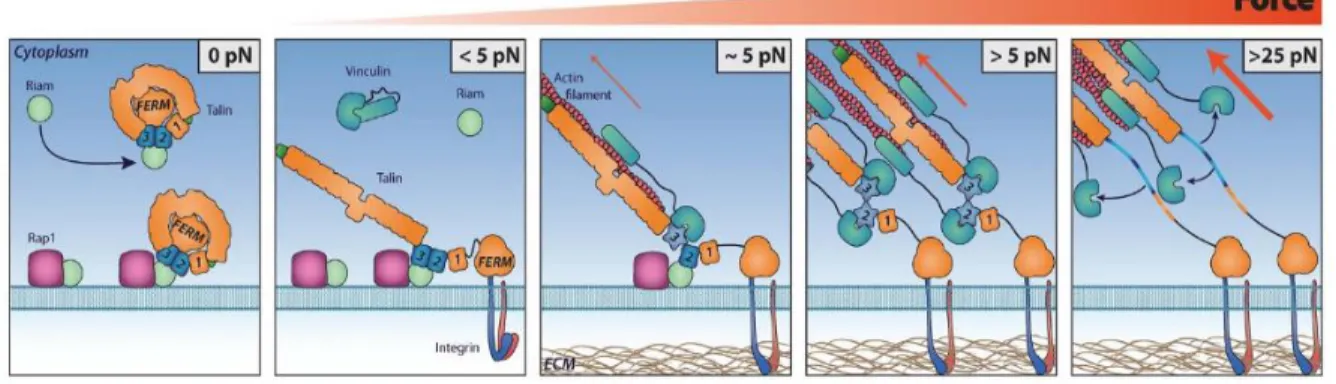
Figure 4: Talin activation and the formation of focal adhesion complexes under increasing applied force. Talin changes binding partners in response to force induced conformational changes. Force plays a key role in driving the formation of FA.0–5pN: RIAM recruits autoinhibited talin to the plasma membrane in a Rap1 via synergistic binding of RIAM to the R2–R3domains of talin. At the membrane talin autoinhibition is relieved. Talin can then activate integrins.,5pN: Only when talin has engaged the integrins and also captured the retrograde flow of actin is force exerted on talin. At,5 pN, the force of a single actomyosin contraction, the R3 domain is destabilized, and this reduces RIAM binding whilst exposing the high affinity VBS which then bind vinculin strengthening the adhesion. 5pN: With more vinculin cross-linking the adhesion can withstand greater force exposing further VBS. 25pN: At sufficiently high forces vinculin is displaced, resulting in unfolding of the VBS to a random coil. Reprinted with permission from 2014 Yao et al. Originally published in Scientific Reports.
The rod domain of talin consists of 13 α-helical bundles (R1-R13) that connect to F-actin directly through two actin-binding sites (ABS2 and ABS3), and indirectly through vinculin, and other adapter molecules such as RIAM, DLC1, and Kank (Figure 3).112 These connections between ECM, integrin, talin, and actin allow for the transmission of cell generated contractile forces and forces derived from externally applied strains.
When talin is under force, the α-helical bundles in the talin rod domain unfold. This allows for new binding sites to be exposed and disrupts the binding of proteins to the folded state. For example, RIAM binds to the R3 folded domain. However, under force unfolding of R3, vinculin is recruited and binds to the unfolded R3 domain (Figure 4).113,114 Vinculin also binds actin, causing increased stability of talin in its unfolded state under higher forces. Each talin α-helical bundle
19
operates as a mechanical switch and opens under different levels of tension. This allows for complex, time-dependent responses to tension exerted across the integrin-actin complex.
Vinculin is the best characterized cytoskeletal linkage protein to talin and can bind to 11 vinculin binding sites throughout the talin rod.115 Binding of vinculin to talin stabilizes the rod domain in its open state, even after tension is reduced (Figure 4).116 This results in the force necessary for talin refolding to be lower than the force needed for unfolding, allowing for stabilization of talin in the unfolded state.117 Vinculin also creates an additional links to F-actin which allows for higher force transmission and increased stabilization of the unfolded talin rod domain.118
Due to mechno-effector proteins playing a key role in the transmission of forces between the ECM and the cells actin cytoskeleton, researchers have tried to parse talins specific contributions to mechanotransdution in cells that are under force during normal and pathological conditions.
20 2.2 Talin in the body
In vertebrates there are two talin genes, Tln1 and Tln2, which encode very similar proteins (74% amino acid sequence identity). Tln1 is ubiquitously expressed in adults, while Tln2 expression is dominant in the heart, brain, and skeletal muscle.52 While Tln1 and Tln2 play the same role in cells, Tln2 has a stronger affinity for F-actin through its co-localization with β1D integrins.119,120 This allows Tln2 to make stronger bonds.121 As such, cells that are under constant forces, such as cardiomyocytes, have higher expression of Tln2 than Tln1, which leads to the unequal expression of Tln2 throughout different cell types. During development, Tln2 knockout mice develop normally, and only incur a mild skeletal myopathy at 3 months of age due to a defect in their myotendinous junctions (MTJs) in skeletal muscle.122 Tln1 knockout mice, however, are embryonically lethal at E8.5-9 due to gastrulation defects.111 This indicates that Tln1 is an essential protein for development, and Tln2 cannot completely compensate for loss of Tln1.
In adult cells, Tln1 and Tln2 switching has shown that talins can partially compensate for each other when one is removed. This was demonstrated in mouse skeletal muscle tissue where knockdown of Tln2 in vivo resulted in successful assembly of integrin complexes at costameres and MTJs.123 However, with aging, defect in MTJs occurred. Likewise, when only Tln1 was knockdown in mouse muscle cells, successful assembly of integrin complexes at costamere and MTJs was seen, with MTJ defects occurring over time.124 This shows that while talins can partially offset each other, they cannot completely compensate. When Tln1 and Tln2 were both removed from skeletal muscle, mice died shortly after birth, illustrating that talins are required for intact integrin function during muscle development and growth.123 Interestingly, these mice had a phenotype similar to β1-integrin KO mice, suggesting that removal of talin from skeletal muscle also inhibited integrin functions. This is consistent with findings in vitro that knockdown of Tln1 and Tln2 in mouse fibroblasts resulted in cells unable to form integrin adhesions,125 and in Drosophila where all of the adhesive functions of βPS (orthologous to β1) required talin to form.110
21 2.3 Talin in the heart
In the heart, talin has been primarily studied in cardiomyocytes, with a focus on talins role in cardiac hypertrophy. During development, both Tln1 and Tln2 are highly expressed in embryonic cardiomyocytes. However, Tln2 becomes the dominant form of adult cardiomyocytes with minimal expression of Tln1.126 In the adult mouse heart, Tln2 is localized to myocyte costameres, where Tln2 colocalizes with β1D integrins, allowing cardiomyocytes to support the high and constant forces that they must endure. This expression level is similar to human hearts, with a slightly higher Tln1 expression in human cardiac muscle.126
Under basal conditions, Tln2-null (Tln2-/-) mice have normal cardiac structure and function up to 12 months of age.127 In these mice, a decrease in β1D integrins coincided with a two-fold increase of Tln1 in the costameres of cardiomyocytes, illustrating that Tln1 can functionally replace Tln2 in cardiomyocytes under basal conditions. This was also observed in a mouse model of cardiomyocyte-specific deletion of Tln1 (Tln1CM-/-). Tln1CM-/- mice had normal basal cardiac structure and function,126 suggesting that Tln1 and Tln2 function in similar roles in cardiomyocytes, and this is protectively redundant under basal conditions. However, during disease, loss of cardiomyocyte Tln1 is beneficial, and blunts cardiac hypertrophy and interstitial fibrosis in response to prolonged pressure overload of the heart, suggesting that during disease, Tln1 and Tln2 play different roles in cardiomyocyte response to injury.
Under pressure overload of the mouse heart, Tln1 protein levels were increased in WT mice 4-weeks post-transverse aortic constriction (TAC) in cardiomyocytes and whole heart tissue, with no change in Tln2 expression.126 This is consistent with protein levels of Tln1 and Tln2 taken from patients with end-stage, non-ischemic dilated cardiomyopathy (DCM) and suggests that Tln1 upregulation plays a key role in cardiomyocyte response to injury.126 When TAC was performed on the Tln1CM-/- mice, ablation of Tln1 in cardiomyocytes resulted in a blunted response to TAC;
22
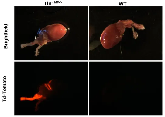
with a decrease in HW/BW ratio, cardiomyocyte area, and a reduction in fibrosis.128 This showed that cardiomyocyte Tln1 alters acute biomechanical signaling and offers an apparent beneficial response to cardiac remodeling after pressure overload of the heart. When the Tln1CM-/- mouse was crossed with the global Tln2 null mouse (Tln2-/-; Tln1CM-/-), rapid cardiac dysfunction occurred. Tln2-/-;Tln1CM-/- mice developed dilated cardiomyopathy with defects in integrin adhesion complexes and abnormal costameres that resulted in death by 25 weeks of age, illustrating that cardiomyocytes need a form of talin to respond to pressure-overload injury of the heart (Figure 5).127 While deletion of talins in cardiomyocytes has been evaluated, the effect of talin deletion in CFs in unknow.
Figure 5: Loss of Tln1 and Tln2 in cardiomyocytes leads to dilated cardiomyopathy. Histological (H&E and trichrome staining) analysis showed cardiac dilation with fibrosis in 8-week-old male mice that were Tln2-null, with Tln1 knockdown in cardiomyocytes as compared to Tln2 null mice. Reprinted with permission from Manso, et al. Originally published in PNAS.
Tln2-/-;Tln1CM-/- Tln2-/-
23
Chap ter 3
Chapter 3
Mouse Models of Pressure Overload Injury to the Heart
Text for Chapter 3 was adapted from Noll NA, Lal H, Merryman WD. Mouse Models of Heart Failure with Preserved or Reduced Ejection Fraction. The American Journal of Pathology, Vol. 190, No. 8, August 2020.
3.1 Introduction
Mouse models of heart failure (HF) have been utilized to improve our understanding of the various aspects and etiologies of HF towards the goal of developing novel treatment strategies.
Mice are the most commonly used animal models in HF research, as they share the majority of their genes with humans, and approximately 85% of the protein-coding regions to the human genome.129 Additionally, mice provide unique experimental advantages, such as the ability to impose genetic alterations, short breeding cycles, and relatively low housing costs. Numerous murine models of HF have been developed through a combination of genetic modifications, administration of pharmacological compounds, and/or surgical approaches to recapitulate human disease.129,130 Mouse models allow for the study of specific risk factors of and treatment strategies for HF without some of the confounding effects of comorbidities seen in other animal models.
Over the last decade, a large increase in mouse models of HF has increased our knowledge of both HF with preserved ejection fraction (HFpEF) and HF with reduced ejection fraction (HFrEF), many of which are highlighted in this chapter and summarized in Table 2.
24 3.2 Mouse models of HFpEF
There are many different models of HFpEF. These models strive to recapitulate the chronic disease progression and risk factors associated with the development of HFpEF, including hypertension, obesity, diabetes, and gaining. Some of these models, if permitted to run long enough, may also lead to the development of HFrEF.
3.2.1 Hypertension
Hypertension is one of the main underlying conditions that leads to HFrEF in humans.4,23 Hypertension, which causes broad changes in inflammation and metabolism, can cause myocardial stiffness and diastolic dysfunction.131 This is additionally exacerbated when hypertension results in increased pressure in the left ventricle (LV), resulting in the expansion of fibroblasts, hypertrophy of vascular smooth muscle cells, and pathological deposition of interstitial collagen. This leads to increased myocardial wall stress, which causes LV hypertrophy in an attempt to compensate for the increased pressure.132 The most commonly used mouse model to study hypertension-induced HFpEF is the administration of deoxycorticosterone acetate (DOCA) while providing high-salt (1% NaCl) drinking water. This model causes increased sodium and water reabsorption in the kidneys, leading to high blood pressure through a decrease in the renin- to-aldosterone ratio. This model has also been shown to be mouse strain-dependent, as C57BL/6 mice are less susceptible to renal damage and hypertension than the 129/Sv strain133. Additionally, renal impairment is more severe in males than in females134. The DOCA model results in cardiac hypertrophy, ventricular fibrosis, upregulation of the hypertrophy markers atrial and brain natriuretic peptides (NPPA, NPPB), and infiltration of inflammatory cells into the cardiac tissue134.
Angiotensin II (Ang II) administration has also been used to induce hypertension and chronic kidney disease in mice. Sustained elevation of Ang II levels in the circulation results in
25
Ang II-mediated vasoconstriction, hypertension, aldosterone secretion, TGF-β-mediated fibrosis, and inflammation - all of which contribute to the development of cardiac hypertrophy. This model has contributed to many cardiovascular discoveries, including that sildenafil, an inhibitor of PDE5A, improves LV performance, reduces adverse remodeling, and diminishes infiltration of inflammatory cells during Ang II-induced HFpEF.135 Additionally, the Ang II model has been combined with the DOCA salt and uninephrectomized models in an attempt to overcome the resistance of C57BL/6 mice to chronic kidney disease (CKD) and hypertension development136,137.
3.2.2 Pulmonary hypertension
Diastolic dysfunction, as experienced in HFpEF, is the most frequent cause of pulmonary hypertension (PH).138 PH is commonly found in deteriorating HFpEF, and is therefore closely associated with worse outcomes and mortality in patients with HFpEF.139 During HFpEF, chronically elevated filling pressure in the LV causes backward pressure in the pulmonary arteries, resulting in vascular remodeling and increased pulmonary arterial pressure, pulmonary vascular resistance, and right ventricular hypertrophy that are associated with PH.140 PH exacerbates the LV diastolic dysfunction that is already occurring in the heart.141 As a result, mouse models of PH were developed to study how PH leads to increased diastolic dysfunction.
AKR/J, NON/shiLtJ, and WSB/EiJ mice, when placed on a high-fat diet for 20 weeks, develop elevated right ventricular systolic pressure and LV end-diastolic pressure while having a preserved EF.142 These findings illustrate that these mice develop biventricular hypertrophy, HFpEF, and PH.
3.2.3 Type 2 diabetes
Cardiovascular complications are the leading cause of diabetes-related morbidity and mortality.143 Diabetes mellitus, or Type 2 diabetes (T2D), is non-insulin-dependent and results from a combination of insulin resistance and β-cell secretory defects.144 Complications associated
26
with T2D include increased coronary heart disease and accelerated atherosclerosis due to associated risk factors of hypertension, obesity, and dyslipidemia145. Db/db and ob/ob mice are the most commonly used T2D that are based on leptin-receptor deficiency or lack of functional leptin, respectively.146 In both mouse models, circulating leptin is taken up in the hypothalamus, causing an increase in appetite, body weight, and decreased energy expenditure. This results in both models having severe, early onset obesity at four-weeks of age and the development of hyperinsulinemia and T2D by 15 weeks.147 Cardiac hypertrophy, increased LV mass and diastolic dysfunction occur in these mice as myocardial oxygen consumption is increased, resulting in decreased cardiac efficiency.146,148,149 Ob/ob mice experience contractile dysfunction; however, db/db cardiomyopathies are more pronounced.150 The major disadvantage of the db/db and ob/ob mouse models is that while there is a robust phenotype of T2D, there are potentially confounding side effects from altered leptin signaling. In db/db mice, there is no altered tyrosine kinase signaling changes in cardiomyocytes, a variation from decreased signaling seen in human muscle tissue.151 In ob/ob mice, the innate and acquired immune response is repressed, potentially resulting in an altered response to acute and chronic injury to the heart.152
3.2.4 Type 1 diabetes
Type 1 diabetes (T1D) is defined by the National Institute of Diabetes, Digestive and Kidney Disease as an autoimmune disease in which the immune system attacks and destroys insulin-producing pancreatic β-cells, resulting in an absolute insulin deficiency.153 The autosomal dominant mutant INS-gene is one known human genetic cause of T1D and serves as a reproducible model of T1D in mice.154 The Akita mouse (Ins2Akita+/-) is heterozygous for the Ins2 gene mutation, wherein all males develop T1D after weaning age. At 5-6 weeks of age, the Akita mice develop hyperglycemia (which is similar to humans who develop T1D between 15 and 25 years of age).155 At 12 weeks, these mice have an increase in the circulating HF markers NPPA
27
and NPPB with diastolic dysfunction and a decrease in the radial strain occurring between 3 and 6 months.155,156
To look at the acute onset of T1D in mice, Streptozotocin (STZ), which directly kills pancreatic β-cells, is administered, inducing chronic T1D. High doses of STZ can cause toxicity outside of the pancreas, so a low continuous dose of STZ is recommended. The STZ mouse model results in hyperglycemia seven to 14 days after the first injection.157 STZ induces early diastolic and vascular dysfunction, which is progressively exacerbated by the development of diabetes, leading to systolic dysfunction and HFpEF, accompanied by abnormal patterns of mitral valve inflow and pulmonary venous flow.158,159 While the STZ model of T1D produces a robust imitation of the disease, it does not capture the autoimmune aspect of the development of T1D in humans.
Acute onset of T1D can also be studied without the use of toxins by using OVE26 mice.
OVE26 mice overexpress calmodulin in pancreatic β-cells, resulting in mice with diabetic nephropathy and severe early onset of T1D during the first week of life. OVE26 mice can live for one year with no insulin treatment and will maintain near-normal body weight.160 These mice spontaneously develop diastolic dysfunction with an increase in end-systolic interventricular septum thickness and end-systolic left ventricular posterior wall thickness. When treated with Ang II, OVE26 mice have exacerbated cardiac hypertrophy, with an increase in LV mass and NPPA expression.161
3.2.5 Obesity
Obesity is a complex chronic disease resulting from the accumulation of several physiological changes over a long period of time and is associated with many other risk factors in the development of HF (e.g. hypertension, diabetes, and psychosocial stress). In the lab, diet- induced obesity in mice has been developed as the standard practice to probe the pathologic
28
contributions of an imbalance of food intake, basal metabolism, and energy expenditure.162 C57BL/6J mice on a high-fat diet closely parallel patterns of progression and metabolic irregularities found in human obesity. After two weeks, C57BL/6 mice have decreased rates of glucose oxidation and glycolysis, which further develops into obesity and T2D.163 At 20 weeks, a 20 to 30% increase in body weight occurs alongside cardiac dysfunction, elevated filling pressures, myocardial fibrosis, and exercise intolerance.164 This model was used to discover the importance of Akt and mTOR signaling in obesity.165 Physiosocial stress is recognized as an independent risk factor for cardiovascular disease, and when it is added to a high-fat diet model of obesity, cardiac dysfunction will occur.166 This model results in prominent interstitial fibrosis, apoptosis of CMs, remodeling of the larger coronary branches, and augmented oxidative stress in the LV.166
3.2.6 Aging
HF is disproportionately distributed among the elderly, as over half of all patients hospitalized with HF are over the age of 75, with 50% presenting with diastolic dysfunction.167 The senescence-accelerated prone (SAM) mouse, derived from inbreeding AKR/J mice, recapitulates many common geriatric disorders evident in elderly human populations.168 The SAM model is comprised of both a senescence-prone (SAMP) and senescence-resistant (SAMPR) control strain. The SAM strains are the best-studied strains regarding HFpEF, as they result in age- dependent diastolic dysfunction in the absence of systolic dysfunction. Additionally, there is an increase in pathological fibrosis and the production of the pro-fibrotic cytokines TGF-β and connective tissue growth factor.169