80
Chapter 4: Spin state tuning at pseudotetrahedral d
7ions:
examining the structural and magnetic phenomena of 4-coordinate [BP
3]Co
II-X systems
The text of this chapter is reproduced in part with permission from:
Jenkins, D. M.; Peters, J. C. J. Am. Chem. Soc. 2003, 125, 11162-11163.
Copyright 2003 American Chemical Society
81 4.1 Introduction
Stereochemical and electronic structure phenomena of the first row transition ions are central issues in coordination chemistry. These properties are strongly coupled, and the experimental determination of one often intimates a great deal about the other. For instance, knowledge of a complex’s solid-state crystal structure can reveal its electronic ground state configuration. The four-coordinate first row transition ions Fe(II), Co(II), and Ni(II) are exemplary. Each is high spin when approximately tetrahedral, occupying S = 2, S = 3/2, and S = 1 ground states, respectively. By contrast, when these same ions feature square planar structures, low spin (Co(II) and Ni(II)),1 or intermediate spin (Fe(II)),2 ground states are manifest. Knowledge of the interplay between stereochemistry and electronic structure thus lies at the heart of our ability to anticipate magnetic phenomena from key structural parameters. Indeed, the assignment of local stereochemical environments within complex metalloenzyme active sites is often achieved by the interpretation of spectroscopic data.3,4 Moreover, chemical reactivity can be dramatically affected by subtle spin-state/stereochemical relationships, as in numerous biocatalytic transformations.5 Inorganic complexes that expose new insights regarding the relationship between stereochemistry and electronic structure are therefore of broad concern.
From the perspective of Ligand Field Theory (LFT), one of the best-studied transition ions is Co2+.6 The most prominent coordination numbers encountered for this ion are four through six. Six coordinate pseudooctahedral species typically populate high spin configurations, though several low spin systems under the influence of unusually strong ligand fields have been characterized.7 A host of six-coordinate systems also
82
exhibit spin crossover phenomena in the solid-state.8 Sandwich and mixed-sandwich complexes (e.g., [Cp]2Co, [Cp]CoL3+, [Tp]Co[Cp]; [Tp] = tris(pyrazolyl)borate) constitute a spectroscopically and magnetically rich subset of the octahedral Co(II) family.9 Five-coordinate Co2+ ions exhibit both trigonal bipyramidal and square pyramidal limiting structures. In contrast to the octahedral systems, these five-coordinate ions are most commonly low spin,10 though again, both spin forms are well-documented, as are systems that exhibit spin crossover in the solid-state.11,12 Four-coordinate Co(II) systems are nominally either pseudotetrahedral or square planar, though a great many species are known to adopt structures that are highly distorted from these limiting structure types.13 Nevertheless, prior to recent studies undertaken by our laboratory,14, ,15 16 all of the four-coordinate cobalt(II) systems that were known to exhibit low spin ground state configurations were classified as square planar. Ions of approximate tetrahedral geometries, whether nearly perfect Td symmetry (e.g., CoCl42-) or species better described as pseudotetrahedral, distorted tetrahedral, or trigonal pyramidal, without exception had been classified as high spin.17, , ,18 19 20
Our group has been exploring the nature of highly covalent pseudotetrahedral first row transition ions (L3M-X) supported by relatively strong field tris(phosphino)borate ligands ([BP3]M-X). The generic abbreviation used to denote these anionic tris(phosphino)borate ligands is [BP3]. [PhBP3] and [PhBPiPr3] designate the [PhB(CH2PPh2)3]- and [PhB(CH2PiPr2)3]- anions, respectively (Figure 4.1). These [BP3]M-X systems are striking in their propensity to populate low spin electronic configurations. For example, we have characterized a series of L3Fe-Nx species that can accommodate low spin ground state configurations for cases where (i) the iron center is
83
either di-,21 tri-,22,23 or tetravalent24 (i.e., d6, d5, or d4), and (ii) a favorable interaction exists with the Nx-type ligand that is characterized by one sigma and two pi bonds (e.g., S = 0, {[PhBP3]FeII≡NR}-; S = ½, [BP3]FeIII≡NR;22,23 S = 0, [PhBPiPr3]Fe≡N). When the degree of π-bonding is attenuated, as is the situation for the divalent halides [BP3]Fe-X,22,25 amides [PhBP3]Fe-NRR’,26 alkyls [PhBPiPr3]Fe-R,27 and diazenidos [PhBPiPr3]Fe-N=NR, rigorously high spin (S = 2) ground states are invariably populated.
By contrast, several divalent cobalt ions supported by these [BP3] platforms populate low spin (S = ½) ground state electronic configurations, even in the absence of a strongly π-bonding X ligand. For example, we have reported that in solution the simple halides [PhBP3]CoX (X = I, Br, Cl) each exhibit a doublet ground state that is predominantly populated at room temperature. The observed ground spin states of these complexes contrasts not only the numerous tetrahedral and distorted tetrahedral complexes studied previously, but also tripodal borate Co(II) systems that are structurally very similar.
These systems include Theopold’s28 and Moro-oka’s29 S = 3/2 [Tp’’]CoX ([Tp’’] = hydrotris(3-isopropyl-5-methylpyrazoly1)borate species and Riordan’s30 S = 3/2
[Tttert-butyl]CoX ([Tttert-butyl] = [PhB(CH2StBu)3]-) derivatives. Moreover, a number of
peculiar observations have been reported within the [BP3]CoII-X family. For instance, a complex featuring an aryloxide X-type ligand, [PhBP3]CoO(2,6-Me2-Ph) (3.4), exhibits a quartet rather than a doublet ground state. In addition, iodide and chloride complexes of the [PhBPiPr3] anion ([PhBPiPr3]CoI and [PhBPiPr3]CoCl) appear to populate rigorously high spin ground states. Each of these observations is somewhat counter-intuitive. The [PhBPiPr3] anion is more electron-releasing than [PhBP3], and on the basis of electronic considerations its Co2+ complexes should be more likely to populate the low spin
84
configuration than [PhBP3]Co(II) systems. The same is true of the aryloxide ligand. A more strongly π−donating aryloxide linkage might be expected to more favorably confer a low spin ground state configuration than a halide ligand.
B P P P
Ph [PhBPiPr3]
Ph Ph
B P P Ph Ph
P Ph
Ph
Ph [PhBP3]
Figure 4.1. Chemical structures of [PhBP3] and [PhBPiPr3].
Perceiving a need to broaden our appreciation of how the interplay between local stereochemistry, the L3 donor ligand-field strength, and the nature of the X-type ligand work to confer a specific electronic configuration, we set out to systematically characterize a host of pseudotetrahedral d7 [BP3]Co-X ions amenable to structure/spin-state correlations. Herein we present the results of this study. Previous studies that have attempted to correlate steric factors with spin-state preferences have emphasized d6 octahedral systems (e.g., (L3)2Fe(II)).31 The systems described in this chapter afford the first opportunity to examine spin-state preferences in four-coordinate pseudotetrahedral L3MX structures by correlating an observed spin-state to the identity of a single X-type ligand, or the identity of an L3 donor scaffold.
85
A qualitative sketch of the d-orbital splitting diagrams anticipated for the various limiting stereochemical structures is shown in Figure 4.2. Structures A and B illustrate the most familiar coordination geometries for four-coordinate Co2+ ions. These structure types are square planar (A) and tetrahedral (B) and give rise to low spin (S = ½) and high spin (S = 3/2) ground state configurations, respectively. An intramolecular distortion that interconverts A and B is denoted as the “classic case” in Figure 4.2. This phenomenon is well-known for Co(II) ions. Configurational and spin-state equilibria in solution between Co(II) ions of these two limiting structure types is a phenomenon that was lucidly described by Holm and Everett nearly four decades ago.18,20a Also, stereochemical tuning of Co(II) complexes using macrocyclic tetradentate ligands can dictate one configuration versus another, and, therefore, different ground spin-states, as exemplified by Lippard and co-workers using tropocoronand ligands.
Figure 4.2. Qualitative stereochemical structures and d-orbital splitting diagrams relevant to the four-coordinate structures.
86
The interconversion between structures A and B is severe and is likely difficult to access in the crystalline state. A gentler distortion, denoted as the “new case” in Figure 4.2, is one of axial character and produces the pseudotetrahedral structure type C. Under three-fold symmetry (C3v) a distortion of this type stabilizes an orbital of a1 symmetry and provides a d-orbital splitting diagram comprised of 1a1 + 2e. This is a familiar orbital arrangement and has been frequently used to describe the electronic structures of sandwich ([CpR]2M) and mixed-sandwich ([CpR]ML3) complexes.9,32 For sandwich complexes, the a1 orbital is most typically placed slightly above a lowest-lying degenerate e-set, though the relative positioning of the lowest three orbitals (a1 + e) has been debated. An important point to underscore is that, to a first approximation, pseudotetrahedral complexes of structure type C (i.e., those typically supported by tripodal L3 donor sets) are electronically best described using a crude “two-over-three”
d-orbital splitting diagram akin to that of sandwich complexes like [Cp]2Fe. A tetrahedral splitting diagram is less appropriate. Therefore, while ligands that favor monomeric L3MX structures are quite often referred to as “tetrahedral enforcers,” owing to the pseudotetrahedral stereochemistry they confer, from an electronic structure perspective these ligands might be more appropriately regarded as “octahedral enforcers.” The tripodal ligand enforces the requisite axial distortion that gives rise to an approximate two-over-three splitting of the d-orbitals under idealized three-fold symmetry. The ground state electronic structures of three-fold symmetric [Tp”]CoII-X complexes are appropriately assigned as 4A2g,28,29,33 but, as discussed further below, these ground states bear a closer electronic relationship to high spin octahedral complexes such as [Tp]2Co34 than to high spin tetrahedral complexes such as [Cs]2[CoCl4].35 These general ideas help
87
to account for the relative ease with which complexes of the high spin structure type C can crossover to a related but low spin structure type D given appropriate choice of the donor ligand set.
4.2 Results and discussion
4.2.1 Synthesis and routine solution characterization of [BP3]CoII-X complexes
To more thoroughly examine structure/spin-state relationships within the [BP3]CoIIX system we prepared a series of [BP3]CoII-X complexes that feature O-atom and S-atom X-type linkages. Table 4.1 lists each of the [BP3]Co complexes featured in the present study, along with their numerical designations, color, and electrochemical characterization data. The magnetic characterization data are incorporated in Table 4.2. A fair number of alkoxide, aryloxide, thiolate, and arylthiolate derivatives of cobalt have been described previously, and several L3MX systems that are structurally relevant to the present cobalt derivatives warrant specific mention. For instance, tris(pyrazolyl)borate ([Tp]) derivatives of cobalt28,29,33 that feature X-type linkages related to the present systems have been reported. There are also several neutral tris(phosphine) Co(I) complexes, for example (PPh3)3CoOPh,36,37 though we are unaware of any four-coordinate P3CoIIX species other than those supported by [BP3] ligands. A tripodal amine donor ligand system that supports complexes with a single aryloxide ligand on cobalt has also been described.38
The choice of [BP3]CoII-X complexes that feature O-atom and S-atom X-type linkages was due to the relative ease with which steric and electronic parameters could be tuned in a systematic fashion, and to the ease with which their Co(II) complexes could be generated and purified. Other X-type linkages were considered, for example alkyls and
88
amides, but these types of complexes have proven to be synthetically problematic within the [BP3]Co-X family. Attempts to prepare them has led to side reactions indicative of undesirable redox chemistry rather than clean metallation.
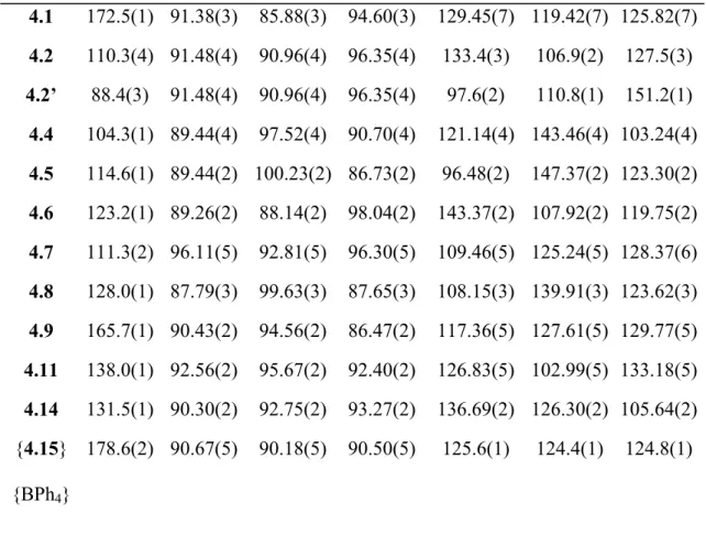
Table 4.1. Summary of color and electrochemical data.
Complex Name and Number Color CoII/CoIII, CoI/CoII (mV)
[PhBP3]CoI, 3.1 Green 10, -920
[PhBP3]CoOSiPh3, 4.1 Purple -360, -1290 [PhBP3]CoO(4-tBu-Ph), 4.2 Red-brown -390, -1330
[PhBP3]CoO(C6F5), 4.3 Olive green NA, NA
[PhBP3]CoSPh, 4.4 Red -160, -1120
[PhBP3]CoS(2,6-Me2-Ph), 4.5 Red -170, -1100 [PhBP3]CoS(2,4,6-iPr3-Ph), 4.6 Red-brown -80, -1190 [PhBP3]CoS(2,4,6-tBu3-Ph), 4.7 Red -60, -1080
[PhBP3]CoSSiPh3, 4.8 Green -210 (irreversible), -1010 [PhBP3]CoOSi(4-NMe2-Ph)3, 4.9 Red -360, -1300
[PhBP3]CoOSi(4-CF3-Ph)3, 4.10 Blue -60 (irreversible), -1080 [PhBP3]CoOCPh3, 4.11 Blue-green -300, -1310
[PhBPiPr3]CoI, 4.12 Green 60, -1250
[PhBPiPr3]CoOSiPh3, 4.13 Purple 100 (irreversible), -1690 [PhBPiPr3]CoSSiPh3, 4.14 Green -140 (irreversible), -1330
{[PhBP3]CoOSiPh3} {B(3,5-(CF3)2-Ph)4}, {4.15}{B(3,5-(CF3)2-Ph)4}
Green Oxidation product of 4.1
89
Table 4.2. Summary of magnetic data.
Complex Number Evans Method (µeff in BM, C6D6, 298 K)
SQUID χmT (cm3 K mol-1) at 20 K, 300 K
3.1 2.8 0.82, 1.01
4.1 3.4 0.47, 1.45
4.2 3.4 0.90, 1.90
4.3 3.8 1.32, 2.13 (at 240 K)
4.4 2.4 0.40, 0.49
4.5 2.3 0.50, 0.51
4.6 2.8 0.46, 0.63
4.7 3.9 2.05, 2.21
4.8 2.5 0.47, 0.44
4.9 3.5 0.46, 1.56
4.10 3.9 1.22, 1.95
4.11 3.8 2.08, 2.28
4.12 4.1 1.83, 1.90
4.13 4.3 2.19, 2.36
4.14 4.0 0.47, 1.23
{4.15}
{B(3,5-(CF3)2-Ph)4}
Diamagnetic Diamagnetic
90
The family of complexes shown in Table 4.1 is conveniently accessible via the use of the soft thallium reagents TlEAr and TlEZAr3 (E = O or S, Z = Si or C, Eq. 4.1).
The typical method for preparation of these thallium reagents involves a metathesis reaction between commercially available thallium ethoxide and the desired phenol, arylthiol, silanol, or silylthiol.39 The types of alcohols and thiols amenable to this method of preparation are restricted to those that have pKa values lower than that of the ethanol byproduct (pKa = 15.9). The addition of one equivalent of the desired thallium reagent as a THF solution to a THF solution of [BP3]CoX (X = I or Cl) affords the substituted product in high crude yield with TlX as an easily separable byproduct. Filtration of the crude reaction mixture followed by crystallization, typically by vapor diffusion of petroleum ether in benzene, provides each of the desired complexes in crystalline form in modest to high yields.
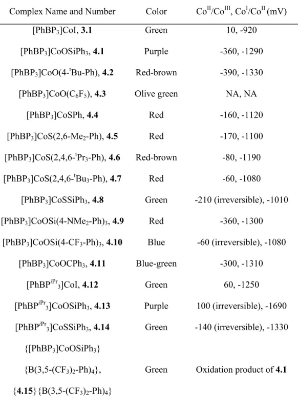
Despite the paramagnetic nature of these Co(II) derivatives, 1H NMR spectroscopy aids in their characterization. While the signature proton resonances are listed for each isolated complex in the Experimental Section, we examined the paramagnetically shifted 1H NMR spectra of the iodides [PhBP3]CoI (3.1) and [PhBPiPr3]CoI (4.12) in some detail, as a representative sample of this family of complexes. Both complexes exhibit solution spectra (see Figure 4.3) consistent with approximate C3 symmetry at room temperature, as only a single set of resonances arises from the phosphine donor arms. T1 relaxation times can be used as a guide to determine the relative distances of ligand-based protons from a coordinated metal center containing unpaired spin.40 By measuring a T1 relaxation time for each proton resonance shown in
91
Figure 4.3 (top and bottom), and correlating these relaxation times with the integrated number of protons corresponding to each resonance, we are able to assign the spectrum of 3.1 with a high degree of confidence. The spectrum of 4.12 suffers from some ambiguity due to certain resonances having similar T1 relaxation times and integration values. Notably, the chemical shift range of the resonances observed for 4.12 is much broader than that of 3.1, likely due to their different respective spin states (vide infra).
Figure 4.3. 1H NMR spectra of 3.1 and 4.12 in C6D6. Assignments are based on the correlation between single scan integration values and T1 relaxation times for each resonance. For 3.1: (i) PhB(CH2PPh2)3, (ii) meta P(C6H5)2, (iii) ortho B(C6H5), (iv) meta and para B(C6H5), (v) ortho P(C6H5)2, (vi) para P(C6H5)2. For 4.12: (i and ii) PhB(CH2PiPr2)3 and P(CH(CH3)2)2, (iii and vii) P(CH(CH3)2)2, (iv) ortho B(C6H5), (v) para B(C6H5), (vi) meta B(C6H5).
92 4.2.2 Electrochemical data
The electrochemical response of each complex featured in this study was examined by cyclic voltammetry in THF solution using either [nBu4N][PF6] or [nBu4N][ClO4] as the supporting electrolyte, a glassy carbon working electrode, a platinum wire counter electrode, and a Ag/AgNO3 reference electrode. The potentials of well-defined redox processes were recorded versus an external ferrocene standard and are listed in Table 4.1.
The electrochemical data are generally unremarkable and hence only a few comparative comments are warranted. For those [PhBP3]Co(II) complexes featuring a Co-OR linkage, specifically complexes 4.1, 4.2, 4.3, 4.9, 4.10, and 4.11, fully reversible Co(II/III) and Co(I/II) redox processes are observed. Relatively little shift in the potential of either redox event is observed within this family, with the exception of the p-CF3-substituted silyloxide species [PhBP3]CoOSi(4-CF3-Ph)3, 4.10. For this complex the Co(I/II) redox event is anodically shifted by ca. 300 mV, and the Co(II/III) redox event is irreversible. These differences are likely due to the electron-withdrawing CF3
substituent, which destabilizes the higher-valent Co(III) state but renders the lower-valent Co(I) state more accessible.
The [PhBP3]Co(II) arylthiolate complexes 4.4-4.8 also exhibit well-behaved Co(II/III) and Co(I/II) redox events. These thiolate species are, as might be expected, easier to reduce and more difficult to oxidize than their aryloxide relatives. Again, only a small degree of variance is observed for the potentials amongst the arylthiolate family of complexes. Two subtle differences worth noting are (i) complexes [PhBP3]CoS(2,4,6-iPr3-Ph), 4.6, and [PhBP3]CoS(2,4,6-tBu-Ph), 4.7, are approximately
93
100 mV more difficult to oxidize than [PhBP3]CoSPh, 4.4, and [PhBP3]CoS(2,6-Me2-Ph), 4.5, presumably reflecting the difference in electron-releasing character between the arylthiolate substituents; and (ii) it is ca. 100 mV more difficult to reduce 4.6 than 4.7, an observation that is difficult to rationalize in simple terms. The triphenylsilylthiolato derivative [PhBP3]CoSSiPh3 (4.8) is the most easily reduced species (-1010 mV), but displays an irreversible oxidation event around -210 mV. The reduction potential recorded for complex 4.8 can be compared with that of its [PhBPiPr3]-supported congener [PhBPiPr3]CoSSiPh3 (4.14). The latter complex 4.14 is ca. 300 mV more difficult to reduce (-1330 mV) due to its more electron-releasing P3 donor scaffold, but still displays an irreversible Co(II/III) process. Comparison of the redox processes observed between [PhBP3]CoOSiPh3, 4.1, and [PhBPiPr3]CoOSiPh3, 4.13, reveals another curiosity of note. While 4.13 is appreciably more difficult to reduce (by ca. 400 mV), as should be expected, its oxidation to Co(III) is electrochemically irreversible. By contrast, {[PhBP3]CoIIIOSiPh3}+ is electrochemically accessible and stable (vide infra). This is difficult to rationalize, except to suggest that a putative {[PhBPiPr3]CoIIIOSiPh3}+ species may be more prone to loss of the triphenylsilyl substituent in the presence of a fluorinated counter-anion from the electrolyte.
Finally, it is interesting to compare the electrochemical data recorded for these cobalt systems with that of a related series of recently reported [BP3]Ni-X systems.41 For example, a reversible Ni(I/II) reduction event is observed for the complex [PhBP3]NiOSiPh3 at -1.47 V, which is ca. 180 mV more negative than the Co(I/II) event of 4.1. More striking is how difficult it is to oxidize the nickel systems to the Ni(III) state.
For the complex [PhBP3]NiOSiPh3, the first oxidative process is encountered at a
94
potential that is ca. 700 mV more positive than for its cobalt analogue 4.1. This large difference appears to reflect the relative instability of a d7 versus a d6 electronic configuration within the [BP3]Ni-X and [BP3]Co-X platforms, respectively.
4.2.3 Chemical oxidation of [PhBP3]CoOSiPh3 to produce {[PhBP3]CoOSiPh3}{BAr4}
Reversible oxidation waves for these [BP3]CoII-X derivatives suggest that their chemical oxidation might afford the corresponding trivalent {[BP3]CoIII-X}+ products, which would comprise a structurally unusual class of pseudotetrahedral Co(III) complexes (type E in Figure 4.2). We have prepared and thoroughly characterized one such example pertinent to the present study: {[PhBP3]CoOSiPh3}{BAr4}, {4.15}{BAr4} (Ar = C6H5, 3,5-(CF3)2-C6H3). The addition of THF to a solid mixture of 4.1 and {[Cp]2Fe}{BAr4} effects a rapid oxidation process to generate the diamagnetic, green product {4.15}{BAr4} (Eq. 4.2). Cationic {4.15}{BAr4} exhibits a sharp singlet in its 31P NMR spectrum at 64.6 ppm and also a sharp singlet in the 19F NMR spectrum at -58.5 for the tetra(3,5-bis(trifluoromethyl)phenyl)borate salt derivative. The combined 1H and 31P NMR spectra obtained for this system reveal that it is threefold symmetric in solution on the NMR time scale. The diamagnetic ground state of {4.15}+ suggests that its electronic configuration is likely related to the diamagnetic imide [PhBP3]Co≡N-p-tolyl, which is an S = 0 Co(III) species featuring a bona fide Co-N triple bond linkage.42 However, whereas {4.15}+ can be reduced at a potential of ca. -360 mV, the imide species [PhBP3]Co≡N-p-tolyl is stable to reduction at potentials as low as ca. – 3.0 V, reflecting both the difference in charge and the weaker strength of the π-bonding in {4.15}+.
95
4.2.4 Structural characterization and stereochemical classification of [BP3]CoII-X derivatives
Solid-state crystal structures have been determined for many of the cobalt complexes listed in Table 4.1. These results are summarized by their core structure representations, collected in Figure 4.4 and Figure 4.5, and by a list of salient bond distances and angles, collected in Table 4.3. In each structure, the tris(phosphino)borate is κ3-bound to a monomeric cobalt center. The pseudotetrahedral structures can be broadly divided into two separate classes based on the average length of the Co-P bonds. As will be corroborated by the SQUID and EPR data discussed below, complexes featuring average Co-P bond distances between 2.15 Å and 2.25 Å (e.g., 4.5 and 4.9) are characteristic of low spin ground states (type D in Figure 4.2), whereas complexes with average Co-P bond distances between 2.30 Å and 2.35 Å (e.g., 4.7 and 4.11) are characteristic of high spin ground states (type C is Figure 4.2).
96
Figure 4.4. Displacement ellipsoid representations (50%) of the core structures of [PhBP3]CoSPh (4.4); [PhBP3]CoS(2,6-Me2-Ph) (4.5); [PhBP3]CoS(2,4,6-iPr3-Ph) (4.6); [PhBP3]CoS(2,4,6-tBu3-Ph) (4.7); [PhBP3]CoO(4-tBu-Ph) (4.2);
[PhBP3]CoOSiPh3 (4.1); [PhBP3]CoSSiPh3 (4.8); [PhBP3]CoOSi(4-NMe2-Ph)3
(4.9); [PhBP3]CoOCPh3 (4.11); and [PhBPiPr3]CoSSiPh3 (4.14). The 4.2 and 4.2’
structures show the disorder in the –O(4-tBu-Ph) ligand, which is bound either η1 (left) or η3 (right) to the cobalt center. See Table 4.3 for bond lengths and angles.
97
B1
B2 Co
Si O
P3 P1 P2
Figure 4.5. Displacement ellipsoid representations (50%) of {[PhBP3]CoOSiPh3}{BPh4}, {4.15}{BPh4}. Hydrogen atoms and solvent molecules have been omitted for clarity. See Table 4.3 for bond lengths and angles.
The basic stereochemical structures observed in the solid-state can be further classified, at least qualitatively, as having one of two general structure types that we will refer to throughout as either umbrella distorted or off-axis distorted (Figure 4.6). In an umbrella distorted structure, the X-type ligand is regarded as axial and trisects the three Co-P linkages, coincident with the B-Co vector. Distortions of an umbrella type are common for four-coordinate complexes supported by tripodal ligands, and it may be said that such ligands in fact enforce the umbrella distortion.28,30 In an off-axis distorted structure, the X-type ligand cants away from the imaginary vector running through the B and Co atoms to such an appreciable extent that it is better regarded as an equatorial
98
ligand rather than an axial ligand. Four-coordinate complexes that exhibit an off-axis distortion are less frequently encountered and appear to arise from the population of a low spin ground state, as discussed in greater detail below.
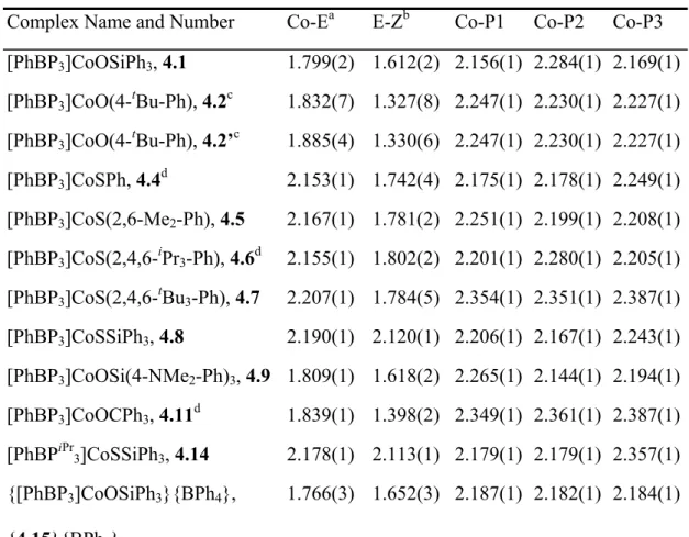
Table 4.3. X-ray diffraction table showing key bond lengths (Å) and angles (deg) for 4.1, 4.2, 4.4, 4.5, 4.6, 4.7, 4.8, 4.9, 4.11, 4.14, and {4.15}{BPh4}.
Complex Name and Number Co-Ea E-Zb Co-P1 Co-P2 Co-P3 [PhBP3]CoOSiPh3, 4.1 1.799(2) 1.612(2) 2.156(1) 2.284(1) 2.169(1) [PhBP3]CoO(4-tBu-Ph), 4.2c 1.832(7) 1.327(8) 2.247(1) 2.230(1) 2.227(1) [PhBP3]CoO(4-tBu-Ph), 4.2’c 1.885(4) 1.330(6) 2.247(1) 2.230(1) 2.227(1) [PhBP3]CoSPh, 4.4d 2.153(1) 1.742(4) 2.175(1) 2.178(1) 2.249(1) [PhBP3]CoS(2,6-Me2-Ph), 4.5 2.167(1) 1.781(2) 2.251(1) 2.199(1) 2.208(1) [PhBP3]CoS(2,4,6-iPr3-Ph), 4.6d 2.155(1) 1.802(2) 2.201(1) 2.280(1) 2.205(1) [PhBP3]CoS(2,4,6-tBu3-Ph), 4.7 2.207(1) 1.784(5) 2.354(1) 2.351(1) 2.387(1) [PhBP3]CoSSiPh3, 4.8 2.190(1) 2.120(1) 2.206(1) 2.167(1) 2.243(1) [PhBP3]CoOSi(4-NMe2-Ph)3, 4.9 1.809(1) 1.618(2) 2.265(1) 2.144(1) 2.194(1) [PhBP3]CoOCPh3, 4.11d 1.839(1) 1.398(2) 2.349(1) 2.361(1) 2.387(1) [PhBPiPr3]CoSSiPh3, 4.14 2.178(1) 2.113(1) 2.179(1) 2.179(1) 2.357(1) {[PhBP3]CoOSiPh3}{BPh4},
{4.15}{BPh4}
1.766(3) 1.652(3) 2.187(1) 2.182(1) 2.184(1)
99
Table 4.3 (cont.)
Complex
Number Co-E-X P1-Co-P2 P1-Co-P3 P2-Co-P3 P1-Co-E P2-Co-E P3-Co-E 4.1 172.5(1) 91.38(3) 85.88(3) 94.60(3) 129.45(7) 119.42(7) 125.82(7) 4.2 110.3(4) 91.48(4) 90.96(4) 96.35(4) 133.4(3) 106.9(2) 127.5(3) 4.2’ 88.4(3) 91.48(4) 90.96(4) 96.35(4) 97.6(2) 110.8(1) 151.2(1)
4.4 104.3(1) 89.44(4) 97.52(4) 90.70(4) 121.14(4) 143.46(4) 103.24(4) 4.5 114.6(1) 89.44(2) 100.23(2) 86.73(2) 96.48(2) 147.37(2) 123.30(2) 4.6 123.2(1) 89.26(2) 88.14(2) 98.04(2) 143.37(2) 107.92(2) 119.75(2) 4.7 111.3(2) 96.11(5) 92.81(5) 96.30(5) 109.46(5) 125.24(5) 128.37(6) 4.8 128.0(1) 87.79(3) 99.63(3) 87.65(3) 108.15(3) 139.91(3) 123.62(3) 4.9 165.7(1) 90.43(2) 94.56(2) 86.47(2) 117.36(5) 127.61(5) 129.77(5) 4.11 138.0(1) 92.56(2) 95.67(2) 92.40(2) 126.83(5) 102.99(5) 133.18(5) 4.14 131.5(1) 90.30(2) 92.75(2) 93.27(2) 136.69(2) 126.30(2) 105.64(2) {4.15}
{BPh4}
178.6(2) 90.67(5) 90.18(5) 90.50(5) 125.6(1) 124.4(1) 124.8(1)
Table 4.3 footnotes. a–E represents the fourth, non-phosphine atom directly bound to the Co center, either O or S; b–Z represents the non-cobalt atom bound to E, which is either C or Si; c–4.2 shows the bond distances and angles for the η1 conformation. 4.2’ shows the bond angles and distances for the η3 conformation; d–
There are two crystallographically independent molecules in the unit cell.
100
Rigorously distinguishing between structures that arise from these two limiting distortions is not readily apparent by inspection. An elegant method known as the continuous symmetry measure13,43 proves very useful in this regard because it allows one to quantitatively discuss how close a given molecular geometry is to an idealized structure type. For example Alvarez and co-workers have used this approach to quantitatively compare true geometric structures to those of idealized tetrahedra or square planes. Under a continuous symmetry measurement, the distance (i.e., deviation) of a given molecule from an idealized polyhedron of a symmetry point group defined as G is numerically defined as S(G). A perfect tetrahedron therefore has an S(Td) = 0, and a perfect square plane has an S(D4h) = 0. Construction of a 2-D plot of S(G) values can then be used to show that a perfect tetrahedron has an S(D4h) = 33.3, and a perfect square plane has an S(Td) = 33.3. As should be obvious, a trigonal pyramidal structure is geometrically much closer to a tetrahedron than to a square plane. This is reflected by its respective S(G) values; it features a relatively small S(Td) by comparison to a large S(D4h) value (S(Td) = 3.57; S(D4h) = 34.87).
Figure 4.6. Limiting distortions relevant to the pseudotetrahedral structure types.
101
Plotting the data for the X-ray structures shown in Figures 4.4 and 4.5 on a 2-D continuous symmetry map allows us to see the deviations. Comparing the species on a S(Td) versus S(D4h) map shows that the complexes we have prepared are all reasonably close to an ideal tetrahedron (Figure 4.7). Complexes on the upper left portion of the graph exhibit a typical umbrella distortion and are high spin (vide infra). This class includes complexes [PhBP3]CoS(2,4,6-tBu3-Ph) (4.7), [PhBP3]CoOCPh3 (4.11), and [PhBPiPr3]CoI (4.12). The ambient temperature solid-state structure of [PhBP3]CoOSiPh3
(4.1) is also in this class. The upper right box features low spin umbrella complexes including the siloxides: 4.1 (at 98 K), [PhBP3]CoOSi(4-NMe2-Ph)3 (4.9), and {[PhBP3]CoOSiPh3}{BPh4} ({4.15}{BPh4}) and the iodide complex we previously reported, [PhBP3]CoI (3.1). The complexes we denote as off-axis structure types are somewhat distinct from the [PhBP3]CoII-X complexes we have described in previous studies (lower right box in Figure 4.7).14,15 These off-axis complexes all incorporate a thiolate as the fourth ligand. Furthermore, all of the off-axis species (4.4, 4.5, 4.6, 4.8, and 4.14) are low spin at 98 K. Not surprisingly, these five complexes feature one elongated Co-P bond in an axial position and two shorter Co-P bonds in the equatorial positions.
Given that all of these P3CoIIX species show only a small distortion from an ideal tetrahedron, it is worth examining whether some of the species are better described as trigonal pyramidal. A continuous symmetry plot mapping the deviations from an ideal trigonal pyramid and a tetrahedron is shown in Figure 4.8. Almost all of the complexes fall within the middle portion of the graph, implying roughly equal distortions from both idealized geometries. Complexes 4.1, 4.9, 4.12, and {4.15} have less distortion from a
102
tetrahedron than complex 4.5, which, while distorted from both idealized geometries, is slightly closer to a trigonal pyramid. It is admittedly difficult to tease out a definitive difference between these two ideal geometries for the structures described. The dotted line shown in Figure 4.8 qualitatively draws the same distinction illustrated by the previous graph (Figure 4.7). The complexes on the left side feature typical umbrella distortions, and the complexes on the right feature off-axis distortions. The single discrepancy between this plot and the plot in Figure 4.7 is that this symmetry map suggests that complex 3.1 belongs to the off-axis class instead of the umbrella class.
None of the complexes we have prepared is truly close to a trigonal pyramidal geometry since the equatorial L-Co-L angles are inequivalent. Known examples of trigonal pyramidal Co(II) species have been assigned as high spin and often feature a tetradentate ligand with three equivalent tripodal arms and one axial donor ligand giving equivalent L-Co-L angles near 120°.17
103
Figure 4.7. The calculated continuous symmetry deviation for each molecule is plotted on a continuous symmetry map of S(Td) (tetrahedral) versus S(D4h) (square planar). The complexes can be assigned to one of two classes based on their deviations from these two idealized structure types. The umbrella class can be subdivided into high spin and low spin complexes. Complexes with two components on the symmetry map (a and b) have two asymmetric molecules within the unit cell. The crystal structure of [PhBP3]CoOSiPh3 (4.1) was solved at two different temperatures.
104
Figure 4.8. The calculated continuous symmetry deviation for each molecule is plotted on a continuous symmetry map of S(Td) (tetrahedral) versus S(C3v) (trigonal pyramidal). Complexes to the left of the dashed line can be assigned as umbrella distorted while those on the right can be assigned as off-axis distorted. Complexes with two components on the symmetry map (a and b) have two asymmetric molecules within the unit cell. The crystal structure of [PhBP3]CoOSiPh3 (4.1) was solved at two different temperatures.
Complex 4.2 does not follow the generalized descriptions discussed above since the aryloxide ligand is disordered over two positions, one that exhibits η1-bonding to the
105
cobalt center (4.2) and one that displays η3-bonding (4.2’) (Figure 4.4).44 To our knowledge, an η3-binding mode of an aryloxide ligand is unprecedented for cobalt complexes. In the η3-bonding mode (4.2’), the Co-O bond distance is 1.885(4) Å, and the Co-C bond distances are 2.277(5) and 2.341(5) for C(46) and C(47), respectively.
Furthermore, the Co-O-C(46) bond angle is highly bent (88.4(3)°) to accommodate favorable π-bonding to the aryl ring.
Two other solid-state structures are worth discussing in more detail. The solid-state structure determined for diamagnetic {4.15}{BPh4} shows an umbrella distortion (Figure 4.5). The complex is markedly three-fold symmetric and represents an ideal example of structure type E, as shown in Figure 4.2. The Co-P distances display a variance of only 0.005 Å, and the average of the three Co-P distances is short at 2.185 Å (Table 4.3). The Co-O bond distance (1.766(3) Å) is only 0.03 Å shorter than in 4.1, and the Co-O-Si bond angle is almost perfectly linear (178.6(2)°). [PhBPiPr3]CoSSiPh3 (4.14) features an off-axis distortion. However, the axial elongation is much more pronounced for complex 4.14 than for the other off-axis complexes. In this case, the axial Co-P bond is greater than 0.17 Å longer than the other two Co-P bonds.
4.2.5 Magnetic characterization (SQUID and EPR) of [BP3]CoII-X derivatives
As mentioned in the introduction, experimental evidence for the preferred low spin ground state configuration in a L3CoII-X structure type was first provided by the complex [PhBP3]CoI (3.1). In the solid-state this complex displays the distorted structure type represented as D in Figure 4.2. The related chloride and bromide complexes [PhBP3]Co-X are also low spin species in their monomeric form in solution, but they give rise to dimeric structures in the crystalline state and are therefore of little utility to the
106
present discussion. In contrast to these low spin [PhBP3]Co(II) halides, we have previously assigned quartet ground states to the complexes [PhBPiPr3]CoI (4.12), [PhBPiPr3]CoCl, and [PhBP3]CoO(2,6-Me2-Ph) (3.4) (type C in Figure 4.2). These assignments, considered collectively, suggested to us the possibility that the low and high spin ground states of pseudotetrahedral L3CoIIX structure types may in fact lie closer in energy (i.e., ∆HHS/LS = kBT) than had been previously anticipated.15,25
For the collection of [PhBP3]CoIIX complexes presented in this chapter (see Table 4.2) it is clear that a low spin ground state is most typically, though not always, preferred.
Moreover, this spin preference is in contrast to Co(II) species supported by the [PhBPiPr3] ligand, where the high spin configuration more typically dominates.
SQUID and EPR data have been collected for all of these [BP3]Co(II) complexes.
Rigorously high spin species include complexes 4.7, 4.11, 4.12, and 4.13. Each of these complexes adopts a structure that falls into the upper left portion of the symmetry plot shown in Figure 4.7, exhibiting a typical umbrella distortion. Complexes 3.1, 4.4, 4.5, 4.6, and 4.8 provide examples of rigorously low spin species. The species 4.1, 4.9, 4.10, and 4.14 display spin crossover phenomena in the solid-state. The low spin Co(II) derivatives, type D in Figure 4.2, gives rise to stereochemical structures exhibiting both umbrella and off-axis distortions. The interpretation of the magnetic data for 4.2 and 4.3 is more complex due to the potential for η3 interactions from the X-type ligand.
In examining the [PhBP3] supported thiolates (4.4-4.8), it is apparent that the preferred ground state is a 2E. Each complex, excluding 4.7, has values of χmTav (cm3 K mol-1) from 10-300 K slightly above the spin only value of χmT = 0.375 for a single unpaired electron: 4.4, 0.41; 4.5, 0.50; 4.6, 0.51; 4.8, 0.45. The solid-state magnetic
107
moment of complexes 4.4 and 4.6 very gently increases as the temperature of each sample is raised above 250 K. We collected data from 4 K to 300 K, and then back to 4 K for complex 4.6, and this gentle curvature at higher temperatures is reproducible. It appears likely that partial population of an S = 3/2 state is present at higher temperatures.
A similar curvature is also observed for [PhBP3]CoI (3.1). The low spin thiolates 4.4, 4.5, 4.6, and 4.8 each exhibit an off-axis distortion in the solid-state and short average Co-P bond distances. The EPR data for these three species corroborates their doublet assignments, exhibiting an axial (g║ > g⊥) EPR signal centered near g = 2 (Figure 4.9).
On the other hand, complex 4.7 exhibits a χmTav (10-300 K) value of 2.11 cm3 K mol-1 between 10 K and 300 K, an amount that is slightly greater than the spin only value for an S = 3/2 system (1.88 cm3 K mol-1). The low temperature EPR confirms this assignment by showing two signals, one at g ≈ 2 and a second signal at low field centered at g ≈ 5.8 (Figure 4.9). The S = 3/2 ground state of 4.7 must be due to high steric crowding by the bulky X-type thiolate ligand. Population of the quartet spin state expands the average Co-P bond distances thereby alleviating unfavorable steric contacts.
108
Figure 4.9. Glassy toluene EPR spectra (20 K) of [PhBP3]CoSPh (4.4) (▬), [PhBP3]CoS(2,6-Me2-Ph) (4.5) (▬), [PhBP3]CoS(2,4,6-iPr3-Ph) (4.6) (▬), and [PhBP3]CoS(2,4,6-tBu3-Ph) (4.7) (▬). Instrumental parameters for the spectra can be found in the Experimental Section.
Many pseudotetrahedral Co(II) complexes supported by [Tp] ligands have been prepared, and all of them are high spin.28,29 Given the preference we have found for [BP3]Co-thiolates to populate low spin ground states, we were curious as to whether a thiolate ligand might confer the low spin configuration to a [Tp]Co(II) system. [Tp]Co thiolates have been prepared previously, and while magnetic data was not reported, they were presumed to be high spin.33a We therefore prepared one example of such a complex, [Tp3,5-Me2]CoS(2,6-Me2-Ph) (4.16), and obtained low temperature magnetic and structural data to accurately determine a ground spin state (Figure 4.10, see Experimental Section
109
for synthesis). The SQUID magnetometry data unequivocally shows that 4.16 populates a high spin ground state, with χTav (10 – 300 K) = 2.41 cm3 K mol-1.
Figure 4.10. Displacement ellipsoid representations (50%) of [Tp3,5-Me2]CoS(2,6-Me2-Ph), 4.16. Hydrogen atoms and solvent molecules have been omitted for clarity. Selected interatomic distances (Å) and angles (deg): Co-N1, 2.026(1); Co-N3, 2.021(2); Co-S, 2.273(1); N1-Co-N1’, 93.39(8); N1-Co-N3, 93.16(5); N1-Co-S, 134.57(5); N1’-Co-S, 114.07(5);
N3-Co-S, 119.04(6).
Whereas thiolate ligands typically confer low spin ground states in the case of [BP3]Co(II) derivatives, weaker field siloxide ligands form complexes that exhibit spin crossover, as evinced by the complexes [PhBP3]CoOSiPh3 (4.1), [PhBP3]CoOSi(4-NMe2-Ph)3 (4.9), and [PhBP3]CoOSi(4-CF3-Ph)3 (4.10). Each of these complexes exhibits an umbrella distortion at low temperature. The structure of 4.1 has been examined both at 98 K (see Figure 4) and at 298 K, and while the average of the Co-P bond distances is expanded at 298 K (reflecting population of the high spin state, vide infra) both X-ray data sets confirm the umbrella distortion (Figure 4.7). Changing
110
the substituents at the para position on the siloxide aryl rings dramatically effects the spin state population at a given temperature (Figure 4.11). The presence of the electron-withdrawing CF3 group in 4.10 serves to lower the Tc of the spin crossover event. As shown in Figure 4.11, there is a strong temperature dependence of the moment of 4.9, and a relatively well-defined partial hysteresis is evident centered around 150 K.
A more gradual and fully reversible change in χmT is observed above 170 K. This magnetic behavior is distinct from the data collected for 4.1, which shows gradual crossover (Figure 4.11). EPR spectra were collected at 20 K for 4.1, 4.9, and 4.10 and the spectra are consistent with the low temperature SQUID data obtained for each sample.45 The EPR spectrum of 4.1 shown in Figure 4.12A exhibits an axial signal (g1 = 2.21, g2 = 2.05, g3 = 2.03) featuring well-defined hyperfine coupling (ICo = 7/2) as well as superhyperfine coupling to phosphorus (3 x P, IP = ½). The observation of phosphorous coupling reflects the highly covalent character of these systems.46 The octet pattern (A1(Co) = 105 gauss) in the g1 region of the spectrum suggests a monomeric species in solution (Figure 4.13). Noticeably absent from the spectra of 4.1 and 4.9 are any low field signals that would signify the presence of a high spin Co(II) component.47 The EPR spectrum of 4.10 (Figure 4.12C) is more interesting. A broad but discernable signal at low field (near g = 5.5) is present at 20 K, suggesting the presence of a high spin component, as is also evident from the solid-state SQUID data. The hyperfine coupling, in the g = 2 region of the spectrum, can be attributed to the presence of the low spin component of 4.10 in the glass, in analogy to the spectra of 4.1 and 4.9. The effect of solvent in the crystal lattice on the spin crossover process was measured for 4.1 in the solid-state and was found to be minimal.
111
Figure 4.11. SQUID magnetization plot of χmT versus T: [PhBP3]CoOSiPh3
(4.1) (♦), [PhBP3]CoOSi(4-CF3-Ph)3 (4.10) (●), [PhBP3]CoOCPh3 (4.11) (▲), [PhBP3]CoSSiPh3 (4.8) (□), and [PhBP3]CoOSi(4-NMe2-Ph)3 (4.9) as the temperature was raised (∆) and lowered (■).
0 100 200 300 400
Field (mT)
A B C
D
Figure 4.12. Glassy toluene EPR spectra (20 K) for (A) [PhBP3]CoOSiPh3 (4.1) (▬), (B) [PhBP3]CoOSi(4-NMe2-Ph)3 (4.9) (▬), (C) [PhBP3]CoOSi(4-CF3-Ph)3
(4.10) (▬), and (D) [PhBP3]CoOCPh3 (4.11) (▬). Instrumental parameters for the spectra can be found in the Experimental Section.
112
Figure 4.13. Experimental (▬) and simulated (▬) EPR spectra of [PhBP3]CoOSiPh3 (4.1). Instrumental parameters for the experimental spectrum can be found in the Experimental Section. The simulated parameters are as follows: g1 = 2.21, g2 = 2.05, g3 = 2.03; a1(Co) = 105 gauss, a2(Co) = 12 gauss, a3(Co) = 65 gauss; a1(P) = 28 gauss, a2(P) = 27 gauss, a3(P) = 34 gauss.
Interestingly, the trityloxide complex [PhBP3]CoOCPh3 (4.11) exhibits a highly bent Co-O-C bond angle of 138.0(1)º, compared with the angle of 172.5(1)º for 4.1 and 165.7(1) for related 4.9. Each of the Co-P bond distances in this umbrella distorted species are also expanded (Co-Pav = 2.37 Å). Moreover, each of the Co-P bond distances is expanded (Co-Pav = 2.37 Å), suggestive of a high spin ground state in accord with its solid-state SQUID (Figure 4.11) and glassy toluene EPR data (Figure 4.12D).
The magnetic data is complicated for 4.2 and 4.3 due to the possibility of η3 interactions of the axial ligand. The solid-state crystal structure obtained for 4.2 at 98 K reveals the presence of two distinct conformational isomers. One of these isomers is a four-coordinate pseudotetrahedral species with a Co-O-Cipso angle of 110.3(4)º. The other
113
isomer is nominally five-coordinate and displays an η3-binding mode of the aryloxide ligand that provides an acute Co-O-Cipso angle of 88.4(3)º. SQUID magnetization data for 4.2 are shown in Figure 4.14. It is clear that the sample predominantly populates a doublet state at low temperature, though a weak signal at low field is discernable in the glassy toluene EPR spectrum of the sample. As the sample is warmed the χmT value gradually rises and at 300 K almost complete crossover to the high spin component is evident. Perhaps the simplest explanation of these data is that the pseudotetrahedral isomer of 4.2 is high spin at all temperatures, and that the isomer that exhibits an η3-bonding mode is low spin at all temperatures. The magnetic data would then reflect variable populations of the two conformational isomers as a function of temperature in both solid and solution. Consistent with this explanation is the fact that the two other pseudotetrahedral aryloxide and alkoxide complexes we have examined, [PhBP3]CoO(2,6-Me2-Ph) (3.4) and [PhBP3]CoOCPh3 (4.11), both exhibit high spin ground states, and the fact that five-coordinate cobalt(II) systems supported by phosphine ligands invariably populate low spin ground states. The fluorinated aryloxide complex [PhBP3]CoO(C6F5) (4.3) is less likely to exhibit π-bonding to the aryl ring due to its electron-withdrawing nature, though interactions with the ortho fluorines of the aryl group cannot be discounted. A similar four-coordinate/five-coordinate equilibrium may explain the change in spin state that is observed in the SQUID data (Figure 4.14).
114
Figure 4.14. SQUID magnetization plot of χmT versus T for [PhBP3]CoO(4-tBu-Ph) (4.2) as the temperature was raised (♦) and then lowered (□), and data for [PhBP3]CoO(C6F5) (4.3) (▲) as the temperature was raised.
The complexes supported by the [PhBPiPr3] ligand feature two high spin complexes, [PhBPiPr3]CoI (4.12) and [PhBPiPr3]CoOSiPh3 (4.13), that exhibit the umbrella distortion, and one thiolate complex, [PhBPiPr3]CoSSiPh3 (4.14), that exhibits an off-axis distortion and is low spin (vide infra) at low temperature. Structural and magnetic data have been reported previously for [PhBPiPr3]CoI (4.12). This complex exhibits a high spin ground state configuration and is therefore distinct from its low spin analog [PhBP3]CoI (3.1). SQUID data collected on a polycrystalline sample of 4.13 are plotted in Figure 4.15A. The sample is clearly a S = 3/2 system (χmTav (10 K - 300 K) = 2.30 cm3 K mol-1) and obeys the Curie-Weiss law. Similar magnetic behavior was reported for iodide 4.12. Magnetization data for the thiolate derivative 4.14 are plotted in Figure 4.15B. The temperature dependence of its magnetic moment is more complex.
Below 100 K the sample appears to be low spin (χmTav (10 K - 100 K) = 0.50 cm3 K
115
mol-1). A gradual rise in χmT is observed above 100 K and a maximum value of 1.23 cm3 K mol-1 is reached at 300 K, the highest temperature at which the data could be recorded.
The magnetic behavior of the sample is fully reversible. The appearance of a gradual spin crossover phenomenon is similar to the rise in magnetic moment that was observed for thiolates 4.4 and 4.6 near room temperature. An interesting observation is that the solution moment of 4.14 at room temperature is 4.0 µB, consistent with a fully high spin population. This moment is different from that determined at low temperature by SQUID analysis of the polycrystalline sample, and we therefore elected to further analyze 4.14 by EPR spectroscopy as a powder and as a frozen glass. These data, along with the glassy toluene EPR spectrum of 4.13, are shown in Figure 4.16. The 20 K glassy toluene spectra of 4.13 and 4.14 (Figure 4.16A and 4.16B) each show spectra characteristic of S = 3/2 species. The low field signal present in the glassy spectrum of 4.14 is absent in its powder spectrum at 20 K (Figure 4.16C). There appears therefore to be a stronger preference to populate the high spin configuration in solution for this thiolate complex. A different solution conformation of 4.14 may exist than the one that is observed in its solid-state structure at 98 K. Differences in spin behavior between solution and solid samples are not uncommon for spin crossover systems.48 Even with the stronger donor ligand [PhBPiPr3] these complexes favor the high spin state unless there is a fourth ligand that is an unusually strong donor such as a thiolate ligand. The extreme Co-P axial bond elongation in 4.14 likely reflects a steric compensation that allows the doublet ground state to be populated.
116
Figure 4.15. SQUID magnetization plot of χmT versus T for (A) [PhBPiPr3]CoOSiPh3 (4.13) (▲), and (B) [PhBPiPr3]CoSSiPh3 (4.14) as temperature was raised (♦) and then lowered (□).
0 100 200 300 400
Field (mT)
A B
C
Figure 4.16. Glassy toluene EPR spectra (20 K) of (A) [PhBPiPr3]CoOSiPh3
(4.13) (▬), and (B) [PhBPiPr3]CoSSiPh3 (4.14) (▬). Powder sample EPR spectrum (20 K) of (C) [PhBPiPr3]CoSSiPh3 (4.14) (▬). Instrumental parameters for the spectra can be found in the Experimental Section.
117
4.2.6 Theoretical analysis of [BP3]CoII-X derivatives
The electronic structure features of umbrella and off-axis geometries share certain similarities, though there are a number of characteristics that distinguish them. To more thoroughly consider the case of their d7 electronic structures we have undertaken the theoretical DFT examination of one representative complex from each structural subgroup. For this study we chose the siloxide complex [PhBP3]CoOSiPh3 4.1 as representative of the umbrella subgroup, and thiolate [PhBP3]CoS(2,6-Me2-Ph) 4.5 as representative of the off-axis subgroup. Single point electronic structure calculations (DFT) were performed using (i) the experimentally determined X-ray coordinates for each complex as the ground state geometrical structure under (ii) the assumption of a doublet electronic ground state. Each of the structures was subsequently allowed to relax into a theoretically determined global minimum in the absence of geometric constraints, but still under the assumption of a doublet ground state electronic configuration. For each complex, the frontier molecular orbitals obtained by both methods of analysis are qualitatively quite similar. There are, however, noteworthy structural differences between the experimentally and theoretically determined structures.
For pseudotetrahedral 4.1 the predicted frontier molecular orbitals from the geometry-restricted calculation are shown in Figure 4.17. Orbitals calculated for the DFT-optimized structure were calculated and are very similar to those shown here. The singly occupied molecular orbital (SOMO) is energetically well-separated from the lowest unoccupied orbital (LUMO). The lobal representations of the frontier orbitals containing significant d-orbital character map well with those we have sketched qualitatively in Figure 4.2, though the SOMO is predicted by DFT to lie much closer in
118
energy to the lower set of orbitals than to the LUMO. The SOMO and LUMO orbitals are nearly orthogonal to one another and align reasonably well along the plane containing the Co-O-Si vector. The lowest-lying three orbitals consist of one orbital of dz2-parentage (HOMO-2) and two orbitals that are canted away from the Co-O-Si vector. While the low symmetry of the structure inevitably gives rise to d-orbital mixing, the lowest-lying pair of orbitals can be very crudely described as dxy and dx2-y2 type orbitals, where the z-axis is assumed to be coincident with the Co-O-Si vector. Interestingly, there are two high-lying [BP3]-centered orbitals (HOMO, HOMO-1) comprised within the frontier manifold that would have been difficult to anticipate in the absence of the calculation.
Figure 4.17. Molecular orbitals derived from a single point energy DFT calculation of [PhBP3]CoOSiPh3 (4.1) assuming a doublet ground state and the crystallographically determined atomic coordinates.
119
DFT-minimization of the geometry of 4.1 gives rise to a conformationally similar structure (4.1-DFT), with the noteworthy distinction that distortion of one of its Co-P bond distances is grossly exaggerated (2.22 Å, 2.25 Å, 2.43 Å). Despite this structural distinction, the calculation still points to a LUMO that is energetically well-separated from a lower-lying set of d-type orbitals that includes the SOMO. This conclusion is in accord with DFT studies for 4.5. Table 4.4 compares the core bond lengths and angles of the solid-state structures to the DFT determined structures.
Table 4.4. Experimental and calculated bond lengths and angles for 4.1, 4.5, and {4.15}.
4.1 Exptl Calcd 4.5 Exptl Calcd {4.15} Exptl Calcd Bond Lengths (Å)
Co-O 1.799 1.849 Co-S 2.167 2.223 Co-O 1.766 1.776 Co-P1 2.156 2.429 Co-P1 2.251 2.418 Co-P1 2.187 2.236 Co-P2 2.284 2.224 Co-P2 2.199 2.271 Co-P2 2.182 2.241 Co-P3 2.169 2.251 Co-P3 2.208 2.286 Co-P3 2.184 2.234
Bond Angles (deg)
Co-O-Si 172.5 162.8 Co-S-C46 114.6 119.1 Co-O-Si 178.6 178.7 P1-Co-P2 91.38 88.9 P1-Co-P2 89.44 89.4 P1-Co-P2 90.67 90.8 P1-Co-P3 85.88 94.8 P1-Co-P3 100.23 95.9 P1-Co-P3 90.18 90.8 P2-Co-P3 94.60 88.8 P2-Co-P3 86.73 89.5 P2-Co-P3 90.50 90.5
P1-Co-O 129.45 116.7 P1-Co-S 96.48 99.9 P1-Co-O 125.6 124.4 P2-Co-O 119.42 138.2 P2-Co-S 147.37 145.4 P2-Co-O 124.4 126.4 P3-Co-O 125.82 118.1 P3-Co-S 123.30 122.1 P3-Co-O 124.8 123.5
120
Lobal representations of the frontier molecular orbitals of the off-axis thiolate complex 4.5 based upon a single-point electronic structure calculation are displayed in Figure 4.18. The LUMO is again energetically well-separated from a set of low-lying d-type orbitals. Certain comparative differences do arise with respect to the MO structure of 4.1. The LUMO now appears to be coincident with the trigonal plane defined by two Co-P vectors and the Co-S vector, and the SOMO is directed with a lobe that is pointed towards the axial P donor ligand. The shape of the SOMO explains why the axial phosphine ligand is appreciably elongated in the crystal structure of 4.5. The DFT-minimized structure, 4.5-DFT, dramatically exaggerates this elongation, as was also observed for the case of 4.1. The HOMO and HOMO-1 orbitals are no longer ligand centered but now comprise d-type orbitals with additional contributions from the equatorial thiolate ligand. The phasing suggests that the interaction is of Co-S π*
character in each case. The lowest-lying orbital is difficult to distinguish from the HOMO and HOMO-1 orbitals, and the high degree of mixing due to the low symmetry of the system is evident.
121
Figure 4.18. Molecular orbitals derived from a single point energy DFT calculation of [PhBP3]CoS(2,6-Me2-Ph) (4.5) assuming a doublet ground state and the crystallographically determined atomic coordinates.
The transition state for dissociation of one phosphine ligand should look similar to the highly distorted structures we have calculated for 4.1 and 4.5. In this context it is interesting to note that the d7 low spin half-sandwich complex [Tp]Co[Cp*] has been characterized, and dissociation of one of its pyrazolyl donor arms is indeed observed in the solid-state.9e,f The authors have suggested that in solution an equilibrium between the κ2- and κ3-binding modes is present.9f The crystal structure of 4.14 is interesting for comparison in that it provides an experimentally determined ground state structure featuring one Co-P bond that is strikingly elongated by comparison to the other two. In effect, the crystal structure of this complex is a better match for the theoretical structure
![Figure 4.1. Chemical structures of [PhBP 3 ] and [PhBP iPr 3 ].](https://thumb-ap.123doks.com/thumbv2/123dok/11697434.0/5.918.315.661.243.548/figure-4-1-chemical-structures-phbp-phbp-ipr.webp)


![Figure 4.4. Displacement ellipsoid representations (50%) of the core structures of [PhBP 3 ]CoSPh (4.4); [PhBP 3 ]CoS(2,6-Me 2 -Ph) (4.5); [PhBP 3 ]CoS(2,4,6- i Pr 3 -Ph) (4.6); [PhBP 3 ]CoS(2,4,6- t Bu 3 -Ph) (4.7); [PhBP 3 ]CoO(4- t Bu-Ph) (4.2);](https://thumb-ap.123doks.com/thumbv2/123dok/11697434.0/17.918.190.788.91.719/figure-displacement-ellipsoid-representations-structures-phbp-cosph-phbp.webp)
![Figure 4.5. Displacement ellipsoid representations (50%) of {[PhBP 3 ]CoOSiPh 3 }{BPh 4 }, {4.15}{BPh 4 }](https://thumb-ap.123doks.com/thumbv2/123dok/11697434.0/18.918.335.647.86.477/figure-displacement-ellipsoid-representations-phbp-coosiph-bph-bph.webp)