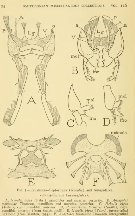
A single large mass of ventral fibers of the mandible (V), representing the combined ventral muscles of the leg coxa (A), form a particularly efficient ventral adductor. The intermandibular ligament, to which the adductor muscles of the jaw are attached, is not a structure restricted to crayfish.

NO. I JAWS OF MANDIBULATE ARTHROPODS — SNODGRASS 7 chitinous endoskeletal substance derived from the inner surface of
The jaw portion of the appendage is transverse and ends with a broad, dentate gnathal lobe (gnL). That the mandibles are appendages of the same segment in all arthropods below the mandible is generally beyond doubt.
NO. I JAWS OF MANDIBULATE ARTHROPODS SNODGRASS II (1935) has shown that the relation is very simple, and her explanation
The change merely involves a shift of the gnathal lobe from a position aligned with the mandibular axis (Fig. 1 C) to a position approximately perpendicular to the axis (G). The first condition occurs in the Chilopoda, the second. Fig. 1 H) is characteristic of the Diplopoda and Symphyla.
NO. I JAWS OF MANDIBULATE ARTHROPODS — SNODGRASS 1
CRUSTACEA
NO. I JAWS OF MANDIBULATE ARTHROPODS SNODGRASS 1
In the Ostracoda, the mandibles have a variety of forms, but are more generalized than those of the Arthropods by retaining a segmented palpus which is two-layered. By the activity of the maxillary epipodites, water currents are drawn through the chambers within the conch valves.
NO. I JAWS OF MANDIBULATE ARTHROPODS SNODGRASS I
From the base of every second jaw projects a large apodemal arm (D, Ap), but the relationship of the muscles to the apodeme has not been determined. In Lepasanscrifera, nephropores (E, npr) are not at the base of the lobes, but lie behind them.
NO. I JAWS OF MANDIBULATE ARTHROPODS — SNODGRASS 23
NO. I JAWS OF MANDIBULATE ARTHROPODS — SNODGRASS 25 appears to be a secondary outgrowth of the gnathal lobe rather than
I THE JAWS OF MANDIBULATE ARTHROPODS—SNODGRASS 25seems to be rather a secondary outgrowth of a bony lobe than. Each mandible is articulated dorsally (a) at the base of the inner lamella of the carapace fold and ventrally with a small process on its anterior margin (A, B, c) with a narrow postantennal wing of the epistome. The ventral muscles of both lower jaws (A, V) are united by a strong median ligament, and most of their fibers (iV) are adductors.
The anterior border of the base of the mandible, close to the epistomal articulation (c), is somewhat raised and bears a low, flange-like apodemal ridge (Ap).

NO. I JAWS OF MANDIBULATE ARTHROPODS SNODGRASS 35
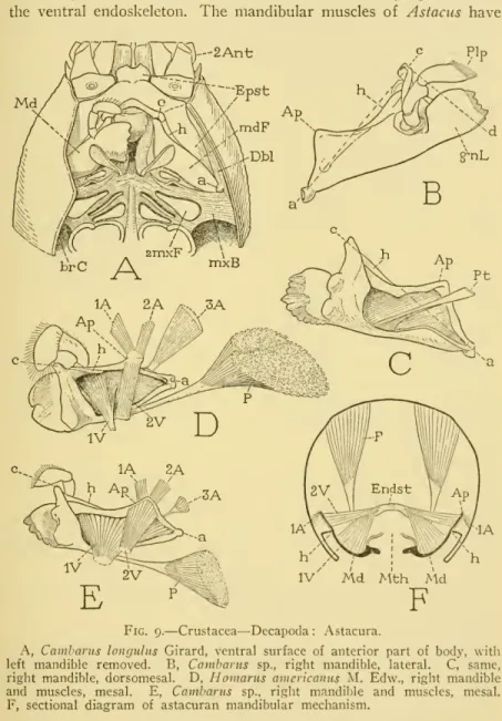
The mandibular muscle of astacurans presents the same functional fiber groups as seen in Macrobrachium, but in a reptantian. The third muscle of the anterior series (3A), inserted laterally from the apodeme, arises dorsally on the carapace and is preserved. A second and quite distinct group of ventral fibers (D, E, 2V) is attached to the inner side of the mandibular apodeme, forming a muscle opposite the lateral adductors (iA, 2A) attached to the outer side of the mandibular apodeme.
The mechanism of the astacuran jaws can be easily understood from the diagram, Figure 9F.
NO. I JAWS OF MANDIBULATE ARTHROPODS — SNODGRASS 39
Their structure and mechanism are so similar in anomuran and brachiuran decapods that the mandibles of both groups can be treated together. In Astacura, as already noted, the mandibles are articulated laterally on the inner walls of the carapace folds (fig. 9A, a), and behind them are the broad, horizontal pleural bridges {mxB) connecting the sternal region of the second segment maxilla with carapace. In Petrolistheseriomerus (fig. 12A, B) the sclerotic bridges (mxb) themselves do not reach the head and appear as small lateral extensions from the termi of the maxillary foramina that carry the mandibu-.
11 A, B; 16 D, H, t) which extend anteriorly along the folds on the sides of the mouth and support the mandibles mesally at the base of the gnathal lobe (Fig. 16H).
NO. I JAWS OF MANDIBULATE ARTHROPODS SNODGRASS 41
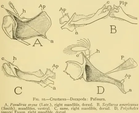
In Anomurah, the mandibles of Petrolisthes (Fig. 12A, B) and Pagurus (E) are similar to the mandibles of the palinuran Polycheles (Fig. The small, thin lobes on the opposite jaws are widely separated and apparently only palps can be functional organs. but the axis of the jaw can never become completely transverse due to the intervention of the first maxilla between the mouth and the articulation point of the mandible on the ventral skeleton (Fig. 15 H; 16 H, iMx).
Borradaile (1922) in Carcinus maenas and Cochran (1935) in Callinectes sapidus described the muscles of the mandibles of brachiura, but these writers overlooked a small group of fibers representing the ventral adductor of Astacure (Fig. 9D, E, iV).

NO. I JAWS OF MANDIBULATE ARTHROPODS — SNODGRASS 47
By the same process the third muscles of the anterior series (C, jA) are attached to the lower jaw (by a long tendon) below the base of the apodeme (A, B, 3At). The second, much larger group of ventral fibers {2V) is attached mesally on the upper margin of the mandibular apodeme. The "molar" surfaces of the mandibles were not observed to have any grinding effect on the food.
The mechanics of the crab mandible would indicate that the jaws are simply a pair of very efficient pincers, but, as in decapods generally, they have.
NO. I JAWS OF MANDIBULATE ARTHROPODS SNODGRASS 49 but now on a doubly articulated axis instead of on a single dorsal
I JAWS OF MANDIBUULATE ARTHROPODS SNODGRASS 49but now on a doubly articulated axis instead of on a single dorsal one.

NO. I JAWS OF MANDIBULATE ARTHROPODS SNODGRASS 5 1
CHILOPODA
The chilopod mandibles appear to initiate a new line in jaw evolution, as their structure is unmatched anywhere. In normal conditions, the anterior parts of the mandibles are largely hidden above the edge of the labrum, and are covered inferiorly by the broad palps of the first maxilla. The anterior articulation (fig. 18A, c) is by means of a knob or hook on the lateral surface of the jaw, some distance back from the anterior end, which is held loosely in the notch (F, g) between the epipharyngeal arm . (/) and the hypopharyngeal arm of the corresponding premandibular sternal sclerite of the head (Fit) supporting the hypopharynx.
The different positions of the two articular points relative to the axis of the chilopod mandible bring the hinge line of the.
NO. I JAWS OF MANDIBULATE ARTHROPODS — SNODGRASS 53
On the dorsal surface of the lower jaw (C), the sclerotized wall of the lobe is separated from the basal part of the jaw by an oblique line of flexibility proximal to the articular process (c). Mesally attached to the base of the lobe are two large muscles (C, E), one (/) arising at the base of the lower jaw, the other {iA) by widely spreading fibers on the dorsal wall of the skull. On the other hand, there is a striking similarity between the geophilid mandible and the mandible of pauropods (Fig. 19 A, Md).
The basal musculature of the mandible of Lithobius (Fig. 18C) includes a dorsal muscle {2A), perhaps a rotator, attached to the posterior end of the mandible, and three groups of ventral fibers (V).
NO. I JAWS OF MANDIBULATE ARTHROPODS — SNODGRASS 55 The fibers of the first ventral group (iV) arise on the head apodeme
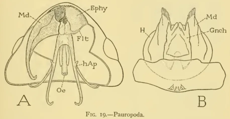
PAUROPODA
The head apodemes of the pauropods, as shown by Silvestri (1902) and by Tiegs (1947), are long, slender arms extending backward through the head into the former. What significance this fact may have for the taxonomic position of the Pauropoda, the writer leaves to the phylogenists. The two dorsal muscles that arise on the roof of the head are attached anteriorly and posteriorly to the base of the male.
The fourth muscle, the ventral protractor, originates from the hanging plate (fultura) of the apodeme and.
SYMPHYLA AND DIPLOPODA
NO. I JAWS OF MANDIBULATE ARTHROPODS — SNODGRASS 59
THE ENTOGNATHOUS APTERYGOTE HEXAPODS
The horizontal position of the mouthparts makes the primitive anterior surfaces dorsal, the posterior surfaces ventral. In Protura and Collembola each mandible is connected posteriorly to the cranial margin, as in Chilopoda, by a thin rod in the lateral wall of the gnathal pouch. All ventral fibers of the mandibles, as noted above, arise from the ligamentous bridge between the intermaxillary sternal brachia (D, Lg).
The "tentorial" fibers are differentiated into a broad transverse adductor (9) and into two protractors (5, 6), which extend from the posterior end of the mandible to the anterior branch of the jaw.

THYSANURA
The ephemeropterid mandible is more similar to the mandible of Machilis than that of the other pterygotes. The ligament runs through the base of the hypopharynx behind the roots of the anterior tentorial arms (A, AT). Thus, the ventral muscle mass of the machilid mandible is seen to be the same as that of Lithobius.
The anterior articulation (c) is not with the clypeus, as it is in the pterygote insects, but with a small condyle (F, c) on the ventrally bent anterior angle of the gena (Ge) behind the clypeus, just outside the invagination (at) of.
NO. I JAWS OF MANDIBULATE ARTHROPODS — SNODGRASS 69
PTERYGOTA
The jaws of mandibular arthropods – SNODGRASS 71 are found in the head and mouth parts of the larva. Sternfeld notes that the reduction in size of the mayfly's mouthparts begins in the nymph stage, but the digestive tract is just the opposite. If the insect is prognathic, the axis of the jaw becomes vertical instead of horizontal, but the relationship of the lower jaw to the head does not change.
In biting and chewing insects, a molar surface of the mandible is distinguished from a toothed incisor.

NO. I JAWS OF MANDIBULATE ARTHROPODS — SNODGRASS 73
The mandibular musculature of the cockroach and most other orthopteroid insects except the Acrididae is the same as that of the lepismatid mandibles (fig. 22E) in which each jaw has four separate muscles. Fig. 24B), although the corresponding muscles differ in relative size in the two groups. However, the main movers of the pterygoid mandible are the dorsal muscles (C), including a slender lateral abductor (A) and a large mesal adductor (P). The ventral muscles of the cockroach mandible (B, C, V) insert within the jaw cavity and consist of two distinct fiber groups, one is a fan-shaped muscle (iV) inserted laterally on the mandible, with convergent fibers attached. on a small arm (.r) of the hypopharyngeal suspensorium (HS), the other {2V) a short, thick muscle inserted behind the mandible and attached to the anterior arm of the tentorium (C, Tnt ).
Among the beetles, the mandibles of species that practice extra-oral digestion of the food have no molar surfaces; in the leaf-feeding Scarabaeidae, on the other hand, in which strongly toothed lobes of the maxillae serve as jaws for biting off the leaf surface (fig. 24 E), the mandibles became specialized as masticatory organs by a great development of the molar surfaces and ' n reduction of the incisor processes (F, G, K).
NO. I JAWS OF MANDIBULATE ARTHROPODS — SNODGRASS 75
In Homoptera, the enlarged base of each mandibular stylet (fig. 25 E), where it is attached to the wall of the bearing pouch, is produced into a thin retractor arm (ra) and a broad rapport arm (pa). Therefore, the identity of the reporting muscles of the mandibles depends on the homology of the loral plates. The writer (1938) suggested that the lora belong to the hypopharynx, since their lower ends are united with the hypopharyngeal floor of the suction pump.
Horizontal section of the cicada head through the lower part of the postclypeus (fig. 25B) shows that the proximal ends of the postclypeus (Pclp) and lora (Lor) are individually distinctly bent into the head.
NO. I JAWS OF MANDIBULATE ARTHROPODS — SNODGRASS 77
Likewise, the posterior edges of the loral plates are curved but become thin membranes (o) continuous medially with the ventral wall of the posterior lobe (p) of the hypopharynx. On raising or removing the anteclype (A, Aclp), it is seen that the lower edges of the loral plates (C, Lor) converge behind the anteclypeus and that their broad, sclerotized anterior lamellae are fused to the cytophoretrogen (Sit) of the hypopharynx. Working from the back of the head (D), removing the labium and cutting the lower parts of the maxillary lobes (MxL), the posterior temedian lobe of the hypopharynx (p).
In some Fulgoridae the upper ends of the loral plates appear to be united with the clypeus.
EXPLANATION OF LETTERING ON THE FIGURES ABBREVIATIONS
I JAWS OF MANDIBULATE ARTHROPODS - SNODGRASS 79 presented in terms of orthopteroid structure; they simply indicate expression in terms of orthopteroid structure; they simply show the extreme degree to which modifications of a basic anatomical complex can be carried out in the evolution of a new mechanism.
ALPHABETICAL LETTERING
The phylogenetic origin of the mandibles of insects and their arthropodan relatives.- A contribution to the study of the evolution of arthropoda. Preliminary account of the habits and structure of the Anaspididae, with remarks on another fresh-water crustacean from Tasmania.
NO. I JAWS OF MANDIBULATE ARTHROPODS SNODGRASS 85