Classical force fields have been developed for small organic molecules and later for macromolecules such as proteins, allowing molecular dynamics simulations of all atoms of protein folding events in explicit water solvent. This chapter introduces the essence and effectiveness of the theories and applications of classical force field simulations and quantum chemical calculations.
Quantum Chemical Computations
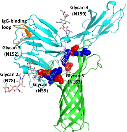
Despite the general requirement of FcγRIa association with the γ-chain for the internalization of the receptor, the interaction of OmpA+ E. This new structure is referred to as OmpA-FcγRIa for the rest of the manuscript.
Results and Discussion
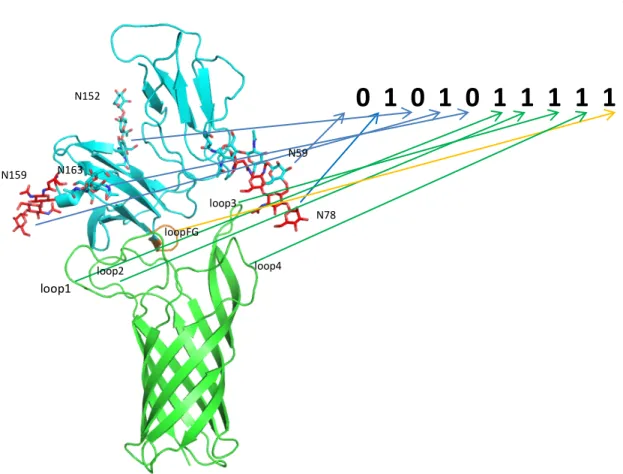
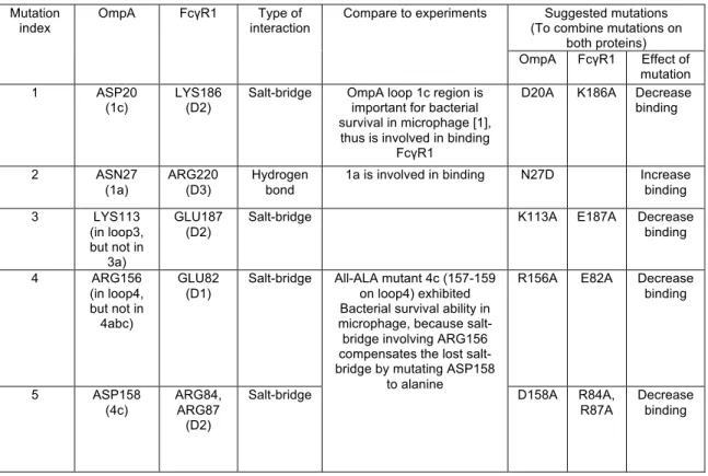
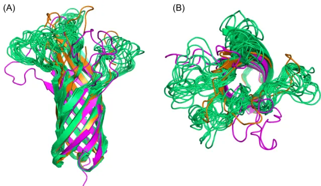
These structures were analyzed on a trial basis to yield 326 positions consistent with the experimental mutation studies (Table 2.1B). The transmembrane region of OmpA was then incorporated into the POPC membrane with the rest of the complex and solvated in water. Although NG4 is close to OmpA, a 50ns MD simulation of OmpA-FcγRIa did not result in any direct interaction between the NG4 glycan and OmpA (Figure 2.7).
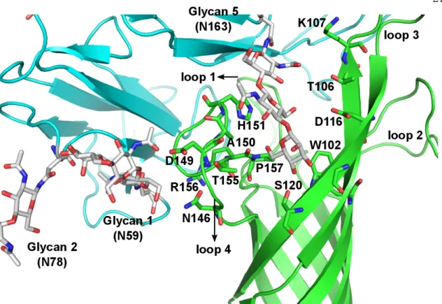
This lack of interaction of NG4 with OmpA was inconsistent with the experimental data since the absence of NG4 also prevented bacterial invasion into macrophages. This new conformation of NG4 does not conflict with any of the protein-protein interface peptides. 24 The NG5 binding site in the barrel between loop 3 and loop 4 of OmpA predicted here coincides with the barrel binding site of GlcNAc1-4GlcNAc epitopes (chitobiose) predicted in our previous work in which we bound the small ligand GlcNAc1- 4GlcNAc to) OmpA22.
In this predicted binding mode for the 3-sugar unit Man1-GlcNAc2 at the NG5 site, the two GlcNAc moieties maintain their interaction with OmpA as in the GlcNAcβ1-4GlcNAc binding mode (Figure 2.6). 4GlcNAcβ1-4GlcNAcβ1 was also included in the minimization, which did not change the interaction of GlcNAcβ1-4GlcNAc with OmpA. Collectively, the simulation studies support the experimental evidence that NG1, NG4, and NG5 interact with loops 1, 3, and 4 of OmpA for binding and provide a detailed 3D structure that can be used to better understand the nature of the protein. protein complex, which we expect will help us develop molecules that can block the formation of this complex and thus prevent neonatal meningitis.
Conclusions
It can also be used to formulate further experimental and computational tests to gain a deeper understanding of the interactions of OmpA with the peptide regions of FcγRIa beyond the glycosylation moieties. We computationally predicted the structures of the transmembrane domain of these two receptors and how different agonists, including steviol glycosides, bind to the binding sites. Taste transduction involves the interaction of taste molecules with cells expressing taste receptors residing in taste buds located in the papillae of.
For each of the two bitter receptors, the helix shapes were optimized within their respective template, all million rotations of each of the seven helices were sampled. We then docked several known agonists to the best conformation of the seven helix bundle for each receptor. The molecular structures of the ligands were constructed with Maestro software or constructed from existing ones.
For each of the best 100 ligand positions in the alanized protein, we then dealanized the mutated residues back to their original hydrophobic identity and optimized their positions along with those of other residues in the binding site using SCREAM66. This results in a unique set of optimized residue side chains for each of the 100 ligand positions. Then, for each of the ten ligand conformations, we selected the lowest binding energy structure, including strain and ligand solvation, that we expected to best represent the binding affinity.
Results and Discussion
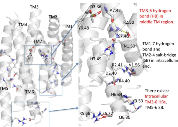
Binding of an agonist breaks the TM2-3 linkage and forms TM3-6 and TM2-6 linkage in the binding site of 2R14. Binding of rubusoside breaks the TM2-3 linkage and forms TM3-6 and TM2-6 linkage in the binding site of 2R14. Molecules in the red box except RebM and RebN are expected to be low-bite targets to be tested experimentally.
Molecules in the red box (excluding RebM and RebN) are predicted to be a low bitter target for experimental testing. The glycan on the –COOH side with more sugar groups is difficult to fit into the binding site and forms strong interactions with the two residues linking TM3-6 at the binding site. According to this rule, 2 molecules on the Priority 1 list and 1 molecule on the Priority 2 list with coordinate (3COOH, 4OH) are the best targets for low bitterness.
Rule #2 says that a large group A on the –COOH side would lead to less bitterness, as illustrated in Figure 3-11. Thus, the two molecules in Priority 2 list with coordinate (4COOH, 2OH) will be among the best targets. If the –OH sugar group (group B) is large, it will be held by the loop region, making it difficult for the –COOH sugar group to form stable interaction with the two residues linking TM3-6 at the binding site.
Conclusions
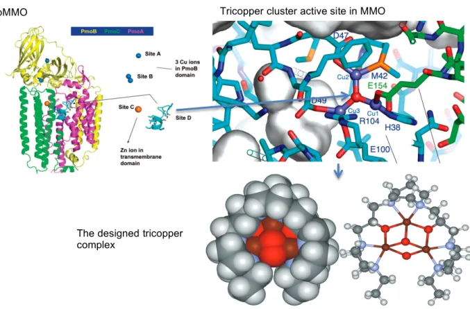
Our results reveal that the facile methane oxidation is mediated by transformations between different structural and spin states of the tricopper cluster. Oxidation of the C-H bonds in alkanes, especially methane oxidation to produce methanol, is still a challenging problem74. A ribbon diagram of the structure is shown in Figure 4.1, with different colors used to depict the three subunits.
Highlighted in the transmembrane domain of the crystal structure at site D is a pocket consisting of a hydrophilic cluster of potential metalligating residues, including His38, Met42, Met45, Asp47, Trp48, Asp49 and Glu100 of PmoA and Glu154 of PmoC. The metal ions must have been removed from site D during the purification of the protein for crystallographic analysis80. Cu(II)Cu(II)Cu(II) capped with “oxo” at position D of the crystal structure.
Highlighted in the transmembrane domain of the crystal structure at site D is a cavity consisting of a hydrophilic cluster of potential metal-ligating residues, including His38, Met42, Met45, Asp47, Trp48, Asp49 and Glu100 from PmoA and Glu154 from PmoC. The coordinated ligands and the geometry of the cluster, including the Cu−Cu and Cu−O distances, are all reasonable, demonstrating the feasibility of pMMO to accommodate a tricopper cluster. Fast atom bombardment mass spectrometry (FAB-MS) of the CuICuICuI-peptide complex Figure 2. A) Productive cycling and B) unsuccessful cycling in the oxidation of methane with O2, mediated by CuICuICuI(7-N-Etppz)1 + complex in the presence of H2O2 as the victim reduction agent.
Results and Discussions .1 The oxygen activation .1 The oxygen activation
Hydrogen peroxide activation
After the methanol product is dissociated from the complex in the previous step, the tricopper compound still has two remaining oxidizing powers. The complex then undergoes the first hydrogen atom transfer across a barrier of 4.0 kcal/mol (TS4). The intermediate from the first PC2 hydrogen transfer undergoes an isomerization where the oxygen in the –OH group breaks one of its bonds to the lower right copper and the hydrogen in the –OH group forms a hydrogen bond with the right-bound –OOH group. copper at the bottom.
After that, the second hydrogen could be transferred from the –OOH group to the –OH group through TS6 with a barrier of 2.4 kcal/mol. The products from the second hydrogen transfer, H2O and O2, can dissociate from the tricopper compound, gaining 30 kcal/mol entropy to compensate for the loss of -37.9 kcal/mol enthalpy in PC5. Values in are given in kcal/mol and are taken relative to state 3 with separate H2O and O2.
Discussion
In Chan's model, the active state is a Cu(II)Cu(II)Cu(III) complex which can activate the C–H bond through a direct insertion of singlet oxene into the C–H bond in a concerted manner.
Conclusions
The activity of Pt-based Karstedt's catalyst capable of olefin hydrosilylation was investigated by calculating free energy surfaces for reaction mechanisms consisting of elementary transformations with literature precedent. Since few data are available on the decomposition or oxidation products of unstable olefin hydrosilylation catalysts, we considered reactions of O2 with catalytic intermediates. This type of reaction is widely used to form Si-C bonds, with an important application in joining hydrosiloxanes with vinyl-terminated cross-linkers.
The catalysts that can catalyze these types of reactions usually involve late transition metals, especially Pt, Pd, Ni, Rh and Co. Chalk and Harrod99,100 proposed a mechanism for platinum-catalyzed hydrosilylation reactions based on simple elementary steps commonly observed in organometallics. chemistry, such as oxidative addition and reductive elimination (Figure 5.1, route a with green arrows). A variation of the Chalk-Harrod mechanism has also been proposed to explain the formation of vinyl silanes.101 This modified version assumes that an olefin attacks on.
Heterogeneous Inhibition of Homogeneous Reactions: Karstedt Catalyzed Hydrosilylation
Introduction
- Computation Details
- Results and Discussion
- Conclusions
Keywords in the Jaguar input file are enclosed in parentheses, follow the detailed method description for readers interested in reproducing the results. For the single point energies of the optimized geometries, a large basis set cc-pvtz(-f)88–91 (basis=cc-pvtz(-f)) is used. The reaction mechanisms of Karstedt's platinum catalyst are modeled using the two sets of simplified substrate side chains, as illustrated in Figure 5.2.
For the silane substrate, group 1 is trimethylsilane, while one of the three methyl groups is replaced by a dimethylsiloxy group. In general, substrates in group 2 are larger than substrates in group 1, representing the local environment of reaction centers in a polymer matrix. The difference in this free energy barrier, ΔΔG between the Markovnikov and anti-Markovnikov transition states, determines the selectivity of the two regioisomers.
The R state can be a sink state after it is formed due to the high barrier to exit through the S transition state of silyl migration to further pass through silane activation and hydride migration through the U transition state. The sensitivity to precatalyst oxygen state A and some resting states have also been studied, such as the examples shown in Fig. The differences of the free energy barriers between the Markovnikov and anti-Markovnikov transition states are calculated for the two substrate groups respectively to explain the regioselectivity change trend between the two substrate groups.