CONTEXT-DEPENDENT EFFECTS OF CCN2 ON β-CELL MASS EXPANSION AND INDICATORS OF CELL STRESS IN THE SETTING OF ACUTE AND CHRONIC
STRESS By
Shannon E. Townsend
Dissertation
Submitted to the Faculty of the Graduate School of Vanderbilt University
in partial fulfillment of the requirements for the degree of
DOCTOR OF PHILOSOPHY In
Molecular Physiology and Biophysics
December 16, 2023 Nashville, TN
Committee:
Richard O’Brien Andrea Page-McCaw
Wenbiao Chen Roger Colbran
ii
ACKNOWLEDGMENTS
The completion of this dissertation would not be possible without a lot of people. I am so grateful to everyone who provided support, guidance, patience, fun, optimism, encouragement, and humor (among other things!) during this whole process.
First and foremost, I must thank my mentor Dr. Maureen Gannon who gave me the opportunity to conduct my dissertation research in her lab. I feel so lucky to have had the opportunity to be mentored by amazing people during my time in academia, and Maureen is no exception. Thank you, Maureen, for providing endless support and
encouragement during both the somewhat easy and tough times. My project was quite a hot mess at times, but you always reminded me about the struggles you had with your project (changing thesis projects four times!), and that gave me the strength and
determination to continue on as we navigated all the unexpected circumstances with reagents, animals and puzzling results. There was never a time in lab where I felt unsupported, and I know that I am the scientist I am today in part due to the wonderful mentoring you provided me during my time at Vanderbilt.
I would also like to thank my thesis committee – my chair Dr. Richard O’Brien and the other members Dr. Andrea Page-McCaw, Dr. Wenbiao Chen, and Dr. Roger Colbran for the very helpful discussions, suggestions, encouragement and feedback.
Through your mentoring, I was able to think more critically about my work and focus more on the positive aspects. Thank you for offering space for me to talk about any concerns and for always being willing to help.
I am fortunate to be a part of a scientific community at Vanderbilt that fosters collaboration. This includes the Molecular Physiology and Biophysics department, the Beta Cell Interest group, and the Diabetes Research and Training Center. It would not have been possible to do my research without the support of multiple cores at
Vanderbilt: the Islet and Pancreas Analysis Core, Molecular and Cell Biology Research Core, the Shared Imaging Resource, and the Division of Animal Care. I also must thank members of other labs such as the Jacobson, Dean, Powers, Hasty and Stein labs that have provided reagents and protocols essential for my thesis work.
iii
I want to greatly thank the members of the Gannon lab that I have had the honor of working with for the past six years, both past and present. The lab has always been a constant source of laughter, fun and encouragement and I’ve felt so lucky to be a
member of such a wonderful lab. Bethany, Ray, Peter and Jen, thank you for being the first to welcome me into the Gannon lab, for patiently teaching me, and for answering all the questions I had starting out. Thank you especially to Jen for providing so much assistance in the past two years as I tried to power through all the experiments to complete this dissertation – you are a BOSS!! Thank you to Karin for being the best emotional support post-doc who always knew to check in when times were tough, and is always happy to answer even the simplest silly question with a smile. Thank you to Darian for being such a positive force in the lab, someone I can chat with in the morning about whatever is on our minds over coffee, and someone who I can always laugh with.
Thank you to Julie for being the best desk-adjacent mate and bringing brightness to our little alcove. Thank you to Chethan, Shilpak and Anthony who, although joined on the tail end of my time in lab, provided guidance, fun and lightheartedness to my lab experience. Finally, thank you to Xiaodong, Ashley and Eric who provided help and feedback throughout their time in lab. I also want to thank the rotation and summer students that have assisted me with my work such as Jade Stanley and Supriya Juneja.
Thank you for helping me achieve this milestone!
I wouldn’t have made it through grad school (and COVID-19) without my friends, both near and far. Thank you to Natalie, Nicole, Esha, Melanie and Kelli for quickly becoming my friends in the first year of IGP. We might have done a bad job keeping in touch sometimes, but even fleeting messages in the group chat provided
encouragement and positivity that kept me continuing on. We’re all in different places now, but I wish you all nothing but the best. I know you’ll be successful and do amazing things! To my friends who I met prior to and during COVID-19 and who have since now become a big second family to me – Koop, Kyle, Mikey, Jesus, Sabrina, Rose, Chris, Bre, Asha, Oct, Rusty and Amit – thank you for being you. Thank you for your
unwavering support, offering your space and time, and reminding me that I am capable of achieving something like this. I was only able to get through some days because I had you all in my corner. Thank you so much for being a part of my life.
iv
Of course, I must thank my family. I am extremely lucky to have two sets of extremely supportive parents. Grad school can be lonely sometimes, but I have never forgotten the love, support and encouragement that my parents have always provided me. Dad, Mom, Rodney, and Symone -- thank you offering your support in regard to whatever career path I decided to pursue. Thank you for working extremely hard so I could have this opportunity to pursue my goals. Thank you for instilling in me the work ethic that I have – it’s the reason I have persevered and made it to this point. Thank you for always being encouraging and for always believing in me and my abilities. Thank you for asking me about the timeline for completion in a way that did not instill great anxiety in me. Thank you for being four strong pillars behind me. I wouldn’t be here without you. To my siblings Alex, Ellie and Matthew – I know I’ve been in school for almost all of your lives, but I cherish the time we get to spend together when I visit home. Thank you for providing comedy and lightheartedness every time I visit home. To Grandma and Aunt Marlene, thank you for your love, support, and always providing a space for me to unwind. I hope I have made you all proud.
v
TABLE OF CONTENTS
Page
Acknowledgements………...ii
List of Figures………....ix
List of Tables………..xi
CHAPTER I. Introduction………..1
General significance……….1
Pancreas development………2
Endoderm specification and initiation of pancreas development…..2
Pancreatic bud formation, outgrowth and branching morphogenesis………..4
Endocrine cell development and differentiation – transcription factors……….7
Embryonic endocrine cell proliferation………13
Islet morphogenesis………...14
Postnatal β-cell mass expansion……….16
Factors that can induce adult β-cell proliferation………..19
Other secreted factors………...22
Diabetes Mellitus………24
Type 1 Diabetes……….….25
Type 2 Diabetes………..31
β-cell loss and dysfunction………....32
Endoplasmic reticulum stress – an overview……….37
ER stress and β cells……….40
Oxidative stress – an overview……….42
Oxidative stress and β cells………..46
Current treatments for Type 2 Diabetes ……….49
Cellular communication network factor 2………53
vi
The role of CCN2 in the embryonic pancreas………58
The role of CCN2 in the adult endocrine pancreas………...59
Thesis overview………...62
II. Materials and Methods……….65
Animals – generation of mouse models………..65
PCR and genotyping………..66
Intraperitoneal glucose tolerance tests (IP-GTT)………..66
High-fat diet studies………66
Islet isolations………..67
Ex vivo islet proliferation assays………..67
Ex vivo islet stress assays………68
Islet gene expression analysis……….68
Kinexus phospho-protein microarray………..69
Assessment of islet function by perifusion……….72
Immunolabeling………..72
β-cell mass………..73
β-cell proliferation………...74
β-cell size……….74
Assessing oxidative and endoplasmic reticulum stress………...74
Statistics………...75
III. Full-length recombinant human CCN2 induces β-cell proliferation ex vivo and stimulates activating phosphorylation events on RSK1 and PLCγ1………....76
Introduction………..76
Results………..78
Discussion………80
IV. Context-dependent effects of CCN2 on β-cell mass expansion and indicators of cell stress in the setting of acute and chronic stress………..84
Introduction………..84
vii
Results……….85
Thapsigargin efficiently induces expression of several ER and oxidative stress markers………...85
β-cell-specific CCN2 induction does not attenuate upregulation of ER stress markers, but induces expression of oxidative stress markers after TG treatment………..86
Discussion………...90
V. CCN2 induction does not attenuate chronic β-cell stress in vivo……….94
Introduction……….94
Results……….96
β-cell-specific induction of CCN2 does not alter cell stress markers in in vivo models of chronic β-cell stress………96
Discussion……….102
VI. Induction of CCN2 during prolonged HFD stimulates β-cell mass expansion………104
Introduction………104
Results………...105
One week of HFD with concurrent CCN2 induction does not alter β- cell proliferation or mass compared to controls………..105
Ten weeks of HFD and concurrent CCN2 significantly increases β- cell mass……….…..110
Discussion……….…112
VII. Induction of CCN2 in db/+ mice results in a significant decrease in β-cell mass……….117
Introduction……….……..117
Results………...117
CCN2 induction from six to eight weeks of age in db/+ mice leads to a significant reduction in β-cell mass………....117
viii
Discussion………..119
VIII. Summary and future directions……….125
Appendix I: Investigating the role of c-Met signaling in CCN2-induced β-cell proliferation………..139
Hepatocyte growth factor (HGF) and c-Met signaling………139
HGF/c-Met signaling and the endocrine pancreas……….140
Generation of mouse model with CCN2 overexpression in the setting of c-Met inactivation……….144
Appendix II: Investigating the role of β1 integrin in CCN2-induced β-cell proliferation………147
Methods……….151
Results and Discussion………...152
IX. References………..154
ix
List of Figures
Figure Page
1-1. Overview models of mouse pancreas organogenesis………..……….6
1-2. Factors that induce changes in β-cell mass…..……….17
1-3. Schematic of traditional and modern Eisenbarth T1D models………26
1-4. Comparison between etiology of Type 1 and Type 2 Diabetes………...28
1-5. Etiologies of Type 2 Diabetes………...34
1-6. Mechanistic timeline of Type 2 Diabetes onset and progression………36
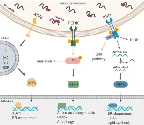
1-7. The three unfolded protein (UPR) pathways………..38
1-8. Nrf2 binding to AREs induces expression of various antioxidant proteins………44
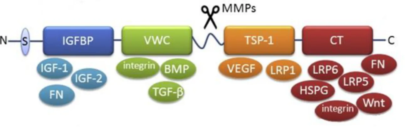
1-9. Structure of CCN2………..55
1-10. CCN2 has differential expression in embryonic and adult pancreata………57
1-11. CCN2 induces β-cell proliferation in settings of β-cell stress………..61
3-1. CCN2 stimulates β-cell proliferation when both domains are present…………...79
3-2. Kinexus phosphorylation network map comparing rhCCN2 versus control……..81
4-1. 10 nM thapsigargin treatment for 24h efficiently induces expression of several ER and oxidative stress markers without affecting expression of key β-cell genes...87
4-2. CCN2 induction during thapsigargin treatment attenuates upregulation of some ER stress genes……….88
4-3. CCN2 induction during thapsigargin treatment causes upregulation of antioxidant genes………89
5-1. CCN2 induction during HFD does not alter β-cell oxidative or ER stress……….97
5-2. CCN2 induction during HFD does not stimulate nuclear translocation of phospho- Nrf2………...98
5-3. db/+ mice exhibit more islet ER and oxidative stress than wild-type controls…100 5-4. CCN2 induction does not alter β-cell oxidative or ER stress in db/+ animals…101 6-1. The RIP-rtTA and TetO-CTGF transgenes do not alter pre-diet weight or weight gained after one week of HFD………106
6-2. CCN2 induction does not alter insulin secretion………..109
x
Figure Page
6-3. CCN2 induction during one week of HFD does not induce α- and β-cell
proliferation, or lead to an increase in β-cell mass after one week………...111 6-4. CCN2 induction during HFD for ten weeks does not alter weight gain on HFD or
glucose tolerance………..113 6-5. CCN2 induction during HFD for ten weeks significantly increases β-cell
mass………115 7-1. CCN2 induction in db/+ mice from four to six weeks of age does not alter glucose
tolerance or stimulate β-cell mass expansion………..118 7-2. CCN2 induction in db/+ mice from six to eight weeks of age does not alter
glucose tolerance but significantly reduces β-cell mass………120 7-3. There are no differences in cell death marker expression, β-cell size, or Pdx1
expression in db/+ mice with/without CCN2 induction from six to eight weeks of age……….122
xi
List of Tables
Table Page
1-1. Types of treatments for Type 2 Diabetes (T2D)……….………52 2-1. Genotyping and qRT-PCR primers utilized in studies………..70
1 CHAPTER I: INTRODUCTION
General significance
The pancreas is an organ with two primary functional roles. One role, governed by the acinar cells of the exocrine pancreas, is to secrete digestive enzymes that aid in food digestion. In humans, the exocrine pancreas comprises approximately 90 percent of the pancreas, while about six percent is comprised of the endocrine pancreas [1].
The endocrine pancreas consists of mini organs known as islets of Langerhans, which contain five distinct hormone-producing cell types: β (insulin), α (glucagon), δ
(somatostatin), ε (ghrelin), and PP (pancreatic polypeptide) cells. Insulin is the main hormone produced by the islet, and it functions to stimulate glucose uptake by insulin- sensitive tissues in the body such as the muscle and liver. This uptake of glucose by these tissues partly contributes to the maintenance of glucose homeostasis specifically by preventing hyperglycemia, which is a hallmark of diabetes mellitus.
Diabetes mellitus is a group of diseases characterized by insufficient insulin output due to either autoimmune destruction of β cells (Type 1 Diabetes), or loss and/or dysfunction of β cells (Type 2 Diabetes). Current treatments of diabetes focus on
managing glucose homeostasis. However, there is currently no cure for these diseases.
With loss and/or dysfunction of β cells being characteristic of diabetes, there is therefore therapeutic potential in increasing or restoring functional β-cell mass. However, islet transplantation is not a feasible cure due to a lack of donor tissue. Researchers are currently examining methods to increase β-cell number through in vivo expansion by identifying factors that can stimulate in vivo β-cell proliferation, with the priority being to characterize the signaling molecules activated by these factors. By understanding the
2
pathways activated to stimulate β-cell proliferation, manipulation of these pathways could further advance efforts to create a cure for diabetes.
Pancreas development
Pancreas organogenesis can be described as an ordered continuum of
molecular and morphological events that include regionalization of the endoderm and early ventral and dorsal bud formation, bud outgrowth and cell specification, endocrine lineage allocation, endocrine proliferation and maturation, and finally islet
morphogenesis. Each process will be described in more detail below.
Endoderm specification and initiation of pancreas development
The exocrine compartment consists of acinar cell clusters and the ductal network which functions to transport digestive enzymes into the rostral duodenum. The
endocrine compartment is composed of the islets of Langerhans, which are spherical cell clusters that produce hormones that work in synchrony to regulate glucose
homeostasis. Insulin secretion stimulates glucose uptake in glucose-sensitive tissues to reduce blood glucose levels, while glucagon secretion stimulates glycogenolysis to raise blood glucose in times of fasting. Acinar, ductal and endocrine cells are all derived from the endodermal germ layer during embryogenesis [2].
Mammalian development is unique in that cells with an endodermal identity are present at two distinct times during development. Primitive or so-called extra-embryonic endoderm arises in the pre-implantation embryo. Primitive endoderm predominantly gives rise to the endoderm layers of yolk sacs, which are crucial for nutrient transport from mother to embryo [3,4]. Definitive endoderm, or the embryonic endoderm, gives
3
rise to all digestive organs including the pancreas and liver [5]. The endodermal gut tube is compartmentalized based on expression of transcription factors and their arrangement along the anterior-posterior axis of the digestive tract. The posterior foregut region encompasses the anterior stomach, liver, and the prepancreatic endoderm. These regions aside from the liver are marked by expression of the
transcription factor pancreatic and duodenal homeobox 1 (Pdx1) [6]. Pdx1 is expressed throughout the endoderm of pancreatic buds and in the antral stomach, rostral
duodenum, and common bile duct [7]. Pdx1 is necessary for development of each of these organs. All cells within the pancreatic epithelium derive from Pdx1-positive cells.
Inactivating mutations in Pdx1 lead to pancreas agenesis in both mice and humans [8,9], while subtler mutations in Pdx1 can lead to a monogenic form of diabetes known as maturity onset diabetes of the young (MODY) [9,10-12]. Pdx1 heterozygosity in mice and humans leads to an increased risk for Type 2 diabetes [10,14]. The initial
presumptive pancreatic area is identified by overlapping expression of Pdx1 and
pancreas transcription factor 1a, or Ptf1a [6,15,16,17]. Ptf1a was originally identified as a regulator of exocrine-specific genes, but it has since been established to be very important for pancreas development as it is necessary for the development of multipotent pancreatic progenitors (MPCs) which are cells that can give rise to all
pancreatic cell types [18-20]. Similar to Pdx1, loss of Ptf1a leads to pancreatic agenesis [15]. Linear tracing analysis has shown that acinar, ductal and endocrine cells are all derived from a Ptf1a-expressing cell [15,21].
The vascular endothelium has been demonstrated to be important for the maintenance of Pdx1 expression, initiation of Ptf1a, dorsal pancreatic bud outgrowth
4
and insulin gene expression [22,23]. This was discovered by co-culture experiments utilizing the vascular endothelium and endoderm, which established that factors from the endothelium support pancreas development. One endothelium-derived factor known to play a role in pancreas development is vascular endothelial growth factor A (VEGF- A). Overexpression of VEGF-A under the control of the Pdx1 promoter leads to an increase in pancreatic blood vessels, as well as an increase in pancreatic islets and ectopic insulin-positive cells in the posterior stomach [24]. Because Pdx1 is expressed in the posterior stomach during this time in development, it is suggested that the endothelium can induce the β-cell fate in competent regions of the endoderm. In contrast, loss of VEGF signaling utilizing a mouse model with a null mutation in VEGF receptor type 2 (VEGFR-2) leads to failure of early insulin- and glucagon-positive cells to develop. Despite this, mice in this model express most pancreatic/endocrine
transcription factors except for Ptf1a, implicating VEGF-A signaling in the initiation of Ptf1a [23].
Pancreatic bud formation, outgrowth, and branching morphogenesis Ventral and dorsal pancreatic bud outgrowth occurs in the initial stages of
pancreatic development. The dorsal pancreatic bud forms at mouse embryonic day 9.5 (e9.5) and human gestational day 26 (hd26), while the ventral bud forms at e10 and hd30 (however, humans have two ventral buds) [25,26]. The period following bud evagination (e8.5-e12.5 in mouse; hd42-49 in human) from the endoderm is known as the primary transition period, and the first known hormone-positive cells scattered among the MPCs can be detected at this time [27]. Early MPCs are marked by
expression of Pdx1, Ptf1a, SRY-box transcription factor 9 (Sox9) and Onecut1 (Oc1).
5
Early MPCs have the potential to differentiate into acinar, ductal or endocrine cells [28].
Oc1 (formerly known as hepatic nuclear factor 6 or Hnf6) is a transcription factor that directly activates the pro-endocrine transcription factor Neurogenin-3 (Ngn3) [29], which will be discussed later in this chapter. Thus, Oc1 is required for endocrine differentiation but is also needed for embryonic pancreatic duct development and later branching morphogenesis [30]. Inactivation of Oc1 leads to a delay in Pdx1 expression, pancreatic hypoplasia, and impaired endocrine and duct cell differentiation [31]. Similarly,
inactivation of Sox9 in the developing pancreas leads to pancreatic hypoplasia and premature depletion of the MPC pool [32].
At ~e12, after growth and elongation of the dorsal and ventral pancreatic buds occurs, the two pancreatic buds fuse. At this time, the pancreas undergoes
morphological and gene expression changes to establish a tip and trunk domain structure. Molecular marker analyses and lineage tracing studies suggest that the tip and trunk domains are both molecularly and functionally different. The tip of the
branching pancreatic epithelium is where most of the outgrowth and elongation occurs.
It is the most outward projecting domain and is composed of secondary MPCs that express Ptf1a, c-Myc, carboxypeptidase A (Cpa1), and Pdx1 and Oc1 at lower levels.
The secondary MPCs in this domain have the capability of giving rise to all three pancreatic cell types (endocrine, duct, and acinar) until e14.5 [28]. However, the
majority of these secondary MPCs in the tip domain differentiate into acinar cells [Figure 1-1]. In the trunk domain, differentiation of endocrine progenitor cells is regulated by juxtacrine Notch-Delta signaling [33,34]. Initially, endocrine/ductal bipotent cells in the trunk express low levels of the Notch receptor and its ligand Delta. The cells with
6
Figure 1-1. Overview models of mouse pancreas organogenesis. A: The upper diagrams depict the branching morphogenesis that occurs during pancreas development, as well as the tip-trunk segregation depicting organization of pancreatic cell lineage. B: The bottom diagram depicts the important transcriptional regulators that are expressed during each stage of pancreas development. Adapted from Pan and Wright (2011). Pancreas organogenesis: From bud to plexus to gland. Dev Dyn 240(3):530-565.
7
increased Delta expression give rise to the endocrine population and leave the
epithelium, while cells with increased Notch signaling remain in the epithelium. The cells that leave the epithelium are endocrine progenitor cells, give rise to all five endocrine cell types, and express Ngn3+ [35,36]. Cells remaining in the epithelium repress expression of Ngn3, and eventually mature into ductal epithelial cells [37].
Endocrine cell development and differentiation – transcription factors
The earliest known marker of an endocrine progenitor is Ngn3, and all endocrine cell types arise from an Ngn3-expressing progenitor cell [35,36]. Bipotential trunk cells with low levels of Ngn3 expression diverge toward the ductal cell fate. In mice, complete inactivation of Ngn3 during development leads to a lack of all endocrine cell types, resulting in neonatal death due to diabetes [35]. These findings support the fact that Ngn3 is vital for the differentiation of all pancreatic hormone-producing endocrine cells.
Interestingly, transgenic Ngn3 overexpression throughout the pancreatic epithelium results in increased differentiation of α cells without a change to the number of other islet endocrine cell types [38,39]. When investigating this phenomenon, it was found that the time of activation of Ngn3 is an important factor in determining endocrine cell fate. For example, early activation of Ngn3 yields α cells, while later activation induces differentiation of β, δ, and PP cells. It has been further demonstrated that a single Ngn3+ progenitor cell is unipotent and can only give rise to one specific endocrine cell type rather than to all [40].
The secondary transition is the time period between e12.5 and e16, and during this time there is a significant increase in the number of endocrine cells budding from the ductal epithelium. The endocrine cells formed during this developmental window
8
eventually contribute to the mature islet. Ngn3 expression peaks during the secondary transition [35]. Because Ngn3 expression is induced by cooperation between Pdx1 and Oc1, it is crucial that proper levels of Oc1 expression are achieved. Loss of Oc1 during development in mice leads to a dramatic decrease in Ngn3 expression, subsequently leading to a significant reduction in insulin and glucagon expression [29]. After cells are committed to the endocrine fate by activation of Ngn3, Oc1 expression is decreased.
Bipotent duct/endocrine progenitor cells with high expression of Oc1 are fated to become ductal cells and Oc1 expression remains in these cells throughout adulthood [41]. If Oc1 expression is experimentally maintained in islets, there is impairment of migration of endocrine cells from the ductal epithelium, disrupted organization of endocrine cell types within the islet, and severely compromised β-cell maturation and function which subsequently leads to overt diabetes [42,43].
The formation of α and β cells is thought to be regulated by the opposing actions of the transcription factors Paired Box 4 (Pax4) and Aristaless-related homeobox (Arx).
Pax4 can be detected by mRNA within the pancreas from e9.5 [44]. It is specifically expressed in first and second wave insulin-producing cells but becomes downregulated soon after birth and is expressed at low levels in adult β cells [45]. Embryonic loss of Pax4 does not result in loss of first wave insulin-producing cells; rather, there is loss of mature β cells, indicating that Pax4 is required during the secondary transition for β-cell differentiation [44,46]. Furthermore, Pax4 null embryos have an increase in the numbers of glucagon-expressing cells [47]. Pax4-expressing cells can give rise to α, β and ε cells, suggesting that Pax4 is expressed in pluripotent endocrine progenitors [48].
9
However, ectopic expression of Pax4 in α cells or pancreatic progenitor cells induces re- specification toward the β-cell fate [49].
While Pax4 acts to direct endocrine progenitors toward the β-cell fate, Arx expression directs endocrine progenitors toward the α-cell fate. Arx is first expressed between e10.5 and e12.5 in scattered cells throughout the pancreatic buds and is later co-expressed with glucagon at e14.5 [49]. Arx acts downstream of Ngn3, and
embryonic loss of Arx results in complete loss of second wave α cells with a
concomitant increase in both β and δ cells [49,50]. Inactivation of both Pax4 and Arx leads to a loss of both β- and α-cell lineages and a concomitant increase in δ cells [50].
Paired box 6 or Pax6 is another transcription factor important for the α-cell lineage.
Pax6 is expressed at e9.5-10.5 in a population of cells in the pancreatic epithelium.
Later in embryogenesis, Pax6 is expressed in cells committed to the endocrine lineage [33,51,52]. Although Pax6 is expressed in both glucagon- and insulin-expressing cells, it is thought to be only essential for α-cell formation. Inactivation of Pax6 causes a
dramatic loss of glucagon-producing cells along with a smaller decrease in other endocrine cell types [53]. This finding suggests that Pax6 is important for the allocation of endocrine progenitor cells to the α-cell lineage, and for the expansion of the
endocrine population as a whole [54,55].
The large Musculoaponeurotic fibrosarcoma (Maf) family of proteins are
transcription factors that were first identified in an avian retrovirus. Two members of the Maf family, MafA and MafB, are crucial for β-cell differentiation and maturation. The expression pattern of both transcription factors differs between human and mouse.
MafA was identified by several groups as an activator of insulin gene expression [56-
10
60]. Expression of MafA begins at e13.5 and continues into adulthood, making it a marker of mature murine β cells [61,62]. In humans, similar to mice, MAFA expression increases in an age-dependent manner and high expression is maintained in β cells throughout adulthood [63]. Although indicated as a critical β-cell maturation factor, global deletion of MafA in mice has no effect on the number of insulin-producing cells generated during embryogenesis. However, loss of MafA in mice leads to defects in β- cell gene expression and postnatal β-cell function, ultimately leading to diabetes [64].
Importantly, reduced MafA/MAFA expression is associated with diabetes progression in both mice and humans. Islets isolated from db/db mice, a model of T2D, have reduced MafA levels and “rescue” of the transcription factor improves GSIS and β-cell mass [65- 68]. Single cell RNA-seq analysis (scRNA-seq) of human β cells demonstrated that β cells that were metabolically inflexible expressed MAFA at a lower level than healthy β cells, solidifying the importance of MAFA in proper β-cell function [69].
The lack of a developmental islet phenotype in MafA knockout animals may be due to compensation by another related Maf family member, MafB [62,70]. MafB is also expressed in developing endocrine cells and is capable of activating insulin reporter gene transcription in tissue culture. During mouse embryogenesis, MafB is expressed in first- and second-wave insulin- and glucagon-producing cells but becomes restricted to α cells soon after birth. In β cells, MafB promotes activation of differentiation genes including Nkx6.1, Glut2, Pdx1, and MafA [62]. Loss of MafB results in a decrease of insulin- and glucagon-positive cells [64]. However, in adult mouse islets, MafB is only expressed in α cells and regulates expression of the glucagon gene [70]. The
11
expression of MAFB differs in humans from mice. In adult humans, in addition to being expressed in α cells, MAFB is co-expressed in β cells with MAFA [70-73].
Other transcription factors involved in specifying the pancreatic endocrine lineage are the members of the NKX class of homeodomain proteins Nkx2.2 and Nkx6.1. IN mice, both Nkx2.2 and Nkx6.1 are expressed in most pancreatic epithelial cells during early embryogenesis. By e15.5, however, Nkx2.2 is restricted to the endocrine cell population and Nkx6.1 is expressed exclusively in insulin-producing cells and other scattered cells in the ductal epithelium [74-76]. Late in gestation, Nkx2.2 can be detected in nearly all hormone-positive cells except for somatostatin-expressing cells.
After birth, both genes are exclusively expressed in the β-cell population. The primary function of Nkx2.2 is as a transcriptional repressor which regulates endocrine
differentiation [77]. Loss of Nkx2.2 leads to loss of insulin-positive cells at any stage examined, with a significant reduction in glucagon-expressing cells and a more modest reduction in pancreatic polypeptide (PP)-positive cells [74]. This occurs without a loss of total endocrine cells due to increased numbers of ghrelin-producing ε cells [47]. These findings demonstrate that Nkx2.2 is required to generate β cells, maintain and expand α- and PP cells, and repress ε-cell fate.
Loss of Nkx6.1 results in dramatic loss of second-wave insulin-positive cells (after e13.5), with no change in the number of other islet endocrine cell types [75].
Therefore, in the absence of Nkx6.1, endocrine cells delegated to be β cells do not adopt an alternate endocrine cell fate. Genetic studies have demonstrated that Nkx6.1 functions downstream of Nkx2.2 to expand and terminally differentiate the β-cell lineage [74]. Transgenic overexpression of Nkx6.1 in β cells using the Pdx1 promoter does not
12
enhance β-cell mass or function [78]. Conversely, loss of both Nkx2.2 and Nkx6.1 results in a decrease of both α- and β cells, indicating a role for both transcription factors in endocrine cell development [79,80].
In addition to its role in pancreas organogenesis, Pdx1 plays a crucial role in later β-cell differentiation and mature function. By late gestation, Pdx1 expression is
maintained at high levels in β cells, with minimal expression in acinar cells [81,82].
While Pdx1 is not required to generate the endocrine cells present during the primary transition, it is specifically required for differentiation of endocrine and exocrine cells between e11.5 and e13.5 [83]. Studies conducted using mice with β-cell-specific Pdx1 inactivation demonstrated that loss of Pdx1 at e11.5 resulted in very early onset diabetes with plasma glucose being significantly higher in Pdx1-mutant pups than control pups at P1 [84]. Examination of β-cell proliferation at e18.5 found that β-cell proliferation was significantly impaired in Pdx1-mutant animals compared to controls, which translated into a decreased number of overall β cells. Interestingly, α-cell proliferation was significantly increased in Pdx1-mutant mice. Analysis of glucose tolerance via IP-GTT in adult mice revealed that male Pdx1-mutant mice had significantly impaired glucose tolerance compared to controls indicating β-cell
dysfunction [84]. This study demonstrated that Pdx1 is required during late gestation for sufficient β-cell mass expansion to occur and for proper β-cell function throughout life.
Other studies have revealed that Pdx1 promotes expression of proinsulin, Glut2 and glucokinase in adult mice which mediates proper glucose-stimulated insulin
secretion. The role of Pdx1 in maintaining expression of these genes is critical, as loss of Pdx1 during adulthood utilizing tamoxifen-inducible Cre recombinase resulted in loss
13
of the key β-cell identity transcription factors Nkx6.1, Ins1, Glut2 and MafA.
Furthermore, loss of Pdx1 in adult β cells results in acquisition of α-cell-like features, demonstrating the importance of Pdx1 in maintaining adult β-cell identity [85].
Embryonic endocrine cell proliferation
There is little proliferation of endocrine cells during early to mid-gestation. However, the percentage of proliferating endocrine cells dramatically increases in late gestation through the early neonatal period [86-89]. For example, during late embryogenesis, β- cell proliferation proceeds at approximately 10% per day in mice [90]. Adequate proliferation is required for sufficient numbers of endocrine cells in adults, but few factors in vivo have been identified that affect embryonic endocrine proliferation.
The Protein kinase R-like endoplasmic reticulum kinase (PERK) is required for both exocrine and endocrine proliferation and function. PERK localizes to the
endoplasmic reticulum (ER) membrane and is hyperactivated in response to ER stress caused by the unfolded protein response (UPR) [91]. Global Eif2ak3 (PERK)
inactivation in mice results in insufficient β-cell mass leading to diabetes, and a progressive loss of exocrine tissue after four weeks of age. Inactivation of Eif2ak3 during embryogenesis leads to decreased β-cell proliferation, leading to decreased β- cell mass in the adult. Gene expression profiling of postnatal day 2 (P2) control and PERK knockout islets revealed that this decreased β-cell proliferation may be due to decreased expression of genes important for progression of the G2 and M phases of the cell cycle such as CyclinA and cyclin dependent kinase 1 (Cdk1) [92]. PERK may also play a role in regulating β-cell maturation or function, as both global and pancreas- wide inactivation of PERK leads to decreased MafA, Pdx-1, and insulin gene
14
expression. Furthermore, inactivation of PERK causes impaired glucose-stimulated insulin secretion (GSIS) [93,94].
β cells may also provide signals that regulate the population of other cell types, specifically the α cell. Removal of Pdx1 in embryonic β cells leads to a significant decrease in β-cell proliferation with a concomitant increase in α-cell proliferation at late gestation [84]. Reciprocal alteration in β- and α-cell proliferation when Pdx1 is
inactivated suggests that embryonic β cells normally provide an inhibitory signal to the α-cell population. Glucagon-positive cells may also send signals to regulate β-cell proliferation as models of glucagon inactivation, such as embryonic global deletion of the glucagon receptor or pro-hormone convertase-2 (PC2), result in increased postnatal β-cell proliferation [95,96].
Islet morphogenesis
At e18.5 and continuing after birth, endocrine cells organize into islets within the acinar parenchyma [27]. Mouse islets have a characteristic architecture with β cells in the core of the islet, with the other cell types such as α and δ cells forming a mantle around the islet. This organization is thought to promote proper cell-cell communication and enhance islet function [97,98]. Although the process of islet morphogenesis is not completely understood, changes in expression of cell adhesion molecules, modifications in extracellular matrix (ECM) proteins, and paracrine and juxtacrine cell-cell
communication events likely play a role in this process. Real-time imaging in pancreatic explants has demonstrated that β cells migrate using extended cytoplasmic filapodia [99], suggesting that migration is an active process. However, it is still not clear whether islets are formed by individual cells which lose connections with neighboring cells,
15
migrate to form clusters, and then reestablish cell adhesions to form islets. One feasible alternative to this process would be groups of cells that migrate in clusters while still maintaining cell-cell contact.
Cell migration requires dynamic regulation of cell-cell, cell-ECM adhesion, and intracellular signaling that regulates the actin cytoskeleton. Islets in mice lacking the epidermal growth factor receptor (EGFR) are elongated and closely opposed to the ductal epithelium [100]. This suggests that EGFR may act to modulate activity of Rac1, a Rho-GTPase involved in migration and adhesion in many cell types. Islets expressing dominant negative Rac1 fail to spread on ECM when treated with EGFR ligands,
indicating that Rac1 may function downstream of EGFR [101]. Integrins,
transmembrane heterodimer receptors that interact with the ECM to affect migration, also play a role in endocrine cell migration. Inhibition of αv integrins in fetal pancreas explants block the emergence of endocrine cells from the ductal epithelium [102].
Furthermore, EGFR and integrin signaling both regulate the activity of matrix
metalloproteinases (MMPs), molecules that can alter the structure of the ECM, which can thus affect the migration of cells [103].
Cell-cell contacts are required for the formation of the typical islet architecture.
One molecule, E-cadherin, is necessary for endocrine cell clustering. β cells that express a dominant-negative form of E-cadherin remain dispersed throughout the pancreas as individual cells [104]. Other adhesion molecules, such as neural cell adhesion molecule (N-CAM), are involved in endocrine cell type segregation [105]. It has been hypothesized that segregation of endocrine cell types, such as α and β cells, is due to differential expression of adhesion molecules [105,106]. This can be observed
16
in the MIN6 and INS-1 immortalized β cell lines, where β cell-only clusters are substantially more cohesive than clusters of the α-TC α cell line. [106].
Postnatal β-cell mass expansion
After birth, β-cell mass increases rapidly and eventually plateaus when an
organism reaches adulthood. There are multiple mechanisms by which β-cell mass can increase postnatally, with the most common being proliferation and hypertrophy [107]
[Figure 1-2]. Proliferation involves an increase in β cells due to replication of existing β cells and has been demonstrated to be the main mechanism by which postnatal β-cell mass increases [108]. Hypertrophy is characterized by an increase in the size of existing β cells. In rats, β-cell proliferation proceeds at a rate of 1-4% per day between the age of 30 to 100 days old [109]. In mature adult mice, β-cell proliferation proceeds at a rate of <1% per day demonstrating the dynamic property of β-cell mass expansion [110]. While these previously described studies were conducted in rodents, it has also been demonstrated that β-cell mass dynamics are similar in humans. For example, human β-cell proliferation peaks around 4% per day during the early neonatal period and decreases to ~0.2% per day in adults under normal physiological conditions [111].
For many years, it was believed that β cells were terminally differentiated cells existing in a G0 state, which is a state in which a terminally differentiated cell is dormant. However, it is now known that adult β-cell proliferation does occur, albeit at low rates, and can be induced as a response to physiological and pathophysiological conditions as well as a multitude of other factors.
Cell proliferation in general is highly regulated through the interaction of a diverse
17
Figure 1-2. Factors that induce changes in β-cell mass. β-cell mass can expand via replication or proliferation, neogenesis, and hypertrophy. β-cell mass can be lost by apoptosis and necroptosis (not shown) and dedifferentiation.
Adapted from Jung et al. (2014). Diabetes Metab J 38(6):426-436.
+Necroptosis
18
set of proteins. Briefly, the cell cycle is a four-stage process that consists of Gap 1 (G1), synthesis (S), Gap 2 (G2) and mitosis (M). Active eukaryotic cells will pass through these different phases during proliferation. During G1, the cell increases in size and duplicates its contents. During S phase, DNA is replicated. During G2, the cell grows and prepares for mitosis. Finally, mitosis occurs, and the result can be two identical daughter cells [112]. In some cases, asymmetric division occurs whereby a stem or progenitor cell generates a daughter cell which has different characteristics [113]. The cell cycle is highly controlled by checkpoints at different stages, which regulate which cells will enter and complete the cell cycle [114]. The main restriction point is located at G1, and cells that pass this point will end up undergoing and completing the cell cycle.
There are many regulators that function at different stages of the cell cycle, with the most important being cyclins and cyclin-dependent kinases (Cdks) [115]. Cyclins are a group of related proteins whose levels fluctuate during the cell cycle, and expression is regulated at the level of protein degradation. Four basic types of cyclins are found during the cell cycle: G1 cyclins, G1/S cyclins, S cyclins and M cyclins [116]. Cyclins form complexes with Cdk serine-threonine kinases, which phosphorylate their
substrates to regulate cell cycle progression [115,116]. The consensus in mammalian cells is that Cdk4 and Cdk6, after transcriptional induction of D-type cyclins in response to mitogenic stimuli, promote entry into the cell cycle [115].
Like most cells, the major restriction checkpoint for both murine and human β cells is the G1 checkpoint [117,118]. Although sharing the same restriction checkpoint, the expression of different cell cycle regulators differs slightly between murine and human β cells. In murine islets, cell cycle progression is controlled by three D cyclins
19
(D1, D2, D3) which bind and activate Cdk4 to permit cell cycle entry. Global deletion of Cdk4 in mice causes abnormalities in β cells including cell hypoplasia, which resulted in diabetes and ketoacidosis [119,120]. Cyclins D1 and D2 are the most highly expressed of the three, and although cyclin D2 is not required for neonatal development, it still plays a role in controlling β-cell growth and replication [121]. Cyclin D1 can compensate for loss of cyclin D2, but loss of both cyclins D1 and D2 leads to uncontrollable diabetes and eventual death in young mice [121]. Conversely, overexpression of cyclin D1
causes increased β-cell proliferation both in vitro and in vivo [122]. It is known that β-cell proliferation declines with age, and the expression levels of cyclin D1 and D2 decline in the same manner, providing evidence for the importance of these cyclins in murine β- cell proliferation. Human β cells express both Cdk4 (at very low levels) and Cdk6 as well as cyclins D1 and D3. While the mouse expresses cyclin D2 in β cells, cyclin D2 is not observed in human β cells [123]. Adenoviral overexpression of Cdk4 in human β cells increases proliferation rate and like mice, adenoviral overexpression of cyclin D1 in human β cells increases β-cell proliferation [123,124].
Factors that can induce adult β-cell proliferation Glucose
Glucose is one of the most potent mitogens for β cells both in vivo and in vitro [125-127]. Studies have shown that both long- and short-term glucose infusion promotes β-cell proliferation in mice. For example, one study in mice utilized a 50%
glucose infusion for 96 hours to maintain elevated blood glucose and found that there was an almost five-fold increase in β-cell proliferation compared to saline infusion [127].
In ex vivo studies, it was demonstrated that culturing human islets in high glucose for 96
20
hours increased β-cell proliferation [128]. Another study utilized streptozotocin (STZ) to deplete β cells in the non-obese diabetic/severe combined immunodeficiency
(NOD/SCID) mouse model, leading to hyperglycemia. Human islets transplanted in this hyperglycemic environment had increased proliferation, again illustrating that glucose acts as a mitogen for β cells [129].
Extracellular matrix proteins
The vascular endothelium, while being important for secreting factors that stimulate pancreas development during embryogenesis, is also involved in β-cell proliferation postnatally. The intra-islet endothelial cells (ECs) are responsible for
depositing the components of the islet vascular basement membrane such as collagen, laminin, fibronectin. Intra-islet ECs also secrete many molecules that affect β-cell proliferation and survival [130]. For example, ECs secrete hepatocyte growth factor (HGF) and cellular communication network 2 (CCN2; formerly known as CTGF) that each enhance β-cell proliferation and survival [131-133]. Intra-islet ECs can also affect β-cell proliferation and survival by modulating the availability of growth factors in the ECM. They secrete matrix metalloproteinases (MMPs) that alter ECM integrity and release sequestered growth factors that affect β-cell proliferation and survival such as fibroblast growth factor (FGF) and VEGF [134].
Collagen, laminin, and fibronectin are the three most abundant and well-studied proteins in the ECM [134]. In mammals, collagen makes up a major portion of the basement membrane and provides structural stiffness and cohesiveness to tissues [135,136]. A study using the INS-1 line, a rat-derived immortalized β-cell line,
demonstrated that cell proliferation was significantly increased after being plated on
21
type IV collagen compared to control [137]. A similar trend is seen when cells are cultured on laminin. Studies using immortalized cell lines and primary islets from both rodents and humans have demonstrated that culturing cells on laminin increases β-cell proliferation and decreases β-cell death [138-142]. Finally, studies have shown that culturing MIN6 cells, a mouse-derived immortalized β-cell line, on fibronectin induces DNA synthesis indicative of cells entering the cell cycle. Furthermore, fibronectin
contains multiple protein-binding domains where growth factors such as FGF and VEGF can bind, suggesting that fibronectin also plays a critical role in sequestering and
regulating accessibility of critical β-cell mitogens [143,144].
Integrins, transmembrane heterodimer receptors for ECM components, are expressed on virtually all cell types. Integrins are composed of one α and β subunit. In the islet, the specific α and β subunits expressed on the different cell types differs among species [145]. Integrins can act synergistically with growth factor receptors including VEGFR, c-Met and EGFR, all of which have been implicated in β-cell
proliferation. One specific integrin subunit, the β1 subunit, is the most studied in general and regarding β-cell proliferation. The β1 subunit is the most promiscuous subunit in that it is capable of binding multiple α subunits, and loss of β1 integrin is embryonically lethal [146]. β1 integrin is highly expressed on β cells and promotes β-cell proliferation through binding of multiple ECM components including laminin, collagen and
fibronectin. Both ex vivo and in vivo studies have demonstrated that β1 integrin inactivation affects β-cell proliferation. For example, blockade of β1 integrin using neutralizing antibodies attenuates the beneficial effects of ECM components on β-cell proliferation and survival [137,139]. In vivo β-cell-specific inactivation of β1 integrin in
22
mice during embryogenesis resulted in significantly decreased β-cell area in adulthood due to decreased embryonic and postnatal β-cell proliferation [147]. Furthermore, tamoxifen-inducible β-cell-specific inactivation of β1 integrin at four weeks of age in mice led to reduced β-cell mass due in part to a decrease in β-proliferation when assessed eight weeks later [148].
Other secreted factors
Fibroblast growth factors (FGFs) have been mentioned previously in the context of pancreas development, but FGFs also play a role in postnatal β-cell proliferation [150]. Many FGF ligands are expressed in adult mouse β cells including FGF1, FGF2, FGF4, FGF5, FGF7 and FGF10. Furthermore, FGF receptor 1 (FGFR1) and FGF receptor 2 (FGFR2) are also expressed in adult mouse β cells [149]. One study sought to examine the roles of FGF1 and FGF2 in β-cell proliferation. In this study, two mouse models were generated with a β-cell-specific inactivation of either FGF1R or FGF2R during embryogenesis. It was revealed that decreased FGF2R signaling did not have detrimental effects on postnatal β-cell mass expansion. However, while FGF1R- deficient mice had the same number of β cells compared to control at birth, loss of FGF1R resulted in a gradual ~25% decrease in the number of β cells by postnatal day (P)-27, demonstrating a role for FGF signaling in postnatal β-cell expansion [149].
VEGF-A is the prototypical member of the VEGF family and has been
demonstrated to be important for β-cell proliferation during embryogenesis. VEGF-A can also induce β-cell proliferation postnatally into adulthood. Loss of VEGF-A during
embryogenesis causes decreased β-cell proliferation and overall reduced β-cell mass when assessed at later in life [150]. In some cases, overexpression of VEGF-A leads to
23
β-cell proliferation. For example, overexpression of VEGF-A utilizing the Pdx1 promoter led to islet hyperplasia when assessed in two-month-old mice [24]. Furthermore,
another study demonstrated that VEGF-A overexpression in adult β cells for two weeks promoted β-cell proliferation [150].
HGF, or hepatocyte growth factor, was previously mentioned as a factor made and secreted by intra-islet ECs. HGF signaling has been implicated in β-cell proliferation and regeneration in many mouse models. β-cell-specific overexpression of HGF in mice resulted in increased β-cell proliferation [152]. Furthermore, HGF plays a role in
maternal β-cell mass expansion. Loss of c-Met, the HGF receptor, in pregnant dams resulted in decreased β-cell proliferation, reduced β-cell mass and an increase in β-cell apoptosis. This ultimately led to gestational diabetes in this model [153]. HGF/c-Met signaling also plays a role in β-cell regeneration. Following partial pancreatectomy, there is an upregulation of c-Met and exogenous intraperitoneal injection of HGF further increases β-cell proliferation. Conversely, inactivation of c-Met in the setting of partial pancreatectomy impaired β-cell proliferation and regeneration [154].
Another factor secreted by intra-islet ECs is platelet derived growth factor (Pdgf).
Pdgf binds to its tyrosine kinase receptors (PDGFα αnd PDGFβ) to control the growth of connective tissue cells including smooth muscle cells and fibroblasts [155,156]. PDGF signaling also promotes proliferation, survival and migration in a multitude of cell types [157]. Classic studies have elucidated that PDGF signaling in cultured islets stimulated DNA synthesis [158,159]. More recent studies have demonstrated an age-dependent expression pattern of Pdgf, with expression of both the receptor and ligand being markedly reduced in murine β cells at 6 weeks and 6 months of age compared to
24
neonatal β cells [160]. β-cell-specific loss of Pdgf during embryogenesis led to a three- fold reduction in β-cell proliferation and a 50% reduction in β-cell mass when assessed at two to three weeks of age in mice [160]. Conversely, β-cell-specific Pdgf
overexpression resulted in significantly increased β-cell proliferation in 14-month-old mice compared to controls, demonstrating that Pdgf signaling activation can sustain adult β-cell expansion in vivo [160].
Pdgf signaling is also implicated in β-cell regeneration – loss of Pdgf in the
setting of β-cell loss by STZ treatment resulted in a failure of β-cell mass restoration due to impairment in β-cell proliferation. Studies conducted using juvenile and adult human islets discovered the same age-dependent expression of PDGF that was observed in mice where PDGF and PDGF receptor expression drastically decreases as age increases. Furthermore, exogenous PDGF treatment was able to induce β-cell proliferation only in juvenile human islets which contrasts with what was observed in mouse studies [160]. However, differences in proliferative capacity and responsiveness to mitogens exist between murine and human β cells, and the disparity between the β- cell response to PDGF may simply be a species difference.
Diabetes Mellitus
Diabetes mellitus is a very complex cluster of diseases and was described as long ago as the ancient Greeks. Multiple forms of diabetes exist, with the two most common being Type 1 diabetes (T1D) and Type 2 diabetes (T2D). T1D is characterized by insufficient insulin output due to the autoimmune destruction of β cells and accounts for 5-10% of diabetes cases. T2D accounts for 90-95% of diabetes cases [161] and is characterized by loss or dysfunction of β-cell mass in the setting of insulin resistance,
25
leading to insufficient insulin output and prolonged hyperglycemia. Another form of diabetes, maturity-onset diabetes of the young (MODY), is caused by genetic autosomal dominant inheritance of mutations in at least 14 identified genes. MODY accounts for 1- 5% of diabetes cases [162,163]. Diabetes is a global health burden with an estimation of 537 million adults (20-79 years) being diagnosed with the disease [164]. As of 2022, in the United States alone there were 37.3 million people estimated to have diabetes, with 8.5 million of those people being undiagnosed [161]. By 2060, the number of adult Americans with diabetes is expected to nearly triple, with the percent prevalence nearly doubling [165]. Diabetes was the seventh leading cause of death in 2019 in the United States, although the number of deaths attributed to diabetes may be underreported [166]. Complications of the disease include blindness, limb amputation, cardiovascular disease, neuropathy, and kidney failure [167]. Thus, it is crucial for effective
therapeutics to be developed while investigation of a cure is ongoing.
Type 1 Diabetes
T1D has been long considered a chronic immune condition characterized by
autoimmune destruction of β cells, but as more information comes to light about the disease, it is now considered a disorder that results from complex interactions between genetic and environmental factors, microbiome, metabolism and the immune system.
While T1D has historically been considered a childhood-onset disease, it is now known that up to 50% of T1D cases are diagnosed in adulthood [168].
In 1984, George Eisenbarth developed a model that illustrates the progression of of T1D relating genetic and environmental factors to progressive loss of β-cell mass and
26
Figure 1-3. Schematic of the traditional and modern models of the
original Eisenbarth of T1D model created in 1984. As more information has come to light about the disease, parameters of the disease and how it
progresses has become more understood. Adapted from Atkinson M. A., and Eisenbarth G. S. (2001). Type 1 diabetes: new perspectives on disease pathogenesis and treatment. The Lancet 358, 221–229.
27
eventual T1D onset [169] [Figure 1-3]. In this since updated model, those with a genetic predisposition to the disease undergo a precipitating event that leads to immunological dysfunction and the eventual loss of β cells due to autoimmune attack. This precipitating event may be different for different people. Overt diabetes is observed when a
significant portion of functional β-cell mass is lost, leading to insufficient insulin output and uncontrolled hyperglycemia [170]. Τhe current standard of care for T1D is
exogenous insulin replacement therapy via multiple daily injections of insulin, or by use of insulin pumps [171]. The use of continuous glucose monitors with insulin pumps, which is referred to in the literature as a closed-loop system, allows for only sufficient amounts of insulin to be injected to maintain euglycemia. The specificity in amount of insulin injected at a given time reduces the hypoglycemic episodes experienced by people with T1D [172]. Precise maintenance of glycemic control still bears a challenge due to the varying pharmacokinetics and absorption rates of currently used insulin analogues. Thus, other therapeutics are being developed to allow for more accurate glycemic control in T1D patients. Some of the therapeutics currently being developed involve immune system modulation, use of stem cell-derived β-like cells, and islet transplantation.
As mentioned previously, T1D is a heritable polygenic disease with risk for the disease varying from 30-70% in identical twins, 6-7% in siblings, and 1-9% for children with a parent with the disease [173,174]. As of 2019, genome-wide association studies (GWAS) have identified more than 50 loci that contribute to the genetic susceptibility of the disease [175]. However, the strongest association with T1D involves variations in classic histocompatibility leukocyte antigens (HLA) class II molecules, which are
28
Figure 1-4. Comparison between the etiology of Type 1 and Type 2 Diabetes. In Type 1 Diabetes, autoimmune attack results in a significant loss of functional β-cell mass which subsequently causes overt hyperglycemia and the onset of T1D due to insufficient insulin output. In Type 2 Diabetes
progression, in the presence of insulin resistance, functional β-cell mass increases. However, eventually β-cell dysfunction and loss occur resulting in insufficient insulin output, hyperglycemia, and overt diabetes. Compared to T1D, hyperglycemia in T2D may be present at a higher functional β-cell mass due to the increased presence of insulin resistance in T2D patients. Adapted from Lorenzo et al. (2021). Int J Mol Sci 22(8), 4239.
29
cell-surface molecules that bind and present antigen to T cell receptors. HLA genes are known to be the most polymorphic in the human genome – classical HLA genes can have thousands of sequences [176]. Class I molecules have also been implicated in the autoimmune destruction of β cells, as it has been demonstrated that these
molecules present antigens to CD8+ T cells, which in turn cause β-cell death by the initiation of cytotoxic T cell killing [177]. Animal models have illustrated the role of class I molecules in T1D. In the NOD mouse model, a mouse model for T1D, loss of
expression of MHC class I molecules renders NOD mice resistant to autoimmune diabetes [178].
Outside of genetic susceptibility, another risk factor for the disease in at-risk populations is the presence of islet autoantibodies. The presence of islet autoantibodies is also considered a marker of the disease. As of 2019, there were over 30 identified autoantigens for T1D [179]. There are four major autoantigens associated with T1D – insulin, GAD65, IA-2 and ZnT8 [180]. GAD65, the 65-kD isoform of glutamic acid decarboxylase (GAD), is expressed in neuroendocrine cells and it has been
demonstrated that higher levels of GAD65 autoantibodies are associated with earlier onset of T1D in genetically susceptible individuals [181]. A study evaluating the diagnostic value of examining Anti-GAD65 autoantibodies found that these
autoantibodies were present in 80% of newly diagnosed children with T1D [182]. IA-2, or islet tyrosine phosphatase-like protein, is a neuroendocrine molecule that is localized to the insulin secretory granules of β cells [183]. ZnT8, or zinc transporter 8, is
necessary for zinc flux into β-cell insulin granules [184,185]. Zinc is co-secreted with insulin and plays both autocrine and paracrine roles in the islet [186]. In genetically
30
susceptible patients, as the number of autoantibodies present increases, so does the risk of developing the disease.
While T1D is thought of as a disease due to malfunction of the immune system, several studies have demonstrated that the β cell plays a role in its own demise. β cells can secrete chemokines, a group of small secreted molecules, which contributes to immune infiltration of the islet. Some examples of chemokines secreted by β cells are CCL2, CCL5, and CXCL10 [187]. All of these chemokines are induced by inflammation which is heavily present in the setting of T1D. CCL2 is involved with monocyte, natural killer (NK) cell, and T-cell recruitment during inflammation [188-190]. CCL2 may be responsible for the influx in macrophages in the islet as studies have demonstrated that increased monocyte recruitment, insulitis and islet destruction occurs during transgenic overexpression of Ccl2 in murine β cells [189]. CCL5 is a chemoattractant for
monocytes, eosinophils and T cells, and blockade of one of the cognate receptors for CCL5 inhibits future immune infiltration and prevents development of diabetes [191].
Finally, CXCL10 mediates chemotaxis in lymphocytes. CXCL10 binds the CXC receptor 4 (CXCR3), and it has been demonstrated that inactivation of CXCR3 delays insulitis in a T1D mouse model. Conversely, overexpression of CXCL10 in mouse islets
accelerated diabetes in a T1D mouse model. [192-194]. CXCL10 is directly toxic to β cells due to signaling through Toll-like receptor-4 (TLR) which leads to pro-apoptotic signaling within the cells [195,196].
There is still much to learn about T1D, and the NOD mouse model has been utilized in efforts to identify therapeutics and a potential cure for T1D [180]. The
autoimmune diabetes phenotype in this mouse model is similar to the course of disease
31
in humans. One strength of the model is that autoimmunity is spontaneous, and mice develop autoantibodies, many of which are present in human T1D, along with circulating autoreactive T cells prior to the onset of disease. One difference between the model and human disease, however, is that the initial autoantigen in the NOD model is insulin while in humans it is thought that disease results from multiple initial autoantigens.
However, progressive loss of β-cell function and mass resembles that of the human disease, and the degree of insulitis increases as disease progresses, although this is not readily observed in humans with long-standing disease [197]. Another difference between the NOD model and human T1D is that overt diabetes occurs primarily in female NOD mice while T1D does not skew towards one sex in humans. Similar to other mouse models of human disease, there has been a failure to translate some basic science findings discovered in the NOD mouse model to humans. Moving forward, along with increasing understanding about T1D in the human population, more understanding needs to be gained about the NOD model in order to bridge the gap between basic science findings and the potential for these findings to be applied in the clinic.
Type 2 Diabetes
Unlike T1D, T2D is not caused by autoimmune attack of the β cells. However, in both of these diseases, loss of functional β-cell mass is key to progression of disease [Figure 1-4]. While T2D does have a stronger link to family history and lineage than T1D, development of the disease depends largely on environmental factors [198]. T2D is primarily linked to insulin resistance and prolonged hyperinsulinemia, with both being associated with poor dietary choices and a sedentary lifestyle. Obesity is heavily linked
32
to T2D as well, with almost 80-90% of T2D patients being overweight or obese [199]. It has been demonstrated that β-cell mass is significantly lower in pancreata from human cadaveric donors who had T2D compared to body mass index (BMI)-matched donors who did not have T2D [200,201]. Furthermore, islets from donors with T2D are smaller and have less than half the islet equivalents of non-diabetic donors [202].
There are multiple mechanisms by which functional β-cell mass can be
insufficient to meet metabolic demand in the setting of T2D [Figure 1-5]. There is great heterogeneity in the human population, and as such people are born with varying
amounts of β-cell mass. Someone born with a low β-cell mass may undergo β-cell mass compensation in the face of metabolic demand, but their increased functional β-cell mass may still be insufficient to meet the increased need for insulin. A lack of functional β-cell mass could also arise if β-cell mass expansion simply fails in the face of
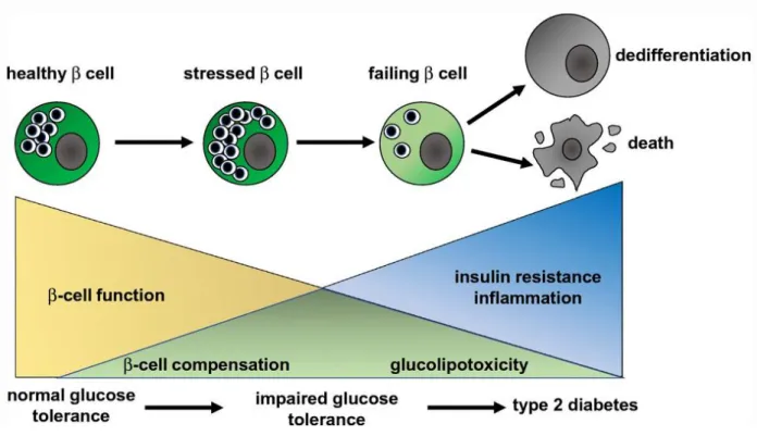
increased metabolic demand. Finally, in the presence of increased metabolic demand, β-cell mass expansion may successfully happen. However, due to the prolonged hyperinsulinemia and the increase of β-cell stress due to increased demand for insulin, β cells may fail, die or lose their identity, all of which can result in loss of functional β- cell mass. In all of these situations, prolonged hyperinsulinemia and insulin resistance would occur, leading to development of T2D [Figure 1-4].
β-cell loss and dysfunction
β-cell loss occurs primarily through dedifferentiation and cell death. Dedifferentiation, also referred to as loss of β-cell identity, is the process by which β cells lose their β-cell- defining transcription factors and occurs due to prolonged exposure to hyperglycemic
33
conditions [203]. Transcription factors expressed by mature adult β cells include Pdx1, Pax6, Nkx6.1, Nkx2.2, and MafA [203-206]. β cells from human T2D donors and T2D mouse models show loss of some of these transcription factors, especially those involved in mature β-cell function such as Pdx1 and MafA [207-209]. Loss of MafA, the last β-cell identity transcription factor to be expressed during β-cell maturation, causes impairments in glucose-stimulated insulin secretion leading to eventual hyperglycemia [210]. Furthermore, in the early stages of human T2D and in the T2D-like db/db mouse model, a model of leptin insufficiency, loss of nuclear MafA is one of the earliest
changes observed in β cells [211]. Another phenomenon that occurs during
dedifferentiation is the induction of so-called disallowed genes, which are genes that are normally actively repressed or not expressed highly in β cells. Increased expression of these disallowed genes is associated with decreased β-cell function. Currently, there have been 60 β-cell disallowed genes identified, and many of them are expressed in immature β cells [212-214]. Therefore, during dedifferentiation, both loss of key identity transcription factors and induction of disallowed genes leads to β-cell dysfunction and thus insufficient insulin output. The processes that induce dedifferentiation are still being elucidated; however, at this point it is known that chronic hyperglycemia and
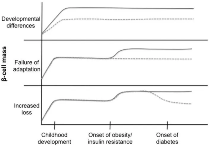
hyperlipidemia can lead to β-cell dedifferentiation and β-cell mass loss [Figure 1-6].
β-cell death can be induced by different mechanisms such as apoptosis and necroptosis, two forms of programmed cell death. Apoptosis results in clearance of cells with minimal damage to the surrounding tissues. However, necroptosis is a type of programmed cell death that combines features of apoptosis and necrosis [215]. Recent studies utilizing a zebrafish model of overnutrition demonstrated that islet inflammation
34
Figure 1-5. Etiologies of Type 2 Diabetes. Diabetes is a disease characterized by loss/dysfunction of β-cell mass, eventually leading to insufficient insulin output and hype