Defining a Role for Prostanoid Receptor EP4 in the Developmental Programming of the Ductus Arteriosus
By
Michael Thomas Yarboro
Dissertation
Submitted to the Faculty of the Graduate School of Vanderbilt University
In partial fulfillment of the requirements for the degree of
DOCTOR OF PHILOSOPHY In
Cell and Developmental Biology December 16th, 2023 Nashville, Tennessee
Approved:
Jeff Reese M.D.
Andrea Page-McCaw Ph.D.
Ken Lau Ph.D.
Michelle Southard-Smith Ph.D.
Ambra Pozzi Ph.D.
ii
© 2023 by Michael T. Yarboro All Rights Reserved
iii Dedication
For a mother who always comforted me when I fell.
For a father who taught me to never stop getting back up.
Finally, for those who lost their struggles to congenital heart disease.
If I could give you my heart I would,
but I suppose my labor will have to do.
iv
Acknowledgments
I thought this would be easier. Not easy, but easier. I was wrong. But learning to not take being wrong personally is perhaps the most practical lesson science can teach us. That and the value of teamwork. I couldn’t have accomplished any of this alone. There are too many people who have touched this project and my growth through it to be fully listed here, but I certainly plan to try.
There are two people who have had the greatest steering impact on my progression as a scientist and my personal growth in the last few years. Jeff Reese, and I have spent so much time together working through experiments, data, conferences, career opportunities. He has devoted so much time to my growth as a scientist and as an individual. I will never be able to thank him enough. I have endured horrible health issues during my training, and he has always been understanding and compassionate beyond what was required. He has stuck up for me when I needed it, but also recognized the moments I needed to stick up for myself. He has been the best mentor I could have imagined. I absolutely could not have made it through this without him, and I hope we continue to collaborate as I move further into the world of vascular biology. The second person is Naoko Boatwright. Naoko is the engine that keeps our lab and several others running. She is an absolute expert at her craft and has mastered so many techniques I don’t think I could list them. But more importantly, she is always attentive and understanding to other members of our group. If someone needs help with an experiment, or coverage when they are ill, Naoko is there without hesitation. She is one of the most impressive people I have ever known, and humble enough that sentence will make her squirm.
She deserves half of this doctorate. Jeff and Naoko welcomed me into their lab like a family and have treated me as such ever since. I cannot thank them enough.
v
With the aforementioned health issues, constant complications with my project, and my own brand of stubborn self-reliance, I have been a difficult student. My committee has been incredibly patient with me throughout it all, especially Andrea, my chair. I have not always followed instructions, and you could have made my life so much harder for it. Thank you for recognizing I was always working towards my goals, even if I wasn’t vocal about it. The shear amount of expertise on my committee is hard to fathom, and still, everyone has always been willing to help me when I needed it. Andrea, Michele, Ambra, Ken; thank you all so much for seeing me through this. You have shaped me into the scientist I am today.
He may not know this, but the person who convinced me to come to Vanderbilt was Chris Wright. The program in developmental biology that Chris leads is the most amazing program I’ve seen at Vanderbilt, with involvement and training opportunities rivaling most departments. The program in developmental biology training grant supported me for two years, but once you become one of Chris’s trainees, he aggressively supports you forever. The PDB journal club is absolutely what allowed me to work through my anxiety about presenting science, and the discussion at the journal club has shaped the critical eye with which I view everything. Thankyou Chris, for always seeing potential in me, even in the moments I didn’t see it in myself.
My project was also supported by an American Heart Association predoctoral fellowship. The mission of the AHA has a lot of personal meaning to me for reasons that will become clear later. It means the world to me for this particular organization to see potential in me as an investigator.
While I may be a student in the Department of Cell and Developmental Biology which deserve their own thanks, my daily interactions have mostly been within the division of neonatology. I have been supported, trained, and had the opportunity to train a slew of rising
vi
faculty, technicians, and undergrads. At the center of this are a few people who really helped me through my project. Mark Hunt is an amazing coordinator for the division. He always has the answers you need and is always willing to lend a hand. Courtney Berger and I made an excellent team, wonderful friends, and I’m so glad to have had the opportunity to work with her.
Deanna Sekulich is a recent addition to the lab, but has picked up technical skills at an amazing pace. I’m glad to have worked with her and am certain she will make an amazing scientist if that is what she chooses to do (no pressure). Elaine Shelton taught me many of the techniques I use on a daily basis and has been an absolutely amazing sounding board for experimental ideas and writing over the years. She has genuinely made me stronger as a scientist.
I have been blessed with an amazing family, especially two amazing parents. I was a challenging kid, but they met those challenges with a tempered hand and well-reasoned word.
I’m the son of a draftsman and a master machinist. My parents taught me early that you could make anything you wanted from nothing with the right application of thought and effort. That you could be whatever you wanted as long as you were willing to take the good with the bad.
They encouraged me at an early age to explore anything that interested me; or rather, they supported me because they knew good and well they couldn’t stop me. To this day, they are always here when I need them and provide absolutely unwavering support. They are good, humble people, and they taught me to be the same. I love them with all I am and am proud to be their son. When I was little, there was no way they could have known this is what I would choose to do with my life. I’m sure they had full expectations I would live a few houses down and be around for Sunday supper. That’s just not the case. Leaving home, and them, is still the hardest thing I’ve ever done. But I’m carrying what they taught me wherever I go, and I wouldn’t be there without them.
vii
I also had amazing grandparents. I spent a lot of my childhood standing across an operating table from my grandfather the veterinarian. He instilled in me a love of biology and a sense of stewardship that guides me. I still use what I learned from him daily in my work. My grandmother was the strongest person I’ve ever known, and it is the resilience I learned from her that has carried me through this. When I was young, my malmal walked me through the woods pointing out all the plants and their uses to heal the sick. She captivated my interest in what nature could show us if we were willing to learn. She also always had my back, even when she probably shouldn’t have. I never knew my palpal, but I feel him every time I play his fiddle, and it has comforted me through many long nights of study and writing.
Throughout this tumultuous process I have been blessed with a phenomenal friend group. They have always been caring and understanding of my circumstances and have offered help without ever expecting something in return. Natalya has always leant an ear, and usually taken my side. When I needed a place to live, she said yes without hesitation. Kevin has always offered a hand and a smile. Megan and I were inseparable through most of grad school and always found the best concerts. Ashley provides a spark of life and levity to everything no matter the circumstances and she has always been able to eek a smile out of me on the longest days. Whitney is always there, without question, no matter the hour, and she knows I’m always there too. My friends from back home have also kept me sane. Ruffin has always been willing to fight harder for me than I am for myself, and I’m not exactly a pushover. Erik consistently brings the creative spark of a fresh perspective to every conversation and is who I go to when I need to think outside the box. Kyle keeps me in check, always providing a dose of critical thought and global perspective. When I need to make sure my head is in the right place; that I’m headed the right direction, these three are my compass. I am so proud of all of you. Thank you for sticking with me.
viii
Four donors provided human tissue for transcriptional analysis as a part of this project.
They deserve our thanks, our respect, and our remembrance.
It is no mistake I found myself in a lab studying congenital heart defects. I was born with several severe congenital heart defects myself. I went through surgery as a toddler, and medical observation until I was 17. During my procedure, I was legally dead for 70 minutes. I came very close to not being here and have already surpassed my pre-surgery life expectancy. It was hard, painful, embarrassing, and uncomfortable, but I was lucky. Others weren’t. Many of the kids I remember playing with in the waiting rooms of my clinics didn’t make it. I still remember their faces. More than anyone or anything else, they are the reason I am here. I feel a deep connection with them and want to do anything in my power to try and prevent others from meeting the same fate or enduring the same struggle. It’s the thought of them that kept me writing on long nights. It’s the thought of them that keeps me moving forward. This is for you. I wish it was more.
Finally, I’d like to acknowledge the publishers that have allowed reprint of published work for the purposes of this dissertation.
ix
Table of Contents
Dedication ... iii
Acknowledgements ... iv
List of Tables ... xii
List of Figures ... xiii
Abbreviations ... xv
Chapter 1 ... 1
INTRODUCTION ... 1
Abstract ... 1
Role of DA in transition to neonatal life/embryology ... 3
Clinical relevance/human epidemiological data ... 6
Contributors to PDA ... 8
Clinical options to treat PDA ... 10
Genetic Landscape of the DA ... 12
Preparation of the DA for Closure and Remodeling ... 13
Mechanisms of Postnatal DA Closure ... 17
Vascular smooth muscle cells in DA function ... 19
Prostaglandin E2 and the DA ... 21
PGE Receptors and the DA ... 27
Nitric Oxide Signaling in DA Vasodilation ... 36
Oxygen Sensing in DA Vasoconstriction ... 38
Summary ... 42
Aims ... 43
Chapter 2 ... 46
TRANSCRIPTIONAL PROFILING REVEALS PUTATIVE TARGETS OF A DEVELOPMENTAL PROGRAM IN THE DUCTUS ARTERIOSUS ... 46
Abstract ... 46
x
Introduction ... 47
Results ... 48
Discussion ... 54
Supplemental Tables ... 65
Chapter 3 ... 72
MOUSE MODELS AND HUMAN SINGLE GENE SYNDROMES ASSOCIATED WITH PATENT DUCTUS ARTERIOSUS (PDA) FURTHER SUPPORT A DEVELOPMENTAL PROGRAM IN THE DUCTUS ARTERIOSUS ... 72
Abstract ... 72
Introduction ... 73
Mouse Models of PDA ... 75
Mouse Models of Premature DA Closure ... 91
Pharmacological Models in Mice ... 92
PDA in Human Genetic Syndromes ... 93
Discussion ... 97
Supplemental Tables ... 105
Chapter 4 ... 121
PGE2 SIGNALING THROUGH EP4 MEDIATES AN UNEXPECTED CHRONIC ROLE IN DA DEVELOPMENT ESSENTIAL FOR ESTABLISHING THE CONTRACTILE PROPERTIES AND REMODELING POTENTIAL REQUIRED FOR DA CLOSURE AFTER BIRTH ... 121
Abstract ... 121
Introduction ... 122
Results ... 124
Discussion ... 145
Conclusion ... 152
Supplemental Tables ... 153
Chapter 5 ... 154
FINAL DISCUSSION, CONCLUSIONS, AND FUTURE DIRECTIONS ... 154
Introduction ... 154
xi
The Transcriptional Landscape of the DA Suggests a Developmental Program ... 156
PGE2-EP4 Plays an Essential Role in Permanent Closure and Remodeling of the DA ... 158
The Oxygen Sensing Mechanisms of the DA Develop Independent of EP4 ... 160
PGE2-EP4 is Likely Essential for Establishing the Mature Contractile VSMCs of the DA ... 162
The Mechanism Through Which PGE2-EP4 Directs DA Development Remains Unclear ... 164
References ... 169
Appendix ... 197
MATERIALS AND METHODS ... 197
xii
List of Tables
Table ... Page
1-1 Factors associated with PDA ... 6
2-1 Summary of Included and Excluded Studies using Microarray to Compare DA and Aorta ... 56
2-2 Summary of Genome and Transcriptome Reads and Alignment ... 57
S2-1 Genes Enriched in the Rodent DA by Three or More Microarrays (Fold Change (FC); DA vs Ao) ... 60
S2-2 Genes Enriched in the Rodent Ao by Three or More Microarrays (Fold Change (FC); DA vs Ao) ... 62
S2-3 Summary of Transcript Reads ... 65
S2-4 Genes Enriched in the Human DA by RNA-seq (Fold Change; DA vs Ao) ... 66
S2-5 Genes Enriched in the Human Ao by RNA-seq (Fold Change; DA vs Ao) ... 68
S2-6 Differentially Expressed Genes Most Highly Expressed in the Human DA by RNA-seq (Fold Change; DA vs Ao) ... 69
S2-7 GO, KEGG, and UP Keywords Common in Human RNA-seq and Rodent Microarrays 70 S3-1 Genetic Models of Patent Ductus Arteriosus (PDA) or in utero DA Closure in the Mouse (n=28) ... 105
S3-2 Human Single-Gene Syndromes Associated with PDA (n=224) ... 108
S3-3 Chromosomal Deletions, Duplications, and Additions Associated with PDA in the Human (N=15) ... 117
S3-4 Mouse Model Genes Associated with Single-Gene PDA Syndromes in Humans (n=10 Genes) ... 118
S3-5 GO, KEGG, and UP Keywords Common Between Human PDA Syndromes and Mouse Models of PDA ... 119
xiii
List of Figures
Figure ... Page
1-1 DA closure in the mouse ... 2
1-2 EP4-mediated late gestational remodeling prepares the DA for closure ... 15
1-3 Eicosanoid synthesis of PGE2 from arachidonic acid ... 15
1-4 Disruption of EP4 during gestation results in a ‘paradoxical’ PDA ... 15
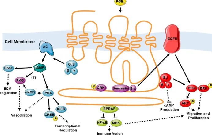
1-5 Downstream signaling mechanisms of the EP4 receptor potentially relevant to the DA ... 28
1-6 Three proposed mechanisms mediate O2 sensing in the DA ... 28
2-1 Venn diagram of compared rodent microarray studies ... 51
2-2 Dendrogram of Human RNA-seq samples ... 52
2-3 Volcano plot of RNA-seq differentially expressed genes ... 53
2-4 Venn diagram of DA vs. Ao genes common between Microarray and RNA-seq analyses ... 54
2-5 ‘Tornadogram’ showing top 30 UniProt (UP) Keywords common between Microarray and RNAseq analyses ... 55
2-6 ‘Tornadogram’ showing top 30 GO Biological Process (BP) terms common between Microarray and RNAseq analyses... 58
2-7 ‘Tornadogram’ showing top 30 GO Cellular Component (CC) terms common between Microarray and RNAseq analyses... 59
3-1 Representative images of various mouse knockout models exhibiting a PDA phenotype ... 100
3-2 Protein-Protein Interaction (PPI) network of effectors in PDA-associated human single-gene syndromes ... 101
3-3 Overlap of mouse models of PDA with associated human single-gene syndromes .... 102
xiv
3-4 ‘Tornadogram’ of top 20 GO Molecular Function (MF) terms common between known
mouse models of PDA and human single-gene syndromes with PDA ... 103
3-5 Graphical Abstract ... 104
4-1 Drug treatment protocols ... 137
4-2 EP4 is the predominant EP receptor in the mouse DA ... 138
4-3 Ptger4 expression is strongly localized to the medial and intimal layers of the patent fetal and closed postnatal (P1) DA... 145
4-4 Use of pressurized vessel myography to assess the mouse DA ... 146
4-5 Selective antagonism of EP4 constricts the DA in utero ... 147
4-6 The role of EP4 in DA patency is dependent on gestational timing ... 139
4-7 Mouse strain has an effect on EP receptor expression and function ... 148
4-8 EP4 is critical during the D17-19 window for proper postnatal DA function ... 140
4-9 EP4 antagonism disrupts cell migration in DA SMCs but not AO SMCs ... 141
4-10 The PDA of EP4 KO mice exhibits an unexpected contractile response to PGE2 ... 142
4-11 Effects of an EP4 KO allele on fetal mice ... 149
4-12 Antagonism of EP1 and EP3 disrupts PGE2 mediated vasoconstriction in the EP4 KO DA ... 150
4-13 The PDA of EP4 KO mice exhibits impaired responses to multiple stimuli ... 143
4-14 The PDA of EP4 KO mice responds effectively to O2 but display an immature phenotype ... 144
phenotype ... 144
4-15 Analysis of maturity markers reveal complexities of DA development ... 151
xv
Abbreviations
(Lp)PLA2) – platelet activating factor acetyl hydrolase/oxidized lipid lipoprotein associated phospholipase A2
AC – adenylyl cyclase
ACE – angiotensin-converting-enzyme AGTR1 – angiotensin II receptor type 1 Akt – protein kinase B
ALK1 – activin receptor-like kinase 1
AMPK – adenosine mono-phosphate activated protein kinase Ao – ascending aorta
Asxl2 – additional sex combs like 2 ATP – adenosine tri-phosphate BMP – Bone morphogenetic protein
BRET – bioluminescence resonance energy transfer BRG1 – brahma-related gene 1
BRM – brahma
cAMP – cyclic adenosine mono-phosphate CGMP – cyclic guanosine mono-phosphate CHD – congenital heart disease
CHF – congestive heart failure CO – carbon monoxide
COX - cyclooxygenase
cPGES – cytosolic prostaglandin E synthase cPLA2 – cytosolic phospholipase A2
CREB – cyclic adenosine mono-phosphate response element binding protein CRISPR – clustered regularly interspaced short palindromic repeats
cSrc – tyrosine-protein kinase Src CX – connexin
CYP – cytochrome P450
xvi CYPOR – cytochrome p450 reductase
DA – ductus arteriosus DAG – diacyl glycerol
DEG – differentially expressed gene DPC – days post-coitus
ECM – extracellular matrix
eNOS – endothelial nitric oxide synthase EP4 – prostanoid receptor EP4
EPAC – exchange factor activated by cAMP EPRAP – EP4 receptor associated protein
ERK – extracellular signal-regulated kinase kinase ET1 – endothelin 1
ETA – endothelin 1 A-type receptor ETC – electron transport chain FBLN1 – fibulin 1
FDR – false discovery rate FGF – fibroblast growth factor
FPKM – fragments per kilobase of transcripts per million FRET – Förster resonance energy transfer
GC – guanylate cyclase
GEF – guanine nucleotide exchange factor
GIVA PLA2 – group IV cytosolic phospholipase A2 GO – gene ontology
GO BP – gene ontology biological process GO CC – gene ontology cellular component GO MF – gene ontology molecular function Gpc3 – glypican 3
GPCR – G protein coupled receptor GTP – guanosine tri-phosphate
xvii
Hand2 – heart and neural crest derivatives-expressed protein 2 HAS – hyaluronic acid synthase
HFOV – high frequency oscillatory ventilation HIF2a – hypoxia inducible factor 2a
HPGD/PGDH– 15-hydroxy-prostaglandin dehydrogenase HPV – hypoxic pulmonary vasoconstriction
ICER – inducible cyclin adenosine mono-phosphate repressor IEL – internal elastic lamina
ILK – integrin-linked kinase
iNOS – inducible nitric oxide synthase IP3 – inositol tri-phosphate
iPLA2 – calcium-independent phospholipase A2 IUGR – intrauterine growth restriction
KO – knockout
LA – ligamentum arteriosum LOX – lysyl oxidase
MAPK – mitogen activated protein kinase MATR3 – matrin 3
MEK – mitogen activated protein kinase kinase MF1 - mesodermal/mesenchymal forkhead 1 MFH1- mesenchymal forkhead 1
MHC – myosin heavy chain MLC – myosin light chain
MLCK – myosin light chain kinase
MLCP – myosin light chain phosphatase MMP – matrix metalloproteinase
mPGES – microsomal prostaglandin E synthase MTHFR – elastin methylenetetrahydrofolate reductase mtNOS – mitochondrial nitric oxide synthase
xviii MYH11 – myosin heavy chain 11
NAD+/ NADH – nicotinamide adenine dinucleotide NFE2 – nuclear factor erythroid 2
NICU – neonatal intensive care unit nNOS – neuronal nitric oxide synthase NO – nitric oxide
NOS – nitric oxide synthase
NSAID – non-steroidal anti-inflammatory drugs NT3 – neurotrophin 3
O2 – diatomic oxygen
PDA – persistent patency of the ductus arteriosus PDE - phosphodiesterase
pGC – particulate guanylate cyclase PGD – prostaglandin D
PGDS – prostaglandin D synthase PGE – prostaglandin E
PGF – prostaglandin F
PGFS – prostaglandin F synthase PGG – prostaglandin G
PGH – prostaglandin H PGI – prostaglandin I
PGT – prostaglandin transporter PKA – protein kinase A
PKC – protein kinase C PKG – protein kinase G PLA2 – phospholipase A2 PLC – phospholipase C
pO2 – partial pressure of oxygen POSTN – periostin
xix
PPHN – persistent pulmonary hypertension of the newborn PPI – protein-protein interaction
PTGER – prostaglandin E receptor
PTGS – prostaglandin-endoperoxide synthase
qRT-PCR – quantitative reverse-transcription polymerase chain reaction RBPJ – recombinant signal binding protein for immunoglobulin κ J Rim4 – recombinant-induced mutation 4
ROS – reactive oxygen species RTK – receptor tyrosine kinase
RT-PCR – reverse-transcription polymerase chain reaction SGBS – Simpson-Golabi-Behmel syndrome
sGC – soluble guanylate cyclase SMC – smooth muscle cell
SNP – single nucleotide polymorphism SNP – sodium nitroprusside
SNP – sodium ntroprusside
sPLA2 – secreted phospholipase A2
SSRI – selective serotonin reuptake inhibitor TAAD – thoracic aortic aneurysm and dissection Tagln – transgrelin
TFAP2B – transcription factor AP2β TGFβ – transforming growth factor β TNF – tumor necrosis factor
TP – thromboxane receptor
TRAF1 – tumor necrosis factor receptor associated factor 1 TXA2 – thromboxane A2
TXS – thromboxane synthase UP Keywords – UniProt Keywords VNCC – vagal neural crest cells
xx VSMC – vascular smooth muscle cell
WT - wildtype
XOR – xanthine oxidoreductase
1 Chapter 1
INTRODUCTION
Abstract:
The ductus arteriosus (DA) is a muscular artery which helps to define the fetal pattern of the circulatory system. After birth, the DA undergoes muscular constriction and remodeling which establish the adult division between the pulmonary and systemic circulation. Fetal DA patency requires acute vasodilatory signaling via the prostaglandin (PGE2) receptor EP4. However, in humans and mice, disrupted PGE2-EP4 signaling in utero causes unexpected persistent patency of the DA (PDA) after birth, suggesting another chronic role for EP4 during development. It is likely that sustained PGE2 signaling via EP4 is responsible for directing the proper development of the DA to enable its closure after birth. This is mediated through highly ordered developmental processes, likely representing a developmental program that is heretofore an unstudied paradigm for DA formation and function.
Introduction:
The DA is a fetal vessel which shunts blood right-to-left past the lungs in utero to protect the developing pulmonary vasculature and direct freshly oxygenated blood from the placenta into the systemic circulation (Figure 1A). Compared to the large elastic arteries which it interconnects (aorta, pulmonary artery), the DA is a large muscular artery with dynamic vasoactive properties. While DA
2
Figure 1. DA closure in the mouse. A) dye perfused outflow tracts of a term CD1 WT pup at birth displaying a patent DA and free flow of dye in a right-to-left fashion from the pulmonary circuit (pulmonary artery) to the systemic circuit (aortic arch). B) at 3hrs after birth, the DA has constricted preventing flow of dye from right-to-left and establishing the divided pulmonary and systemic circuits of the adult circulatory system. DA – ductus arteriosus, AA – aortic arch, PA – pulmonary artery.
3
patency, or openness, is essential during fetal development, its rapid postnatal closure is critical for circulatory transition to neonatal life. DA constriction is an elegant cascade of biological processes requiring acute changes in vascular tone,
transcriptional profiles, fluidity in cell phenotypes, and both prenatal and postnatal structural
remodeling (1, 2). Frequently, disruptions in these genetic, environmental, and developmental processes result in failure of the DA to close, termed PDA. Risk for PDA is multifactorial, consisting of genetic, environmental, immunological, and as of yet unexplained factors. Regardless of cause, PDA poses serious challenges for affected infants, especially more fragile preterm infants. Treatment options are limited, and all convey their own risks of severe side effects. For this reason, an emphasis on preventative medicine, and to that end a deep understanding of the mechanistic underpinnings of DA formation, closure, and transcriptional identity is critical.
Role of DA in Transition to Neonatal Life/Embryology:
The specifics of the DA’s anatomical position and function result from the complex evolutionary transition from water-based respiration to air-based respiration. In the majority of fish species, the circulatory system exists as a singular loop where deoxygenated blood returns from the body, passes through the sinus venosus, through a single atria, and into a single ventricle, which expels it through the aortic arch arteries associated with the gills (3). As blood passes through these arch arteries, it is reoxygenated via gas transfer with the surrounding water. The development of primitive lungs in some fish species allowed them to shift back and forth between water-breathing and air-breathing, but required further modification of the cardiovascular system. In order to separate gill-based water breathing from lung-based air breathing, separate vascular circuits were required. Thus, aquatic
4
species with terrestrial habits developed hearts with septations dividing the atria and ventricles into left and right sides. The most proximal, sixth aortic arch artery to the heart became a muscular shunt which could alternate blood flow between the lungs and other arch arteries, depending on environmental context. This system of alternating between gills and lungs survives in some extant fish such as the lung fishes, gouramis, bichirs, and some species of climbing perch. These should not be confused with other fish species which rely on labyrinthine organs (arapima, beta fish) or highly vascularized swim bladders (alligator gar) for air breathing, as these rely on different circulatory adaptations (4).
Due to the necessity of the DA structure for adaptation to air breathing, it is conserved in five vertebrate classes; amphibians (5), reptilians (6, 7), birds (8, 9), mammals (10), and of course the lobed fin fishes in which they evolved (11). Of note, these groups span all terrestrial life. While there is variation in structure amongst these classes, the unilateral DA of mammals arise from the left sixth arch artery, and the bilateral DA of birds arise from both the left and right sixth arch arteries (12).
Specifically, the vascular smooth muscle cells (VSMCs) of the DA arise from neural crest cells which migrate via aortic arch arteries down the DA and ascending Ao on their way towards the heart where they will contribute to cardiac development (13-15). Endothelium of the DA and ascending Ao are contributed by migration from the second heart field (16). This combination of cellular origins makes the DA and Ao unique amongst the central vasculature, despite their different phenotypes. For this reason, the ascending Ao provides an exceptional control for studies of DA development and physiology. The DA has likely been evolutionarily conserved as a structure due to its indispensable role during embryonic development. Whether a bird embryo developing in an egg or a mammal developing in utero, the fluid-filled environment prevents the use of lungs for respiration. As a result, whether a bird embryo through its chalaza and air cell (17), or a mammal through its umbilical cord and placenta, oxygenation of fetal blood is obtained independent of the underdeveloped, unaerated
5
lungs. As previously mentioned, the vascular and alveolar structures of the lungs develop very slowly, and have high vascular resistance in utero (18, 19), thus a full hemodynamic load is injurious (20). To prevent this, the DA shunts the majority of oxygenated right ventricular outflow ~60% in humans (21), past the developing lungs.
After birth, closure of the mammalian DA is initiated by a cascade of signals arising in part from the newly inflated lungs. The fetal environment is markedly hypoxic, with oxygen (O2) tensions equal to 20-30% of normal adult values (22). Increasing O2 tension exerts a constrictive effect on the DA while also helping to stimulate metabolism of circulating dilatory prostaglandins (23-25). As the DA closes and remodels, the adult circulatory pattern of wholly divided pulmonary and systemic circuits is established (Figure 1B). While some animals do maintain either an open (tortoises (26)) or reversible DA (lungfish (12)), this is not the case with humans or mice. In humans, functional DA closure occurs in 12-24hrs and permanent anatomic closure will occur over the course of 2-3 weeks (27). In mice, functional DA closure occurs in 3-6hrs (Figure 1) and permanent anatomic closure in 2-3 days (28).
PDA disrupts the transition of the cardiovascular system to neonatal life and may have severe hemodynamic consequences. Excessive left-to-right shunting results in pulmonary over-circulation with oxygenated blood being mixed with deoxygenated blood and recirculated through the lungs. This over-circulation can result in pulmonary edema, as well as enlargement of the left side of the heart and a decrease in delivery of oxygenated blood to peripheral tissues known as “ductus steal” (1).
Together, these effects burden the development of the infant and contribute to poor outcomes.
6 Clinical Relevance/Human Epidemiological Data:
Despite decades of research, the exact mechanistic underpinnings of PDA are still poorly understood. Risk factors for PDA have been thoroughly catalogued and provide important insights into DA biology (Table 1). There is evidence for a genetic basis in some forms of PDA, but in most cases, PDA likely results from a combination of prematurity, environmental conditions, or cardiovascular comorbidities. PDA comprises 5-10% of congenital heart disease (CHD) cases in the US (29). While PDA is relatively rare in healthy term infants, it is disproportionately common in preterm (64% at 27-28 weeks) and very preterm infants (87% at 24 weeks) (30, 31). Most at risk are those with particularly low birth weights (80% in infants <1000 grams) (32). It should be noted that low birth weight is itself likely not the causeof PDA or its increased severity. Instead, it is likely that birth weight is a more reliable predictor of gestational maturity than timing in medically complicated pregnancies. With this consideration, prematurity appears to be the most important indicator of PDA prevalence and severity in the newborn. Premature infants also tend to have a high incidence of comorbidities. While extremely premature infants (<28 weeks) comprise only 0.7% of births in the United States (33) together with slightly more mature infants (28-32 weeks) they account for more than half of all infant deaths (34). Without limiting by PDA or other comorbidities, extremely premature infants have a 26% mortality rate during their initial birth hospitalization with each lost week of gestation correlating to lower survival (35, 36). While instances of extremely premature infants have been relatively consistent since 2000 (33), the frequency of the less severe late preterm infants (28- 32 weeks) which historically accounted for ~75% of preterm births, are increasing (37). With preterm infants currently comprising 11.4% of live births in the United States (33) PDA incidence may be increasing not decreasing. With treatment, an isolated PDA has a good prognosis (27), but adverse outcomes can be severe, especially in preterm and low-birth weight neonates where comorbidities tend to define outcomes (38).
7
Established Risk Factors for PDA Other Factors Associated with PDA
Early gestational age IUGR Maternal drugs:
Antihistamine Magnesium ACE inhibitors Anticonvulsants Ca channel blockers Cocaine
Maternal PKU Low birth weight Delay in indocin treatment
RDS Furosemide treatment
Persistence of DA flow Use of HFOV
Sepsis Race:
Caucasian (PT) African American (T) Excess fluid administration
Antenatal NSAID exposure
Initial hypotension Gender:
Male (PT) Female (T) Need for intubation/airway pressure
Lack of antenatal betamethasone Genetic Conditions (T)
(trisomy 21, 18, 13, Char, Holt-Oram, DiGeorge, Noonan, CHARGE, TAAD/PDA) Genetic Susceptibility Familial PDA
Maternal diabetes (T) Prolonged ROM
Birth at high altitude (T) Twins
Congenital rubella (T) Perinatal stress Hypothyroidism (T, PT) Antenatal hemorrhage
Breech Phototherapy
Table 1. Factors associated with PDA. Risk factors for PDA were considered to be well- established if they were identified by studies that sought causative factors for PDA, remained significant after multivariate analysis, or were consistently observed in multiple controlled trials in different patient populations. Other factors that have been shown to have an association with PDA were drawn from single studies, epidemiologic surveys, birth defect registry reports, case reports, or small studies that did not control for confounding variables. PDA at term (T) gestation is regarded as a congenital malformation, but these risk factors may also occur in preterm (PT) infants. Conflicting studies that did not detect an association of PDA with these factors are not presented. Genetic conditions were considered separately. Only a subset of representative citations are shown for risk factors that were consistently identified in numerous studies. (IUGR, intrauterine growth restriction;
HFOV, high frequency oscillatory ventilation; ACE, angiotensin-converting-enzyme) Adapted with permission from Reese et al. (39)
8 Contributors to PDA:
The environment of the womb has a profound effect on the developing tissues of the fetus, including the DA. One historical example of this was the observation of an increased incidence of PDA in the offspring of mothers infected with rubella (40). Congenital rubella syndrome, infection of the fetus with rubella during gestation, has not only been found to increase PDA, but to increase pulmonary hypertension-dependent mortality associated with PDA (41). This increased mortality could be abated via closure of the DA. Interestingly, the congenital rubella syndrome-associated PDA features a tubular-type PDA configuration which makes the DA significantly harder to close via transcatheter occlusion compared to cone-type PDAs (42). While various PDA configurations have been described in detail via echocardiography (43) and a classification system has been adopted to categorize them (44), this is primarily relevant for catheter-based occlusion.
Pharmacological treatments administered to mothers during pregnancy have also been associated with PDA. The importance of developmental timing in fetal PGE2 signaling was first supported by observations that maternal exposure to cyclooxygenase (COX) inhibitors, given as a tocolytic to arrest preterm labor by blocking production of PGE2, results in fetal DA constriction after 30-32 weeks of gestation, but not earlier in pregnancy (45, 46). In contrast, mothers who received COX inhibitors as tocolytics during late- but not mid-gestation had an increased risk of PDA in their offspring (47). This particular finding revolutionized the understanding of the DA and will be expanded on throughout this dissertation. Similarly, the treatment of antenatal depression using selective serotonin reuptake inhibitors such as sertraline or fluoxetine (48) is very common, with prescriptions written in 2-6% of all pregnancies (49-51). Illustrating an important aspect of these maternal-fetal interactions, selective serotonin reuptake inhibitors (SSRIs) readily cross the placenta resulting in fetal levels of 70-80% those of the maternal blood. Maternal SSRI administration, while not directly linked to PDA in infants, was found to predispose neonates to persistent pulmonary hypertension of
9
the newborn (PPHN) (52, 53), a condition promoted by in utero constriction of the DA (54). In one of the first projects I worked on during my Ph.D., our group performed studies on the DA of neonatal mice, finding dose dependent constriction in response to SSRIs, as well as in utero constriction of the DA in SSRI treated pregnant dams (55). Together these findings suggest SSRIs likely contribute to PPHN in humans through in utero constriction of the DA. As another example, antenatal betamethasone, despite being associated with decreased PDA in observational (56-58) and controlled (59) studies increases the vasoconstrictive potential of the DA across all tested timepoints, likely through regulation of genes associated with O2 sensing and muscle contraction (60). This may be problematic in pregnancy, as it could lead to in utero constriction, despite being a powerful strategy to accelerate maturation of other organ systems and decrease comorbidities.
Cardiovascular comorbidities also predispose an infant to PDA, or increased symptoms from PDA. PDA exists as a secondary disorder in ~10% of other congenital heart defects (27). While PDA and the primary defect may arise from a similar genetic or developmental disruption, severe congenital heart defects are often reliant on a patent DA to be compatible with life. Further, many of the more critical congenital heart defects display no outward symptoms until the DA begins to close after birth (4). Specifically, hypoplastic left heart syndrome, transposition of the great arteries, and tetralogy of Fallot are examples of life-threatening conditions which require maintenance of a patent DA (5). These medically induced PDAs are maintained with prostaglandins such as PGE1 until the primary defect can be surgically corrected, at which point the DA is usually ligated or excised, depending on the repair.
Maternal treatment is not the only way that drug exposures can affect PDA. It is now becoming more clear that pharmacological interventions performed in the neonatal intensive care unit (NICU) can affect DA tone, both positively and negatively. It was previously believed that fetal and neonatal sepsis contributed to PDA, as the conditions correlate clinically. However, our group found that while
10
the inflammatory pathways associated with sepsis had little or no effect on tone in the mouse DA, the aminoglycoside antibiotics administered to treat that sepsis resulted in dilation of the DA (61). This is likely through the depression of calcium (Ca2+) flux that mediates gentamicin’s myocardial depressant effects (39, 62-64). Trials using cimetidine, an H2 receptor antagonist generally used as an antacid, in an attempt to decrease lung injury in extremely premature infants failed to decrease lung injury and increased incidence of PDA (65). Our group found this was caused by cimetidine-mediated cytochrome P450 inhibition (CYP), which may disrupt the O2 sensing mechanisms of the DA (39, 66).
Similar effects were found for ranitidine, an H2 receptor antagonist with similar CYP inhibitor properties. Phosphodiesterase (PDE) 3 inhibitors milrinone, amrinone, and the PDE5 inhibitor sildenafil have all been found to have vasodilatory effects on the DA despite their continued use in the NICU (67-69).
Clinical Options to Treat PDA:
The first-line treatment for PDA is administration of COX inhibitors, usually indomethacin or ibuprofen. Indomethacin is relatively COX-1 selective, and tends to have vasoconstrictive effects on the cerebral, renal, and mesenteric vasculature (70). Decreased cerebral blood flow is accompanied by reduced cerebral oxygenation (71) and potentially cerebral ischemia (72). Reduced renal blood flow results in decreased creatinine clearance (73), higher blood urea nitrogen levels (74), decreased urine output (75), and overall decreased kidney function. While there are conflicting reports as to whether prophylactic indomethacin is associated with increased risk for necrotizing enterocolitis (76) or not (77-80), gut hypoperfusion and subsequent mucosal hypoxia provide a route for potential ischemia, ulceration, and bacterial invasion (81, 82). Gastrointestinal perforation has been reported (81). While many of these effects may be abated by the use of ibuprofen, this treatment may be associated with pulmonary issues such as an increased susceptibility to chronic lung disease
11
(requiring supplemental O2 at 28 days of age) or persistent pulmonary hypertension of the newborn, though these findings are hotly debated (83). It is worth noting that while necrotizing enterocolitis, chronic lung disease, and persistent pulmonary hypertension are all common amongst infants with PDA, this may be due to these conditions being more prevalent in extremely premature neonates.
Therapeutically, indomethacin has a closure rate of 80-90% (27), but pharmacologically unresponsive PDAs will require surgical ligation or catheter-based occlusion. Any cardiothoracic surgery comes with extreme risk, but this risk is increased for premature and low birth weight neonates. Unfortunately, it is the extremely premature neonates who are most likely to fail pharmacological treatment. Overall, surgical ligation conveys a mortality rate of 2-20% depending on comorbidities. Ligation is also associated with increased neurodevelopmental impairment, retinopathy of prematurity, and chronic lung disease (84). Additionally, 30-44% of all PDA ligations are followed by acute respiratory and hemodynamic instability in the initial 24hrs post procedure (85, 86). This condition is referred to as post-ligation cardiac syndrome, and it is associated with systemic hypotension, (85, 86) chronic lung disease (87), and by some reports, up to 33% mortality (88). A less fatal but common side effect of ligation is the accidental severing of the left recurrent laryngeal nerve which wraps around the DA in situ, resulting in partial vocal paralysis (89, 90). A relatively new alternative to surgical ligation is percutaneous closure using a catheter-based occlusion device such as the Amplatzer Piccolo (91). In this procedure, a catheter delivers a deployable mesh device which will be placed into the patent lumen of the DA under ultrasound or fluoroscopic guidance, occluding flow and providing a scaffold for surrounding tissue ingrowth to achieve permanent, secure closure.
Clinical trials and multi-center studies are still in progress, so it is difficult to thoroughly evaluate the risk factors of percutaneous closure (92-94). While treatment options all convey risk, the bottom line is an open DA must eventually be closed. Left untreated, pulmonary hypertension may develop, and the mortality rate for untreated PDA increases with age (95-97). All cases of PDA in the adult are
12
unresponsive to COX inhibitors and will likely need treatment with riskier surgical options. Because treatment options for PDA are limited, with such severe side effects, and such a vulnerable population, preventative medicine, and therefore a thorough understanding of the underlying biology is required. The breadth and severity of side effects associated with disrupting the prostanoid pathway further emphasizes the importance of prostanoid signaling in the DA.
Genetic Landscape of the DA:
Developmental programs are sequential cascades of gene and protein expression with subsequent pathway regulation which direct a developmental process. Grasping the genetic and transcriptional landscapes of the DA are critical to identifying potential constituents or regulators of such a program. This starts with understanding the heritability of PDA and what genes/transcripts may affect PDA risk or severity. Both term and preterm PDAs may have a genetic component, with a 5% sibling recurrence rate (98, 99) and a higher correlation between monozygotic twins compared to dizygotic twins (100, 101). While reports have varied, one twin study found that genetic factors and a common gestational environment contributed up to 76% of this variance. Studies on familial PDA and the offspring of consanguineous parentage provide genetic information on chromosome regions that confer risk for PDA (102, 103). In addition, candidate gene studies have identified genetic loci which contribute to syndromic forms of PDA such as transcription factor AP2β (TFAP2B), or whose sequence variants can contribute to isolated non-syndromic cases of PDA (104, 105). Although the genetic predisposition for most PDAs is unknown, a robust understanding of the genes whose perturbation results in PDA may provide key insights into the development and function of the DA critical to the designing new and improved therapies. To this end, many mouse models of PDA have been described. These will be discussed in-depth in chapter 3. Of note, few mouse models of PDA originated as attempts to generate a PDA phenotype. PDA was generally an incidental finding among
13
other cardiovascular anomalies. Knockouts (KOs) of several key genes involved in prostaglandin signaling, including prostaglandin-endoperoxide synthase 1 (Ptgs1);Ptgs2 double KO (106, 107), prostaglandin E receptor 4 (Ptger4) KO (108-110), and the Pgdh KO (24, 111) resulted in PDA.
Ptger4 (EP4) expression was also found to be downregulated in the Prdm6 KO mouse (112), emphasizing the importance of PG signaling pathways in the DA. Microarray studies have also been performed on mouse (113, 114), rat (115-120), sheep (121), chicken (122), and human (123, 124) tissues in efforts to elucidate the transcriptional landscape of the DA. Methodologies varied greatly between these studies with their findings varying in kind. The state of this understanding is, to borrow a phrase, ‘as clear as mud at night.’ Recent studies utilizing next-gen sequencing have attempted to remedy this, and even identified new KO models in the process (125), but a global and comprehensive understanding of this landscape is required to advance the state of PDA treatment.
Preparation of the DA for Closure and Remodeling:
DA closure occurs in three primary steps. Soon after birth, the first stage, “functional closure”
in response to acute signaling changes, occurs. This process takes about 1-3 days in humans or 3- 6hrs in mice and results in a DA that is completely but reversibly occluded by muscular constriction of the vessel wall. Next, a rapid wave of remodeling and “anatomic closure” begins within the lumen and wall of the constricted DA, leaving it irreversibly closed. This generally takes up to a week in humans and about 24-48hrs in mice. Finally, a second wave of neonatal remodeling occurs resulting in fibrosis and necrosis, leaving a new structure, the ligamentum arteriosum (LA), as a remnant of the DA structure. This process can take quite a while, several weeks in humans, and multiple days in mice. It is clear that tissue remodeling, and to that effect cell phenotype, play significant roles in effective DA closure. An underappreciated preliminary stage precedes the others and consists of several key remodeling steps that occur in late gestation and prepare the DA for eventual closure.
14
Critically, in cases of PDA none of these pre- or post-birth remodeling steps occur (126), therefore these processes are likely critical for proper DA function.
DA structural remodeling, and in some ways the process of DA closure, actually begin in the immediate antenatal period. During late development, the DA undergoes a process of extensive remodeling of its extracellular matrix (ECM) and tissue structure which is necessary to prepare the vessel for closure after birth. At the beginning of this process, during mid-gestation, the DA exists as three discrete layers; an adventitia of fibroblasts and myofibroblasts, a continuous muscular media of VSMCs aligned perpendicular to the direction of flow, and an intima composed of a single layer of endothelium, separated from the medial layer by a contiguous sheet of elastin designated the internal elastic lamina (IEL) (Figure 2A). VSMCs such as those in the medial layer of the DA, display a spectrum of phenotypes (127-129), though this is usually simplified as an axis with a sedentary, contractile phenotype on one side and a secretory, migratory, synthetic phenotype on the other. In literature from the 1970s-80s, before the variable smooth muscle cell (SMC) phenotype was fully understood, contractile SMCs are referred to as ‘mature’ with any other phenotypic characteristics deemed ‘immature.’ The exact phenotypic state of DA VSMC populations at this preparatory point in DA development is somewhat unknown. Microarray analysis of laser micro-dissected VSMCs from preterm rat DAs revealed the presence of some mature muscle markers associated with the contractile phenotype, but also extensive expression of synthetic or non-muscle markers (119). This suggests a mixed phenotypic population of DA VSMCs, containing both contractile cells
15
Figure 2. EP4-mediated late gestational remodeling prepares the DA for closure. A) The developing DA, with (right to left) an open lumen, intima composed of a single layer of endothelial cells attached to an intact IEL, media composed of several layers of circumferentially oriented contractile VSMCs, and an adventitia composed of several layers of fibroblasts and thick extracellular matrix. B) Synthetic VSMCs from the DA media begin to secrete hyaluronic acid facilitating migration, disrupt LOX mediated crosslinking of elastin leading to a fragmented IEL, and migrate inward towards the IEL; all reportedly EP4 mediated processes. Endothelial cells begin to detach from the IEL in a possibly hyaluronic acid mediated mechanism. C) Synthetic VSMCs begin to invade the subendothelial space created by complete detachment of endothelial cells from the IEL. D) Intimal thickening progresses as VSMC numbers in the subendothelial space continue to increase, due primarily to migration not replication. Synthetic VSMCs begin to transition back into contractile VSMCs which assume a radial orientation perpendicular to the lumen. Continued disruption of LOX leads to the formation of disorganized partial sheets of elastin throughout the media. E) Intimal thickening is complete, characterized by a thick layer of radially reoriented contractile VSMCs dividing the highly fragmented IEL from the intimal endothelial cells. Endothelial cells may begin breaking basal polarity and growing on top of each other. Fragmented elastin sheets are common in the medial layer with human DA forming a secondary discontinuous elastic lamina in the mid-media. In larger species such as humans, full intimal cushions form, protruding into the lumen of the vessel and disrupting perfusion to the vessel wall. This results in the formation of deposits of fat-rich macrophages called foam cells, and cores of necrotic cells in the subendothelial space. F) Legend.
Figure created using images and assets from Servier Medical Art. Servier Medical Art by Servier is licensed under a Creative Commons Attribution 3.0 Unported License (https://creativecommons.org/licenses/by/3.0/).
16
which maintain DA tone, and a migratory, synthetic subpopulation. At this stage, synthetic VSMCs begin to disrupt the cross-linking of elastin required to maintain the IEL. This is possibly achieved through the EP4-mediated inhibition of lysyl oxidase (LOX) (130, 131).
Upregulation of matrix metalloproteinases (MMPs) by some unknown mechanism and subsequent matrix degradation is also a potential explanation. Unable to adequately maintain itself, the IEL becomes fragmented, with large openings forming across its surface (132, 133). Disorganized patches of elastin begin to form throughout the medial layer, with a second highly fenestrated IEL forming in the middle of the medial layer in humans (132, 134). The endothelial cells of the intimal layer begin to break their bonds with the IEL creating a subendothelial space (Figure 2B) (135, 136) though not in some species, such as dogs (126, 137). Proper regulation of elastin is critical to DA closure after birth, and an intact IEL is associated with PDA in both humans and dogs (137-140).
Natural genetic disruption of elastin assembly in the brown Norway rat (141) produces several vascular disorders (142) including PDA (143). VSMCs migrate through the fragmented IEL into the subendothelial space (Figure 2C) (144). These VSMCs contain large amounts of rough endoplasmic reticulum and were found throughout the subendothelial space and media, lending support to the notion these migratory cells are of the secretory synthetic phenotype. Further, areas of the vessel rich in ‘odd looking’ VSMCs contain less elastin, suggesting these synthetic VSMCs are responsible for elastin degradation in the DA (145).
VSMC migration is facilitated by the EP4-mediated production of hyaluronic acid, a hygroscopic matrix component by the secretory synthetic VSMCs (146-148). This hyaluronic acid is readily detected in the subendothelial region, and may contribute to endothelial cell detachment from the IEL, expansion of the subendothelial space, and migration of synthetic VSMCs into this space (135). EP4- mediated activation of AC increases cyclic adenosine mono-phosphate (cAMP), which activates the serine/threonine protein kinase A (PKA), and subsequently hyaluronic acid synthase (HAS). This has
17
been shown to promote cell migration in cells cultured from rat DA (146). The process of amassing VSMCs in the subendothelial space is called intimal thickening, which is present in most species (Figure 2D). A more extreme version of this occurs in larger species such as humans, with the formation of large intimal cushions which serve to partially occlude the lumen of the DA (Figure 2E)
(132, 136, 149, 150). These intimal cushions are large enough they may prevent complete perfusion into the vessel wall, leading to antenatal cytolytic necrosis, immune recruitment in the formation of mucoid lakes, and necrotic cores of dying SMCs (134). Endothelial cell detachment from the IEL does not occur in some species, including dogs, explaining why some species may lack intimal cushion formation (126, 137). The severity of intimal thickening and disruption of the IEL increases with gestational age (132, 144). The VSMCs within the subendothelial space undergo radial realignment, assuming a contractile phenotype and organizing themselves pointing inward towards the lumen, perpendicular to their normal orientation (144, 151). Large amounts of amorphous extracellular material is also present in glycogen pockets throughout the media at this stage, and may represent un-crosslinked matrix proteins staged for post birth fibrosis of the vessel (144). At this point, the DA is ready for birth and subsequent constriction.
Mechanisms of Postnatal DA Closure:
After birth, a complex cascade of signals and processes lead to the initial, functional closure of the DA. As soon as the neonate leaves the hypoxic environment of the womb, their airway opens, and breathing is initiated. The sudden increase in partial pressure of oxygen (pO2) begins to relax the hypoxic pulmonary vasoconstriction (HPV) mechanism that has been constricting the pulmonary vasculature in utero. Separation from the placenta cuts off a key source of prostaglandins, with remaining PGE2 being rapidly metabolized by 15-hydroxy-prostaglandin dehydrogenase (HPGD)
18
which is highly expressed in the lungs (23). Decrease in PGE2 activation of EP4 leads to a gradual decline in cAMP and reduced activation of myosin light chain phosphatase (MLCP), allowing the cycling of myosin light chain (MLC) in the contractile apparatus. This is further facilitated by internalization of EP4 which reduces the rate of signaling. Simultaneously, O2-sensing mechanisms act to increase internal Ca2+, triggering calmodulin-dependent activation of myosin light chain kinase (MLCK) and increasing MLC cycling. O2 displacement of CO attachment to circulating hemoproteins alleviates another dilatory mechanism, decreasing cyclic guanosine mono-phosphate (cGMP) production and further decreasing activation of MLCP. With MLC free to cycle and MLCK active, the DA VSMCs rapidly constrict, completely occluding the vessel, and establishing the adult circulatory pattern. At this stage, blood may still be present in the lumen of the DA, but the vessel is hemodynamically insignificant, and the systemic and pulmonary circuits no longer mix.
The processes belying the permanent closure of the DA are still unclear. Recent sequencing analysis shows that certain transcripts become upregulated during this window, specifically those associated with matrix and cytoskeletal components (125). It is unclear what role the radially realigned VSMCs in the subendothelial space play. It is thought that intimal cushions serve to occlude the lumen of vessels in species which are too large for lumen obliteration by muscular closure alone.
The need for these cushions is likely dictated by the ratio of circumference to area of the vessel cross section. VSMCs themselves take up space, and it is logical there would be limits to how much they can compress the vessel wall. Once the vessel is completely closed, the endothelial cells begin to necrose. VSMCs which have likely transitioned to a synthetic myofibroblast-like phenotype, begin to produce large amounts of matrix proteins, leading to fibrosis of the tissue. This permanently seals the vessel and prevents medication-induced reopening, or mechanical reopening under pressure.
Finally, following permanent anatomic closure of the DA, the VSMCs undergo necrosis and apoptosis, leaving behind a fibrotic tether between the pulmonary artery and aortic arch. This
19
structure is termed the LA and it will persist through adulthood. The LA is an overlooked structure which provides some support to the positioning of the outflow tracts, but does not appear to serve other functions, although it may remain partially responsive to stimuli (152). Though the process of DA degradation likely begins immediately after permanent closure, this can take some time, with SMCs present two months after birth in the pig DA (153, 154). Though these SMCs are in the process of degenerating, with lipid vacuoles, lysosomes, dilated rough endoplasmic reticulum, and Golgi bodies found in their cytoplasm. Early endothelial cell degeneration was characterized by aggregates of Weibel-Palade bodies; elongated secretory organelles which mediate inflammation and tissue repair in vascular endothelium (155). Fibrosis and lipid vacuole production by degenerating SMCs increases by six months of age. Fibromuscular connective tissues fill the lumen and the boundary between intima and media becomes indiscernible (144). Aforementioned amorphous extracellular material is reduced throughout the media, replaced by crosslinked matrix including collagen fibers (144). Interestingly, the remodeling process of the DA reproduces many characteristics of pathologic atherosclerosis (144, 149, 154). These include intimal thickening-mediated stenosis of the vessel, expansion of intimal VSMCs, degradation of the IEL, and the generation of lipid-filled vacuoles. There are also similarities in immune recruitment to the vessel wall, though this has not been thoroughly studied in the DA (156).
Vascular Smooth Muscle Cells in DA function:
In order to understand the complex cascade of factors that leads to DA closure at birth, it is critical to understand the mechanisms underlying the contraction and relaxation of SMCs. Smooth muscle cells compose the cylindrical walls of many tissues throughout the digestive system, respiratory tract, and reproductive systems (157). We are interested specifically in the VSMCs that compose the medial layers of blood vessels. SMCs lack the striated pattern created by repeating
20
contractile apparatuses in skeletal and cardiac muscle. Instead, SMCs possess a crosshatch pattern of contractile proteins that allow them to shorten and widen on contraction, decreasing the luminal diameter of their given structure. In the vasculature, this equates to vasoconstriction. The contractile apparatus of VSMCs is composed of long actin filaments and myosin chains which span the cell connecting intermediate junctions on opposite faces (157). The formation of cross bridges between the heads of myosin dimers allows them to exert force. These myosin dimers are composed of two heavy chains (MHC) with prominent heads that interact with actin. At the base of these heads are the myosin light chains whose phosphorylation state allows the interaction of MHC and actin. Specifically, phosphorylated MLC activates the actomyosin Mg-ATPase portion of the MHC, allowing cross bridge cycling (158, 159). The phosphorylation state of MLC is generally controlled by receptor signaling, detection of stretch by specialized ion channels, or neuronal signaling. Some muscular arteries, like the DA, maintain a baseline level of MLC phosphorylation giving them vascular tone, or the maintenance of a partially contracted lumen (160). The state of the contractile apparatus in SMCs is largely determined by a balance between MLCK which phosphorylates MLC facilitating contraction, and MLCP which dephosphorylates MLC preventing contraction (161-163). Dilatory factors generally act to increase the activity of MLCP and decrease the activity of MLCK. This points to the most critical factor for muscle contraction of any kind, intracellular Ca2+. The activity of MLCK is largely governed by calmodulin, which binds and activates MLCK following activation by intracellular Ca2+ (162, 164).
This makes Ca2+ critical for significant contraction of SMCs. This Ca2+ can either be imported through the cell membrane, or released from intracellular Ca2+ stores like the sarcoplasmic reticulum (165).
MLCP is also subject to regulation by inhibitors such as RhoA (166), or activators such as its own M110 regulatory subunit (167). 7 transmembrane G protein coupled receptors (GPCRs) can mediate both relaxation and constriction in SMCs, depending on their G protein subunits. The GPCRs discussed here function through heterotrimeric G protein complexes, or large GTPases composed of an α subunit and a β/γ complex (168). Each receptor contains a guanine nucleotide exchange factor
21
(GEF), which exchanges the GDP bound to the alpha subunit for a GTP (169). This elicits a conformational change which leads to the dissociation of the G proteins from the receptor. The activated α subunit will then go on to illicit G protein-specific downstream effects. For instance, the EP1 receptor is bound to a Gqα subunit, which once activated, will activate membrane bound phospholipase C (PLC) to produce inositol triphosphate (IP3) and diacylglycerol (DAG). This IP3 will then bind IP3 receptors on the sarcoplasmic reticulum, initiating the release of Ca2+ into the cell, fostering contraction. DAG on the other hand, activates protein kinase C (PKC) which phosphorylates targets such as L-type Ca2+ channels which import more Ca2+ into the cell. As with all kinases discussed here, kinase isoforms are often tissue specific and illicit specific functions based on context. It is not safe to assume kinases do the same thing in different places. The specific mechanism of each signal will be discussed in detail.
Prostaglandin E2 and the DA:
A critical pathway for maintaining DA patency in utero is prostaglandin signaling, specifically PGE2 activation of the prostanoid receptor EP4. This ligand-receptor interaction is part of a large family of lipid signaling molecules known as eicosanoids, which are produced by the activity of the COX isozymes, and their corresponding receptors. Eicosanoid signals are generally paracrine in nature and mediate communication between cell types within a tissue. PGE spec