Discovering Biological Roles of
Glycosaminoglycans and Protein O-GlcNAcylation Using Chemical Tools
Thesis by
Matthew Everett Griffin
In Partial Fulfillment of the Requirements for the Degree of Doctor of Philosophy
CALIFORNIA INSTITUTE OF TECHNOLOGY Pasadena, California
2017
(Defended June 1, 2017)
ii
© 2017
Matthew Everett Griffin ORCID: 0000-0001-9549-4418
All rights reserved except where otherwise noted.
ACKNOWLEDGEMENTS
I would first and foremost like to express my gratitude to my advisor, Linda Hsieh-Wilson, for providing me with the encouragement, resources, and expertise to tackle such interesting and exciting chemical biology questions. I would also like to thank my committee members, Dennis Dougherty, Rustem Ismagilov, and Paul Sternberg, for their insightful comments and suggestions both on my research and my future career plans. I would like to thank my collaborators Edward Driggers, Dan Mason, Eric Peters, Mona Shahgholi, and Jost Vielmetter as well for their thoughtful contributions.
I would also like to thank the current and former members of the Hsieh- Wilson lab for their intelligence and patience while also making the lab such an enjoyable work environment. In particular, I would like to acknowledge Abigail Pulsipher, whose brilliant and kindhearted nature has been such an integral and cherished part of my formal education and circle of friends. I would also like to especially thank all of the lifelong friends that I have made in the lab: Greg Miller, Elizabeth Jensen, Yao Xiao, John Thompson, Alex Sorum, Yelena Koldobskaya, Cori Jenkins, Andrew Wang, Jean-Luc Chaubard, Sheldon Cheung, Kuang Wei Yang, Young In Oh, Chithra Krishnamurthy, Gloria Sheng, and Song Gil Lee. In addition to your unquestionable intelligence, you have all been a beacon of happiness throughout my time here for which I am indescribably grateful.
In addition to the scientific peers who have helped to shape my time here, I would never have been able to get off the proverbial ground without such a hardworking infrastructure whose work goes far too often unrecognized. In particular, I am very grateful to Elisa Brink, Susan Mucha, Joe Drew, and Agnes Tong, who have always had an open door and open ear for my requests and hopefully infrequent grievances.
iv I am also indebted to my friends outside of lab who have brightened my life during my time here. Although too many to name, I would like to especially thank Kelvin Bates, Christopher Haley, LeeAnn Suen, Matt Rienzo, Annet Blom, Matt Smarte, Marcus Low, Noah Duffy, Renee Arias, Keith Beadle, Belinda Wenke, Andrew Buller, Maddi Kieffer, Chris Marotta, Doug Duquette, Beau Pritchett, Ben Stagg, Alexis Komor, Myles Herbert, and anyone who I have forgotten in my old age.
Team 151 – Helen Segal, Eric Lubeck, and Connie Wang – you have become my California family and have given me some of my happiest memories here.
My Louisiana family – Stephen, Lynn, Andrew, and Lauren – I cannot describe how much I appreciate your encouragement when I have been down, your patience when I have been overbearing, and your love at all times. I love you.
ABSTRACT
Carbohydrates surround nearly every cell in the human body.
Glycosaminoglycans like chondroitin sulfate and heparan sulfate on the cell surface regulate protein ligand engagement and receptor activation to control a variety of biological processes including development, angiogenesis, and neuronal growth. These polysaccharides exert activity through protein binding to their diverse chemical structures. Therefore, the development of methods to tailor glycosaminoglycan populations at the cell surface with defined structures could provide novel approaches to control biological activity. Herein, two new methods to engineer the cell surface glycocalyx with known glycosaminoglycans are reported. Together, these methods provide complementary short- and long-term approaches to change carbohydrate structures at the cell surface to guide neuronal growth and stem cell differentiation. It is also critical to identify unknown protein-carbohydrate interactions that underlie biological phenomena. Novel GAG interactions with the orphan receptor Tie1 and angiopoietin ligands Ang1 and Ang4 are reported herein. These interactions provide the first evidence for a physiological ligand for Tie1 since its discovery over 25 years ago. Moreover, interactions between Ang1 and Ang4 and heparan sulfate are shown to potentiate Tie2 survival signaling, providing novel insights into an important but poorly understood signaling axis.
Within the cell, thousands of proteins are modified by O-GlcNAc glycosylation, a process that is uniquely catalyzed by a single transferase and hydrolase pair unlike many other post-translational modifications. O- GlcNAcylation functions in many biological contexts including transcription, translation, proteostasis, and metabolism. Key to understanding its effects on these physiological phenomena is the discovery of O-GlcNAc modification sites.
However, due to a number of technical challenges, O-GlcNAc proteomics has not
vi progressed nearly as quickly as phosphoproteomics. Thus, developing new methods to enrich O-GlcNAcylated substrates and map modification sites is critical to unravel the myriad functions of O-GlcNAc. Herein, a labeling approach using a chemically cleavable tag is reported as an improved method to capture and release O-GlcNAcylated substrates. Unlike other methods, the cleavable Dde tag used herein is quantitatively removed under mild, neutral conditions and leaves a minimal residual tag on the O-GlcNAcylated peptide to be analyzed. Moreover, the Dde linker outcompetes a UV-cleavable tag that has been previously utilized in the field, identifying 414 unique O-GlcNAcylated peptides. Together, these results highlight the potential usefulness of the new approach developed herein to illuminate novel roles of O-GlcNAcylation in diverse systems.
PUBLISHED CONTENT AND CONTRIBUTIONS
Pulsipher A, Griffin ME, Stone SE, Brown JM, Hsieh-Wilson LC. “Directing neuronal signaling through cell-surface glycan engineering.” J. Am. Chem. Soc.
2014, 136 (19): 6794-6797. doi: 10.1021/ja5005174. Research article.
M.E.G. participated in the conception and execution of experiments, including chemical synthesis of glycosaminoglycan probes and surface longevity experiments, and in writing of the manuscript. This article, including figures, is reproduced in part within Chapter 2 with permission according to the ACS AuthorChoice terms of use.
Pulsipher A*, Griffin ME*, Stone SE, Hsieh-Wilson LC. “Long-lived glycan engineering to direct stem cell fate.” Angew. Chem. Int. Ed. 2015, 54 (5): 1466- 1470. doi: 10.1002/anie.201409258; * denotes equal contribution. Research article.
M.E.G. participated in the conception and execution of experiments, including chemical synthesis of glycosaminoglycan probes, phosphorylation assays, and microarray analysis, and in writing of the manuscript. The article, including figures, is reproduced in part within Chapter 2 with permission from the publisher.
Griffin ME, Hsieh-Wilson LC. “Glycan engineering for cell and developmental biology.” Cell Chem. Biol. 2016, 23 (1): 108-121. doi: 10.1016/j.chembiol.
2015.12.007. Review article.
M.E.G. participated in the conception and writing of the manuscript. The article, including figures, is reproduced in part within Chapter 1 with permission from the publisher.
Griffin ME, Jensen EH, Mason DE, Jenkins CL, Stone SE, Peters EC, Hsieh-Wilson LC. “Comprehensive mapping of O-GlcNAc modification sites using a chemically cleavable tag.” Mol. Biosys. 2016, 12 (6): 1756-1759. doi: 10.1039/c6mb00138f.
Research article.
M.E.G. participated in the conception and execution of all experiments, data analysis, and in writing of the manuscript. The article, including figures, is reproduced in part within Chapter 5 with permission under a Creative Commons Attribution 3.0 Unported License.
viii TABLE OF CONTENTS
Acknowledgements………... ... iii
Abstract ... v
Published Content and Contributions ... vii
Table of Contents ... viii
List of Figures and Tables ... xii
Nomenclature ... xiii
Chapter 1: Glycosaminoglycans as Active Signaling Components of the Extracellular Matrix ... 1
1.1 Glycosaminoglycan Structure and Biosynthesis ... 2
1.2 GAG Binding to the Receptor Tyrosine Kinase Superfamily ... 6
1.3 Altering Cell-Surface GAG Populations ... 8
1.4 References ... 11
Chapter 2: Methods for Short- and Long-Term Glycan Engineering at the Cell Surface to Control Biological Outcomes ... 18
2.1 Abstract ... 19
2.2 Liposomal Delivery of CS GAGs to Control Neuronal Growth ... 20
2.2.1 Approach and Synthesis ... 20
2.2.2 Validation ... 22
2.2.3 Controlling Intracellular Signaling ... 25
2.2.4 Controlling Neuronal Growth ... 27
2.3 HaloTag Anchoring of HS GAGs to Control Stem Cell Differentiation ... 31
2.3.1 Approach and Synthesis ... 31
2.3.2 Validation ... 34
2.3.3 Deconvoluting the Role of HS GAG Sulfation Epitopes in
FGF Activation ... 36
2.3.4 Accelerating Stem Cell Differentation ... 40
2.4 Materials and Methods for Liposomal Experiments ... 46
2.4.1 Chemicals, Biochemical Reagents, Cell Lines, and Animals ... 46
2.4.2 Synthetic Methods ... 46
2.4.3 Liposomal Formation and Characterization ... 49
2.4.4 Biological Processes ... 52
2.5 Materials and Methods for HaloTag Experiments ... 57
2.5.1 Synthetic Methods ... 57
2.5.2 Biological Procedures ... 63
2.6 References ... 72
Chapter 3: Discovering of Orphan Receptor Tie1 and Angiopoietin Ligands Ang1 and Ang4 as Novel GAG-Binding Partners ... 78
3.1 Abstract ... 79
3.2 GAGs are Physiological Ligands for Orphan Receptor Tie1 ... 83
3.2.1 ELISA, Carbohydrate Microarray, and Surface Plasmon Resonance ... 83
3.2.2 Identification of the Tie1-GAG Binding Site ... 86
3.2.3 Contextualization of Tie1-GAG Binding ... 92
3.2.4 Towards a Functional Role of Tie1-GAG Binding ... 94
3.2.5 Generation of a Tie1-GAG-Binding Deficient Mouse Model ... 97
3.3 HS GAGs to Angiopoieitn Ligands and Potentiate Tie2 Signaling ... 104
3.3.1 ELISA ... 104
3.3.2 Ternary Complex Formation ... 105
3.3.3 Potentiation of Tie2 Signaling by Glycan Engineering ... 107
3.4 Conclusions ... 109
3.5 Methods and Materials ... 111
3.5.1 General Methods ... 111
x
3.5.2 Protein Expression and Purifications ... 113
3.5.3 Protein-GAG Binding Assays ... 114
3.5.4 Computational Methods ... 117
3.5.5 Biological Assays ... 118
3.5.6 Transgenic Tie1 Mouse Model ... 122
3.6 References ... 128
Chapter 4: Deciphering the Importance of O-GlcNAc Glycosylation Using Chemical Tools ... 136
4.1 O-GlcNAc Glycosylation of Proteins ... 137
4.2 Methods to Identify Sites of O-GlcNAcylation ... 140
4.2.1 Complications in the Analysis of O-GlcNAcylation ... 140
4.2.2 Affinity Enrichment of O-GlcNAcylated Substrates ... 141
4.2.3 O-GlcNAc Enrichment by Replacement ... 142
4.2.4 O-GlcNAc Enrichment by Chemical Functionalization ... 144
4.3 References ... 149
Chapter 5: Chemically Cleavable Tagging Method for the Enrichment and Detection of O-GlcNAc Glycosylated Proteins and Peptides ... 157
5.1 Abstract ... 158
5.2 General Approach ... 159
5.3 Method Development ... 162
5.3.1 Validation of Chemically Cleavable Linker ... 162
5.3.2 Comparing Dde and PC Linkers ... 165
5.3.3 Validation Using Known O-GlcNAcylated Proteins ... 165
5.4 Extension to Native O-GlcNAcylation Sites ... 168
5.4.1 Choosing a Workflow ... 168
5.4.2 Digestion-First Method ... 172
5.4.3 Comparing Dde and PC Linkers with Endogenous O-GlcNAc Sites ... 174
5.5 Conclusions and Outlooks ... 176
5.6 Materials and Methods ... 188
5.7 References ... 199
Appendix A: Expedient Synthesis of Chondroitin Sulfate Glycomimetic Polymers ... 204
A.1 Rationale ... 205
A.2 General Chemical Methods ... 206
A.3 Norbornenyl Linker Synthesis ... 207
A.4 Production of the CS Disaccharide Building Block ... 210
A.5 Elaboration to CS Sulfation Epitope Monomers ... 217
A.6 CS Polymerization ... 222
A.7 References ... 224
xii LIST OF FIGURES AND TABLES
F1-1 Structures of GAG family members ... 3
F2-1 Schematic of liposomal delivery method of glycan engineering ... 21
F2-2 Synthesis of aminooxy-functionalized CS GAGs ... 22
F2-3 Optimization of lipid composition of liposomes for membrane fusion ... 23
F2-4 Quantifying cell surface labeling by liposomal delivery ... 24
F2-5 Biophysical characterization of conjugated liposomes ... 24
F2-6 Validation of cell functionalization with CS-E via liposomal delivery ... 26
F2-7 Potentiation of Akt signaling via cell surface glycan engineering ... 27
F2-8 Surface lifetime of lipid anchored molecules ... 28
F2-9 Controlling neurite outgrowth via cell surface glycan engineering ... 29
F2-10 Schematic of HTP glycan engineering approach ... 32
F2-11 Synthesis of 2-13 and F-CL ... 33
F2-12 Surface lifetime of HTP-anchored molecules ... 35
F2-13 Synthesis of B-HS and B-HS-CL ... 35
F2-14 Glycan engineering with HS GAGs ... 36
F2-15 Chemical structures of HS GAGs. ... 37
F2-16 ERK1/2 activation by cell surface engineering ... 38
F2-17 Ternary complex formation between FGF2 and FGFR1 with HS ... 39
F2-18 Ternary complex formation between FGF8b and FGFR3c with HS ... 41
F2-19 Accelerated stem cell differentiation with glycan engineering (qRT-PCR) ... 42
F2-20 Accelerated stem cell differentiation with glycan Engineering (immunofluorescence) ... 43
F2-21 Mature neuronal processes on differentiated mESCs ... 44
F3-1 Domain structure of Tie1 and Tie2 ... 81
F3-2 General mechanism of Tie/Ang signaling ... 82
F3-3 Carbohydrate enzyme-linked immunosorbent assay for
Tie1-GAG binding ... 84
F3-4 Carbohydrate microarrays to examine sulfation specificity of Tie1-GAG binding ... 84
F3-5 Carbohydrate surface plasmon resonance to examine kinetics of Tie1-GAG binding ... 85
F3-6 Tie1 truncations to determine Tie1-GAG binding domain ... 87
F3-7 Homology model of Tie1 ... 88
F3-8 Docking of CS-E and HS hexasaccharides to Tie1 ... 89
F3-9 Amino acids in putative GAG binding site of Tie1 ... 90
F3-10 Sequence alignment of N-terminal Tie1 gene ... 91
F3-11 Carbohydrate ELISA of Tie1 mutant constructs ... 94
F3-12 HS-dependent Tie1 binding to endothelial cells ... 94
F3-13 Tie1 association with Tie2 and α5-integrin ... 96
F3-14 Direct observation of Tie1/Tie2 heterocomplexes by PLA ... 97
F3-14 Validation of guide RNAs in HEK-293T cells ... 100
F3-15 CRISPR/Cas9-mediated in vivo Tie1 mutations ... 102
F3-16 Carbohydrate ELISA using angiopoietin ligands ... 105
F3-17 Ternary complex formation of Tie2-Ang-HS ... 106
F3-18 Production of an HTP-expressing endothelial cell lines ... 108
F3-19 Potentiation of Tie2 signaling by glycan engineering ... 109
F4-1 O-GlcNAc cycling on proteins ... 138
F4-2 Beta-elimination and Michael additions (BEMA) ... 143
F4-3 Common carbohydrates used in MOE ... 145
F4-4 Chemoenzymatic labeling ... 146
F5-1 Two-step chemoenzymatic approach ... 160
F5-2 Cleavable biotin tags used for O-GlcNAc site identification ... 162
F5-3 Validation of Dde linker 5-4 ... 163
F5-4 Stability of Dde linker 5-4 ... 164
xiv
F5-5 Comparison of Dde linker 5-4 and PC linker 5-1 ... 166
T5-1 Validation of 5-4 to identify O-GlcNAc sites ... 168
F5-6 Synthesis of Cy3-Dde-biotin 5-6 ... 170
F5-7 Long-term stability of Dde linker ... 171
F5-8 Long-term stability of Dde linker in solution ... 172
T5-2 O-GlcNAc sites identified from HEK-293T lysates ... 177
FA-1 Synthesis of norbornene linker ... 207
FA-2 Synthesis of core CS monomer building block ... 210
FA-3 Synthesis of CS sulfation epitope monomers. ... 217
NOMENCLATURE
Ang (Angpt) Angiopoietin
AFn AlexaFluor n
Ala alanine
B biotin/biotinylated
BCA bicinchoninic acid
BEMA(D) β-elimination Michael addition (DTT)
BMP bone morphogenic protein
Boc tert-butyloxycarbonyl
Bp base pair
BSA bovine serum albumin
CBP CREB-binding protein
CCD charge coupled device
ChABC chondroitinase ABC
CHO Chinese hamster ovary
Chst carbohydrate sulfotransferase
CID collision-induced dissociation
CL chlorohexyl linker
c-fms colony stimulating factor 1
CREB cAMP response element binding
CRISPR clustered regularly interspaced short palindromic repeats
CS-(A/C/E) chondroitin sulfate (A/C/E)
CuAAC copper-catalyzed azide-alkyne cycloaddition
Cys cysteine
Cy3 cyanine 3
DAPI 4’,6-diamidino-2-phenylindole
DCM dichloromethane
xvi
Dde 1-(4,4-dimethyl-2,6-dioxocyclohex-1-ylidene)ethyl
Ddv/ivDde 1-(4,4-dimethyl-2,6-dioxocyclohex-1-ylidene)isovaleryl
deX de-X-sulfated
DIPEA N,N-diisopropylethylamine
DLS dynamic light scattering
DMEM Dulbecco’s modified Eagle medium
DMF N,N-dimethylformamide
DNA deoxyribonucleic acid
DOPE 1,2-dioleoyl-sn-glycero-3-phosphatidylethanolamine
DOTAP 1,2-dioleoyl-3-trimethylammoniumpropane chloride
DS dermatan sulfate
dsDNA double stranded DNA
DTSSP 3,3’-dithiobis(sulfosuccinimidyl propionate)
DTT dithiothreitol
EDC 1-ethyl-3-(3-dimethylaminopropyl)carbodiimide
EDTA ethylenediaminetetraacetic acid
EGTA ethylene glycol-bis(β-aminoethyl ether)tetraacetic acid
Efn ephrin
EGF epidermal growth factor
En embryonic day n
ELISA enzyme-linked immunosorbent assay
Eph erythropoietin-producing human hepatocellular receptor
ERK extracellular signal-regulated kinase
(m)ESC (murine) embryonic stem cell
ETD electron transfer dissociation
Ext1 exostosis type 1
F 5-(((2-(carbohydrazino)methyl)thio)acetyl)aminofluorescein
FBS fetal bovine serum
Fc fraction crystallizable
FACS fluorescence-assisted cell sorting
FGF(R) fibroblast growth factor (receptor)
FNIII fibronectin-like III
GAG glycosaminoglycan
Gal galactose
GALE UDP-galactose 4’-epimerase
GalN galactosamine
GalNAc N-acetylgalactosamine
GalNAz N-azidoacetylgalactose
(Y289L) GalT (Y289L) β-1,4-galactosyltransferase
gDNA genomic DNA
GFP green fluorescent protein
Glc glucose
GlcA glucuronic acid
GlcN glucosamine
GlcNAc N-acetylglucosamine
GlcNAz N-azidoacetylglucosamine
Gly glycine
HA hyaluronic acid OR hemagglutinin tag
HBSS Hank’s buffered saline solution
HCD higher-energy collisional dissociation
HDR homology-directed repair/recombination
HEK human embryonic kidney
hep heparin
HEPES 4-(2-hydroxyethyl)-1-piperazineethanesulfonic acid
HLB hydrophilic-lipophilic-balanced
HS heparan sulfate OR horse serum
HTP HaloTag protein
HUVEC human umbilical vein endothelial cell
xviii
ICC immunocytochemistry
IdoA iduronic acid
IF immunofluorescence
Ig immunoglobulin domain
Ile isoleucine
IRDye800 infrared dye 800
ketoGal 2-acetonyl-2-deoxygalactose
KS keratin sulfate
Leu leucine
LTQ linear trap quadrupole
LSM laser scanning microscope
LV lentivirus
LWAC lectin weak affinity chromatography
ManNAz N-azidoacetylmannosamine
MAPK mitogen-activated protein kinase
MEF murine embryonic fibroblasts
MeOH methanol
MES 2-(4-morpholino)ethanesulfonic acid
Met methionine
MOE metabolic oligosaccharide engineering
MS mass spectrometry
NaAsc sodium ascorbate
NANOG homeobox transcription factor Nanog
Ndst N-deacetylase N-sulfotransferase
NGF nerve growth factor
NHEJ non-homologous end joining
NHS N-hydroxysuccinimide
NMR nuclear magnetic resonance
Nrp1 neuropilin 1
nt nucleotide
OGA O-GlcNAcase
(s)OGT (short form) O-GlcNAc transferase
PAM protospacer motif
PARP1 poly-ADP ribose polymerase 1
PBS(T) phosphate buffered saline (Tween 20)
PC photocleavable
PC12 pheochromocytoma 12 (cell line)
PCR polymerase chain reaction
PDGFR platelet-derived growth factor receptor
PEG polyethylene glycol
PFK1 phosphofructokinase I
(CS/HS)PG proteoglycan
Phe phenylalanine
PI3K phosphatidylinositol 3-kinase
PLA proximity ligation assay
P/S penicillin/streptomycin
PTM post-translational modification
qRT-(PCR) quantitative reverse transcription / real time
RNA ribonucleic acid
RNAi RNA interference
RTK receptor tyrosine kinase
SCX strong cation exchange
SDHA succinic dehydrogenase A
SDS-PAGE sodium dodecyl sulfate protein acrylamide gel electrophoresis
S.E.M. standard error of the mean
Ser serine
SOX1/2 (sex determining region Y)-box 1/2
SPR surface plasmon resonance
xx
ssODN single stranded oligonucleotide
Stat5 signal transducer and activator of transcription 5
TBS(T) Tris buffered saline (Tween 20)
TEM transmission electron microscopy
TFA trifluoroacetic acid
THPTA tris(3-hydroxypropyltriazolylmethyl)amine
Thr threonine
Tie1/2 tyrosine kinase with immunoglobulin-like and EGF-like domains 1/2
TNFα tumor necrosis factor α
Trk tropomyosin receptor kinase
Trp tryptophan
TUJ1 β-tubulin III
Tyr tyrosine
UDP uridine diphosphate
UV ultraviolet
VEGF(R) vascular endothelial growth factor (receptor)
(s)WGA (succinylated) wheat germ agglutinin
Wnt wingless-type MMTV integration site family member
Xyl xylose
C h a p t e r 1
Glycosaminoglycans as Active Signaling Components of the Extracellular Matrix
Portions of this chapter are published as:
Griffin ME, Hsieh-Wilson LC. “Glycan engineering for cell and developmental biology.” Cell Chem. Biol. 2016, 23: 108-121. doi: 10.1016/j.chembiol.2015. 12.007.
Review article.
2 1.1 Glycosaminoglycan Structures and Biosynthesis
Carbohydrates are generally thought of as a fuel source for life. However, these molecules also function in many other roles necessary for survival including development, angiogenesis, and neuronal growth.1-5 In particular, carbohydrates at the cell surface can strongly regulate signal transduction and cellular activity.
This feat is achieved in large part through their structural diversity, which allows them to selectively bind to a variety of different proteins and in turn modulate their functions. Unsurprisingly, the dysregulation of cell-surface carbohydrate production and presentation can contribute to a variety of diseases including inflammation and cancer progression.6, 7 Therefore, discovering relationships between the chemical structures of carbohydrates, the proteins to which they bind, and the resulting biological functions is critical both for the basic understanding of many physiological processes and for the prevention and treatment of various pathologies.
Cell-surface carbohydrates exist in a variety of forms and are classified based on their overall size, membrane anchor, monosaccharide composition, glycosidic connections, and further modifications of the monosaccharide residues.8 Of particular interest is the class of carbohydrates known as glycosaminoglycans (GAGs), which exist as linear polysaccharides of generally 20 to 200 repeating disaccharide units.8 GAGs and their attached proteins, known collectively as proteoglycans, are almost ubiquitously present at the cell surface either anchored in the cell membrane or secreted into the extracellular matrix; however, the
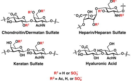
Figure 1.1 Structures of GAG family members. GAGs are made of repeating disaccharide units.
Three of the four families can be sulfated at different hydroxyl groups along the polysaccharide.
chemical composition of GAG structures varies significantly between cell types.
GAGs can be subdivided into four main classes based on their monosaccharide components: (1) chondroitin sulfate and dermatan sulfate, (2) heparin and heparan sulfate, (3) keratan sulfate, and (4) hyaluronan (Figure 1-1). Chondroitin sulfate and dermatan sulfate (CS/DS) contain a repeating disaccharide unit of a hexuronic acid (GlcA for CS and both GlcA and IdoA for DS) and GalNAc.
Heparin and heparan sulfate (hep/HS) are made of a mixture of GlcA and IdoA (~10% GlcA for heparin and 10-50% GlcA for HS) along with GlcNAc, which can be deacetylated as GlcN. Keratan sulfate (KS) exists as repeats of Gal and GlcNAc.
These three GAG structures are synthesized in the Golgi apparatus on their proteoglycan cores and can also be differentially sulfated on their hydroxyl groups. The fourth class of GAGs, hyaluronan or hyaluronic acid (HA), is unique in that it is not attached to a protein structure and is generally much larger than
O O O
R1O HO
O
AcHN O2C
OR1
OR1 O
R1 = H or SO3
R1O O
NHR2 OR1 O O
O OR1 OH O2C
O
R2 = Ac, H, or SO3 O O
O HO
OH AcHN
OR1 O OR1
HOO O
O HOO HOO
AcHN O2C
OH
OH O Chondroitin/Dermatan Sulfate
Keratan Sulfate Hyaluronic Acid
Heparin/Heparan Sulfate
4 the other three classes, existing upwards of 5 MDa or more in mass and 20 µm in length.9 Furthermore, the repeating unit of HA (GlcA and GlcNAc) is assembled at the cell membrane by hyaluronic acid synthases and is completely unsulfated.
For the purpose of brevity, only CS/DS and hep/HS will be discussed further.
The repeating patterns of CS/DS and hep/HS can be classified further by their sulfation patterns. In mammals, CS and DS can be sulfated on the GlcA/IdoA-C2, GalNAc-C4, and GalNAc-C6 hydroxyl positions. Hep and HS can be modified at the GlcA/IdoA-C2, GlcN-C3, and GlcN-C6 hydroxyl positions as well as the free amine on GlcN. These modifications are catalyzed by carbohydrate sulfotransferases that reside in the Golgi apparatus.10 Sulfate groups can be removed by sulfatases; however, this generally occurs in lysosomes after GAG internalization during degradation.11 Only two enzymes, HS 6-O-endosulfatases Sulf1 and Sulf2, are known to modify GAG structures once they are secreted to the cell surface.12
CS/DS and hep/HS biosynthesis follow similar pathways.13, 14 First, a common tetrasaccharide (Xyl-Gal-Gal-GlcA) is appended to Ser residues of various proteins. Usually, this occurs at Ser-Gly/Ala-X-Gly sequences;15 however, this motif is not universally found at all modification sites. Next, the first N- acetylhexosamine residue is attached to the core tetrasaccharide. This step commits the growing strand to either CS/DS (GalNAc) or hep/HS (GlcNAc). The identity of the attached GAG is dependent on a number of factors including the protein core.16 Although certain proteins like versican and glypicans are modified
only by CS or HS, respectively, others like syndecans can contain both structures simultaneously, whereas some proteins like neuropilin-1 have different structures attached to the same Ser residue on different copies of the protein.17, 18 The chains are then elongated by a number of polymerizing enzymes. For CS, GlcA can be epimerized to IdoA at this point to produce regions of DS. For hep/HS, GalNAc is first de-N-acetylated and N-sulfated, after which GlcA residues can be epimerized to IdoA. Finally, sulfotransferases modify hydroxyl groups along the length of the carbohydrate backbone to produce the mature sulfated polysaccharide.
Importantly, these modification reactions do not proceed to completion, increasing heterogeneity of the final structure. Moreover, other than substrate preferences exhibited by the modifying enzymes, it is relatively unclear how the cell orchestrates structural heterogeneity of the produced GAG polysaccharides.
It is hypothesized that regions of high and low sulfation density exist along the oligosaccharide, but little structural information is directly available due to the difficulties in GAG sequencing.19-21 Nevertheless, it is through these different structures that GAGs exert their biological activity by binding to proteins. Thus, to fully understand the biological activity of GAGs, it is critical (1) to discover the interactions between proteins and specific GAG structures and (2) to design methods to change GAG structures at the cell surface to alter and perhaps control biological function.
6 1.2 GAG Binding to the Receptor Tyrosine Kinase Superfamily
To control intracellular activity, GAGs must interact with transmembrane proteins on the cell surface to transfer external information across the cell membrane. One of the largest groups of these proteins is the receptor tyrosine kinase (RTK) superfamily.22, 23 Made up of 58 receptors organized into 20 subfamilies, RTKs are characterized by the presence of an intracellular tyrosine kinase domain that is activated by receptor dimerization, causing cross- phosphorylation of tyrosine residues on opposite receptors and leading to downstream activation of signaling pathways.23 Their extracellular domains are much more structurally diverse, allowing RTKs to function in a variety of fundamental biological processes including cell survival and motility.23-26 Moreover, their dysregulation has been linked to a variety of disease states including cancer, diabetes, and atherosclerosis.27-29
RTKs generally function through binding to extracellular ligands.
Interestingly, many of the RTK subfamilies have been linked to interactions with GAGs – either through binding to soluble protein ligands or to the receptor itself.30-41 One of the most famous examples of the involvement of GAGs is the ternary complex formed by fibroblast growth factor 1 (FGF1), FGF receptor 1 (FGFR1), and hep/HS.30 As illustrated by crystallographic studies, hep/HS binds to the receptor-ligand complex using a binding site that spans both proteins in a 2:2:2 stoichiometric ratio, cooperating to stabilize the active receptor dimer.
Carbohydrate microarray studies have also illustrated that FGFR1 binding to
hep/HS is facilitated by FGF2 binding,36 providing mechanistic evidence for ternary complex assembly. Other examples of GAG binding to ligands and receptors include the vascular endothelial growth factor 1 (VEGF1) and VEGF receptor 1 (VEGFR1) system.32, 42 Surface plasmon resonance (SPR) analyses have shown that VEGFR1 but not VEGFR2 can directly interact with hep/HS. As seen before with FGF2/FGFR1, VEGF1 binding to hep/HS facilitates the formation of a ternary complex containing VEGFR2. More recently, CS-E has been demonstrated to bind to members of the erythropoietin-producing human hepatocellular (Eph) family of receptors.40, 41 CS-E binding to EphA4 and EphB3 can facilitate receptor activation without the canonical ephrin (Efn) ligands, highlighting a novel mechanism of action for RTK signal transduction mediated by GAG binding.
Together, these results illustrate only a small portion of the knowledge gained from the discovery of GAG-RTK interactions and their biological consequences.
However, our understanding of the connections between GAG binding and RTK signaling is far from complete. Given the sheer size of the RTK superfamily and the diverse biological settings where they function, it is quite possible that many new GAG-RTK interactions with significant biological ramifications remain still undiscovered.
8 1.3 Altering Cell-Surface GAG Populations
A variety of approaches have been developed to alter GAG structures at the cell surface and observe the resulting biological phenotype. The majority of these methods are reductive, meaning that they remove GAG structures through biosynthetic inhibition or degradation, and occur through genetic manipulation.
These approaches include gene deletion or knockout, gene knockdown by RNAi, and gene overexpression. Genetic methods offer excellent spatial and temporal control, enabling the precise manipulation of specific genes in a cell-specific and inducible manner. However, because of the linear synthesis of the GAG backbone and the substrate specificity of individual sulfotransferases, the genetic disruption of a single enzyme may lead to dramatic changes in GAG populations and are generally unsuitable to probe in importance of individual sulfation epitopes. For example, N-deacetylation and N-sulfation of GlcNAc in HS biosynthesis is critical for further sulfation reactions. Therefore, knockout of the responsible enzyme N- deacetylase and N-sulfotransferase 1 (Ndst1) leads not only to decreases in N- sulfation but also O-sulfation at all other positions.2, 43 Similarly, the production of the CS-E epitope by carbohydrate sulfotransferase 15 (Chst15) requires the activity of Chst11 to first add a sulfate group to the GalNAc-C4 hydroxyl position and produce the CS-A epitope. Deletion of Chst11 leads to the loss of both the CS- A and CS-E motifs.44 Moreover, knocking out GAG enzymes can lead to developmental defects or embryonic lethality, which can hinder the identification of functions in adult organisms.45 Nonetheless, important discoveries have been
made regarding the necessity of GAGs for proper development using genetic approaches. For instance, the importance of HS in bone maturation has been demonstrated by the production of a mouse model deficient in HS biosynthesis due to a hypomorphic mutation in the HS polymerase Ext1.46 These mice exhibited improper endochondral ossification during development, and this phenotype was attributed to the importance of HS binding with the growth factor Indian hedgehog.
An alternative reductive approach is the use of GAG degrading enzymes to selectively remove carbohydrates at the cell surface. As with genetic approaches, direct delivery or transgenic expression of the enzyme can be finely controlled to provide spatiotemporal selectivity. However, these enzymes lack fine substrate specificity and will at least partially degrade all CS or HS GAGs depending on the enzyme used. Furthermore, the longevity of this approach depends greatly on the stability of the enzyme, and long-term experiments may require multiple deliveries of the enzyme to avoid complications from newly synthesized GAGs.
However, GAG degrading enzymes are invaluable tools to quickly and effectively remove nearly all GAGs of a specific subpopulation. One promising application of this method has been the delivery of chondroitinase ABC (ChABC) derived from the bacterium Proteus vulgaris to sites of spinal cord injury.47-49 Reactive astrocytes produce large quantities of CS after injury to inhibit axonal regeneration,50 and direct injection or viral delivery of ChABC has been associated with neuronal regrowth and functional recovery.47, 48
10 Recently, complementary additive approaches have been developed to overcome the obstacle of structural selectivity found in reductive methods. In these methods, known collectively as de novo glycan display, carbohydrate or glycomimetic structures are directly inserted into plasma membranes using approaches such as lipid insertion, liposomal fusion, or protein conjugation.51-56 These techniques provide excellent control over glycan structure, allowing known epitopes to be displayed for functional analysis. However, exogenous sugars are typically displayed alongside the native glycan population, which could obscure the biological effects of the newly added carbohydrates. To address this complication, de novo glycan display methods can be used in combination with reductive approaches to minimize the contributions of interfering endogenous carbohydrates. The versatility of the technique also allows for the display of a wide range of carbohydrate-based structures, including glycomimetics such as synthetic glycopolymers, glycans appended to simplified proteins, or even the glycan component of glycoproteins alone. As a relatively new field, de novo glycan display has only been applied to a limited number of biological contexts. For example, anchoring of lactosyl or cellobiosyl-containing glycopolymers in the cell membrane by passive lipid insertion was used to examine galectin-mediated crosslinking and aggregation.52 However, prior to the work outlined in Chapter 2, the ability to elicit biological activity as a function of glycan structures at the cell surface was unknown. Our work53, 54, along with other, simultaneous publications in the field55, 56, demonstrated that the display of defined carbohydrate structures
at the cell surface could be used to drive multiple biological processes including immunoevasion, neuronal outgrowth, and stem cell differentiation. Together, these results highlight the utility of de novo glycan display as a novel tool to directly connect carbohydrate structure and biological function unlike other existing methods.
1.4 References
1. X. Lin. Functions of heparan sulfate proteoglycans in cell signaling during development. Development. 2004, 131: 6009-6021.
2. J. R. Bishop, M. Schuksz, J. D. Esko. Heparan sulphate proteoglycans fine-tune mammalian physiology. Nature. 2007, 446: 1030-1037.
3. R. V. Iozzo, J. D. San Antonio. Heparan sulfate proteoglycans: heavy hitters in the angiogenesis arena. J Clin Invest. 2001, 108: 349–355.
4. K. Sugahara, T. Mikami. Chondroitin/dermatan sulfate in the central nervous system. Curr Opin Struct Biol. 2007, 17: 536-545.
5. G. M. Miller, L. C. Hsieh-Wilson. Sugar-dependent modulation of neuronal development, regeneration, and plasticity by chondroitin sulfate proteoglycans. Exp Neurol. 2015, 274: 115-125.
6. K. R. Taylor, R. L. Gallo. Glycosaminoglycans and their proteoglycans: host- associated molecular patterns for initiation and modulation of inflammation. FASEB J. 2006, 20: 9-22.
12 7. R. Sasisekharan, Z. Shriver, G. Venkataraman, U. Narayanasami. Roles of
heparan-sulphate glycosaminoglycans in cancer. Nat. Rev. Cancer. 2002, 2:
521-528.
8. J. D. Esko, K. Kimata, U. Lindahl. Proteoglycans and Sulfated Glycosaminoglycans. In: A. Varki et al., Eds., Essentials of Glycobiology. 2nd Ed. Cold Spring Harbor (NY): Cold Spring Harbor Laboratory Press; 2009.
9. D. Vigetti et al. Hyaluronan: biosynthesis and signaling. Biochim Biophys Acta.
2014, 1840: 2452-2459.
10. N. Gamage et al. Human sulfotransferases and their role in chemical metabolism. Toxicol Sci. 2006, 90: 5-22.
11. H. H. Freeze. Genetic Disorders of Glycan Degradation. In: A. Varki et al., Eds., Essentials of Glycobiology. 2nd Ed. Cold Spring Harbor (NY): 2009.
12. W. C. Lamanna et al. Heparan sulfate 6-O-endosulfatases: discrete in vivo activities and functional co-operativity. Biochem J. 2006, 400: 63-73.
13. T. Mikami, H. Kitagawa. Biosynthesis and function of chondroitin sulfate.
Biochim Biophys Acta. 2013, 1830: 4719-4733.
14. K. Sugahara, H. Kitagawa. Heparin and heparan sulfate biosynthesis. IUBMB Life. 2002, 54: 163-175.
15. R. Raman, V. Sasisekharan, R. Sasisekharan. Structural insights into biological roles of protein-glycosaminoglycan interactions. Chem Biol. 2005, 12: 267- 277.
16. K. Prydz. Determinants of Glycosaminoglycan (GAG) Structure. Biomolecules.
2015, 5: 2003-2022.
17. R. V. Iozzo, L. Schaefer. Proteoglycan form and function: A comprehensive nomenclature of proteoglycans. Matrix Biol. 2015, 42: 11-55.
18. Y. Shintani et al. Glycosaminoglycan modification of neuropilin-1 modulates VEGFR2 signaling. EMBO J. 2006, 25: 3045-3055.
19. J. T. Gallagher. Multiprotein signalling complexes: regional assembly on heparan sulphate. Biochem Soc Trans. 2006, 34: 438-441.
20. M. Ly et al. The proteoglycan bikunin has a defined sequence. Nat Chem Biol.
2011, 7: 827-833.
21. N. Volpi, R. J. Linhardt. High-performance liquid chromatography-mass spectrometry for mapping and sequencing glycosaminoglycan-derived oligosaccharides. Nat Protoc. 2010, 5: 993-1004.
22. J. Schlessinger. Cell signaling by receptor tyrosine kinases. Cell. 2000, 103:
211-225.
23. M. A. Lemmon, J. Schlessinger. Cell signaling by receptor tyrosine kinases.
Cell. 2010, 141: 1117-1134.
24. T. N. Sato et al. Distinct roles of the receptor tyrosine kinases Tie-1 and Tie-2 in blood vessel formation. Nature. 1995, 376: 70-74.
25. P. Duchek, P. Rorth. Guidance of cell migration by EGF receptor signaling during Drosophila oogenesis. Science. 2001, 291: 131-133.
26. P. Duchek, K. Somogyi, G. Jekely, S. Beccari, P. Rorth. Guidance of cell migration by the Drosophila PDGF/VEGF receptor. Cell. 2001, 107: 17-26.
27. E. B. Pasquale. Eph receptors and ephrins in cancer: bidirectional signalling and beyond. Nat Rev Cancer. 2010, 10: 165-180.
28. J. Boucher, A. Kleinridders, C. R. Kahn. Insulin receptor signaling in normal and insulin-resistant states. Cold Spring Harb Perspect Biol. 2014, 6.
14 29. K. V. Woo et al. Tie1 attenuation reduces murine atherosclerosis in a dose-
dependent and shear stress-specific manner. J Clin Invest. 2011, 121: 1624- 1635.
30. J. Schlessinger et al. Crystal structure of a ternary FGF-FGFR-heparin complex reveals a dual role for heparin in FGFR binding and dimerization.
Molecular Cell. 2000, 6: 743-750.
31. C. Rolny, D. Spillmann, U. Lindahl, L. Claesson-Welsh. Heparin amplifies platelet-derived growth factor (PDGF)-BB-induced PDGF alpha-receptor but not PDGF beta-receptor tyrosine phosphorylation in heparan sulfate- deficient cells. Effects on signal transduction and biological responses. J Biol Chem. 2002, 277: 19315-19321.
32. D. Xu, M. M. Fuster, R. Lawrence, J. D. Esko. Heparan sulfate regulates VEGF165- and VEGF121-mediated vascular hyperpermeability. J Biol Chem.
2011, 286: 737-745.
33. L. E. Kemp, B. Mulloy, E. Gherardi. Signalling by HGF/SF and Met: the role of heparan sulphate co-receptors. Biochem Soc Trans. 2006, 34: 414-417.
34. V. Koprivica et al. EGFR activation mediates inhibition of axon regeneration by myelin and chondroitin sulfate proteoglycans. Science. 2005, 310: 106- 110.
35. M. W. Barnett, C. E. Fisher, G. Perona-Wright, J. A. Davies. Signalling by glial cell line-derived neurotrophic factor (GDNF) requires heparan sulphate glycosaminoglycan. J Cell Sci. 2002, 115: 4495-4503.
36. C. J. Rogers et al. Elucidating glycosaminoglycan-protein-protein interactions using carbohydrate microarray and computational approaches. Proc Natl Acad Sci USA. 2011, 108: 9747-9752.
37. P. B. Murray et al. Heparin is an activating ligand of the orphan receptor tyrosine kinase ALK. Sci Signal. 2015, 8: ra6.
38. G. Bezakova, M. A. Ruegg. New insights into the roles of agrin. Nat Rev Mol Cell Biol. 2003, 4: 295-308.
39. T. Arai, A. Parker, W. Busby, Jr., D. R. Clemmons. Heparin, heparan sulfate, and dermatan sulfate regulate formation of the insulin-like growth factor-I and insulin-like growth factor-binding protein complexes. J Biol Chem.
1994, 269: 20388-20393.
40. C. J. Rogers et al. Chondroitin sulfate E influences retinotopic mapping via EphB3. in preparation. 2017.
41. G. J. Miller et al. Chondroitin sulfate E mediates axonal inhibition via EphA4 activation. in preparation. 2017.
42. M. Teran, M. A. Nugent. Synergistic binding of vascular endothelial growth factor-A and its receptors to heparin selectively modulates complex affinity. J Biol Chem. 2015, 290: 16451-16462.
43. E. Forsberg, L. Kjellen. Heparan sulfate: lessons from knockout mice. J Clin Invest. 2001, 108: 175-180.
44. M. Klüppel, T. N. Wight, C. Chan, A. Hinek, J. L. Wrana. Maintenance of chondroitin sulfation balance by chondroitin-4-sulfotransferase 1 is required for chondrocyte development and growth factor signaling during cartilage morphogenesis. Development. 2005, 132: 3989-4003.
45. S. Mizumoto, S. Yamada, K. Sugahara. Human genetic disorders and knockout mice deficient in glycosaminoglycan. Biomed Res Int. 2014, 2014: 495764.
46. L. Koziel, M. Kunath, O. G. Kelly, A. Vortkamp. Ext1-dependent heparan sulfate regulates the range of Ihh signaling during endochondral ossification. Developmental Cell. 2004, 6: 801-813.
16 47. E. J. Bradbury et al. Chondroitinase ABC promotes functional recovery after
spinal cord injury. Nature. 2002, 416: 636-640.
48. R. R. Zhao et al. Lentiviral vectors express chondroitinase ABC in cortical projections and promote sprouting of injured corticospinal axons. J Neurosci Methods. 2011, 201: 228-238.
49. G. García-Alías, S. Barkhuysen, M. Buckle, J. W. Fawcett. Chondroitinase ABC treatment opens a window of opportunity for task-specific rehabilitation.
Nat Neurosci. 2009, 12: 1145-1151.
50. L. L. Jones, R. U. Margolis, M. H. Tuszynski. The chondroitin sulfate proteoglycans neurocan, brevican, phosphacan, and versican are differentially regulated following spinal cord injury. Exp Neurol. 2003, 182:
399-411.
51. D. Rabuka, M. B. Forstner, J. T. Groves, C. R. Bertozzi. Noncovalent cell surface engineering: incorporation of bioactive synthetic glycopolymers into cellular membranes. J Am Chem Soc. 2008, 130: 5947-5953.
52. B. Belardi, G. P. O'Donoghue, A. W. Smith, J. T. Groves, C. R. Bertozzi.
Investigating cell surface galectin-Mediated cross-linking on glycoengineered cells. J Am Chem Soc. 2012, 134: 9549-9552.
53. A. Pulsipher, M. E. Griffin, S. E. Stone, J. M. Brown, L. C. Hsieh-Wilson.
Directing neuronal signaling through cell-surface glycan engineering. J Am Chem Soc. 2014, 136: 6794-6797.
54. A. Pulsipher, M. E. Griffin, S. E. Stone, L. C. Hsieh-Wilson. Long-lived engineering of glycans to direct stem cell fate. Angew Chem Int Ed. 2015, 54: 1466-1470.
55. J. E. Hudak, S. M. Canham, C. R. Bertozzi. Glycocalyx engineering reveals a Siglec-based mechanism for NK cell immunoevasion. Nat Chem Biol. 2014, 10: 69-75.
56. M. L. Huang, R. A. A. Smith, G. W. Trieger, K. Godula. Glycocalyx remodeling with proteoglycan mimetics promotes neural specification in embryonic stem cells. J Am Chem Soc. 2014, 136: 10565-10568.
18 C h a p t e r 2
Methods for Short- and Long-Term Glycan Engineering at the Cell Surface to Control Biological Outcomes
Portions of this chapter are published as:
Pulsipher A, Griffin ME, Stone SE, Brown JM, Hsieh-Wilson LC. “Directed neuronal signaling through cell-surface glycan engineering.” J. Am. Chem. Soc.
2014, 136: 6794-6797. doi: 10.1021/ja5005174. Research article.
and
Pulsipher A*, Griffin ME*, Stone SE, Hsieh-Wilson LC. “Long-lived glycan engineering to direct stem cell fate.” Angew. Chem. Int. Ed. 2015, 54: 1466-1470.
doi: 10.1002/anie.201409258; * denotes equal contribution. Research article.
2.1 Abstract
Cell-surface GAGs are mediators of a variety of critical signaling events and can dictate the biological activity of cell populations through their interactions with cell-surface receptors and soluble ligands. Many approaches to connect structural determinants of the cellular glycocalyx with biological function employ reductive approaches by reducing or eliminating glycan structures from the extracellular matrix. However, a forward approach in which specific glycans are anchored onto plasma membranes would allow more control over the exact carbohydrate structures being displayed at the cell surface. Moreover, this tactic would provide direct evidence for biological activity as a result of a carbohydrate structure. Here, two methods are presented to modify cell surfaces with defined carbohydrate structures. The first method employs the functionalization of CS polysaccharides with an aminooxy group and conjugation to ketone-displaying liposomes to directly fuse with the cell membrane. The second approach describes the transgenic expression of a transmembrane HaloTag construct, which can covalently bond with chlorohexyl-functionalized HS polysaccharides.
These two complementary methods provide both short- and long-term display of known carbohydrates, which allow for the direct control of cellular fate in both neuronal growth and stem cell differentiation.
20 2.2 Liposomal Delivery of CS GAGs to Control Neuronal Growth
2.2.1 Approach and Synthesis
Although some strategies for engineering cell-surface glycans have been reported,1-8 the remodeling of cell membranes with complex polysaccharides such as GAGs has not been demonstrated. Moreover, the application of these methodologies prior to our work has been largely limited to imaging2-5 or studying cell-surface phenomena, such as receptor clustering.6 Here we developed a method to display specific sulfated GAG structures on cell surfaces using a liposomal fusion strategy. Tailoring membranes with the CS-E sulfation epitope activated growth factor-mediated signaling pathways and enabled the fine-tuned modulation of neuronal growth. These findings demonstrate that chemically controlling the presentation of exogenous glycans on cell surfaces can induce sustained effects on cellular signaling and function. Our studies also highlight the potential for glycan engineering to modulate complex cellular events, and they provide a powerful, new tool for remodeling cell membranes with a wide variety of important biomolecules.
We chose to utilize liposomes as glycan carriers due to their biocompatibility, ease of preparation, low cytotoxicity, and tunable biophysical properties.9 Elegant studies have used glycan-presenting vesicles for intracellular antigen delivery10, 11 or sugar-encapsulated vesicles with folate receptors for cell-specific metabolic labeling.4 However, only two liposomal methods have been developed to our
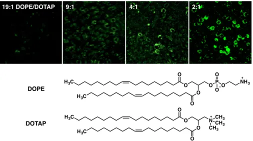
Figure 2-1. Schematic of liposomal delivery method for glycan engineering. Liposomes containing a ketone functional handle are produced and then functionalized with GAGs. Liposomes are then added to cells, where they fuse with the cell membrane to display carbohydrates on the cell surface.
knowledge for the cell-surface display of exogenous molecules, namely fluorophores.12, 13 We expanded on these methods in an effort to incorporate large, sulfated GAGs into cell membranes (Figure 2-1). To promote membrane fusion and surface presentation of the glycans rather than intracellular uptake, we used cationic 1,2-dioleoyl-3-trimethylammoniumpropane chloride (DOTAP) and neutral 1,2-dioleoyl-sn-glycero-3-phosphatidylethanolamine (DOPE) as our primary lipids. Phosphatidylethanolamine lipids are an abundant, natural component of the neuronal cell membrane, and DOPE-based liposomes containing a lipid-functionalized fluorophore have been used to label a variety of cell types, including primary neurons.12 We also incorporated 2-dodecanone into the liposomes to add a ketone handle for appending the glycans via oxime chemistry. CS polysaccharides containing a peptide fragment with an N-terminal amine were readily derivatized with an aminooxy group by coupling them to
Liposome Formation
Functionalization of
Liposomes with GAGs Membrane Fusion with Cells
22
Figure 2-2. Synthesis of aminooxy-functionalized CS GAGs (2-6, 2-7, and 2-8).(a) N- hydroxyphthalimide (1.2 eq), NaHCO3 (1.2 eq), DMF, 60 °C, 2 h, 88%; (b) TFA (3 eq), DCM, 1 h, 99%; (c) EDC (1.1 eq), sulfo-NHS (1.1 eq), 100 mM MES pH 6.0, 300 mM NaCl, RT, 30 min, then polysaccharide-NH2, 50 mM NaHCO3, RT, 12, 60%. (d) hydrazine monohydrate (3 eq), ddH2O, RT, 3 h, 92%.
phthaloyl-protected aminooxyacetic acid followed by cleavage of the phthaloyl group with hydrazine monohydrate (Figure 2-2). The polysaccharides were then incubated with dodecanone-containing liposomes at 25 °C for 3 h to produce GAG-displaying vesicles. The synthetic ease and versatility of this approach represent advantages compared to existing methods, which require the radical- mediated synthesis of polymers end-functionalized with lipids.5-8 With our approach, the lipid reagents are commercially available, and many biomolecules can be derivatized with aminooxy groups, including various glycans, peptides, lipids, nucleic acids, and proteins,14-16 thus providing a general strategy for displaying a diverse range of bioactive molecules.
2.2.2 Validation
Preliminary optimization of liposomal membrane fusion was performed on rat pheochromocytoma (PC12) cells using liposomes functionalized with a
O O O N O
O O
Br O
O O OH N O
O
O NH NO
O
O
O NH H2N O
peptide
peptide
a b c
d
2-1 2-2
CS-E: 2-3 CS-A: 2-4 CS-C: 2-5
CS-E: 2-6 CS-A: 2-7 CS-C: 2-8
Figure 2-3. Optimization of lipid composition of liposomes for membrane fusion. Liposomes were produced with different w/w ratios of DOPE and DOTAP and 10% w/w dodecanone. AF488- hyd was incubated with the liposomes (ddH2O, RT, 3h) to spontaneously react with the presented ketone moiety. PC12 cells were then incubated with the fluorescent liposomes (37 °C, 30 min) and imaged to visualize fusion efficiency.
hydrazide-conjugated fluorophore (AF488-hyd). We found that a 2:1 w/w ratio of DOPE:DOTAP was optimal for membrane fusion, as visualized by fluorescence microscopy (Figure 2-3). To approximate the relative levels of fluorophore incorporation at the cell surface, we incubated liposomes containing varying concentrations of AF488-hyd with PC12 cells on ice for 30 min. Cells labeled with liposomes containing 10 mol % AF488-hyd displayed similar fluorescence signal profiles by fluorescence-assisted cell sorting (FACS) analysis as cells labeled with an anti-CS-E monoclonal antibody17 that detected endogenous CS-E levels (Figure 2-4). These results suggest that this liposomal strategy can incorporate exogenous molecules into cell membranes at levels roughly similar to those of endogenous CS polysaccharides. We next examined whether this approach could be used to
19:1 DOPE/DOTAP 9:1 4:1 2:1
H3C
O O H3C
O O
OP O O
O
NH3
H3C
O O H3C
O O
N CH3 CHCH3 3 DOPE
DOTAP
24
Figure 2-4. Quantifying cell surface labeling by liposomal delivery. PC12 cells were functionalized with liposomes containing different amounts of 2-dodecanone (w/w) that had been reacted with AF-488 and analyzed by flow cytometry. As an approximation, these data were compared to PC12 cells labeled with a CS-E monoclonal antibody.
\
Figure 2-5. Biophysical characterization of conjugated liposomes. (A) Schematic of different liposomes used for characterization. (B) Liposomes were negatively stained with uranyl acetate and imaged using TEM (scale bar = 50 nm). (c) DLS was used to measure average liposome hydrodynamic diameters, which showed increases after functionalization. Zeta potential analysis showed a negative shift in potential after functionalization with sulfated GAGs. (d) Energy dispersion spectroscopy showed the incorporation of sulfur after functionalization with sulfated GAGs.
Counts (x 103)
Rel. Fluorescence Intensity
Untreated 0%
5%
10%
20%
anti-CS-E 10
8
0
100 101 102 103 104
2 4 6
N CH3
NH AF488 O
Liposome Diameter (nm) Zeta Potential (mV)
I 88.2 ± 5.0 69.0 ± 4.3
II 100.7 ± 3.9 56.1 ± 1.9
IIIA 159.6 ± 2.2 -46.2 ± 5.5
IIIE 135.0 ± 2.3 -46.9 ± 0.7
I II IIIA
CS-A Lip (sulfur)
Energy (keV) IIIA Untreated
(sulfur)
Energy (keV)
1 2 3 4 5 6 7 0
I
1 2 3 4 5 6 7 0
O CH3
I II
N CH3 O
NH O
peptide peptide
IIIA and IIIE
A
B
C D
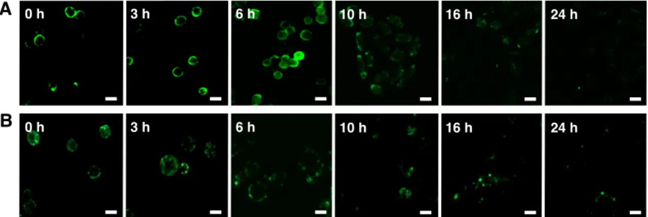
display large GAG polysaccharides on cell surfaces. Liposomes containing 2:1 w/w DOPE:DOTAP and 10% w/w dodecanone were functionalized with CS-E- enriched polysaccharides (∼70 kDa). To characterize their biophysical properties, we used transmission electron microscopy (TEM), dynamic light scattering (DLS), and zeta potential measurements. The liposomes exhibited parameters predicted to favor membrane fusion, including spherical morphologies, average diameters ranging between 132.6 and 159.6 nm, and good stabilities (zeta potentials of ±46−69 mV; Figure 2-5b). The change from positive to negative electrokinetic potential (69 to −46 mV; Figure 2-5c) confirmed successful conjugation of the sulfated polysaccharides. Furthermore, energy dispersive spectroscopy (EDS) verified the presence of sulfur on CS-functionalized liposomes after CS conjugation (Figure 2-5d). To test for membrane fusion, PC12 cells were treated with chondroitinase to remove endogenous CS, incubated with CS-E-modified liposomes, and immunostained with an anti-CS-E antibody.
Importantly, strong immunostaining for CS-E was observed on the surfaces of cells treated with CS-E-functionalized liposomes compared to chondroitinase- treated cells without liposome addition, indicating efficient incorporation of the polysaccharides (Figure 2-6).
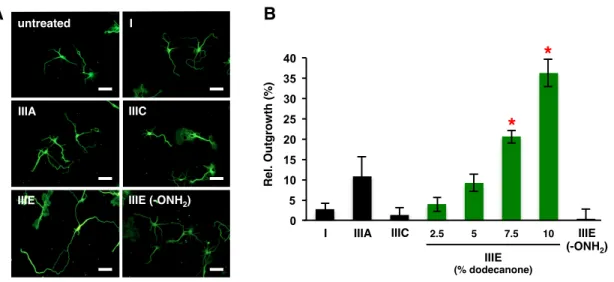
2.2.3 Controlling Intracellular Signaling
Having validated the method, we investigated whether the approach could be used to control cellular signaling pathways. Previous studies from our laboratory