DNA Mechanics and Transcriptional Regulation in the E. coli lac operon
Thesis by
Stephanie Johnson
In Partial Fulfillment of the Requirements for the Degree of
Doctor of Philosophy
California Institute of Technology Pasadena, California
2012
(Defended April 18, 2012)
c 2012 Stephanie Johnson All Rights Reserved
Dedicated to the memory of Jonathan Widom (1955–2011).
Jon was to me a consummate biophysicist, who spoke with equal proficiency the languages of both physics and biology, and whose unique perspective and creativity were evident in all my conversations with him, as well as in the ideas for which he is well known in the broader scientific
community, such as the “mechanical code” to genomes that was a main focus of his work. More admirable even than his scientific prowess, though, was his comportment in interactions with other
scientists: unfailingly polite and considerate despite being at the center of several heated controversies, conveying equal regard and thoughtfulness for a graduate student’s remarks as any professor’s; in Rob’s words in Jon’s obituary, “He loved life, and everyone around him felt happier
and smarter in his presence.”
Acknowledgements
I have loved my time at Caltech and readily acknowledge that this is largely due to the people with whom I have had the privilege of interacting these past six years. First and foremost, my advisor Rob, who has been great fun to work with, and who has been instrumental in both my scientific and personal growth throughout my time at Caltech. Rob’s impact on my professional development is almost too extensive for words, but is probably best exemplified by one of my first interactions with him, as a rotation student in his lab during my first year: I proposed an experiment to try to measure the effects of gyrase without a magnetic trap, and was all set to order the necessary components when Rob rocked my world by asking, “That’s an interesting idea, but why don’t you do a calculation to estimate if the experiment will work or not?” (The calculation gave the experiment a “maybe;” my rotation ended before I could try it.) I have learned an amazing amount from Rob, both about quantitative biology and about bigger life issues (such as, finishing the last 10% is always the hardest!), and I am grateful for how involved he gets with each of his students’
lives. I couldn’t have asked for a better mentor.
I am also indebted to the late Jon Widom, who inspired and closely guided the projects described in this thesis, and who did a lot to shape my idea of what it means to be a scientist; my committee members, Doug Rees, Zhen-Gang Wang, and Niles Pierce, for their advice and guidance on this project throughout my time at Caltech; and Liz Haswell at Washington University in St. Louis, who mentored me on a project that unfortunately did not become part of my thesis, but who taught me a lot about the next stages in an academic career, particularly as I had the opportunity to spend a month in St. Louis just after she started her lab.
It has been a great pleasure to have spent much of the last six years working with (and hanging out
with!) the other members of Rob’s lab, who have become not just scientific collaborators but good friends as well: Lin Han (my predecessor on this project, who laid an amazing amount of foundation for the work that I’ve done), Paul Grayson, Frosso Seirtaridou, Eric Peterson, Dave Wu (never too busy to drop everything and help me with a code bug!), Dave van Valen (who patiently spent many hours explaining math, physics, and Matlab to me), Hernan Garcia (who taught me molecular biology and helped develop the TPM analysis and the second TPM setup), Heun Jin Lee (who often provided much-needed technical advice, especially about surface chemistry and microscopy), Tristan Ursell, Arbel Tadmor, Sidney Cox, Maja Bialecka, Christoph Haselwandter, James Boedicker, Franz Weinert, Dan Jones, Rob Brewster, Mattias Rydenfelt, my fellow TPM-ers, Geoff Lovely and Yi-Ju Chen, and my coauthor and close collaborator on TPM analysis and theory, Martin Lind´en. I also had the opportunity to work with three outstanding undergraduates during my Ph.D. tenure: Kate Craig, who worked with me the summer after my first year when I didn’t know much more than she did, and helped me develop both the TPM technique and the original versions of the “masterscript”
analysis code; Kiefer Aguilar, still a good friend, whom I had the pleasure of watching mature from a kid just out of high school to a young professional college graduate, and who came up with several creative improvements to our TPM assay, such as dual-channel slides; and Chao Liu, who made a lot of progress on the initial stages of the polyA project almost entirely on her own, as she worked with me during one of the busiest times in my grad school career, and who was also a joy to TA with for one quarter of Rob’s APh 161. Linda Song and Pradeep Ramesh, though not technically
“my” undergrads, still helped me quite a bit with teaching and research projects (and were a lot of fun to have around!). I am also very grateful to our admins Linda Scott and Katie Miller, who not only keep the lab from falling apart but have often provided a much-needed sympathetic ear.
The work presented here would not have been possible without the advice and technical expertise of Kathy Matthews at Rice University and Jia Xu in her lab, who spent a week teaching me the Lac repressor purification (and many hours after that helping me debug it back in California); Dan Grilley in Jon Widom’s lab, who helped Yi-Ju and me with making and verifying the DNAs for the polyA project; John Beausang and Phil Nelson at the University of Pennsylvania, who helped
with some of the earlier stages of TPM analysis; and my roommate (for all six years!) Young In Oh in the Hsieh-Wilson lab, Beth Huey-Tubman in the Bjorkman lab, and the Shan lab, for borrowed equipment and advice (especially Beth) on the Lac repressor purification.
I am grateful for the financial support I received from a Virginia Gilloon Fellowship for Women in Science and Engineering, and from a National Science Foundation graduate fellowship.
And finally, a very heartfelt thank you to my family and my friends both in the LA area and in Northern California. As much as I’ve enjoyed my time at Caltech, there were certainly rough parts, and I am incommunicably grateful for the support of my family and friends during those times (as well as for all the laughter during the fun times!). A special thank you to my parents, whose support has taken different forms as I’ve grown up, but has always been overwhelming and unconditional;
and to my brother Matt Johnson and my fianc´e Luke Breuer, who have provided not only emotional but tangible support as well, in the form of lots of IT help and very patient tutorials in math, code writing, and physics (and in some cases, beautifully written code itself).
Abstract
Many gene regulatory motifs in both prokaryotes and eukaryotes involve physical manipulations of the genetic material, often on length scales short enough that the mechanical properties of the DNA significantly impact gene expression. One class of such manipulations, called “action at a distance”, includes transcription factor-mediated DNA looping, in which a binding site some distance away on the DNA is brought into close proximity with the transcription machinery at the promoter. DNA looping is a key component of several important regulatory systems in bacteria, and is crucial to the combinatorial control that is common at eukaryotic promoters regulated by more transcription factors than can physically bind adjacent to the promoter. Here we use a prototypical DNA looping protein, the Lac repressor from E. coli, to explore questions regarding the role of DNA mechanics in DNA looping and combinatorial control, particularly concerning the role of sequence flexibility in short-length-scale looping. We combine a statistical mechanical model of looping by the Lac repressor with a single-molecule technique called tethered particle motion that allows us to quantify this looping, and the systematic tuning of four biologically relevant and experimentally tractable parameters: loop length, loop sequence, repressor-DNA affinity, and repressor concentration. We show that this combination is a powerful approach to measuring repressor-DNA binding affinities and sequence-dependent DNA flexibilities in a way that is orthogonal, and therefore complementary, to conventional ensemble assays. Our results show that the sequence dependence to looping is more complicated than has been observed in other contexts, suggesting that “sequence flexibility” as a general term is misleading, and, we argue, that the measurement of sequence flexibilities depend more strongly than previously appreciated on the shape of the deformation used to make the mea- surement. Finally, we present preliminary results with a more complicated system that is a case
study for broader issues in combinatorial control, and a new hidden Markov model approach, based on variational Bayesian inference, to analyze these more complicated systems, which we hope will allow more precise dissections of, and more robust extraction of kinetic parameters from, tethered particle motion assays.
Contents
Acknowledgements iv
Abstract vii
1 Introduction 1
1.1 The importance of the physical state of the DNA to gene regulation . . . 1
1.2 DNA looping and combinatorial control . . . 4
1.3 The controversial flexibility of DNA at short length scales . . . 7
1.4 The role of sequence flexibility in transcriptional regulation . . . 10
1.5 Thelac operon as a case study for measuring DNA flexibility in the context of DNA looping, and for broader questions of combinatorial control . . . 13
1.6 The single-molecule tethered particle motion assay for studying DNA looping and questions of DNA bendability . . . 15
1.7 Structure of the thesis . . . 18
2 A statistical mechanical model of the in vitro looping probability 24 2.1 Tuning the simple titration curve . . . 25
2.2 The case of multiple looped states . . . 32
2.3 Effect of an inactive fraction of repressor . . . 33
2.4 Effect of the presence of dimers in solution . . . 34
2.5 Effect of cooperative binding of repressor heads . . . 39
2.6 Low repressor concentrations . . . 41
2.7 Calculating relative J-factors . . . 43
2.8 Conclusion . . . 44
3 Precision single-molecule measurements of dissociation constants and J-factors 46 3.1 Improvements over previous work . . . 47
3.1.1 Accurate measurements of dissociation constants and J-factors requires protein purified in-house . . . 47
3.1.2 The PUC306 construct exhibits anomalous behavior even in the presence of protein purified in-house . . . 49
3.2 Computational controls: Dimers at low concentration, the active fraction of repressor, and low repressor concentrations . . . 51
3.2.1 The dimer-to-tetramer transition, and the active fraction of repressor . . . . 52
3.2.2 Data analysis at low repressor concentrations . . . 54
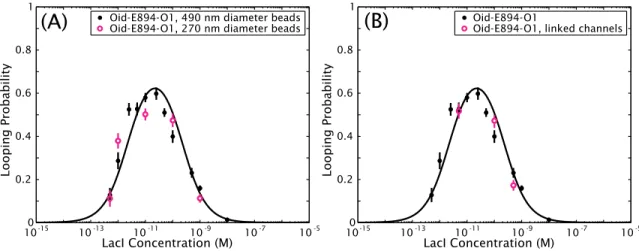
3.3 Experimental controls: Different bead sizes and nonspecific adsorption to chamber walls . . . 55
3.3.1 Smaller beads result in similar looping probabilities . . . 55
3.3.2 No detectable loss of protein to chamber walls . . . 56
3.4 Conclusion . . . 57
4 The sequence dependence of transcription factor-mediated DNA looping 58 4.1 Effect of repressor concentration and operator strength on the looping probability . . 59
4.2 Effect of sequence on the looping probability . . . 63
4.3 Effect of loop length on the looping probability . . . 64
4.4 A need to revisit our understanding of sequence flexibility . . . 67
4.5 Preliminary results with additional sequences . . . 70
4.6 The masking of sequence effectsin vivo by nonspecific DNA-bending proteins . . . . 75
4.7 Conclusion . . . 78
4.A Appendices to Chapter 4 . . . 80
4.A.1 RMS of the unlooped and looped states as a function of concentration and of
loop length . . . 80
4.A.2 Compiling looping predictions from several recent theoretical analysis . . . . 82
5 A kinetic analysis of looping by the Lac repressor 84 5.1 Kinetics of looping by the Lac repressor by conventional methods . . . 87
5.1.1 State lifetimes and missed events . . . 87
5.1.2 Looping rate constants . . . 92
5.1.3 Direct interconversions between looped states? . . . 94
5.2 Preliminary results with a hidden Markov model analysis . . . 96
5.2.1 Overview of a variational Bayesian hidden Markov model analysis of TPM data 96 5.2.2 Examples of results . . . 101
5.3 Conclusion . . . 104
5.A Appendices to Chapter 5 . . . 106
5.A.1 Obtaining kinetic information from dwell time histograms . . . 106
5.A.2 Calculating the dead time of a filter . . . 109
5.A.3 A physical model for the observable distributions . . . 111
6 The three operators of the wild-type lac system: A case study in combinatorial control 116 6.1 A statistical mechanical model of the wild-typelac system . . . 119
6.2 DNA-bending proteins may be essential elements of thelacregulatory system . . . . 124
6.3 Conclusion . . . 128
7 Conclusion 132 Appendices 140 A Detailed derivation of the model that includes the dimer-to-tetramer transition 140 A.1 Assumptions . . . 140
A.2 Derivation ofploop([R]), taking into accountT ⇔2D . . . 142
A.3 Dimers due to damaged protein . . . 148
B DNAs 150 B.1 Constructs containing E8 and 601TA . . . 150
B.2 Constructs containing poly(dA:dT) . . . 153
B.3 Constructs derived from the naturally occurringlac operon . . . 154
C Lac repressor purification 157 D Tethered particle motion: Methods 160 D.1 TPM sample preparation . . . 160
D.1.1 Method summary . . . 160
D.1.2 Detailed protocol . . . 160
D.2 TPM data acquisition and analysis . . . 162
D.2.1 Acquiring data . . . 162
D.2.2 Particle tracking and calculation of the root-mean-squared motion of the bead 163 D.2.3 Determining the looping probability for each trajectory . . . 165
D.2.4 Minimum number of trajectories and minimum observation time . . . 166
D.2.5 Calculating the average looping probability for a set of trajectories . . . 169
D.2.6 Fitting concentration curves . . . 171
D.2.7 Calculating J-factors without concentration curves for each construct . . . 173
E Representative traces 178
Bibliography 183
Chapter 1
Introduction
1.1 The importance of the physical state of the DNA to gene regulation
The publication of the first draft of the sequence of the human genome in 2001 [1], a crucial moment in an effort that began with the first complete genomic sequence of a free living organism (that of the bacteriumHaemophilus influenzae) in 1995 [2], and the publication of numerous other genomes from mice [3] to platypus [4] since then, was in many ways one of the crowning achievements of modern biology. To name two of many revolutionary changes brought about by these fully sequenced genomes, the completion of the human genome ushered in an entirely new era of medical research—
for example, by streamlining the process by which disease genes of unknown biochemical function are identified [1, 5]—and offered a clear path towards a not-so-distant future of highly personalized medical treatment [6]. Fully sequenced genomes have also spawned a host of additional bioinformatic databases that contain information related to, but a level above, the sequence of nucleotides in a genome (e.g., RegulonDB [7] and EcoCyc [8] for E. coli), such as locations of binding sites for transcriptional regulators.
As important as these advances to our understanding of the content of genomes have been, it has become increasingly clear that genomic-sequence and protein-binding-site databanks do not contain the sum total of the information content of a cell’s genome. Rather themechanical propertiesof the DNA polymer in which the genomic sequence information is encoded, and thephysical stateof the
DNA in a cell, are known from many examples to play crucial roles in the regulation of the genetic information encoded by the sequence. For example, cellular differentiation and tumorigenesis often involve the rearrangement of chromatin (the packaged and organized DNA in eukaryotic nuclei), indicating that the localization of a gene in a eukaryotic nucleus can control the level of its output [9, 10, 11]. And it is now clear that mutations to DNA sequence alone cannot account for all aspects of cellular progression from normal to cancerous, but instead that epigenetic changes—including modifications to the structure and organization of the DNA in the cell—play significant roles in the progression of many types of cancers [12].
Perhaps the most telling indicator that genome structure and the mechanical properties of DNA are tightly controlled by cells is the fact that all domains of life express proteins whose sole function seems to be genomic structuring. Eukaryotic genomes are tightly spooled around protein complexes called histones [13], with the resulting DNA-protein complex, called a nucleosome, being the funda- mental unit by which the approximately 3 gigabases of DNA (about 1 meter) are packaged into the roughly 100µm3nucleus [14, 15, 16]. Nucleosomes play a crucial role in the regulation of transcrip- tion as well [14, 15], with genes sequestered into nucleosomes expressed less than genes in the linker DNA that connects adjacent nucleosomes. Bacteria express at least six kinds of “nucleoid-associated proteins” (NAPs), which are thought to package the genome in a similar manner to nucleosomes [17].
Many of these NAPs are DNA-bending proteins—that is, they modify the flexibility of the genomic DNA, not only its organization [17, 18]—and are known to influence gene expression [17, 19, 20].
Mitochondrial genomes (contained in structures called mt-nucleoids) are packaged and organized by nonspecific DNA-bending proteins as well, and there is evidence that the organization of mt- nucleoids changes with cellular metabolic demands [21]. Archea also express at least two kinds of architectural proteins, called chromatin proteins, that compact the genome and probably also in- fluence DNA metabolic processes; one class, called histones, is homologous to eukaryotic histones [22, 23].
Cellular manipulation of the DNA polymer is not restricted to the structuring and packaging of genomes, however. Instead DNA is subjected to a wide variety of physical manipulations in
cellular processes as diverse as the looping events that occur during DNA replication [24, 25], the bending of DNA during recombination [24, 25], and the physical rearrangements of genomic DNA induced by transcription factors [24, 25, 26, 27]. In fact one of the most ubiquitous classes of regulatory architecture found in all domains of life depends upon the physical manipulation of the DNA polymer: so-called “biological action at a distance”, where proteins (often transcription factors) bring two sites separated by some distance on the DNA into close proximity, thus looping the intervening DNA [28, 29, 30].
Interestingly, many of the biological manipulations experienced by DNA, but especially many cases of “action at a distance” in transcriptional regulation, involve bending and twisting the DNA on length scales that are short in comparison with its natural scale of deformation, that is, the per- sistence length (discussed in more detail below) [27, 31]. Eukaryotic DNA is subjected to enormous deformations when packed in nucleosomes, with 147 bp of DNA (already smaller than the persis- tence length) wrapped 1 3/4 times around the histone octamer [13, 26]. Similarly, in the context of prokaryotic transcription factor-mediated DNA looping, not only are such lengths the default in naturally occurring transcriptional networks, but the optimalin vivolengths as determined by the maximal regulatory effect are often at loop lengths smaller than 100 bp [27, 32, 33].
Here we examine the role of the mechanical properties of the DNA polymer, and especially the role of sequence-dependent bendability, in the regulation of gene expression at the level of transcription.
We will focus on the short-length-scale bending that is so prevalent in cellular processes but that, as will be described in more detail below, remains poorly understood. Although many aspects of gene regulation involve such short-length-scale bending, we will focus on the process of DNA loop formation by a prokaryotic transcription factor, with some reference as well to nucleosome positioning, which impacts transcriptional output in eukaryotes, and to the DNA-bending proteins that structure the genome in prokaryotes.
1.2 DNA looping and combinatorial control
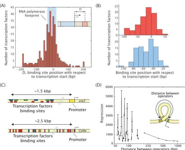
DNA looping, in which two disparate sites on a single DNA molecule are brought together by a single protein or protein complex, is one kind of biological “action at a distance” and occurs in both prokaryotes and eukaryotes, though not necessarily by the same mechanisms in both [25, 26, 28, 29, 30]. The prevalence of loop formation in transcriptional regulation should not be surprising:
given the widespread occurrence of combinatorial control in both eukaryotes and prokaryotes (i.e., the fact that more than one transcription factor at a time often influences the regulatory state of a promoter, as shown for a few key examples in Figs. 1.1(C) and 1.3), it is not surprising that regulatory proteins must bind other sites besides those immediately adjacent to the promoter they regulate [25, 34]. There is only space for one or two regulatory proteins to bind and “touch” the transcription apparatus directly. The side effect of such distal binding is that the DNA has to loop in some way to give access to the promoter of interest.
DNA looping was first discovered in the ara operon in E. coli [35], where it is mediated by a protein called AraC that has two DNA binding domains in the same molecule. When these two domains bind to two sites separated by some distance along the DNA, the intervening DNA is looped out, and the genes of thearaoperon are repressed [35, 36]. Such looping induced by a two- headed DNA binding protein, with one binding site near the promoter of interest and the other some distance away, has since been shown to play a key role in the regulation of several other operons in E. coli, including the lac, deo, and gal operons [26]. It is also a key feature of a well-studied viral protein called the lambda phage repressor, which was the first looping protein whose activity was verified in vitro [26, 37]. In the case of these two-headed looping proteins, looping is thought not only to enable combinatorial control, but also to contribute to efficient transcriptional control by increasing the effective concentration of the transcription factor in the vicinity of the promoter [25].
If one head of a DNA looping transcription factor releases from the DNA, it is more likely to rebind and reform the loop than to dissociate entirely from the DNA, as it is tethered near its binding site by the second head. Additional implications for the role of looping in other aspects of fine-tuning control of transcription continue to be suggested [38, 39].
(A) (B)
D, binding site position with respect to transcription start (bp)
+1 D
-200 -100 0 100 200
0 5 10 15 20 25 30 35 40 45
RNA polymerase footprint
~1.5 kbp
Promoter Transcription factors
binding sites
eve2
~2.5 kbp
Promoter Transcription factors
binding sites
CyIIIa
50 100 250 500 1000
0 1000 2000 3000 4000 5000 6000
Distance between operators (bp) Distance between
operators
(D)
Repression
(C)
0 50
5 10 15 20 25
-100 -80 -60 -40 -20 5
10 15 20
-100 -50 0
0
Binding site position with respect to transcription start (bp)
Number of transcription factors
Number of transcription factors
Figure 1.1: Action at a distance and combinatorial control in prokaryotes and eukaryotes. (A)Many promoters inE.
coliare regulated by one or more transcription factors that bind tens or even hundreds of base pairs away from the promoter they regulate. Shown here are all known transcription factor binding sites inE. coli; many do bind adjacent to the promoter, indicated by the blue box, but a significant number bind some distance away. Data from RegulonDB;
figure courtesy of Hernan Garcia (modified from [40], c2010 Elsevier Ltd). (B)Even at promoters regulated by a repressor (top) or activator (bottom) where that activator or repressor has only one binding site (a subset of the data in (A)), that binding site can be up to 100 bp away from the transcription start site. These cases, even more so than those that make up the rest of the distribution in (A), are suggestive of a key role for loop formation in many regulatory systems. Data from RegulonDB; figure courtesy of Mattias Rydenfelt. (C)Combinatorial control in eukaryotes. In these two well-known examples fromDrosophila(top, adapted from [41]) and sea urchin (bottom, adapted from [42]), not only are many binding sites up to several kilobases away from the promoters, but the inputs from the binding of transcriptional regulators to all of these sites must be integrated to produce the observed output.
In theDrosophilaexample, gradients of several different transcription factors combine to produce the famous “eve stripes” that establish the body plan during development. In the sea urchin example, theCyIIIagene encodes a form of cytoskeletal actin, and is tightly regulated both spatially and temporally during development by a set of at least nine transcription factors, including both activators and repressors, and at least one putative DNA looping protein (the SpGCF1 protein, red boxes) [42]. (D)In this classic example of the effect of loop length on repression of the lacZgene inE. coli(introduced in detail in Fig. 1.3), a significant modulation of gene expression is observed as a function of the spacing of the binding sites (“operators”) that form the boundaries of the DNA loop. The spacings between peaks in repression is roughly 10 bp, the helical repeat of DNA. Such modulation with 10 bp periodicity is a signature of DNA looping. Figure adapted from [27], c2006 Wiley Periodicals, Inc., by Hernan Garcia, based on data from [32].
DNA looping in transcriptional regulation is not limited to those cases in which a two-headed protein is an activator or repressor of the gene. Many genes inE. coliare regulated by transcription factors that bind tens or even hundreds of base pairs away, as shown in the histogram of E. coli transcription factor binding sites in Fig. 1.1(A)), and in fact a significant fraction of these genes seem to be regulated by a single activator or repressor with only a single binding site that is not immediately adjacent to the promoter (Fig. 1.1(B)). Some of these belong to a class of promoters in bacteria that are regulated by an enhancer-dependent mechanism similar to that of eukaryotes, in which a loop forms between a distally bound activator and the promoter of the gene of interest, the most well-known example being the nitrogen-assimilation genes regulated by NtrC [43]. The implication with all of these promoters with distantly bound regulating transcription factors is that they must involve some form of DNA looping to bring the regulatory factors in contact with the transcription machinery at the promoter.
In higher eukaryotes non-adjacent binding sites for transcriptional regulators are the rule rather than the exception, with regulatory sites often located kilobases away from the target promoter [44]. Two well-known examples of eukaryotic promoter regions and their control factors are shown in Fig. 1.1(C). It has long been postulated that these cases of truly long-range action-at-a-distance involve some form of DNA loop formation [29], but it was only within the last decade that such loop formation was demonstrated for eukaryotes in vivo [30, 45]. It appears that in eukaryotes, DNA looping is most often effected not by two-headed looping proteins, as in bacteria, which have the ability in and of themselves to loop DNA; rather, a looped complex is formed by the transcriptional regulator, RNA Polymerase and linker proteins like Mediator or Cohesin [45]. (There are, however, at least two single-protein, bidentate looping complexes in eukaryotes, both involved in cancer in humans, RXR and p53 [46, 47].)
Loop formation is perhaps one of the clearest examples of the importance of DNA mechanics to gene regulation. One of the classic signatures of looping is a modulation of regulatory activity as the distance between the two binding sites for the activator or repressor is changed [25], as shown in Fig. 1.1(D). Regulatory activity at loop lengths shorter than several hundred base pairs shows
peaks and troughs with a periodicity of about 10–11 bp [32, 48], corresponding to the helical period of double-stranded DNA. The interpretation is that minima of loop formation (in the example of Fig. 1.1(D), indicated by troughs in repression) correspond to loop lengths for which the DNA has to betwisted, in addition to beingbent, in order for a loop to form [49, 32, 48, 50, 25]. That is, how twistable the DNA of the loop is, or at least how much it must be twisted—a parameter that can be modulated simply by the addition or removal of one or a couple base pairs in the loop—can have very large effects on the ability of the transcription factor to either activate or repress the gene of interest.
The example shown in Fig. 1.1(D) highlights an intriguing aspect of loop formation that is not as yet thought to be well understood. As noted above, the default loop lengths in many prokaryotic transcriptional networks, and the optimalin vivoloop lengths as determined by maximal regulatory effect, as in the example in Fig. 1.1(D), are often shorter than 150 bp [27, 32, 33]. Even in eukaryotes, where very long loops are more common, the behavior of short DNAs still plays a role in transcrip- tional regulation, in the wrapping of 147 bp sections of the genome almost two full times around the histone cores of nucleosomes. This prevalence of short loops is surprising, given our canonical understanding of DNA as a semi-flexible polymer with a persistence length, a length over which the DNA tends to be straight, of roughly 150 bp [31]. Generally speaking it should be energetically costly to bend DNA at lengths shorter than 150 bp. In part because of the prevalence of short loops and bendsin vivo, however, this canonical behavior of DNA is still a highly controversial issue.
1.3 The controversial flexibility of DNA at short length scales
Despite the clear importance of the short-length-scale mechanical properties of DNA in loop forma- tion as well as the many other cellular processes noted above, there remains both uncertainty and controversy about the ease with which such short DNAs can be deformed [51], and also about the role of sequence at these short scales, particularly in the context of protein-mediated bending or looping [51, 52]. The controversy surrounding short-length-scale DNA bending has been reviewed recently [51] but we will summarize the key points here.
The classic conception of DNA as a semi-flexible polymer is usually encapsulated in the worm- like chain (WLC) model [53], which describes DNA as relatively flexible at long length scales, where entropy dominates the energetics of the polymer, but relatively stiff at short length scales, where elasticity dominates [51]. The parameter that determines the length scale in question is the persistence length, the length over which the polymer is relatively stiff. More precisely, the persistence length is defined as the length over which the tangent vectors of two points on the molecule become uncorrelated. Under typical conditions the persistence length of double-stranded B-DNA is 50 nm, or 150 bp [31], which, as noted above, raises the question of how the short DNA loops and bends that are so prevalent in biology form.
A number of experimental approaches are available for studying the flexibility of DNA [51], but a particularly common one, especially for studying the behavior of DNAs on the order of one or a couple persistence lengths, is ligase-mediated cyclization [54, 55]. In a cyclization reaction, a linear DNA with complementary single-stranded overhangs on either end (“sticky ends”), is mixed with DNA ligase and allowed to close into circles and to form dimers (or, with lower efficiency, higher-order multimers). The activity of the DNA ligase effectively captures a sampling of the ring closure and dimer formation, which are assumed to be fast compared to the activity of the ligase. The outcome of a cyclization reaction is a parameter called thecyclization J-factor, the effective concentration of one end of the DNA in the other, defined as the ratio of the rate of formation of ligated circles to the rate of formation of ligated dimers [54, 56]. A higher J-factor indicates a more flexible DNA.
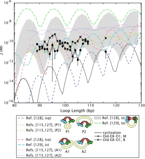
One reason cyclization assays have found such popularity for the study DNA flexibility is that the molecular conformations of all of the players are known, and extensive theoretical work has been done to predict cyclization J-factors as a function of DNA length based on our current models of DNA flexibility. The Shimada-Yamakawa result [57] is one of the most widely used, and will be the basis of comparison for our looping results in Chapter 4 (see Fig. 4.3). As we will see in that chapter, our currently incomplete knowledge about the conformation of the DNA in a protein- mediated loop, in contrast to the simpler case of a ligated DNA minicircle, prevents us from having a similarly clean result for predictedloopingJ-factors. Nevertheless, the Shimada-Yamakawa result
is the usual starting point for discussions of DNA flexibility; and in particular, it predicts that the sub-persistence length loops that are so commonin vivo should be so unfavorable as to form only through the assistance of DNA-bending proteins (such as the architectural proteins discussed in the first section above).
One of the first challenges to the applicability of the Shimada-Yamakawa result at short length scales came from a study by Cloutier and Widom of short DNA fragments derived from the nucleo- some affinity assays discussed in the next section [58]. Usingin vitrocyclization studies (and thus in the absence of any of the DNA-bending proteins presentin vivo), they found the cyclization J-factors for several sub-persistence-length DNAs to be several orders of magnitude higher than predicted by the Shimada-Yamakawa theory. Cloutier and Widom’s result was disputed by Du and coworkers for technical reasons [59], but has continued to inspire controversy and additional experimental and theoretical efforts that attempt to explain the results of Cloutier and Widom (and the subsequent work from others that either support or refute their initial results) [51, 60].
So the question of how short DNA loops and bends formin vivo, and, now,in vitroas well, remains as yet unanswered. Some cases of tightly bent DNA have been solved to a greater extent than the in vitrocyclization results discussed here: for example, the favorable interactions between the DNA wrapped in a nucleosome and the histone proteins around which the DNA is wrapped are sufficient to overcome the energy penalty of wrapping a persistence length of DNA one-and-three-quarters times around the histone core [27]. We will return to this question of the bendability of DNA at short lengths in the context of looping by the Lac repressor in Chapter 4, where we argue that the geometry of a protein-mediated loop and/or flexibility of the looping protein can be sufficient to overcome the energy penalty of bending sub-persistence lengths of DNA into transcription factor-mediated loops.
However we turn now to a less well-studied aspect of short transcription-factor mediated loops, that of the role of sequence, though as we will see this aspect has already been studied, and generated significant controversy of its own, in the context of the tight bends of eukaryotic histones.
1.4 The role of sequence flexibility in transcriptional regula- tion
Although it has been known since the 1980s that the sequence of DNA can impact its flexibility and twistability [61], the implications of this sequence dependence to the mechanical properties of DNA on transcriptional regulation have been studied only in a few select cases. It has been shownin vivo that flexible or pre-bent sequences in (non-loop-forming) promoter regions can increase transcription [62], and that the inclusion of phased A-tracts that introduce static curves into activation loops (such as those mediated by NtrC) can increase transcriptionin vitro[63, 64] andin vivo[65], though activation (and, presumably, loop formation) is surprisingly insensitive to the particular geometry induced by such curved DNAs [63, 66]. In fact an intrinsically curved A-tract DNA is a natural part of thenifLApromoter ofKlebsiella pneumoniae, in a region that is thought to be looped out by NtrC, and this curved DNA is essential for wild-type levels of transcription [65]. Phased A-tracts have been used to examine the effects of intrinsically curved sequences on Lac repressor-mediated DNA loopsin vitro as well; similarly to NtrC loops, it appears that the Lac repressor can accommodate multiple different loop geometries imposed by static bends, with these static bends inducing the formation of hyperstable complexes that remain looped for days [67, 68, 69, 70]. While these studies have provided valuable insights into the role of static bends and loop geometries in loop formation, they have only brushed the surface of the question of what role sequence plays more generally in DNA looping and transcriptional regulationin vitroorin vivo.
Though the role of DNA sequence has not been extensively studied in the particular case of transcription-factor mediated looping, it has become a key parameter in the discussion of a different mechanism of transcriptional regulation, that of nucleosome positioning in eukaryotes [14]. Nucleo- somes do not have a defined binding site sequence and can form on any DNA of sufficient length; but they do preferentially bind to some sequences over others. A number of sequences with very different nucleosome affinities have been identified, some isolated from natural sources and others from nu- cleosome affinity assays with synthetic sequences [14]. It has been argued for both classes that their
-3 -2
-1 0
-1
0
1
2
3
∆∆Gcyc 0 (k
BT)
∆∆G lcun0 k( B)T
5S 601TA
E8 E6 E13
90 95 100 105 110 115
10-12 10-11 10-10 10-9 10-8
DNA length (bp) J (M) cyc
E8TA
(A) (B)
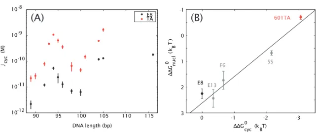
Figure 1.2: Sequences with high nucleosome affinities also have high cyclization J-factors. (A) J-factors for two different sequences, a putatively more flexible sequence called 601TA (red, here abbreviated “TA”) that has a high affinity for nucleosomes, and a random sequence called E8 (black) which has a lower nucleosome affinity, as determined by ligase-mediated cyclization assays. At all lengths tested, DNA minicircles composed of the TA sequence form more readily than those of the E8 sequence. Adapted from [85]. (B)A sequence’s propensity to be wrapped in a nucleosome (here represented by the energy of forming a nucleosomein vitro, ∆∆Gnucl) correlates with its propensity to form DNA minicircles (here represented by the energy of forming these minicircles, ∆∆Gcyc). E8, E13, and E6 are all synthetic random sequences; 5S is a strong natural nucleosome positioning sequence from sea urchin. Adapted from [58].
nucleosomal affinities stem from different intrinsic flexibilities, and not in response to some other in vivo condition or to a property specific to nucleosome binding [58, 71, 72]: because nucleosomes involve tight bending of short DNAs, sequences with high intrinsic flexibilities are thought to de- crease the energy of nucleosome formation, yielding the observed positioning preferences. Though a corollary hypothesis, that sequence flexibility confers preferences for nucleosome positioning and/or occupancy in vivo, is quite controversial [15, 73, 74, 75, 76, 77, 78, 79, 80], the original in vitro claim has nevertheless led not only to many theoretical and experimental studies on the relationship between sequence and flexibility [51, 52, 81, 82, 83, 84], but also to the elucidation of numerous sequence “rules” that can be used to predict the likelihood that a nucleosome will prefer certain sequences over others [14]. Algorithms that predict nucleosome positions based on these sequence rules are highly predictivein vitro, even for sequences from organisms that do not themselves con- tain nucleosomes [77]. The outstanding question in the field is how predictive these sequence rules, and sequence effects in general, are of nucleosome positionsin vivo, as compared to other potential nucleosome positioning factors such as chromatin remodeling complexes [15, 78].
More importantly for the purposes of this study, the claim that a sequence’s nucleosome affinity stems from its degree of intrinsic flexibility has also led to the determination of certain sequences that are claimed to be highly flexible in a general sense. For example, Cloutier and Widom characterized a sequence selected from a chemically random pool of sequences, which they called 601TA, and which they showed to have a significantly higher affinity for nucleosomes than a synthetic random sequence called E8 [58, 85, 86]. In fact the 601TA sequence, which we will henceforth abbreviate TA, is the strongest known nucleosome positioning sequence, either synthetic or natural [14, 86], and is often used in in vitro assays to ensure the localization of nucleosomes at a precise, desired position (e.g., [87]).
Like other previously-described nucleosome positioning sequences [71, 72], the argument that the TA sequence’s high affinity for nucleosomes stems from a high intrinsic flexibility is based largely on the results of in vitro ligase-mediated DNA cyclization assays (described in the previous section).
For short DNAs at least, relative to the persistence length of 150 bp, more flexible sequences should cyclize more readily than other sequences, and therefore should have higher J-factors. Cloutier and Widom showed this to be the case for the TA versus E8 sequences, as shown in Fig. 1.2(A), and moreover they correlated the cyclization J-factors for a number of sequences with the energy required to form a nucleosome with these sequences, as shown in Fig. 1.2(B) [58, 85].
It is generally assumed that cyclization assays are a useful tool for measuring sequence flexibility in some general sense and for learning about DNA looping as well [29, 58, 84, 88], with an implication that sequences that appear to be more flexible in cyclization assays might likewise lead to increased loop formation, just as they have been found to increase nucleosome formation. That is, if TA and E8 differ in mechanical bendability in some general sense, then TA should increaseloopingby a bacterial transcription factor just as it increases nucleosome binding and cyclizes more readily than E8. Therefore these sequences, which yield such strong sequence effects in two other in vitro assays, should be ideal for addressing the question of how sequence affects DNA looping in vitro and, perhaps,in vivo. As we will see, the question of a sequence’s flexibility is actually more subtle than these nucleosome formation and cyclization assays reveal.
1.5 The lac operon as a case study for measuring DNA flex- ibility in the context of DNA looping, and for broader questions of combinatorial control
In this work we exploit insights about DNA flexibility garnered from one class of genetic regulation where it has been studied extensively, that of nucleosome formation, to make predictions about how a different class of mechanical deformations in regulatory biology, that of DNA looping by a prokaryotic transcription factor, will be altered by these same sequences. As described in the next section, we test these predictions experimentally with a single-molecule assay in conjunction with ideas from statistical mechanics for the case of one of the most well-known transcriptional regulators in bacteria, that of the Lac repressor, though there are clear implications for other prokaryotic and eukaryotic regulatory motifs as well.
The discovery in 1961 by Jacob and Monod of genes whose products regulated the transcription of other genes [89] led to a restructuring of our understanding of both the content and management of cells’ genomes. Thelacoperon inE. colihas since become a paradigm of genetic regulation at the level of transcription initiation [90] and continues to be an area of intense research even after more than 40 years (for just two of many examples that illustrate several outstanding questions about this system, see the recent work of [38, 70]).
Thelacoperon, shown in Figure 1.3, encodes a set of three structural genes involved in the uptake and metabolism of the sugar lactose, and one regulatory gene whose product controls transcription initiation at the single promoter for the polycistronic mRNA encoding the three structural genes [91].
The product of the regulatory gene, called the Lac repressor or LacI, is a 154 kDa homotetramer whose binding to a site on the DNA (the O1 operator) overlapping thelacZYApromoter (also called Plac) prevents the binding of RNA Polymerase to the promoter, thereby decreasing transcription [91, 95]. However, when lactose is present, a derivative of lactose binds to the repressor and changes its conformation such that its affinity for O1 is significantly decreased. As a result the repressor no longer out-competes the polymerase for binding to the promoter and transcription can readily occur
380 bp O1
O3 O2
71 bp lacI CAP Plac
Pi lacZ lacY lacA
~1200 bp
3072 bp 1251 bp 609 bp
Figure 1.3: Schematic of the lac operon. The three structural geneslacZ, lacY, and lacA are transcribed as a polycistronic mRNA and encode the proteins β-galactosidase, lactose permase, and galactoside acetyltransferase.
These proteins break lactose into galactose and glucose, transport lactose into the cell, and acetylate galactosides respectively [91] (the natural substrate(s) and specific role of galactoside acetyltransferase in the operon are unknown [92]). ThelacIgene, located upstream of the three structural genes and under the control of a separate promoter, encodes the Lac repressor, which can bind to any two of the operators O1, O2, and O3 simultaneously (see also Fig. 1.4) [93, 94, 32]. O1 overlaps the promoter for the three structural genes and impedes the ability of RNA polymerase to transcribe thelacZ, lacY, andlacAgenes [91]. The operon also contains a binding site for the CAP-cAMP complex, a positive regulator of the operon, between O1 and O3 [91, 95]. Although the protein coding region for the Lac repressor ends before O3, transcription of thelacIgene is known to continue into the regulatory region of thelacZYApromoter, with the regulatory region of thelacpromoter possibly serving as the terminator of transcription fromPi[96].
[38, 91]. Thus the function of the repressor is to coordinate the transcription of lactose-metabolizing proteins with the presence of lactose as a carbon source.1 Additionally, a separate positive regulation mechanism involving cyclic AMP (cAMP) and the CAP protein coordinates transcription of thelac operon with the presence or absence of the preferred carbon source, glucose [90, 91].
After this elegant repressor-mediated model of transcriptional regulation was proposed, it was determined that there are actually two additional Lac binding sites in the general region of thelac promoter [97, 98] (Fig. 1.3). Originally termed “pseudo-operators” because they did not seem to affect binding of the Lac repressor to DNA in vitro [99], it later became clear that both of these auxiliary operators (as they are now known) and the main operator must be present for maximal levels of transcriptional repressionin vivo[93], a puzzle later solved by the discovery that each dimer of the tetrameric repressor binds a separate site on the DNA, thereby forming a loop in the DNA [49, 93, 94, 100]. Not only, as mentioned above, does such looping offer the advantage of increased local concentration of the repressor, in the case of repression, as with the Lac protein, looping further sequesters the polymerase binding site on a curved DNA fragment, enhancing the repressor’s ability to prevent initiation [25].
The Lac repressor, because it has been so extensively studied, offers one of the best case studies
1Even when the repressor is fully active transcription of the operon is not completely inhibited, as small amounts ofβ-galactosidase and lactose permease are necessary to produce the lactose derivative that inactivates the repressor upon first exposure to lactose [91, 38].
for examining the role of DNA mechanics in loop formation and transcriptional regulation. The Lac repressor has been a popular choice in many studies with synthetic looping constructs, both in vivo and in vitro, where the naturally occurring three-operator architecture is usually replaced by synthetic two-operator versions, and often with completely non-natural sequences comprising the intervening loop DNA (e.g., [32, 49, 50, 101]). Moreover, because the naturally occurring architecture does in fact contain more than two binding sites, with the potential to form multiple loops, plus the binding site for a transcriptional activator (CAP), the Lac repressor and the wild-type regulatory region offer a convenient potential case study in broader studies of combinatorial control.
In this work we will use looping by the Lac repressor in ain vitrosingle-molecule assay, described in the next section, as a tool to probe the role of DNA mechanics in loop formation, specifically the role of the two DNA sequences described in the previous section, and to gain insight into the interplay between sequence flexibility and transcriptional regulation by action at a distance both in vitro and in vivo. The bulk of this work will make use of the kinds of synthetic two-operator looping constructs that are typically used in studies with the Lac repressor. However, Chapter 6 will discuss the extension of our looping assay to the full, three-operator construct that forms the natural architecture and demonstrate our ability to dissect more complicated architectures as well.
1.6 The single-molecule tethered particle motion assay for studying DNA looping and questions of DNA bendability
Single-molecule biophysics has provided a new generation of insights into the molecular machines that underlie cellular dynamics. One of the most important classes of such experiments has focused on the interaction between DNA and its protein binding partners, as in the looping experiments that we describe here. Many (though not all) of the single-molecule techniques that can be used to monitor loop formation and other deformations in DNA in real time rely on the imaging of microscopic reporter particles or “beads” which are attached through specific, non-covalent small- molecule interactions to the DNA and/or to the protein of interest. These beads, which can be
imaged under a microscope (while the molecular players cannot), act as reporters of the underlying molecular dynamics.
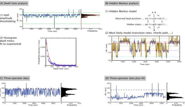
In this work we use the single-molecule tethered particle motion (TPM) technique to study looping by the Lac repressor [102, 103, 104, 105]. As shown schematically in Fig. 1.4(A), a TPM experiment consists of a linear piece of DNA attached at one end to a microscope coverslip and at the other to a microsphere. The dynamics of the microsphere then serve as a readout of the hidden underlying dynamics of the DNA and its partner proteins. In the specific case of looping by the Lac repressor that we are considering, when two operators are present on the DNA tether and repressor is introduced into the sample, the repressor can bind the two operators simultaneously and stabilize a loop in the tether. This loop reduces the effective length of the tether and so reduces the extent of the bead’s Brownian motion. As a result, the formation and breakdown of loops can be observed either directly, by measuring the bead’s distance from the coverslip [106], or indirectly, as will be done here, by measuring the root-mean-squared motion of the bead in the plane of the coverslip [102, 103]. These measurements result in a telegraph-like signal (see examples in Appendix E) and can be converted into the probability of the system being in the looped state: one useful definition of the looping probability is that it is the total time spent in the looped state divided by total observation time. TPM has been used to examine processes ranging from DNA looping by transcription factors [104, 107, 108, 109], as discussed here, to the dynamics of recombination proteins [110, 111] and restriction enzymes [111], and other processes associated with translation and DNA rearrangement [112]. Looping by the Lac repressor in particular has also been extensively studied by TPM [104, 108, 109, 113, 114, 115, 116].
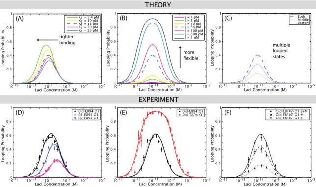
Here, however, we go beyond previous uses of TPM to study DNA looping by combining this single-molecule experiment with a statistical mechanical model and the systematic variation of four biologically relevant parameters (Fig. 1.4(B)): repressor-operator affinity, loop length, loop sequence, and repressor concentration. In all previous Lac repressor studies with TPM, or other single-molecule techniques such as FRET [67, 68, 69, 70], only one or a couple loop lengths, operators, and repressor concentrations were studied. In many cases, therefore, the repressor-operator dissociation constants
O2 TA SL
DI
E 144 bp 89-116 bp 172 bp O1
O1
Oid ... E8
TUNABLE BIOLOGICAL PARAMETERS:
Operator Strength
Loop Length
Loop Sequence
Repressor Concentration
reporter bead
microscope slide tethered
DNA binding
sites
tunable DNA region
looping protein
(B)
EXPERIMENTAL SETUP:
(A)
BE AD
Figure 1.4: Schematic of the tethered particle motion (TPM) assay. (A) DNA looping is observed as a result of changes in the Brownian motion of the tethered bead [102, 103, 104, 105]: looping decreases the effective length of the DNA tether, which decreases the bead’s root-mean-squared (RMS) motion. Most of the work here will be concerned with a parameter derived from the RMS motion, the looping probability, which we define as the time spent in the looped state(s) divided by the total observation time. Chapter 5 looks at kinetic parameters that can also be determined from the motion of the bead. (B)Four distinct tunable biological parameters: 1. Repressor binding site, or operator. Most of the work here uses the strong, synthetic “Oideal” (Oid) operator, the strongest naturally occurringO1 operator, and the weaker naturally occurringO2operator (see Chapter 6 for studies that also involve the weakest naturally occurring operator,O3). 2. Loop length. The wild-typelacoperon contains the three operators O1,O2, andO3, which have the potential to generate three loops of different lengths (see also Fig. 1.3): theO1-O2
loop is 380 bp, theO1-O3 loop is 71 bp (shorter than the persistence length of DNA), and the O2-O3 loop is 472 bp. In our synthetic constructs (see Chapters 3 and 4) we use two operators and systematically tune the distance between them as shown in the figure. 3. Loop sequence. Most of the work discussed here will focus on two sequences,
“E8” and “TA”. “E8” refers to a synthetic random sequence, “TA” to a synthetic nucleosome positioning sequence (part of the 601TA sequence [86]). The TA sequence has a higher cyclization J-factor than E8 and is wrapped into nucleosomesin vitromore readily than E8 [58, 85]. See Section 4.5 for discussion of an additional sequence also related to nucleosome formation, and Chapter 6 for a discussion of the sequences of the wild-typelacregulatory region. 4.
Lac repressor concentration. One of the key tools we will use in this work is the concentration titration, where the looping probability is measured as a function of the repressor concentration. DNA constructs will be referred to with the operator closest to the microscope slide listed first; operator and loop sequences are given in Appendix B. The promoter-containing DNAs of Fig. 4.2 are identical to those shown here except that the O1 operator closest to the bead has been replaced by O2, 36 bp of the loop closest to this O2 operator are replaced by thelacUV5 promoter sequence, and the length of the flanking DNA between O2 and the bead is 139 bp rather than 172 bp. Fig. B.4 shows the flanking regions of the three-operator constructs of Chapter 6.
were assumed (as opposed to measured) in order for a looping J-factor to be calculated. Here we are describing a new way of measuring both the operator dissociation constants and the relative flexibilities of different DNA sequences as contained in the looping J-factor, by tuning both repressor concentration and operator strengths, with a rigorous comparison between these experiments and the theoretical models we have developed. We will argue here that only through this systematic tuning of parameters and interplay between theory and experiment is it possible to uncover some of the surprises, particularly about sequence-dependent flexibility in vitroandin vivo, that will be detailed in the following chapters.
The most important of the parameters that we will tune in this study for the purpose of the main goal of this work, that of investigating the role of sequence flexibility in loop formation, is the flexibility of the DNA in the loop, which is captured in a parameter called the looping J-factor. The looping J-factor is analogous to the cyclization J-factor, introduced above, obtained in the ligation- mediated cyclization assays which are commonly used to measure DNA flexibility at short lengths, and can be thought of as the effective concentration of one end of the loop in the vicinity of the other [56, 57]. The J-factor therefore provides a measure of the energetics of bending the DNA into the loop. The approach we have developed here allows us to measure these looping J-factors in a way that provides quantitative insights into how each of the four biologically important parameters we test affects DNA looping and permits us to contrast the role of sequence in DNA cyclization and nucleosome formation with that of looping. As we will see, we find that the two sequences discussed above, E8 and TA, which have significantly different propensities for forming DNA minicircles inin vitrocyclization assays or for forming nucleosomes, create a more complicated sequence dependence in the context of DNA loop formation than has been previously appreciated.
1.7 Structure of the thesis
The remainder of this thesis is organized as follows:
In Chapter 2 we develop the theoretical framework that both drives our experimental design and allows us to interpret our experimental results. We begin by analyzing in detail the effects that
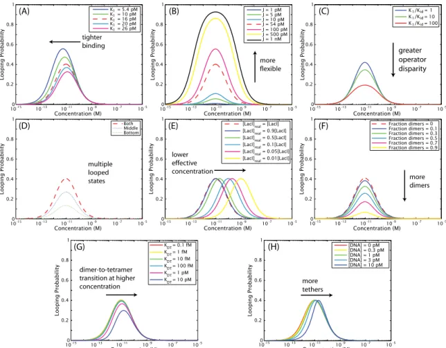
100-15 10-13 10-11 10-9 10-7 10-5 0.2
0.4 0.6 0.8 1
Concentration (M)
Looping Probability
K = 5.4 pM K = 10 pM K = 16 pM K = 20 pM K = 26 pM
tighter binding
(A) 11
1 1 1
100-15 10-13 10-11 10-9 10-7 10-5 0.2
0.4 0.6 0.8 1
Concentration (M)
Looping Probability
[LacI]real = [LacI]
[LacI]real = 0.9[LacI]
[LacI]real = 0.5[LacI]
[LacI]real = 0.1[LacI]
[LacI]real = 0.05[LacI]
[LacI]real = 0.01[LacI]
lower effective concentration
(C)
100-15 10-13 10-11 10-9 10-7 10-5 0.2
0.4 0.6 0.8 1
Concentration (M)
Looping Probability
J = 1 pM J = 5 pM J = 10 pM J = 54 pM J = 100 pM J = 500 pM J = 1 nM
more flexible
(B)
Figure 1.5: Predictions of our statistical mechanical model (Eq. (2.1)) for the effect of intentionally or unintentionally tuning various parameters of the system on the looping probability that we measure with TPM, described in more detail in Chapter 2. One of the key experimental tools we will use in this work is the concentration titration, in which the looping probability is measured as a function of repressor concentration, and so these concentration titrations are the lens through which we view the predictions of the model as well. (A)Prediction of the model for the effect of changing the affinity between the repressor and one of the operators, expressed as a change in one of the two repressor-operator dissociation constants (K1). As will be demonstrated mathematically in Eq. (2.3) and Eq. (2.5), decreasing theKd of one operator both increases looping and shifts the maximum of looping to lower repressor concentrations. (B)Prediction of the model for the effect of changing the J-factor of the loop, that is, changing its flexibility. As derived in Eq. (2.5), increasing the J-factor leaves the maximum of looping unchanged but increases looping at all concentrations. (C)In Chapter 2 we consider the effect not only of deliberately tuning the four parameters of Fig. 1.4, as we show here in (A) and (B), but also of unintentional parameter “tuning” caused by potential experimental artifacts. For example, we asked what a concentration titration would look like if there were a discrepancy between the actual concentration of repressor in the TPM chamber, and the concentration we believed we pipetted into the chamber. One example of how such a discrepancy could arise is the loss of repressor from solution by adsorption to the chamber walls. We find that the effect of such a concentration titration is a simple horizontal translation that leaves therelativevalues of the dissociation constants and J-factors unchanged, but does affect our measurement of theirabsolutevalues. The possible loss of protein to chamber walls is tested explicitly in Chapter 3 and found to be negligible.
the four experimentally tunable parameters of Fig. 1.4 (repressor concentration, loop length, loop flexibility, and operator strength) should have on the looping probability that we observe by TPM.
These predictions are summarized in Fig. 1.5(A–B). We then turn to consequences of potential but unintended experimental effects on the looping probability, such as those caused by the adsorption of protein to the TPM chamber walls, as shown in Fig. 1.5(C). The theoretical explorations of this chapter make specific and testable predictions for the changes to the looping probability we might observe through these intentional and unintentional experimental changes.
In Chapter 3 we relate the work presented here to previous work from the Phillips lab, describing the improvements necessary to report accurate dissociation constants and J-factors, as will be done in later chapters. We also present several experimental and computational controls and other veri- fications of the validity of our combined theory plus TPM approach, most of which are motivated by the considerations of Chapter 2 and are explicit tests of the predictions of that chapter. For example, we designed an experiment to test whether we lose protein to the chamber walls, such that
10-150 10-13 10-11 10-9 10-7 10-5 0.2
0.4 0.6 0.8 1
LacI Concentration (M)
Looping Probability
Oid-E894-O1 Oid-TA94-O1
(B)
10-150 10-13 10-11 10-9 10-7 10-5 0.2
0.4 0.6 0.8 1
LacI Concentration (M)
Looping Probability
Oid-E894-O1 O1-E894-O1 O2-E894-O1
(A) (C)
90 95 100 105 110 115
0 0.2 0.4 0.6 0.8 1
Loop Length (bp)
Looping Probability
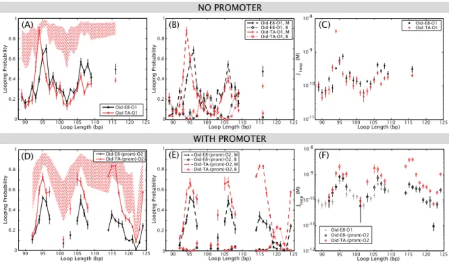
120 125 Oid-E8-O1 Oid-TA-O1
Figure 1.6: The sequence dependence of looping is more complicated than has been observed in cyclization and nucleosome affinity assays, as suggested by these results from Chapter 4. (A) We first demonstrate the strength of our combined statistical mechanical model and TPM approach for measuring biologically important parameters such as dissociation constants and J-factors, by showing that the effect of changing the strength of one operator agrees well with the theoretical predictions of Chapter 2, and that the dissociation constants we measure agree well with values obtained from bulk biochemical assays. Shown here is the looping probability as a function of repressor concentration, for 94 bp of the random E8 sequence flanked by three combinations of operators. As predicted in Fig. 1.5(A), increasing the strength of binding to one of the operators increases looping and shifts the maximum of looping to lower repressor concentrations. Curves are fits of Eq. (2.1) to the data, from which we obtain dissociation constants for the operators and J-factors for the DNA in the loop. (B)Our model is also robust to changing the J-factor: as predicted by Fig. 1.5(B), changing the sequence of the loop to the putatively more flexible sequence TA does not change the dissociation constants or the location of the maximum, but does increase looping at all concentrations. We find the J-factor for this 94 bp loop of TA to be about 10 times larger than that of a 94 bp loop of E8, qualitatively consistent with our expectations from cyclization and nucleosome affinity assays that TA is more flexible in some general sense than E8. (C)However, when we measure the looping probability of these two sequences at fixed repressor concentration but varying loop length, we find a sequence dependence to looping only at the 94 bp used in the concentration titrations, highlighting the importance of systematic experiments tuning several experimental parameters in order to fully capture the behavior of the system. The red hatched region indicates a prediction for where the TA data were to fall if the TA sequence were as m