Dorsal-Ventral Patterning and Gene Regulation in the Early Embryo of
Drosophila melanogaster
Thesis by Mayra Garcia
In Partial Fulfillment of the Requirements for the Degree of
Doctor of Philosophy
California Institute of Technology Pasadena, California
2012
(Defended May 17, 2012)
© 2012
Mayra Garcia
All Rights Reserved
To my parents, Pedro and Aureliana, for always being supportive
and
my sisters, Analu and Ybette, for being great rolemodels
Acknowledgments
First I would like to thank my advisor, Angela Stathopoulos, who has been very supportive throughout my graduate career. She is a wonderful advisor and was always available for guidance when I needed help. She also challenged and pushed me to do my best. By leading by example; she has shown me that with a lot of hard work and sometimes a little bit of luck you can accomplish a lot.
I would, also, like to thank all of the past and current members of the Stathopoulos lab who have provided excellent advice and discussion throughout the years. Especially, Snehalata Kadam who has been a wonderful bay-mate and was always available when I had genetics questions. Also, Desirea Mecenas who was very helpful when I first joined the lab and did not know my way around.
I give many thanks to my advisory committee, Marianne Bronner, Carl Parker, and Ellen Rothenberg. I enjoyed meeting with them and talking about my work because they always had great feedback. I am especially indebted to Carl Parker who helped me by sharing large numbers of embryos with me and showing me how to make nuclear extracts.
My undergraduate advisor, Jennifer Armstrong, and the other professors at Scripps College, who did an excellent job preparing me for graduate school, also deserve my sincere gratitude.
Also, I must thank my family who has always been very supportive and understanding. I know they do not really understand what I have been doing for the past seven years, but nonetheless, they are still proud of me. I do not think I would have made it to graduate school without having wonderful parents who taught me to work hard and
not give up. They have, also, allowed me to disappear for long stretches at time when I was overly involved in my work and only gave me a tiny bit of grief for it.
Lastly, I would like to thank Tony for allowing me to get away from my work for a while especially when things became stressful.
Abstract
In order for an embryo to develop and form properly, the anterior-posterior and dorsal- ventral axes must be specified. This is accomplished by controlled regulation of gene expression that allows for the activation and repression of tissue specific genes.
Patterning of the Drosophila dorsal-ventral axis is an excellent model for understanding how axis specification is controlled. The dorsal-ventral axis is patterned by a nuclear gradient of Dorsal that is highest in ventral regions of the embryo. Dorsal activates genes in a concentration dependent manner to establish early patterning of the embryo. The patterns are refined by interactions between Dorsal and other activators as well as repressors in both dorsal and ventral regions of the embryo. Until recently there was only evidence for repressors acting in ventral regions of the embryo but our studies and other recent studies have provided evidence to suggest that several repressors act in dorsal regions of the embryo to refine Dorsal target genes. Here we show that an element, the A-box, previously identified in the cis-regulatory module (CRM) of the gene intermediate neuroblast defective (ind), is necessary and sufficient to mediate dorsal repression and is also involved in activation of ind. We conducted an affinity chromatography assay and identified factors that bound the A-box element. One of the factors that bound to this element, Grh, activates ind. We also identified several chromatin-remodeling factors that may function to silence ind in dorsal regions of the embryo. Our results also indicate that a second tier of repression that is independent of the A-box element, mediates repression of ind via Dpp-signaling. We extended our studies to the CRMs of ventral neuroblast defective (vnd) and short gastrulation (sog).
Using a chimeric CRM repression assay, we found that strong and weak dorsal repression
are also mediated by the vnd and sog CRMs, respectively. This suggests that limiting amounts of Dorsal are not sufficient to establish the dorsal borders of dorsal-ventral patterning genes as was previously believed, and rather, repressors are used to establish these borders.
Contents
Chapter 1. Introduction 1
Chapter 2. Lateral Gene expression in Drosophila Early Embryos is Supported by Grainyhead-mediated Activation and Tiers of Dorsally- Localized Repression 21
Abstract 22
Introduction 23
Results 28
Discussion 51
Materials and Methods 58
Chapter 3. Identification of Chromatin Factors that Bind the ind Cis- Regulatory Module 63
Abstract 64
Introduction 65
Procedure 67
Results and Discussion 70
Chapter 4. Repressors Play a Role in Establishing the Dorsal and Ventral Borders of Neurogenic Ectoderm Genes 78
Abstract 79
Introduction 80
Results 82
Discussion 90
Chapter 5. Discussion 94
Appendices 101
Appendix A. Supplemental Information: Chapter 2 101
Appendix B. Supplemental Information: Chapter 3 104
Bibliography 105
Chapter 1
Introduction
Embryonic development is a very complex and elegant endeavor. Starting from a single cell, an embryo must become a complex organism with axis polarity and distinctive organ structures. Amazingly the embryo does this with a limited tool kit that it adapts and reorganizes to fit its particular needs. The same signaling pathways are used over and over again throughout embryonic development to activate or repress sets of tissue specific genes that will provide information to the embryo and aid in its development.
After the single cell divides into multiple cells, it forms a hollow ball of cells called a blastula. Or in the case of Drosophila melanogaster, a syncytial blastoderm is formed in which one cell holds several nuclei, which are located along the periphery of the cell. The first step in distinguishing these cells is axis specification. The dorsal- ventral as well as the anterior-posterior axis of the developing embryo must be determined in order for proper development to proceed. If this does not occur properly, the embryo’s development will not progress and the embryo will not survive. To accomplish this task the embryo uses morphogen gradients; in fact, throughout its development, the embryo uses morphogen gradients to pattern the axis of body parts and organ structures. Often the same morphogens are reused to pattern different body parts.
For example, BMP/Dpp signaling is used to pattern the dorsal-ventral axis of the embryo as well as the anterior-posterior axis of the wing disc (Holley et al., 1995; Ingham and Fietz, 1995). Since morphogens play such a large role in development, they are well studied, and it is important to further understand their strengths and limits in patterning fields of cells.
It was once believed that morphogens are sufficient to provide the positional information necessary to pattern fields of cells (Wolpert, 1996). While there is evidence that morphogens, when ectopically expressed, can trigger expression of certain genes in a set pattern (figure 1.1), it is becoming increasingly apparent that other factors, some of which are triggered by the morphogen gradient, are also important in patterning (Balaskas et al., 2012; Liberman et al., 2009; Stathopoulos and Levine, 2005). Our data suggests morphogen gradients are only necessary for initiating gene expression and often in a broader domain than the final pattern. Once the expression is initiated, other factors (some dependent and some independent of the morphogen) refine the expression patterns.
In the following chapters, we provide evidence to support this idea. Here we give a brief summary of morphogen gradients. Then we use the Dorsal morphogen gradient as an example of how a morphogen is used to initiate expression patterns, while other factors (including repressors) and signaling pathways are used to refine these patterns.
Morphogen Gradients
A morphogen is a signaling molecule that imparts positional information and organizes a field of cells into a pattern by activating different sets of genes in a concentration-dependent manner. In the simplest example, morphogen concentrations are established by diffusion of a ligand from a localized region. The receptor for the ligand is generally ubiquitously expressed. At the source, the ligand concentration is higher and more receptors are bound leading to higher levels of signal (Rogers and Schier, 2011; Teleman et al., 2001).
The effectors of morphogen gradients are generally transcription factors that are activated, phosphorylated, or translocated into the nucleus by morphogen initiated signaling transduction. The effector then activates the transcription of cell specific genes that leads to axis specification and later differentiation of cells (Ashe and Briscoe, 2006;
Rogers and Schier, 2011; Teleman et al., 2001).
The idea of morphogen gradients was first introduced over a century ago.
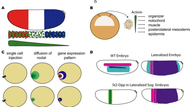
Thomas Hunt Morgan first presented the idea of gradients being used for pattern formation with his studies on regeneration. The idea continued for 40 years and was revived with further works advancing the idea in the 1960s (Wolpert, 1996). In 1968, Wolpert published his theory for solving the French flag problem, or rather how to get three sequential rows of different cell types, using a gradient (figure 1.1 A). Without a means of visualizing morphogen gradients, early studies were focused on using tissue culture studies in which different concentrations of a signaling molecule were added to see if a concentration-dependent response was elicited. Studies in Xenopus showed that treating presumptive ectoderm with increasing amounts of the morphogen Activin created different types of mesodermal tissue (Green et al., 1992; Green and Smith, 1990) (figure 1.1 B).
Studies were also focused on proving that morphogens could act at a distance.
One such study showed that a Nodal-related TGF-β protein was able to pattern the mesoderm when injected into a single cell of an early stage embryo. Cells close to the site of injection expressed genes that were typical to high levels of the signal, while cells that were further away expressed genes that were typical to low levels of the signal.
When lower amounts of the morphogen were injected only low level signaling genes
were expressed. This study was important because it showed that the signal was capable of acting at a distance from the injection site (Chen and Schier, 2001) (figure 1.1 C).
Many studies have explored how morphogen gradients are formed. Evidence supporting active transport by secretion and endocytosis has been shown in many systems by mutation to the endocytosis machinery, which affects how far the gradient can spread (Teleman et al., 2001).
Comprehending how cells interpret the information provided by the gradient is another key question in understanding morphogens. The answer must lie, at least in part, in a concentration-dependent mechanism. In fact, binding studies in Drosophila in both anterior-posterior and dorsal-ventral patterning have shown that genes that respond to lower amounts of signal contain higher affinity binding sites while those that respond to higher amounts of a signal contain lower affinity binding sites (Driever et al., 1989;
Stathopoulos and Levine, 2004). By this mechanism genes that have low affinity binding sites can only be activated by the highest concentrations of the effector. Now that gradients can be visualized and quantified, it has become increasingly clear that gradient information alone does not provide the positional information necessary for patterning (Balaskas et al., 2012; Liberman et al., 2009; Reeves et al., 2012). Instead, interactions between the target genes and combinatorial interactions between the morphogen effector and other transcription factors come together to pattern the embryo. These interactions include but are not limited to feed-forward loops, cross repression, positive feedback, autoregulation, reciprocal inhibitor gradients, and temporal integration (Ashe and Briscoe, 2006).
Figure 1.1. Ectopic expression of morphogens can pattern fields of cells.
Several experiments have been conducted in an attempt to show that morphogen gradients are sufficient to pattern the embryo.
(A) The French Flag Model proposed by Wolpert states that a morphogen gradient can provide positional information to a field of cells. The morphogen (green circles) diffuses from its source to form thresholds of signal. In the cartoon, high levels of the morphogen activate the red expression pattern. While, intermediate and low levels of the morphogen activate the white and blue expression patterns, respectively. In this model there is no need for other inputs as the gradient thresholds are sufficient for patterning.
(B) A study in Xenopus was conducted in which the animal cap was removed from early blastulas. The cells were dissociated and treated with increasing concentrations of the morphogen, Activin. The cells were then assayed for the presence of tissue specific gene markers. It was determined that increasing levels of Activin alone were able to specify five different tissue types.
(C) In a study to see if a morphogen can act at a distance, Nodal was injected into a single cell of a zebra fish embryo. The morphogen shown in green was able to diffuse and formed a gradient. The embryo was assayed for gene expression and it was found that a pattern of gene expression was formed with genes that respond to high levels of Nodal being expressed at the injection site and genes that respond to lower levels being expressed further from the injection site. When lower concentrations of Nodal were injected a pattern was not formed and only the genes that respond to low levels of Nodal were expressed.
(D) In Drosophila it was shown that Dpp signaling was capable of patterning the neurognic ectoderm. The neurogenic ectoderm genes depicted in the cartoon are msh (pink), ind (purple), and sog (teal). The lateralized embryo was created using an activated Toll receptor. In the lateralized embryo sog is expressed almost throughout the entire embryo. When Dpp was ectopically expressed in the eve.stripe2 domain it was necessary to use a sog- background, because Sog inhibits Dpp. In this background the Dpp signal was able to diffuse and in areas where the signal was the highest ind was repressed allowing msh to be expressed, thus creating a pattern in the lateralized embryo.
Dorsal-Ventral Patterning and the Dorsal Nuclear Gradient
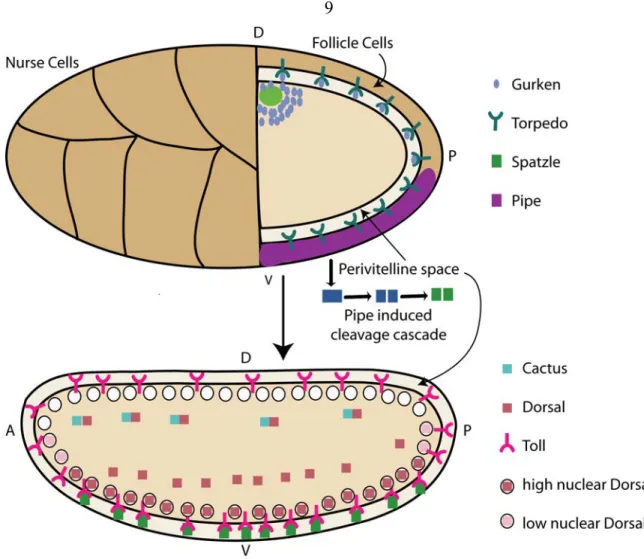
Dorsal-ventral pattering in the Drosophila embryo begins in the oocyte. The oocyte is surrounded by a group of cells called follicle cells, except at the anterior region where it is bordered by a group of cells called nurse cells. The nurse cells deposit Gurken and other factors into the ooctye; in late stages of oogensis, the nucleus and Gurken migrate to the dorsal-anterior region of the oocyte (Neuman-Silberberg and Schupbach,
1993). Gurken is the ligand for the Egf receptor Torpedo, which is located in the follicle cells. Gurken activates Torpedo imparting a dorsal fate to these cells. Both Gurken and Torpedo mutants result in ventralized embryos (Neuman-Silberberg and Schupbach, 1993). Torpedo signaling results in the limited expression of Pipe to the ventral follicle cells, where it activates a proteolytic cascade through an unknown mechanism (Sen et al., 1998). The cascade ends with the processing of Spätzle, which is the ligand of the Toll receptor (Morisato, 2001). Activation of the Toll receptor results in degradation of Cactus that binds to the transcription factor Dorsal to sequester it in the cytoplasm. Once Dorsal is released it can be transported into the nucleus, where it activates gene expression (Belvin et al., 1995). This establishes a nuclear gradient of Dorsal in the early syncytial blastodem, which will persist up to gastrulation (figure 1.2).
The Dorsal gradient does not follow the classical statues for formation of a morphogen gradient. In this case the ligand, Spätzle, is secreted into the perivitelline space by the follicle cells, which are not part of the embryo proper; but nonetheless, it follows many of the same themes used in interpretation of gradients for patterning.
Ultimately, the effector of the signaling is Dorsal, which forms a nuclear gradient and activates target genes in a concentration dependent manner.
Figure 1.2. Patterning of the eggshell and embryo.
First the eggshell of the drosophila embryo is patterned, and this leads to the patterning of the embryo. The developing oocyte is shown in the upper cartoon and the embryo is shown in the lower one. Gurken (blue circles) is secreted into the oocycte by the nurse cells, and the nucleus and Gurken both migrate to the anterior-dorsal region of the embryo. Gurken is secreted into the perivitelline space where it binds the Egf receptor Torpedo. Activation of Torpedo signaling results in limited expression of Pipe (purple) to ventral follicle cells. Pipe initiates a cleavage cascade that results in the cleavage of Spätzle. Spätzle is secreted into the perivitelline space where it binds to the Toll receptor located in the embryo membrane. Activation of the Toll pathway leads to the
degradation of Cactus (blue square), resulting in the translocation of Dorsal into the nucleus. A gradient is formed with high levels of Dorsal in the ventral nuclei and lower levels in intermediate nuclei. There is little-to-no Toll signaling in the Dorsal region of the embryo resulting in little-to-no nuclear Dorsal.
The nuclear gradient of Dorsal divides the embryo into three tissue types;
mesoderm is specified by high levels of nuclear Dorsal, where as, the neurogenic ectoderm and non-neurogenic ectoderm are specified by intermediate and low/no levels of nuclear Dorsal, respectively (Stathopoulos and Levine, 2004) (figure 1.3 B). The genes that specify the non-neurogenic ectoderm are expressed in the dorsal part of the embryo and are repressed by Dorsal, while genes in ventral and lateral regions of the embryo are activated by Dorsal. Over 30 genes have been identified as Dorsal target genes (Stathopoulos et al., 2002). For several of these genes the cis-regulatory modules (CRM) were studied and proved to be useful in deciphering the mechanisms used in interpretation of the Dorsal gradient (Ip et al., 1992b; Liberman and Stathopoulos, 2009;
Markstein et al., 2004; Stathopoulos and Levine, 2005).
Analysis of Dorsal binding sites within CRMs revealed binding sites of both high and low affinity. The low affinity binding sites were located in the CRMs of genes such as snail (sna), twist (twi), and heartless (hrt), which are expressed in the ventral most part of the embryo and specify the mesoderm. Presumably, these genes can only be expressed in regions with high levels of nuclear Dorsal. The Dorsal binding sites for these genes were also closely associated with Twist binding sites, which suggest the use of a feed- forward loop in the regulation of Dorsal target genes (Ip et al., 1992b; Jiang et al., 1991).
The genes that are expressed in the neurogenic ectoderm contain high-affinity Dorsal binding sites and while some also contain Twist binding sites, they are not as closely associated to the Dorsal binding sites (Stathopoulos and Levine, 2004). Some of the genes expressed in the presumptive neurogenic ectoderm are expressed in broad patterns [short gastrulation (sog) and rhomboid (rho)], while others appear to be carved out by repressors and are restricted to fewer cells [single minded (sim) and intermediate neuroblast defective (ind)](Morel and Schweisguth, 2000; Stathopoulos and Levine, 2005). In fact cross repression, which is seen as a theme in the regulation of other morphogen gradients, is seen in interpretation of the Dorsal gradient (Cowden and Levine, 2003). To this effect, the genes that respond to high levels of the gradient, which specify ventral regions of the embryo, repress genes that respond to lower levels of the gradient in a ventral-dominant fashion. Thus sna represses the genes that are expressed in the neurogenic ectoderm, and within the neurogenic ectoderm genes expressed in more ventral locations repress genes that are expressed more dorsally (figure 1.3 A).
Dynamics of the Dorsal Nuclear Gradient
A recent study has shown that the early expression patterns of dorsal-ventral target genes are closely related to the dorsal gradient and changes in the patterns are seen with changes in the Dorsal nuclear gradient (Reeves et al., 2012). As cell divisions occur, Dorsal is shuttled in and out of the nucleus; the amplitude of the gradient also increases with each successive nuclear cycle. The target genes respond to Dorsal dynamics; at the end of nuclear cycle 13 the target genes are repressed in ventral regions but then in early nuclear cycle 14, when the gradient is being reestablished after nuclear
division, the genes are derepressed in ventral regions (Reeves et al., 2012) (figure 1.3 A).
Presumably at these early stages, the broad expression patterns are solely reliant on activation by Dorsal; and at later stages, interactions between the target genes and inputs from other signaling molecules and transcription factors refine and maintain the patterns.
The Dorsal Gradient is Sufficient to Activate Genes that are Expressed along the Dorsal- Ventral Axis
A study found that if an activated Toll receptor was placed at the anterior pole of the early embryo, it was sufficient to create a Dorsal gradient along the anterior-posterior axis (Huang et al., 1997). This ectopic gradient of nuclear Dorsal was able to activate the expression of its target genes. The patterning of the genes was similar to the patterning observed in the dorsal-ventral axis. This would suggest that Dorsal alone is sufficient to pattern the dorsal-ventral axis, although applying a gradient response interpretation model, such as the French flag model (figure 1.1 A), would be an oversimplification because the patterns are not solely established by different concentrations of Dorsal.
We know that there are other inputs such as combinatorial interactions between Dorsal and Twist and cross-repression between the Dorsal target genes that provide positional information for pattern formation. These interactions that are dependent on Dorsal still occurred in the ectopic gradient. Also, combinatorial interactions between Dorsal and Zelda have been shown to be important in the expression of sog. Zelda is ubiquitously expressed and thus would be available to interact with ectopic Dorsal to activate dorsal-ventral patterning genes along the anterior-posterior axis (Liberman and Stathopoulos, 2009).
Many of the interactions and signaling pathways that pattern the dorsal-ventral axis are either directly or indirectly dependent on Dorsal. Another example is Egfr signaling, which is dependent on Dorsal activation of the Egfr ligand, vein (vn) and the Egfr ligand (spitz) processor rho (Ip et al., 1992a; Schnepp et al., 1996) (figure 1.3 C).
There is no denying that a gradient is necessary for patterning. If only one level of Dorsal is present, then only one tissue type is formed and the axis is not patterned properly (figure 1.1 D), but patterning is more complicated than genes being activated by different threshold levels of a gradient. A more complete model is that the Dorsal nuclear gradient establishes the early expression patterns and also a base for signaling and combinatorial interactions, this along with other maternally deposited factors present in the embryo patterns the dorsal-ventral axis (figure 1.3). As development proceeds, the gene regulatory network established by Dorsal is self-sufficient, allowing patterning and development to continue without the need for the gradient to be maintained.
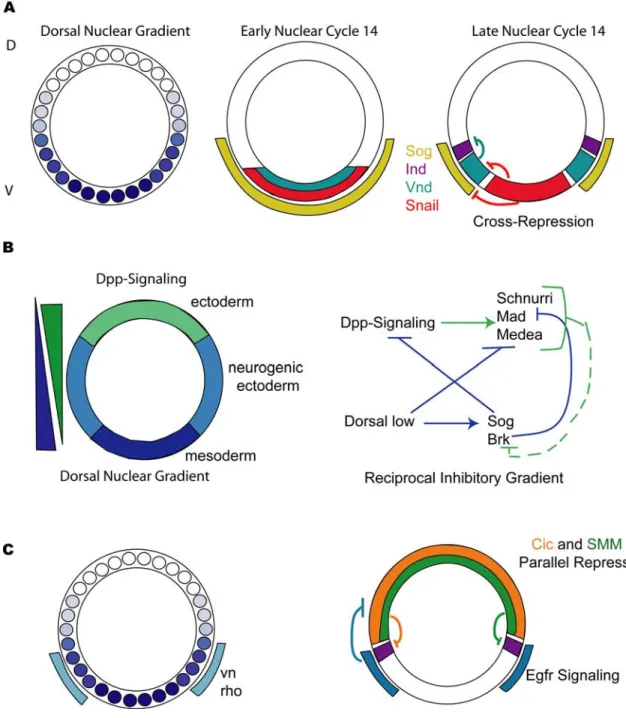
Figure 1.3. Interpretation of the Dorsal gradient and patterning of the dorsal- ventral axis of Drosophila embryos.
The Dorsal nuclear gradient establishes gene expression, which is then refined by interactions such as cross-repression and reciprocal inhibitory gradients.
(A) The schematic depicts a cross section through the center of the embryo. The small circles represent nuclei and the different shades of blue represent increasing levels of the
Dorsal nuclear gradient, with the highest levels of Dorsal found in the most ventral nuclei. In early nuclear cycle 14 the genes expressed along the dorsal-ventral axis, sog (yellow), sna (red), and vnd (teal) are expressed in overlapping regions in the ventral part of the embryo. At this point they are only reliant on the forming Dorsal gradient that is being reestablished after nuclear division. In late nuclear cycle 14 Snail represses vnd and sog restricting their expression to ventral-lateral and lateral regions of the embryo, respectively. At this stage, ind (purple) is expressed in lateral regions of the embryo; it is repressed by Vnd and Sna in ventral-lateral and ventral regions of the embryo.
(B) The embryo is divided into three tissue layers; the ectoderm (green) in dorsal regions of the embryo, the neurogenic ectoderm (light blue) in lateral regions of the embryo, and the mesoderm (dark blue) in ventral regions of the embryo. A reciprocal inhibitory gradient of Dpp-signaling (BMP-signaling) opposes the Dorsal nuclear gradient. This gradient is formed by inhibition of Dpp directly by Sog and indirectly by Brk. Dorsal is also believed to bind to the CRMs of Dpp target genes to repress their expression. Due to the presence of Schnurri-Mad-Medea (SMM) binding sites in neurogenic ectoderm CRMs it is believed that Dpp signaling represses them. Thus, the Dpp and Dorsal gradients act antagonistically to each other.
(C) Egfr signaling plays a role in patterning the neurogenic ectoderm. Dorsal activates expression of vn and rho in ventral-lateral regions of the embryo. They function to turn on Egfr signaling in this region of the embryo. Egfr signaling is believed to inhibit Cic (pink); this allows for the activation of ind (purple), as it frees it from repression by Cic.
There is a second tier of repression that limits the dorsal border of ind, this is mediated by Dpp-signaling acting through the SMM binding site (green).
The Role of Dpp-Signaling in Dorsal-Ventral Patterning of the Early Embryo
Opposing the Dorsal nuclear gradient there is a BMP signaling step gradient with high levels in dorsal-most regions and lower levels in dorsal-lateral regions of the embryo. This gradient is created by graded localization of the ligand, Dpp; while the receptors Thickveins and Punt are ubiquitously expressed (Ashe, 2005; Mizutani et al., 2006). Dpp binds to the receptors either as a homodimer or as a heterodimer with Screw (Scw). Graded Dpp signaling functions to pattern the non-neurogenic ectoderm and might also function to repress genes that are expressed in the neurogenic ectoderm (Ashe, 2005;
Mizutani et al., 2006).
Dpp is localized to dorsal and dorsal-lateral regions of the early embryo.
Although the expression pattern of dpp is generally uniform, a step gradient of Dpp signaling is formed with high levels of signaling in dorsal most regions of the embryo and lower levels of signaling in dorsal-lateral regions of the embryo (Irish and Gelbart, 1987; Ray et al., 1991; Shimmi et al., 2005). There is evidence to suggest that Sog binds to Dpp and inhibits it from binding the receptors (Yu et al., 1996). In lateral regions of the embryo a Dpp-Scw-Sog-Tsg complex is formed which is transported to dorsal regions of the embryo (Shimmi et al., 2005). The protease Tolliod cleaves this complex resulting in higher levels of signaling in dorsal regions of the embryo (Canty et al., 2006; Serpe et al., 2005). In lateral regions of the embryo, where Sog is present, Dpp rebinds Sog keeping signaling levels low. In contrast, Sog is absent in dorsal regions, which allows for high levels of Dpp-Scw heterodimers to accumulate resulting in high levels of signaling. Sog is activated by Dorsal, thus the Dorsal gradient indirectly interacts with and helps to shape the Dpp morphogen gradient (figure 1.3 B). It is believed that Dpp
effectors are repressed by Brinker, adding yet another level of interaction between the Dorsal and Dpp gradients (Jazwinska et al., 1999; Rushlow et al., 2001).
Dpp signaling is also thought to act on Dorsal target genes and is believed to aid in patterning the neurogenic ectoderm. This mechanism is highly conserved between vertebrates and invertebrates, although in vertebrates the opposing gradient in the neural tube is Sonic Hedgehog and not Dorsal. It has been shown that, in a lateralized background, activation of Dpp signaling in an anterior-posterior stripe can create a pattern of neurogenic ectoderm genes (Mizutani et al., 2006) (figure 1.1 D). At the ectopic source of Dpp, the most dorsal neurogenic gene, msh, is expressed with the second most dorsal neruogenic gene, ind, being expressed in the rest of the embryo (figure 1.1 D). In contrast, when Dpp signaling is lost or reduced there is not a dramatic change to the neurogenic ectoderm genes, suggesting that while Dpp signaling can and likely does contribute to patterning of the neurogenic ectoderm, it is not absolutely necessary (Von Ohlen and Doe, 2000). In the following chapters, we provide evidence for the presence of other regulatory factors that are independent of Dpp signaling, that act dorsally to pattern genes in the neurogenic ectoderm. The presence of these factors clarifies some of the results that have been seen when investigating the role of Dpp signaling in regulation of the neurogenic ectoderm. Unlike ectopic activation of Dpp signaling, loss of Dpp signaling has little to no effect on gene expression in the neurogenic ectoderm. The lack of an observable phenotype in Dpp mutants is likely due to the presence of factors that are independent of Dpp signaling that compensate for the loss of Dpp signaling.
Egfr Signaling also Plays a Role in Patterning the Neurogenic Ectoderm
Egfr signaling is present in the presumptive neurogenic ectoderm, and it is essential for proper patterning. The Egfr receptor is ubiquitously expressed at this stage and the ligand vein is activated by Dorsal in lateral regions of the embryo (Schnepp et al., 1996). rho, which activates Egfr signaling by cleaving the Egfr ligand Spitz, is also activated by Dorsal in a domain similar to vein (Bier et al., 1990; Ip et al., 1992a).
During later stages of development neuroblasts develop a unique identity based on where they are located and differentiate into specialized neuroblast accordingly. Their location is determined based on which proneural gene they express and whether they receive Egfr signaling. In Egfr mutants expression of ind is lost and consequently no intermediate neuroblast are formed (Skeath, 1998). The medial neuroblast, are still specified and express vnd, but the lack of Egfr signaling causes them to display some traits specific to lateral neuroblast. The lateral neuroblasts, which are specified by msh, expand into regions where intermediate neuroblasts would normally form. Thus it is clear that Egfr signaling plays and important role in patterning the dorsal-ventral axis of the neurogenic ectoderm. Even though Egfr signaling is only necessary for the activation of ind, it has a dramatic affect on the patterning of the neurogenic ectoderm; as loss of just one of the neurogenic ectoderm genes results in patterning defects of the entire tissue.
The Egfr receptor is a receptor tyrosine kinase (RTK) with many downstream effectors including transcriptional activators and repressors. Initially it was unclear whether Egfr signaling activated ind directly or whether it inhibited a repressor allowing ind to be expressed. A recent publication and our work featured in the following chapter support the latter case, suggesting Egfr signaling is responsible for inhibiting a repressor
that binds an 16 bp repeated sequence (the “A-box” element) present in the ind CRM (Ajuria et al., 2011; Stathopoulos and Levine, 2005) (figure 1.3 C).
Insights into Dorsal-Ventral Patterning via CRM Analysis
Analysis of CRMs has proven useful in understanding how genes are regulated and how axis specification is determined. In chapter 2, we analyze the ind CRM, which drives expression of a sharp dorsal-ventral stripe in the presumptive neurogenic ectoderm. We show that an element, we call the “A-box” is both necessary and sufficient to mediate repression in dorsal regions of the embryo and thus maintains proper patterning of the neurogenic ectoderm by refining the dorsal border of ind. We conducted affinity chromatography to identify factors that bind the A-box element; our analysis resulted in the identification of Grh, which we believe acts as an activator rather than a repressor. We also show that a second tier of repression, acting to define the dorsal boundary of ind, is mediated by Dpp signaling and acts on the ind CRM via a Schnurri-Mad-Media complex (SMM) binding site (figure 1.3 C). Thus, the pattern of ind is initiated by Dorsal and Grh activation and is refined by tiers of repressors in both ventral and dorsal regions of the embryo.
Our analysis of the A-box element reveled that as well as the activator Grh several chromatin factors also bound the A-box element. Chapter 3 summarizes the results from the affinity chromatography analysis and discusses a possible role for chromatin remodeling factors in regulating ind expression and patterning of the dorsal-ventral axis of the early embryo.
In chapter 4 we extend our CRM analysis to vnd and sog. We show that dorsal repressors may play a role in regulating the expression pattern of vnd and, to a lesser extent, sog. We also provide a more detailed discussion of the combinatorial interactions known to pattern the dorsal-ventral axis.
In chapter 5 we discuss the implications of our studies and discuss future directions to determine how genes are regulated to establish axis specification.
Chapter 2
Lateral Gene Expression in Drosophila Early Embryos is Supported by Grainyhead-mediated Activation and Tiers of Dorsally-Localized Repression*
*This chapter, first published in Plos One in 2011, was written by Mayra Garcia and Angelike Stathopoulos.
Abstract
The general consensus in the field is that limiting amounts of the transcription factor Dorsal establish dorsal boundaries of genes expressed along the dorsal-ventral (DV) axis of early Drosophila embryos, while repressors establish ventral boundaries. Yet recent studies have provided evidence that repressors act to specify the dorsal boundary of intermediate neuroblasts defective (ind), a gene expressed in a stripe along the DV axis in lateral regions of the embryo. Here we show that a short 12 base pair sequence (“the A- box”) present twice within the ind CRM is both necessary and sufficient to support transcriptional repression in dorsal regions of embryos. To identify binding factors, we conducted affinity chromatography using the A-box element and found a number of DNA-binding proteins and chromatin-associated factors using mass spectroscopy. Only Grainyhead (Grh), a CP2 transcription factor with a unique DNA-binding domain was found to bind the A-box sequence. Our results suggest that Grh acts as an activator to support expression of ind, which was surprising as we identified this factor using an element that mediates dorsally-localized repression. Grh and Dorsal both contribute to ind transcriptional activation. However, another recent study found that the repressor Capicua (Cic) also binds to the A-box sequence. While Cic was not identified through our A-box affinity chromatography, utilization of the same site, the A-box, by both factors Grh (activator) and Cic (repressor) may also support a “switch-like” response that helps to sharpen the ind dorsal boundary. Furthermore, our results also demonstrate that TGF-β signaling acts to refine ind CRM expression in an A-box independent manner in dorsal-most regions, suggesting that tiers of repression act in dorsal regions of the embryo.
Introduction
During development the embryo is patterned by the localized expression of genes to discrete parts of the embryo. Such tight spatial regulation of gene expression is necessary to set the boundaries that distinguish different cell types required for proper development. One mechanism to impart spatial information is to regulate gene expression through transcription factors that are spatially localized. Alternately, localized activation of signaling pathways in particular domains can also influence the boundaries of gene expression.
In Drosophila melanogaster, the dorsal-ventral (DV) axis of the pre-gastrula embryo is patterned by a nuclear gradient of the NF-κB homologous transcription factor Dorsal (Reeves and Stathopoulos, 2009). High levels of nuclear Dorsal are present in ventral regions of the Drosophila embryo and nuclear levels decrease progressively toward more dorsal regions. Due in part to these differing nuclear Dorsal levels, different domains of gene expression are established along the DV axis to specify different cell types (Stathopoulos et al., 2002). In the ventral most regions of the embryo, high concentrations of nuclear Dorsal drive expression of genes such as twist and snail (sna) to specify the presumptive mesoderm. In ventral lateral regions of the embryo, intermediate levels of Dorsal activate genes such as rhomboid (rho) and ventral neuroblast defective (vnd) and low levels of Dorsal support expression of genes such as short gastrulation (sog) in broad lateral domains of the embryo (that encompass both ventral-lateral and dorsal-lateral regions) to specify distinct domains within the presumptive neurogenic ectoderm (Bier et al., 1990; Ip et al., 1992a; Jimenez et al., 1995). Lastly, as Dorsal can also function as a repressor, the expression of some genes such as zerknüllt (zen) are
limited to dorsal regions of the embryo, leading cells in this domain to adopt amnioserosa and non-neurogenic dorsal ectoderm cell fates (Jiang et al., 1993; Jiang et al., 1992;
Stathopoulos et al., 2002). Even though Dorsal provides positional information through its dorsal-ventrally modulated nuclear gradient, combinatorial interactions of transcription factors are very influential towards DV patterning. Specifically, Dorsal regulates gene expression together with other transcription factors, such as the bHLH factor Twist and the early ubiquitous activator Zelda (e.g. Ip et al., 1992b; Liang et al., 2008; Liberman and Stathopoulos, 2009).
More and more evidence suggests that signaling pathways also help to define gene expression patterns in the early embryo. For example, the expression domains of several Dorsal target genes cannot be explained by changing Dorsal levels (and/or the localization of any other previously characterized transcription factors). Additionally, it is well understood that signaling molecules provide positional information to help define the very specific expression domain encompassed by the gene single-minded (sim). sim is expressed as a stripe of a single cell width present in ventrolateral regions of the embryo, within cells located between the presumptive mesoderm and neurogenic ectoderm boundary. sim expression is supported by combinatorial interactions of Dorsal and Twist transcription factors and also through Notch-dependent signaling (e.g. Morel and Schweisguth, 2000).
Along similar lines, the gene intermediate neuroblast defective (ind) is expressed in dorsal-lateral regions of the embryo in a stripe of 5–7 cells in width, which is narrower than the broad domain encompassed by sog. Genetic studies support the view that refined ind expression is supported by inputs from both Dorsal and Epidermal growth
factor receptor (Egfr) signaling, suggesting that limiting amounts of both of these inputs help delineate ind expression boundaries (Von Ohlen and Doe, 2000). The Egfr gene is ubiquitously expressed in embryos but the receptor is activated locally in ventrolateral regions by the ligands Vein and Spitz (Rutledge et al., 1992; Schnepp et al., 1996).
Several binding sites for the ETS transcription factor, which mediates Egfr signaling, are also found in the ind cis-regulatory module CRM, but it has not been shown if they are required for activation or whether an indirect mechanism is used for activation of ind expression via Egfr signaling (Stathopoulos and Levine, 2005).
No other gene in the Drosophila embryo described to date shares the same expression domain as ind, yet understanding how the ind expression domain is regulated may have far-reaching implications. Interestingly, the genes that pattern the ventral nerve cord of Drosophila and the neural tube of higher vertebrates share a conserved organization and function (Cornell and Von Ohlen, 2000; Mizutani et al., 2006).
Specifically, the gene ventral neuroblast defective (vnd)/Nkx2.2 is expressed ventral to ind/Gsh, and the gene muscle specific homeobox (msh)/Msx1/2 is expressed dorsally to ind (Chu et al., 1998; Isshiki et al., 1997; Weiss et al., 1998). Experiments conducted in the Drosophila embryo have suggested that the ventral boundaries of these genes are set following a “ventral dominance rule,” in which the more ventral genes repress expression of the more dorsal genes (Cowden and Levine, 2003). In contrast, it had been proposed that the dorsal boundaries of these genes result from limiting amounts of the activator, Dorsal, present in distinct domains along the DV axis (reviewed in Stathopoulos et al., 2002). However, recently it was discovered that the ind gene is expressed in a domain along the DV axis where the Dorsal gradient appears uniform without a clear transition
that would be capable of setting a dorsal border (Liberman et al., 2009). A previous analysis of the ind CRM suggested evidence for a dorsally-acting repressor which could explain how the dorsal boundary of ind is specified (Stathopoulos and Levine, 2005).
Direct evidence for repressor action within dorsal regions of the early embryo was found through analysis of the cis-regulatory region of ind (Stathopoulos and Levine, 2005). A 1.4 kb (kilobase) DNA fragment located ~2 kb downstream of the ind coding sequence was found to support expression in a refined stripe within lateral regions of the embryo, in a pattern comparable to the endogenous gene. However, the promoter proximal half of the ind CRM drove expression of a reporter gene within a broad pattern, one that extends into ventral-lateral as well as dorsal-lateral regions, suggesting that the distal half contains repressor binding sites. Using a chimeric CRM assay designed to detect repression along the dorsal-ventral axis by silencing of an associated even-skipped stripe 3/7 CRM (eve.stripe3/7), this previous study found that the 1.4 kb ind CRM mediates repression of eve.stripe3/7 in dorsal and ventral regions of the embryo. A specific search for an element supporting dorsal repression was conducted and identified a 111 base pair (bp) region of the ind CRM, which supported dorsal-lateral and dorsal repression of eve.stripe3/7. A 12 bp sequence was highlighted, as it repeats twice within these 111 bp, and was called the A-box (WTTCATTCATRA). Importantly, in this previous study, when the A-box was mutated in the context of a minimal element supporting repression in dorsal regions (i.e., 267 bp fragment), repression of the eve.stripe3/7 CRM was lost. Presumably transcription factors bind to the A-box element to help establish the dorsal boundary of the ind gene, but their identities remained unknown.
Additional evidence also suggests that TGF-ß signaling may also regulate the ind expression domains, but whether or not this signaling pathway functions through the A- box element was not known. Decapentaplegic (Dpp) is a TGFß/BMP homolog that is limited in its expression to dorsal regions of the embryo and functions as a morphogen to support patterning of the amnioserosa, at higher levels in dorsal-most regions of the embryo, and the non-neurogenic ectoderm, at lower levels in dorsal-lateral regions of the embryo (Ferguson and Anderson, 1992). A previous study found that in mutants in which Dpp signaling is expanded into lateral regions of the embryo, ind expression is lost (Von Ohlen and Doe, 2000). Likewise, ectopic expression of dpp in lateralized embryos that exhibit expanded ind expression throughout the embryo was able to repress ind in the domain where Dpp signaling was presented (Mizutani et al., 2006). Also, the ind CRM contains a 15 bp DNA sequence implicated in TGF-β signaling-mediated repression (Stathopoulos and Levine, 2005). Similar sites have been shown to mediate repression by recruiting a Dpp-dependent Schnurri/Mad/Medea (SMM) protein complex, but SMM dependent repression of ind has never been shown and in fact this mechanism of repression has only been shown to act at later stages of development (Dai et al., 2000;
Pyrowolakis et al., 2004).
Therefore, to gain further insight into how patterning is controlled along the dorsal-ventral axis of Drosophila embryos, we tracked the repression activity supported by different DNA elements associated with the ind CRM. We found that the A-box element facilitates both activation and repression of ind and propose that this helps to mediate a sharp border. In addition, we found that TGF-β signaling supports ind
repression in dorsal-most regions of the embryo through the SMM site located within the ind CRM that is distinct from the A-box.
Results
Chimeric CRM Assays Can Help Identify and Track Repression Activity Associated with CRM Sequences
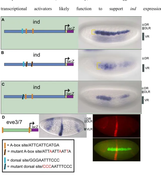
In order to gain insights into how the boundaries of dorsal-ventral patterning genes are set, we deconstructed the cis-regulatory element of ind to find direct evidence for dorsal repression activity. We utilized a chimeric cis-regulatory module (CRM) assay, using eve.stripe3/7 and ind CRMs in order to determine whether repressors are present within either of these sequences to help refine the domains of expression (Stathopoulos and Levine, 2005). The ind CRM supports expression along the DV axis in a lateral stripe, comparable to the endogenous gene ((Figure 1A; Stathopoulos and Levine, 2005). In turn, the eve.stripe3/7 sequences supports expression of two stripes located along the anterior-posterior (AP) axis of embryos (figure 2.1 B) (Small et al., 1996). When two CRMs are placed in tandem upstream of a reporter gene (i.e. lacZ), if additive expression is observed this result indicates that either repressors are not present or they are not located in range to act on the adjacent CRM; conversely, if non-additive expression is observed this indicates repressors are present and function to silence activators associated with both CRMs. Previously, using a chimeric CRM assay, it was shown that the 1.4 kb ind CRM drives repression of eve.stripe3/7 (Figure 1C;
Stathopoulos and Levine, 2005). In this case non-additive expression is observed; the eve.stripe3/7 CRM is repressed in ventral regions by snail and vnd repressor sites located
in the ind CRM and by unknown transcription repressors in dorsal regions. Concurrently, the ind CRM is repressed by Knirps, through sites in the eve.stripe3/7 CRM, forming a gap in the ind expression pattern. It was suggested the unknown transcription repressors located in dorsal regions act through a pair of 12 bp A-box sequences located within the 1.4 kb ind CRM. Here we examined the function of the A-box sequence more closely.
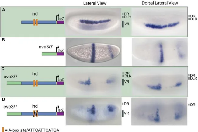
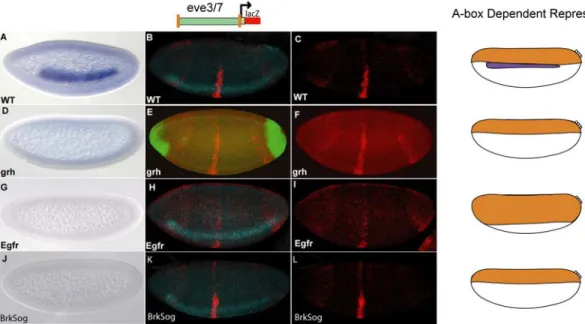
Figure 2.1. The ind CRM contains binding sites that mediate repression in dorsal regions.
lacZ reporter expression was visualized within cellularized embryos (late stage 5) by in situ hybridization using a digoxigenin-labeled antisense lacZ riboprobe. In this and all subsequent figures, embryos are oriented with anterior to the left. In addition, embryos are oriented to show views of lateral, dorsal on top, (left image) and dorsal (right image) domains. The repression domains are outlined to the right of each image: DR = dorsal
repression, DLR = dorsal lateral repression, and VR = ventral repression. The schematic depicts the chimeric CRM combinations used: (A) The 1.4 kb ind CRM drives expression of lacZ in a 5–7 cell lateral stripe representative of ind expression; (B) The 0.5 kb eve.stripe3/7 CRM drives expression of lacZ in two anterior-posterior stripes representative of eve.stripe3/7 expression; (C) The eve.stripe3/7-ind chimeric CRM drives expression of lacZ in a non-additive fashion showing repression of eve.stripe3/7 in dorsal, dorsal lateral, and ventral regions; (D) The eve.stripe3/7-mut-A-box-ind chimeric CRM supports non-additive expression with repression of eve.stripe3/7 in dorsal and ventral regions but not dorsal lateral regions.
The A-box Element Mediates Repression of ind in Dorsal-Lateral Regions of the Embryo, while Other Sequences Support Repression in Ventral and Dorsal-Most Regions of the Embryo
When we mutated both of the A-box sites in the context of the full-length ind CRM and assayed the fragment’s ability to repress expression of the associated eve.stripe3/7 CRM, repression of eve.stripe3/7 was lost in dorsal lateral regions (figure 2.1 D, compare with 2.1 C). This result demonstrated that these two A-box sequences are necessary to mediate dorsal-lateral repression of eve.stripe3/7 by the ind CRM. Next, we assayed the full-length ind CRM with two mutant A-boxes alone and found that lacZ reporter expression was expanded into dorsal-lateral regions; giving a broad, patchy, and diffuse pattern not a sharp stripe of 5–7 cells in width representative of ind (figure 2.2 B, compare with 2.2 A).
However, even in the absence of the A-box sites, repression was retained in dorsal-most and ventral regions of the embryo when the A-box was mutated in the context of the full- length CRM (figure 2.2 B), as well as in the chimeric CRM assay of ind and eve.stripe3/7 CRMs (figure 2.1 D). These results suggest that the A-box sequences mediate dorsal- lateral repression, but that there might be other repressor binding sites in the ind CRM which mediate repression in dorsal-most and ventral regions of the embryo. Vnd and Snail binding sites within the ind CRM most likely mediate the repression observed in ventral regions (Cowden and Levine, 2003). In contrast, while we were able to track repression in dorsal-most regions, the identity of the responsible transcription factors was unknown.
A-box Elements Limit Expression in Dorsal-Lateral and Dorsal Regions of Embryos Another important question is whether the A-box elements are sufficient to cause repression of the eve.stripe3/7 CRM, as perhaps multiple sequences within the ind CRM are necessary to support repression. To investigate this, we flanked the eve.stripe3/7 CRM with the A-box element (i.e., A-box.eve.stripe3/7.A-box) and observed clear repression in dorsal-lateral regions, as expected, and also within dorsal regions of the embryo (figure 2.2 D). Weak repression was also observed in ventrolateral regions at lower frequency (data not shown). This result suggests that A-box sequences are sufficient to support repression in dorsal-lateral regions, but also contribute to repression in dorsal-most and ventrolateral regions of the embryo.
The expression supported within the eve.stripe3/7 domain did extend a few cells above the endogenous ind dorsal boundary in the context of the A-box.eve.stripe3/7.A-
box reporter. This may indicate the chimeric CRM assay is limited in its ability to track repression activity as the stripe of expression also extended a few cells above ind when the full length ind CRM was assayed in tandem to eve.stripe3/7. Alternatively, sharp definition of the ind dorsal boundary may require more input than localized repressor activity.
The Dorsal Transcription Factor Only Partially Supports Activation of ind
We investigated the activation of the ind expression pattern by mutagenizing the sole match to the Dorsal binding site consensus present within the ind 1.4 kb CRM (figure 2.2 C). ind is not expressed in dorsal mutants (Von Ohlen and Doe, 2000), thus, we expected loss of the sole Dorsal binding site would severely impair reporter expression. Instead, we found that the expression pattern driven by the mutated CRM is very similar to that driven by the wild-type CRM, except for a gap in the expression pattern (figure 2.2 C).
Early ind expression, at the start of cellularization, exhibits a smaller gap in expression at 40% egg length (Stathopoulos and Levine, 2005) which is likely mediated by anterior-posterior patterning factors. In reporter constructs, repression within this domain is more apparent with the 1.4 kb ind CRM sequence is oriented in the opposite direction relative to the promoter in reporter constructs (data not shown). The function of activators, including Dorsal and others that act through the A-box sequence, are likely required to counterbalance this repression.
Our results suggest that Dorsal binding contributes to ind activation but that other activators also influence ind expression. Furthermore, chromatin immunoprecipitation
(i.e., ChIP-seq) experiments did not detect Dorsal binding in the genome at the ind CRM (Ozdemir et al., 2011), which indicates Dorsal may not bind to the ind CRM (or that it is a very transient interaction). Collectively, these results suggest that additional transcriptional activators likely function to support ind expression.
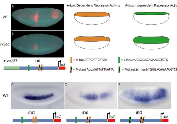
Figure 2.2. The A-box sites are necessary for dorsal lateral repression and sufficient for dorsal and dorsal-lateral repression.
Site-directed mutagenesis was used to mutate regulatory sites in the ind CRM. The CRMs depicted in the schematic were used to drive expression of lacZ in embryos that were analyzed by in situ hybridization using a lacZ anti-sense riboprobe. Cellularized embryos of stage 5 are oriented to show a lateral view, with anterior to the left and dorsal on the top. The yellow brackets mark the height of the expression pattern. The repression domains are outlined to the right of the image: DR = dorsal repression, DLR = dorsal lateral repression, VR = ventral repression, VLR = ventral lateral repression. (A) The 1.4 kb ind CRM drives expression of a lateral stripe of 5–7 cells in width comparable to ind expression. (B) The 1.4 kb mut-A-box-ind CRM drives expression of 7–10 cell width lateral stripe that is diffuse, weak, and expanded compared to the ind CRM. (C) The 1.4 kb mut-dorsal-ind CRM drives expression that has a gap and is weak in posterior regions compared to the ind CRM. (D) The eve.stripe3/7 CRM flanked by A-box sites (A-box-eve.stripe3/7-A-box) shows repression in dorsal, dorsal-lateral, and ventral-lateral regions. In the fluorescent image lacZ expression is shown in red and endogenous ind expression is shown in green, as detected by multiplex fluorescent in situ hybridization (Kosman et al., 2004).
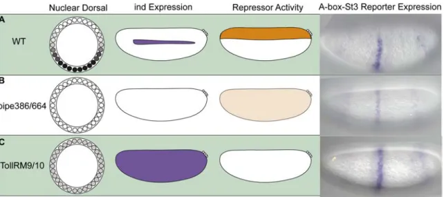
Dorsalized and Lateralized Embryos Provide Insights into the Localization of the A-box Repressor Activity
Next we introduced the lacZ reporter gene containing the eve.stripe3/7 CRM flanked by A-box sequences (i.e., A-box-eve.stripe3/7-A-box) into different mutant backgrounds to test whether the repressor activity associated with the A-box sequence is influenced by altered DV positional information. Maternal mutant backgrounds exist that
affect the levels of nuclear Dorsal (i.e., low or absent) to create lateralized or dorsalized embryos, respectively. Expression of Dorsal target genes are affected such that certain genes expressed by a particular level of Dorsal, normally refined in expression to distinct domains along the DV axis, are instead expressed ubiquitously or absent in either of these mutant backgrounds. In sum, our aim was to determine whether the repressor activity was responsive to changes in Dorsal levels, providing additional evidence that the repressor activity we had tracked was indeed functioning in a DV localized manner.
In pipe mutants, Dorsal is not able to enter the nucleus thus Dorsal target genes are not activated, resulting in dorsalized embryos (Stathopoulos et al., 2002; Stein et al., 1991). In this mutant background, endogenous ind is not expressed. We assayed the A- box-evestripe3/7-A-box lacZ reporter construct in the pipe mutant background and found that expression of lacZ was retained but severely dampened (figure 2.3 B compare with 2.3 A). This result suggests that some repressor activity is present ubiquitously in dorsalized embryos but most likely it is less active, because only partial repression of the reporter is observed.
We also examined reporter expression in Tollrm9/10 embryos, which have a partially active form of the Toll receptor allowing low levels of Dorsal to enter the nucleus throughout the embryo (Stathopoulos et al., 2002). In this background ind is expressed throughout the embryo, suggesting that repressors are unable to refine the ind pattern in this background. We also observed strong uniform expression of the lacZ reporter in the eve.stripe3/7 domain indicating that in this background the repressor activity is gone (figure 2.3 C).
The A-box element clearly supports repression in dorsal regions of the embryo and is responsive to mutations altering DV pattern (figure 2.3). These results suggest the A-box associated repressor exhibits localized expression in dorsal regions of the embryo and/or that its activity is modulated by signaling pathways that exhibit differential activation along the DV axis.
Figure 2.3. Dorsalized and lateralized embryos provide insights into the A-box repressor domain of activity.
The depictions show the Dorsal nuclear gradient within embryo cross-section schematics, whereas ind expression and the putative repressor activity are schematized within lateral views. Expression of the A-box-eve.stripe3/7-A-box reporter gene was examined by in situ hybridization in (A) wild-type, (B) pipe384/pipe664 mutants, and (C) TollRM9/TollRM10 mutants. The in situ images show lacZ expression as such: (A) Repression of lacZ is shown in dorsal regions of the embryo in WT embryos. (B) Weak repression of lacZ is shown throughout the embryos from pipe mutant females (i.e., dorsalized embryos). (C) A lack of repression of lacZ is shown in embryos from TollRM9/10 mutant females (i.e., lateralized embryos).
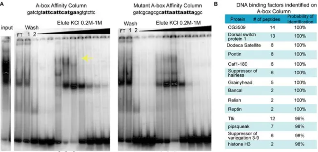
Affinity Chromatography and Mass Spectrometry Identifies Putative A-box Binding Factors
In order to provide molecular insight into the mechanism by which ind expression in dorsal regions is limited, we set out to identify the factor that binds the A-box element choosing affinity chromatography using a 22 bp oligonucleotide containing the A-box sequence (12 bp) and endogenous flanking regions (5 bp on either side). As a control, we also compared binding with that obtained with a mutant A-box sequence modified in 3 of 12 bp, which we showed does not support dorsal repression when assayed in the context of a chimeric CRM assay in vivo (see figures 1.1 D, 1.2 B) and containing different flanking region].
We used affinity chromatography to purify proteins that recognize the A-box or mutant A-box sequence from early embryonic nuclear extracts age 0–6 hours. The A-box binding activity was tracked throughout a number of biochemical separations (see figure 2.S1 and materials and methods). There were several factors that bound to both columns but some of the binding was specific to the A-box (figure 2.4 A). Cold competition with the A-box versus the mutant A-box confirmed the binding observed was specific to the A-box (data not shown). With advances in mass spectroscopy, we could analyze a complex sample containing a number of proteins. Therefore, at this step, we analyzed samples isolated from either the A-box column or the mutant A-box column by mass spectrometry.
Focusing on factors that only bound the A-box column (figure 2.4 B), we selected targets for future analysis. Several transcription factors were found specifically associated with the A-box, and not the mutant A-box column. Furthermore, several
chromatin-related factors bound to the A-box column but failed to bind the mutant A-box column (figure 2.S2). This suggested to us that the repressor activity associated with the intact A-box sequence may be composed of a large complex of proteins including chromatin components; a role for chromatin in supporting expression in the early Drosophila embryo is unclear (see discussion).
Figure 2.4. Affinity chromatography and mass spectrometry was used to identify factors that bind the A-box element.
(A) shows the EMSAs preformed using γ32P-labeled A-box oligonucleotides on nuclear extract fractions after they were affinity purified with the A-box column and the mutant A-box column. FT denotes the flow through which did not bind to the column. The yellow arrow marks the area where the A-box specific binding was found. The stars mark the samples used for mass spectrometry identification. (B) The table lists the DNA binding factors that bound to the A-box element column but not the mutant A-box column. The “# of peptides” corresponds to the number of unique peptides that contributed to the protein identification. The probability of identification was calculated
by the program Scaffold used to identify the proteins by mass spectrometry analysis and corresponded to the likelihood a correct match was made.
The Grainyhead Transcription Factor Binds to the A-box sequence and is Required to Support ind Expression
In order to narrow down a list of factors to examine in this preliminary analysis, we focused on identifying factors that bind specifically to the A-box DNA sequence. We conducted EMSAs on the following factors, which contain a predicted DNA-binding domain, and for which cDNAs were available: ATP-dependent chromatin assembly factor large subunit (Acf1), Structure specific recognition protein (Ssrp), CG3509, Grainyhead (Grh), Dorsal switch protein 1 (Dsp1) and Pipsqueak (Psq) (data not shown).
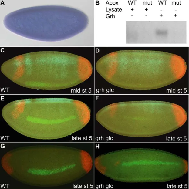
Of these factors, only Grh exhibited binding to the 22 bp oligonucleotide, containing the 12 bp A-box and endogenous sequences.
Using in vitro translated proteins in EMSAs, we further analyzed Grh and found that while it bound the A-box element it did not bind to the mutant A-box element (figure 2.5 B, full gel figure 2.S3). We, therefore, conducted additional analysis on Grh as it seemed a likely candidate to support the A-box repression activity. The grh gene is maternally and zygotically expressed (Huang et al., 1995; Weber et al., 1995), and by in situ hybridization we confirmed that it is ubiquitously expressed in the early embryo (figure 2.5 A). While some evidence exists that grh transcripts are localized to dorsal and lateral regions of the embryo (Huang et al., 1995), we could not detect such a localized expression domain by in situ hybridization even though a number of different riboprobes were designed to detect grh transcripts.
We generated grh germline clone females in order to deplete both maternal and zygotic grh expression from embryos. The conventional method of creating germline clones (Perrimon, 1998), which relies on flipase catalyzed mitotic recombination in the context of transheterozygous FRT ovoD (dominant female sterile mutation) and FRT grh chromosomes, for example, could not be used because ovoD within the commonly used FRT ovoD chromosome is most likely inserted at the grh locus. FRT ovoD in combination with all grh alleles tested are zygotically lethal, but no lethality was observed with ovoD insertions located on other chromosomes. Thus, it was necessary to make germline clones in females of the genetic background FRT grh/FRT GFP.
Embryos obtained from these females were manually screened for absence of GFP (Luschnig et al., 2004), thus allowing isolation of embryos containing the mutant form of grh. To ensure that grh zygotic transcripts were absent, females containing germline clones were mated to males containing appropriate balancer chromosomes to allow detection in the early embryo (i.e., FRT grh/ Cyo ftz-lacZ; see Materials and Methods).
Because manual hand sorting of embryos was required, only a small number of embryos could be examined, but multiplex in situ hybridization allowed us to examine the expression of multiple genes simultaneously. Therefore, in addition to examining the effect of loss of grh on ind expression, we also assayed whether this mutation affected expression of two other genes, tailless (tll) and zen. In a previous study, embryos devoid of grh maternal message were produced X-ray irradiation induced mitotic recombination;
tll was found to be expanded in grh mutant embryos obtained in this manner (Liaw et al., 1995). However, we failed to see expansion of tll in embryos lacking both maternal and/or zygotic grh; a similar negative result was recently reported (Harrison et al., 2010).