Replaces C42-P
November 1996
Vol. 15 No. 3
Erythrocyte Protoporphyrin Testing; Approved Guideline
This document contains recommendations for the measurement, reporting, and interpretation of erythrocyte protoporphyrin using hematofluorometric and extraction measurement methods.
ABC
C42-A
THIS NCCLS DOCUMENT HAS BEEN REAFFIRMED
AS AN APPROVED CONSENSUS DOCUMENT
NCCLS...
Serving the World's Medical Science Community Through Voluntary Consensus
NCCLS is an international, interdisciplinary, nonprofit, scope, approach, and utility, and a line-by-line review of its standards-developing and educational organization that technical and editorial content.
promotes the development and use of voluntary consensus
standards and guidelines within the healthcare community. It Tentative A tentative standard or guideline is made available is recognized worldwide for the application of its unique for review and comment only when a recommended method consensus process in the development of standards and has a well-defined need for a field evaluation or when a
guidelines for patient testing and related healthcare issues. recommended protocol requires that specific data be collected.
NCCLS is based on the principle that consensus is an effective It should be reviewed to ensure its utility.
and cost-effective way to improve patient testing and
healthcare services. Approved An approved standard or guideline has achieved
In addition to developing and promoting the use of voluntary reviewed to assess the utility of the final document, to ensure consensus standards and guidelines, NCCLS provides an open attainment of consensus (i.e., that comments on earlier and unbiased forum to address critical issues affecting the versions have been satisfactorily addressed), and to identify quality of patient testing and health care. the need for additional consensus documents.
PUBLICATIONS NCCLS standards and guidelines represent a consensus opinion An NCCLS document is published as a standard, guideline, or materially affected, competent, and interested parties obtained
committee report. by following NCCLS’s established consensus procedures.
Standard A document developed through the consensus less stringent than applicable regulations. Consequently, process that clearly identifies specific, essential requirements conformance to this voluntary consensus document does not for materials, methods, or practices for use in an unmodified relieve the user of responsibility for compliance with applicable form. A standard may, in addition, contain discretionary regulations.
elements, which are clearly identified.
Guideline A document developed through the consensus
process describing criteria for a general operating practice, The comments of users are essential to the consensus procedure, or material for voluntary use. A guideline may be process. Anyone may submit a comment, and all comments used as written or modified by the user to fit specific needs. are addressed, according to the consensus process, by the Report A document that has not been subjected to con-sensus including those that result in a change to the document when review and is released by the Board of Directors. published at the next consensus level and those that do not CONSENSUS PROCESS appendix to the document. Readers are strongly encouraged The NCCLS voluntary consensus process is a protocol document. Address comments to the NCCLS Executive establishing formal criteria for: Offices, 940 West Valley Road, Suite 1400, Wayne, PA
! The authorization of a project
! The development and open review of documents
! The revision of documents in response to comments by volunteer for participation in NCCLS projects. Please contact
users the NCCLS Executive Offices for additional information on
! The acceptance of a document as a consensus standard or guideline.
Most NCCLS documents are subject to two levels of consensus–"proposed" and "approved." Depending on the need for field evaluation or data collection, documents may also be made available for review at an intermediate (i.e.,
"tentative") consensus level.
Proposed An NCCLS consensus document undergoes the first stage of review by the healthcare community as a proposed standard or guideline. The document should receive a wide and
consensus within the healthcare community. It should be
on good practices and reflect the substantial agreement by
Provisions in NCCLS standards and guidelines may be more or
COMMENTS
NCCLS committee that wrote the document. All comments,
result in a change, are responded to by the committee in an to comment in any form and at any time on any NCCLS
19087, USA.
VOLUNTEER PARTICIPATION
Healthcare professionals in all specialities are urged to
committee participation.
T
HE NCCLS consensus process, which is the mechanism for moving a document through two or more levels of review by the patient testing community, is an ongoing process. Users should expect revised editions of any given document. Because rapid changes in technology may affect the procedures, bench and reference methods, and evaluation protocols used in testing, users should replace outdated editions with the current editions of NCCLS documents. Current editions are listed in the NCCLS Catalog, which is distributed to member organizations, or to nonmembers on request. If your organization is not a member and would like to become one, or to request a copy of the NCCLS Catalog, contact the NCCLS Executive Offices. Telephone: 610.688.0100;Fax: 610.688.0700.
Erythrocyte Protoporphyrin Testing; Approved Guideline
Abstract
Erythrocyte Protoporphyrin Testing; Approved Guideline (NCCLS document C42-A) is a comprehensive document for use by laboratorians who perform erythrocyte protoporphyrin (EP) determinations; its aim is to reduce/eliminate the lack of uniformity in current measurement practices. The biochemistry and pathology of EP are discussed, the history of EP determinations is summarized, and the applications of the test are defined. The document recommends the adoption of a specific molar absorptivity constant for the standardization of EP methods and the universal adoption of reporting units expressed as the molar ratio of protoporphyrin to heme. Detailed methods for the measurement of EP by extraction and hematofluorometry are included, and the interpretation of EP results is discussed.
[NCCLS. Erythrocyte Protoporphyrin Testing; Approved Guideline. NCCLS document C42-A (ISBN 1-56238-306-X). NCCLS, 940 West Valley Road, Suite 1400, Wayne, Pennsylvania 19087, USA 1996.]
C42-A
ISBN 1-56238-306-X
November 1996 ISSN 0273-3099
Erythrocyte Protoporphyrin Testing; Approved Guideline
Volume 16 Number 8
Noel V. Stanton, M.S.
Dean Brown, M.S.
Peter R. Dallman, M.D.
Elaine Gunter, M.T.(ASCP) Robert F. Labbe, Ph.D.
Patrick J. Parsons, Ph.D.
Ray Yip, M.D.
ABC
This publication is protected by copyright. No part of it may be reproduced, stored in a retrieval system, or transmitted in any form or by any means (electronic, mechanical, photocopying, recording, or otherwise) without written permission from NCCLS, except as stated below.
NCCLS hereby grants permission to reproduce limited portions of this publication for use in
laboratory procedure manuals at a single site, for interlibrary loan, or for use in educational programs provided that multiple copies of such reproduction shall include the following notice, be distributed without charge, and, in no event, contain more than 20% of the document's text.
Reproduced with permission, from NCCLS publication C42-A, Erythrocyte Protoporphyrin Testing; Approved Guideline. Copies of the current edition may be obtained from NCCLS, 940 West Valley Road, Suite 1400, Wayne, Pennsylvania 19087, USA.
Permission to reproduce or otherwise use the text of this document to an extent that exceeds the exemptions granted here or under the Copyright Law must be obtained from NCCLS by written request. To request such permission, address inquiries to the Executive Director, NCCLS, 771 East Lancaster Avenue, Villanova, Pennsylvania 19085, USA.
Copyright ©1996. The National Committee for Clinical Laboratory Standards.
Suggested Citation
[NCCLS. Erythrocyte Protoporphyrin Testing; Approved Guideline. NCCLS document C42-A (ISBN 1-56238-306-X). NCCLS, 940 West Valley Road, Suite 1400, Wayne, Pennsylvania 19087, USA 1996.]
Proposed Guideline April 1995
Approved Guideline
November 1996
ISBN 1-56238-306-X ISSN 0273-3099
Committee Membership
Area Committee on Clinical Chemistry
Daniel A. Nealon, Ph.D Johnson & Johnson Clinical
Chairholder Diagnostic Systems, Inc.
Rochester, New York
Basil T. Doumas, Ph.D. Medical College of Wisconsin
Vice Chairholder Milwaukee, Wisconsin
Subcommittee on Erythrocyte Protoporphyrin Testing
Noel V. Stanton, M.S.
Wisconsin State Laboratory of HygieneChairholder Madison, Wisconsin
Dean Brown, M.S. Perkin Elmer
Acworth, Georgia
Peter Dallman, M.D. University of California, San Francisco San Francisco, California
Elaine Gunter, M.T.(ASCP) Centers for Disease Control and Prevention Atlanta, Georgia
Robert F. Labbe, Ph.D. University of Washington Seattle, Washington
Patrick J. Parsons, Ph.D. New York State Dept. of Health Albany, New York
Ray Yip, M.D. Centers for Disease Control and Prevention
Atlanta, Georgia Advisors
Rita Ellerbrook, Ph.D. Helena Laboratories, Inc.
Beaumont, Texas
Reginald M. Griffin, Ph.D. Environmental Science Associates, Inc.
Bedford, Massachusetts Phil Guadagno Helena Laboratories, Inc.
Beaumont, Texas
Ronald B. Schifman, M.D. Tucson Veterans Administration Medical Center Tucson, Arizona
Samuel Schwartz, M.D. University of Minnesota
Minneapolis, MinnesotaAdvisors
(Continued)
Bill Walker Aviv Biomedical, Inc.
Lakewood, New Jersey
Sharon S. Ehrmeyer, Ph.D. University of Wisconsin
Board Liaison Madison, Wisconsin
Denise M. Lynch, M.T.(ASCP), M.S. NCCLS
Staff Liaison Wayne, Pennsylvania
ACTIVE MEMBERSHIP (as of 1 October 1996) Sustaining Members
American Association for Clinical Chemistry Bayer Corporation
Beckman Instruments, Inc.
Becton Dickinson and Company Boehringer Mannheim
Diagnostics, Inc.
College of American Pathologists Coulter Corporation
Dade International Inc.
Johnson & Johnson Clinical Diagnostics
Ortho Diagnostic Systems Inc.
Professional Members
American Academy of Allergy Asthma & Immunology American Academy of Family Physicians
American Association of Bioanalysts
American Association of Blood Banks
American Association for Clinical Chemistry American Association for Respiratory Care
American Chemical Society American Medical Technologists American Public Health
Association
American Society for Clinical Laboratory Science
American Society of Hematology American Society for Microbiology American Society of Parasitologists, Inc.
American Type Culture Collection, Inc.
Australasian Association of Clinical Biochemists
Canadian Society of Laboratory Technologists
Clinical Laboratory Management Association
College of American Pathologists
College of Medical Laboratory Technologists of Ontario Commission on Office Laboratory Accreditation
Corps professionnel des Iowa State Hygienic Laboratory technologistes médicaux du Massachusetts Department of
Québec Public Health Laboratories
Institut für Stand. und Dok. im Michigan Department of Public Med. Lab. (INSTAND) Health
International Federation of National Institute of Standards Clinical Chemistry and Technology
International Society for Ohio Department of Health Analytical Cytology Oklahoma State Department of Italian Society of Clinical Health
Biochemistry Ontario Ministry of Health
Japan Association of Medical South African Institute for
Technologists Medical Research
Japanese Committee for Clinical Swedish Institute for Infectious Laboratory Standards Disease Control
Joint Commission on Accreditation of Healthcare Organizations
National Academy of Clinical Biochemistry
National Society for Histotechnology, Inc.
Ontario Medical Association Laboratory Proficiency Testing Program
Sociedade Brasileira de Analises Clinicas
Government Members
Armed Forces Institute of Haven, CT
Pathology Beckman Instruments, Inc.
Association of State and Becton Dickinson and Company Territorial Public Health Becton Dickinson Consumer Laboratory Directors Products
BC Centre for Disease Control Becton Dickinson
Center for Preventive Medicine Immunocytometry Systems
(France) Becton Dickinson Microbiology
Centers for Disease Control and Systems
Prevention Becton Dickinson Primary Care
China National Centre for the Diagnostics
Clinical Laboratory Becton Dickinson VACUTAINER Commonwealth of Pennsylvania Systems
Bureau of Laboratories Behring Diagnostics Inc.
Connecticut Department of Behring Diagnostics Inc. - San Public Health & Addiction Jose, CA
Services bioMérieux Vitek, Inc.
Department of Veterans Affairs Biometrology Consultants Deutsches Institut für Normung Bio-Rad Laboratories, Inc.
(DIN) Biosite Diagnostics
FDA Center for Devices and Boehringer Mannheim Radiological Health Diagnostics, Inc.
FDA Division of Anti-Infective Boehringer Mannheim GmbH
Drug Products Bristol-Myers Squibb Company
Health Care Financing CASCO Standards
Administration ChemTrak
INMETRO Cholestech
Instituto Scientifico HS. Raffaele Ciba Corning Diagnostics Corp,
(Italy) A Chiron Company
Industry Members
Abbott Laboratories ABC Consulting Group, Ltd.
Advanced Care Products Division (Div. Ortho Diagnostic Systems Inc.) aejes
Bayer Corporation - Elkhart, IN Bayer Corporation - Middletown, VA
Bayer Corporation - Tarrytown, NY
Bayer Corporation - West
Ciba Corning Diagnostics Corp, Medical Laboratory Automation The R.W. Johnson
A Chiron Company - Inc. Pharmaceutical Research
Electrophoretic Products MediSense, Inc. Institute (Div. Ortho Diagnostic Ciba Corning Diagnostics Corp, Medix Biochemica Systems Inc.)
A Chiron Company - Merck & Company, Inc. Schering Corporation International Operations Metra Biosystems Schleicher & Schuell, Inc.
Ciba Corning Diagnostics Corp, Micro Media Systems Inc. (Div. Second Opinion A Chiron Company - Irvine, CA Medical Specialties Inc.) SenDx Medical, Inc.
Ciba Corning Diagnostics Corp, Nellcor Puritan Bennett Sherwood Medical Company A Chiron Company - Reagent Neometrics, Inc. Showa Yakuhin Kako Company,
Systems Nissui Pharmaceutical Co., Ltd. Ltd.
Clinical Lab Engineering Norfolk Associates, Inc. Sienna Biotech
COBE Laboratories, Inc. North American Biologicals, Inc. Sigma Chemical Company Cosmetic Ingredient Review Olympus Corporation SmithKline Beecham
Coulter Corporation Optical Sensors, Inc. Corporation
Cytometrics, Inc. Organon Teknika Corporation SmithKline Diagnostics, Inc.
CYTYC Corporation Orion Diagnostica, Inc. (Sub. Beckman Instruments, Dade International - Deerfield, IL Ortho Diagnostic Systems Inc. Inc.)
Dade International - Glasgow, Otsuka America Pharmaceutical, Streck Laboratories, Inc.
DE Inc. Sysmex Corporation
Dade International - Miami, FL Pfizer Inc TOA Medical Electronics
Dade International - Procter & Gamble TOSOH Medics, Inc.
Sacramento, CA Pharmaceuticals, Inc. Unipath Co (Oxoid Division)
DAKO A/S The Product Development Group The Upjohn Company
Diagnostic Products Corporation Radiometer America, Inc. Vysis, Inc.
Diametrics Medical, Inc. David G. Rhoads Associates, Wallac Oy
Difco Laboratories, Inc. Inc. Warner-Lambert Company
Enterprise Analysis Corporation Rhône-Poulenc Rorer The West Company Epoch Pharmaceuticals Roche Diagnostic Systems Wheaton PharmaTech Eppendorf, Netherler Hinz GmbH (Div. Hoffmann-La Roche Wyeth-Ayerst
Donna M. Falcone Consultants Inc.) Xyletech Systems, Inc.
Fujisawa Pharmaceutical Co. Roche Laboratories (Div. Zeneca
Ltd. Hoffmann-La Roche Inc.)
Gen-Probe Glaxo, Inc.
H&S Consultants
Health Systems Concepts, Inc.
Helena Laboratories Higman Healthcare
Hoechst Marion Roussel, Inc.
Hybritech, Incorporated Hycor Biomedical Inc.
i-STAT Corporation Integ, Inc.
International Biomedical Consultants
International Remote Imaging Systems (IRIS)
Johnson & Johnson Clinical Diagnostics
LifeScan, Inc. (Sub. Ortho Diagnostic Systems Inc.) Lilly Research Laboratories Madych Associates, Inc.
Mallinckrodt Sensor Systems Medical Device Consultants, Inc.
Trade Associations
Association of Medical Diagnostic Manufacturers Health Industry Manufacturers Association
Associate Active Members
Affinity Health System (WI) Allegheny University of the Health Sciences (PA)
Allergy Testing Laboratory (TX) Alton Ochsner Medical
Foundation (LA)
American Oncologic Hospital (PA)
Associated Regional &
University Pathologists (UT) Astra Research Center Boston (MA)
Baptist Medical Center - Montclair (AL)
Battelle (OH)
BC Children’s Hospital (Canada) Bethesda Hospital (OH)
Bristol Regional Medical Center (TN)
Brooks Air Force Base (TX) Broward General Medical Center (FL)
Canterbury Health Laboratories (New Zealand)
CENTREX Clinical Laboratories (NY)
Chester County Hospital (PA) Childrens Hospital Los Angeles (CA)
Children's Hospital Medical Center (Akron, OH) Children's Hospital Medical Center (Cincinnati, OH) Children's Hospital - New Orleans (LA)
City of Hope National Medical Center (CA)
City Hospital (WV)
The Cleveland Clinic Foundation (OH)
Coler Memorial Hospital (NY) Commonwealth of Kentucky CompuNet Clinical Laboratories (OH)
Dean Medical Center (WI) Dhahran Health Center (Saudi Arabia)
Diagnostic Systems Laboratories, Inc. (TX) Dianon Systems, Inc. (CT) Duke University Medical Center (NC)
Dwight David Eisenhower Army Medical Center (Ft. Gordon, GA)
Easton Hospital (PA) East Texas Medical Center Ellis Fischel Cancer Center (MO)
Elmhurst Memorial Hospital (IL) Mobile Infirmary Association Elyria Memorial Hospital (OH) (AL)
Evanston Hospital (IL) Montgomery Regional Medical Federal Medical Center (MN) Center (AL)
Fort Leonard Wood Army Montreal Children’s Hospital Community Hospital (MO) (Canada)
Grady Memorial Hospital (GA) Mount Sinai Hospital (NY) Great Smokies Diagnostic Mount Sinai Hospital (Toronto,
Laboratory (NC) ON, Canada)
Harris Methodist Fort Worth National Genetics Institute (CA)
(TX) National Institutes of Health
Hartford Hospital (CT) (MD)
Heritage Hospital (MI) National Naval Medical Center Hopital Saint Pierre (Belgium) (MD)
Hunter Area Pathology Service Naval Hospital Cherry Point (NC)
(Australia) New Jersey Department of
Incstar Corporation (MN) Health
Institute for Transfusion The New York Blood Center
Medicine (PA) New York State Department of
Iowa Methodist Medical Center Health
Japan Association Clinical New York State Library Reagents Ind. (Tokyo, Japan) North Carolina Laboratory of Kaiser Permanente (CA) Public Health
Kenora-Rainy River Regional North Carolina School of
Laboratory Veterinary Medicine
Program (Dryden, ON, Canada) North Central Bronx Hospital Laboratorio Clinico Borinquen (NY)
(PR) North Shore University Hospital
Laboratory Corporation of (NY)
America (NC) Northwestern Memorial Hospital
Lahey Hitchcock Medical Center (IL)
(MA) Ocean County Medical
Lancaster General Hospital (PA) Laboratories (NJ)
Lawrence Memorial Hospital Our Lady of Lourdes Hospital
(MA) (NJ)
Loma Linda University Medical Our Lady of the Resurrection
Center (CA) Medical Center (IL)
Maine Medical Center Palo Alto Medical Foundation Malcolm Grow USAF Medical (CA)
Center (MD) PAPP Clinic P.A. (GA)
Martin Army Community Pathogenesis Corp. (WA) Hospital (Ft. Benning, GA) Pathology Associates Martin Memorial Medical Center Laboratories (CA)
(FL) Permanente Medical Group (CA)
Maryview Medical Center (VA) Polly Ryon Memorial Hospital McKennan Hospital (SD) (TX)
M.D. Anderson Hospital & Polyclinic Medical Center (PA) Tumor Institute (TX) Puckett Laboratories (MS) MDS Laboratories (Etobicoke, Queens Hospital Center (NY)
ON, Canada) The Queen’s Medical Center (HI)
The Medical Center of Ocean Ravenswood Hospital Medical
County (NJ) Center (IL)
Medical College of Virginia Rhode Island Department of
Hospital Health Laboratories
Melbourne Pathology (Australia) Riverside Clinical Laboratories Memorial Medical Center (IL) (VA)
Mercy & Baptist Medical Center Riverside-San Bernardino County
(LA) Indian Health (CA)
Mercy Hospital (MN) St. Anthony’s Hospital (FL) Methodist Hospital of Indiana St. John Hospital and Medical Methodist Hospitals of Memphis Center (MI)
(TN) St. John's Hospital (IL)
St. Luke’s-Roosevelt Hospital UNC Hospitals (NC) University of Utah Medical
Center (NY) University of Alberta Hospitals Center
St. Mary of the Plains Hospital (Canada) University of Virginia Medical
(TX) University of California, San Center
St. Mary’s Regional Medical Francisco VA (Albuquerque) Medical
Center (NV) University of Cincinnati Medical Center (NM)
St. Paul Medical Center (TX) Center (OH) VA (Indianapolis) Medical Center St. Paul Ramsey Medical Center University Community Hospital (IN)
(MN) (FL) VA (Jackson) Medical Center
San Francisco General Hospital University of Florida (MS)
(CA) University of Hawaii at Manoa VA (Miami) Medical Center (FL)
Shadyside Hospital (PA) University Hospital (Gent) VA (Milwaukee) Medical Center
Shanghai Center for the Clinical (Belgium) (WI)
Laboratory (China) University Hospital (London, VA (Perry Point) Medical Center
Shore Memorial Hospital (NJ) ON, Canada) (MD)
Sinai Hospital of Detroit (MI) University Hospital (IN) Veterans General Hospital SmithKline Beecham Clinical University Hospital of (Republic of China)
Laboratories (GA) Cleveland (OH) Warde Medical Laboratory (MI)
SmithKline Beecham Clinical The University Hospitals (OK) Wilford Hall USAF Medical Laboratories (TX) University of Medicine & Center (TX)
SmithKline Beecham Clinical Dentistry, NJ University William Beaumont Hospital (MI)
Laboratories (WA) Hospital Wisconsin State Laboratory of
Specialty Laboratories, Inc. (CA) University of Michigan Hygiene
Stanford Health Services (CA) University of Nebraska Medical York Hospital (PA)
SUNY @ Stony Brook (NY) Center Zale Lipshy University Hospital
Travis Air Force Base (CA) (TX)
Tripler Army Medical Center (HI)
OFFICERS BOARD OF DIRECTORS
A. Samuel Koenig, III, M.D., Carl H. Blank, Dr.P.H. Robert F. Moran, Ph.D.,
President Wyoming Department of FCCM, FAIC
Family Medical Care Health Chiron Diagnostics Corporation
William F. Koch, Ph.D., Carl A. Burtis, Ph.D. David E. Nevalainen, Ph.D.
President Elect Oak Ridge National Laboratory Abbott Laboratories National Institute of Standards
and Technology Sharon S. Ehrmeyer, Ph.D. Donald M. Powers, Ph.D.
F. Alan Andersen, Ph.D., Diagnostics
Secretary Helen M. Free, D.Sc.
Cosmetic Ingredient Review Bayer Corporation Eric J. Sampson, Ph.D.
Donna M. Meyer, Ph.D., Elizabeth D. Jacobson, Ph.D. and Prevention
Treasurer FDA Center for Devices and
St. Joseph Hospital Radiological Health Marianne C. Watters,
Charles F. Galanaugh, Past Kenneth D. McClatchey, M.D., Parkland Memorial Hospital
President D.D.S.
Becton Dickinson and Loyola University Medical Ann M. Willey, Ph.D.
Company Center New York State Department of
John V. Bergen, Ph.D., Executive Director
University of Wisconsin Johnson & Johnson Clinical
Centers for Disease Control
M.T.(ASCP)
Health
Contents
Page
Abstract . . . i
Committee Membership . . . v
Active Membership . . . vii
Foreword . . . xiii
1 Introduction . . . 1
2 Scope . . . 1
3 Precautions . . . 1
3.1 Universal Precautions . . . 1
3.2 Instrument Hazards . . . 1
4 Biochemistry . . . 2
4.1 Definitions . . . .2
4.2 Abbreviations . . . .4
4.3 Biosynthesis . . . .4
4.4 Regulation . . . .4
5 History of Analytical Determination of Protoporphyrin . . . 4
6 Applications of the Erythrocyte Protoporphyrin Test . . . 6
6.1 Lead Poisoning . . . .7
6.2 Iron Deficiency . . . .8
7 Millimolar Absorptivity: A Historical Perspective. . . 9
8 Recommended Procedure for the Quantitative Fluorometric Determination of Erythrocyte Protoporphyrin After Extraction . . . 10
8.1 Principle of the Method . . . 10
8.2 Apparatus . . . .11
8.3 Protoporphyrin IX Standard Solutions . . . 12
8.4 Specimen Requirements . . . 14
8.5 Method for the Determination of Erythrocyte Protoporphyrin by Extraction . . . .14
8.6 Calculations . . . .16
8.7 Reference Intervals . . . 16
8.8 Quality Assurance . . . 16
8.9 HPLC Analysis . . . 18
9 Front-Surface Fluorometry (Hematofluorometry) . . . 18
9.1 Principle of Operation. . . 18
9.2 Materials and Reagents . . . 19
9.3 Specimen Requirements . . . 19
9.4 Instrument Calibration . . . 20
9.5 Operating Procedure: General . . . .20
9.6 Operating Procedure: Instruments That Use a Derivatizing Reagent . . . .21
9.7 Results . . . 22
9.8 Interferences . . . .23
9.9 Quality Assurance/Quality Control (QA/QC). . . 23
10 Interpretation of Erythrocyte Protoporphyrin Values . . . 26
10.1 Erythrocyte Protoporphyrin Reporting Units . . . 26
10.2 Reference Intervals . . . 26
10.3 Interpretation of Erythrocyte Protoporphyrin Results . . . 27
Appendix A: Preparation of Quality Control Materials for Extraction Methods . . . 29
Appendix B: Proficiency Testing and Laboratory Certification . . . .30
References . . . 32
Bibliography . . . 37
Summary of Comments and Subcommittee Responses . . . 38
Related NCCLS Publications . . . .41
Foreword
In the past twenty years, there has been tremendous change in the application and analytic
methodology of erythrocyte protoporphyrin (EP) testing. Before the early 1970s, testing for EP was generally restricted to research settings. The discovery of an association between EP and blood lead created a substantial demand for the test and, in 1978, the Centers for Disease Control (now the Centers for Disease Control and Prevention) recommended EP as the primary screening test for childhood lead poisoning. It was also learned that EP is a sensitive indicator of iron status, which provides another useful application for the EP test.
This interest in EP led to the development of assay methods suitable for the clinical laboratory: first a two-step extraction procedure followed by conventional fluorometry, and later the introduction of compact, dedicated front-face fluorometers (hematofluorometers), which allowed an immediate, simple, and inexpensive test result using a drop of whole blood.
However, these analytic methods are not without problems. When hematofluorometers (HF) were introduced, primary standards did not exist, and the available secondary standards proved to be unreliable. Generally, the standardization of HF is traceable to extraction results, but discrepancies between these methods are well documented in the literature. In fact, the standardization of
extraction methods has been contentious, due, in large part, to the lack of stability of protoporphyrin in solution and a lack of consensus as to the molar absorptivity of the molecule. In addition, a lack of uniformity in reporting units exists, with results reported either as some ratio of protoporphyrin to hemoglobin or as a simple weight per whole blood volume concentration.
This document addresses these problems. It provides recommendations for uniform procedures, recommends the universal adoption of molar ratio reporting units, and gives detailed procedures for analysis by both extraction and hematofluorometry.
The concentration of blood lead considered acceptable has been reduced below the point at which EP is affected, which resulted in a decline in demand for this test. EP remains useful, however, in determining the duration and extent of lead exposure, as well as having utility in the characterization of iron status. This NCCLS document provides a thorough discussion of all aspects involved in the laboratory determination of this clinically important analyte.
Key Words
Erythrocyte protoporphyrin, hematofluorometer, iron deficiency, lead poisoning, zinc protoporphyrin.
Erythrocyte Protoporphyrin Testing; Approved Guideline
1 Introduction
Because there is an overall lack of uniformity in measurement practices for erythrocyte proto-porphyrin (EP) testing, the analysis of EP is an area that is in need of guidelines.
Differences in the basis for measurement between the two most commonly used methods, extraction and hematofluorometry, have also been a source of confusion. This document provides recommended methods for achieving valid results using either method. To improve comparability between methods, enhance diagnostic utility, and conform to Système International d' Unités (SI units), the subcommittee recommends that all EP
measurements be reported in units that compare the abundance of EP to the abundance of heme in the specimen (micromole per mole).
As of 1993, most laboratories were stan- dardizing EP measurements based on an inaccurate reference value. The committee recommends that the dimethyl ester (DME) hydrolysis preparation technique be used for the preparation of protoporphyrin IX (PPIX) standards and that 297 L @ mmol @ cm be-1 -1
adopted as the correct millimolar absorptivity (m,) for PPIX. Detailed directions are
provided. The revised value for m, will result in new reference intervals, which are given.
2 Scope
This document is useful to all laboratories that perform EP tests; users of hematofluorometry in nonlaboratory settings will benefit from its use as well.
Recommended methods for extraction
(Section 8) and hematofluorometry (Section 9) of EP test procedures are given. The
millimolar absorptivity (m,) of protoporphyrin IX (PPIX) is redefined (Section 7). This will result in changes in calibration and reference intervals for EP. New reference intervals are given (Section 10). The history of method development for EP (Section 5) and the biochemistry of porphyrins (Section 4) are described. Terminology (Section 4), sections on the causes of elevated porphyrin
results (Section 6) and interpretation (Section 10), and a brief description of liquid
chromatography for the differentiation of the various porphyrins (Section 8.9) are provided.
The appendices address the preparation of QA materials and the availability of proficiency- testing programs.
Although NCCLS documents generally use units that are fully acceptable within the Système International d’Unités (SI), these do not always coincide with the units
recommended by the International Union of Pure and Applied Chemistry (IUPAC) and by the International Federation of Clinical Chemistry (IFCC) for reporting results of clinical laboratory measurements. NCCLS documents also include the IUPAC/ IFCC- recommended units of volume (L) and
substance (molecular) concentration (mol/L) in parentheses, where appropriate.
3 Precautions
3.1 Universal Precautions
Because it is often impossible to know which might be infectious, all patient blood
specimens are to be treated with universal precautions. Guidelines for specimen handling are available from the U. S. Centers for Disease Control and Prevention
[MMWR 987;36(suppl 2S):2S–18S]. NCCLS document M29-T2, Protection of Laboratory Workers from Infectious Disease Transmitted by Blood, Body Fluids, and Tissue–Second Edition; Tentative Guideline, deals specifically with this issue.
3.2 Instrument Hazards
As described in the manufacturer's instrument manual, and in relevant sections of this document, when using instrumentation, take precautions to avoid safety hazards and biohazards. For additional information, refer to NCCLS document I17-P, Protection of Laboratory Workers from Instrument Biohazards; Approved Guideline.
Figure 1. Structure of porphyrin and porphyrinogen and a list of the substituents that identify the different porphyrins. From Labbe RF, Lamon JM. Chemistry of porphyrins. In: Tietz NW, ed.
Textbook of Clinical Chemistry. Philadelphia: WB Saunders, 1986. Reprinted with permission.
4. Biochemistry
While reading the following sections, refer to Figure 1 for relevant molecular structures and to Figure 2 for an outline of the heme bio- synthetic pathway.1
4.1 Definitions
Erythrocyte protoporphyrin, n- The total the metal substrate in states of insufficient non-heme (iron-free) protoporphyrin (3, 7, 12, iron. Ferrochelatase can be inhibited
17- tetra-methyl-8, 13-divinyl-2, 18-porphine- transiently in severe, acute lead poisoning. It dipropionic acid) present in erythrocytes. It is not inhibited significantly in chronic lead includes both metal-free protoporphyrin and poisoning (EC 4.99.1.1).
zinc protoporphyrin (ZP). Erythrocyte
protoporphyrin is a product of blood porphyrin Hematofluorometry, n- An analytical
analysis in which the commonly used acid procedure based on the measurement of front extraction solvent liberates the chelated zinc surface fluorescence of blood (erythrocytes) to yield the larger pool of metal-free with exposure to long wavelength ultraviolet
protoporphyrin. This term is recommended as a replacement for "free erythrocyte
protoporphyrin (FEP)," which should be reserved for specific reference to native, metal-free porphyrin.
Ferrochelatase, n- The enzyme that catalyzes the chelation of iron (II) by protoporphyrin with the liberation of two protons.
Alternatively, the enzyme utilizes zinc ion as
Figure 2. The heme biosynthetic pathway showing the distribution of enzymes and intermediates between the mitochondria and cytoplasm. B PO = pyridoxal phosphate. The enzyme uro'gen synthase6 4 has recently been termed "porphobilogen synthase." The intermediate, designated by "X" between porphobilinogen and uro'gen III, has been identified as hydroxy methyl bilane. From Labbe RF, Lamon JM. Chemistry of porphyrins. In: Tietz NW, ed. Textbook of Clinical Chemistry. Philadelphia: WB Saunders, 1986. Reprinted with permission.
light. The hematofluorometer is a dedicated Porphyrins, n- Porphyrins are derivatives of instrument that is designed specifically to porphin, which is a macrocyclic tetrapyrrole measure the zinc protoporphyrin content of bonded by four methane groups. The porphin red blood cells (RBCs). derivatives have various substituents that Heme, n- Most often refers to iron-protopor- four pyrrole rings. Of most clinical interest are phyrin chelates, as in hemoglobin (Hb) or uroporphyrin (8 carboxyl groups),
cytochromes. Used generically, heme also coproporphyrin (4 carboxyl and 4 methyl designates any iron II (ferrous) porphyrin groups), and protoporphyrin (2 carboxyl, 2 chelate, e.g., coproheme. Iron III (ferric) vinyl, and 4 methyl groups).
protoporphyrin as the chloride is hemin, and
as the hydroxide is hematin. Porphyrin precursors, n- Include *-aminolevu- Millimolar absorptivity, n- The absorbance of
a substance at a concentration of 1 mmol/L, measured with a light path of 1 cm. This absorptivity is a constant characteristic of the absorbing substance and the particular wave-
length of radiation used. Porphyrinogens, n- Porphyrinogens are Non-heme protoporphyrin, n- Refers to all porphyrins. They occur as intermediates in forms of the porphyrins not chelated with heme bio-synthesis. Partially reduced forms either iron (II) or iron (III). are non-fluorescent but are colored.
occupy the eight peripheral positions on the
linic acid, a product of succinate and glycine condensation/decarboxylation, and
porphobilinogen, a monopyrrole formed by the condensation/cyclization of two molecules of
*-aminolevulinic acid.
reduced, colorless, nonfluorescing forms of
Protoporphyrin IX (PPIX), n- Protoporphyrin IX Intermediates of clinical interest in the por- is derived from series III precursors and it is phyrin biosynthetic pathway include the pre- the naturally occurring isomer (protoporphyrin cursors, *-aminolevulinic acid and
has 15 possible isomers). It is the immediate porphobilinogen, and the tetrapyrroles, precursor to heme. This is commonly but uroporphyrinogen, coproporphyrinogen, and erroneously referred to as the IX isomer. protoporphyrinogen, which lead to the NOTE: PPIX is the term commonly used in the acquired and congenital disorders, excess porphyrin literature and it is the form used in formation and oxidation of the porphy- this document. It does not conform to the rinogens to porphyrins leads to their IUPAC-recommended nomenclature, deposition in tissues with a resultant protoporphyrin 9 (PP 9). photosensitivity. As the terminal reaction of Zinc protoporphyrin, n- The naturally occurring iron (II) to form heme is catalyzed by
byproduct of heme formation that ferrochelatase. When protoporphyrin is being accumulates in increased concentrations when synthesized in the young red cells at a faster inadequate iron (II) substrate is available for rate than iron (II) is being delivered, which is
ferrochelatase. defined clinically as a state of relative
4.2 Abbreviations DME: dimethyl ester
EDTA: ethylenediaminetetraacetic acid
EP: erythrocyte protoporphyrin
FA: free acid
Hb: hemoglobin Primary regulation of the porphyrin/heme
HCl: hydrochloric acid pathway occurs at the level of d-aminolevu-
Hct: hematocrit linate synthetase (EC 2.3.1.37), which is sub-
HF: hematofluorometer ject to two different influences. The free m,: millimolar absorptivity heme pool in the cell acts either at the PPIX: protoporphyrin IX transcription level to regulate enzyme QA: quality assurance formation or through feedback inhibition of
QC: quality control preformed enzyme. Although it has been
RBC: red blood cell investigated less frequently, evidently a 8-max: wavelength of absorbance second site of control occurs at the level of
maximum porphobilinogen synthase (EC 4.2.1.24;
ZP: zinc protoporphyrin
.
4.3 BiosynthesisMention of naturally occurring porphyrins first appears in the medical literature late in the 19th century. The Nobel prize was awarded to Hans Fisher in 1930 for the complete organic synthesis of protoporphyrin and heme.
Investigations of protoporphyrin biosynthesis began with the advent of radioisotopes in biochemical research in the early 1950s.
During the next two decades, the biosynthetic pathway and its regulation were described in detail, largely through the investigations of Neuberger and Shemin and their colleagues.
Porphyrin metabolism is now reviewed thoroughly in many excellent references. 2-5
formation of protoporphyrin. In certain
the pathway, the protoporphyrin chelation of
iron-deficient erythropoiesis,4 ferrochelatase can catalyze a greater formation of zinc protopor-phyrin (ZP). Note that lead interferes with iron availability, which might be due to inhibition of iron reductase enzymes.
4.4 Regulation
*-aminolevulinate dehydratase), which is a branch point in metabolism and is influenced by iron status. In pathologic states,
genetically altered enzymes can occur and create rather unexpected patterns of porphyrin accumulation and/or excretion. Various
feedback inhibitions of pathway enzymes cause these changes, which have much diagnostic importance.
5 History of Analytical Determination of Protoporphyrin
Protoporphyrin assay techniques have devel- oped in response to both perceived needs and instrumentation availability. Initially, studies of blood, urine, and stools from uncommon inborn errors in porphyrin metabolism
(porphyrias) were based on visual absorption Procedures for internal QC of both samples or fluorescence emission spectroscopy for and standards were also recommended. In semiquantitative assays. Whole blood or6 1976, an Ad Hoc Committee for Methodology washed RBCs were extracted into a 3:1 and Quality Control for Protoporphyrin IX mixture of ethyl acetate: glacial acetic acid (or Standards was convened at the Centers for ethyl ether: acetic acid) and then into a 5 to Disease Control and Prevention (CDC) in 25% hydrochloric acid (HCl) solution to Atlanta, GA. The committee was created to separate fluorescing porphyrins from heme. establish uniform guidelines for the
Colorimetry or spectrophotometry was later7 8 standardization of extraction procedures and employed for quantitative analysis. Schwartz9 the reporting of test results. It was at this modified the extraction procedure for selective meeting that a m, of 241 L @ cm @ mmol Generally, 2- to 10-mL blood samples were consensus.
recommended for these procedures.
Increased concentration of red cell porphyrin rapid screening technique for EP was the has long been known to occur in subjects observation that EP concentrations are
with lead poisoning, and more recent studies elevated in cases of iron deficiency. Because show that lead interferes with several steps in this is a common, treatable disorder, much heme biosynthesis. An increased awareness attention has been given to using EP as an of the widespread health hazard associated indicator of iron deficiency.
with increased lead exposure made it necessary to augment blood lead analysis with a rapid, sensitive, and inexpensive screening method to monitor the significant biological effects of lead toxicity. A blood lead determination is an indicator of the dose to which one has been exposed. However, biological variation allows for different tolerances among different persons and segments of the population. Increased EP level has been considered a useful indicator of the effect of lead on the subject at previously accepted, allowable thresholds adopted for lead screening programs. Also, EP proved to be a rapid test that could be conducted in the field with little chance of contamination when compared to blood lead determination.
EP methods quickly evolved. Sassa's adapta- tion of earlier methods took advantage of the sensitive spectrofluorometers that became available and permitted quantitative analysis of protoporphyrin in as little as 2 µL of blood. 10 The limitation of this method was that it required the use of a research-grade spectro-fluorometer that could not readily be adapted to field use. Piomelli 11described a micro method, including a dried blood spot filter paper modification, that could be used on a filter fluorometer, which was better adapted for mass screening. Chisolm and Brown 12further optimized the instrumental parameters and chemical extraction procedures in a "Selected Method."
Validation of each step of the extraction and calibration procedures were emphasized.
-1 -1
Another factor that supported the need for a
2
An important finding by Lamola et al was that in normal erythrocytes, the major molecular form of protoporphyrin is zinc protoporphyrin (ZP). 14Previous analytical methods for EP had converted ZP to metal-free EP during the ex- traction process.
Blumberg et al 15describe a dedicated front-surface fluorometer that could be used to detect ZP, the major form of protoporphyrin in erythrocytes. This instrument, the
hematofluorometer (HF), greatly simplified the analytical procedure and made mass screening in outpatient settings possible. However, proper standardization has been an area of long-standing difficulty 16(see Section 9).
Chisolm and Brown17 developed an acetone- extraction procedure specific for ZP. Also, they observed that in many disease states—such as severe lead poisoning, iron deficiency, sickle cell disease, or
erythropoietic protoporphyria—a highly
variable fraction (approximately 20 to 99%) of the protoporphyrin was not chelated to zinc.
They confirmed that this free fraction was contributed by the protoporphyrin found in an increased reticulocyte layer, as reported by Watson et al18and Chisolm and Brown 17report equivalent quantitative results for EP
measured by the HCl-extraction method and ZP measured by the acetone-extraction method in reticulocyte-free blood samples.
7
determination of copro- and protoporphyrin. reported by Mauzerall was adopted by13
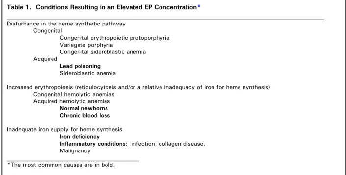
Table 1. Conditions Resulting in an Elevated EP Concentration*
Disturbance in the heme synthetic pathway Congenital
Congenital erythropoietic protoporphyria Variegate porphyria
Congenital sideroblastic anemia Acquired
Lead poisoning
Sideroblastic anemia
Increased erythropoiesis (reticulocytosis and/or a relative inadequacy of iron for heme synthesis) Congenital hemolytic anemias
Acquired hemolytic anemias Normal newborns Chronic blood loss Inadequate iron supply for heme synthesis
Iron deficiency
Inflammatory conditions: infection, collagen disease, Malignancy
*The most common causes are in bold.
Poor interlaboratory agreement for EP
prompted a reassessment of the published m, for PPIX and ZP. If indeed the protoporphyrin being measured by the extraction technique were the same as that measured by the HF, then the values obtained from extraction techniques should have agreed with the ZP values measured by the front surface
techniques. However, this was not the case.
In 1985, the United States Public Health of the Bureau of Health Care Delivery and Assistance and the CDC consulted with a number of experts in Washington, DC, to review the calibration procedures and the reporting of test results. One recommendation was to re-examine the m, for PPIX. In 1989, Gunter et al 19proposed that the m, for PPIX be 297 L @ cm-1 @ mmol (see Section 7).-1 Other investigators described the quantitative determination of metal-free versus ZP.
Different species of individual porphyrins can be separated by high performance liquid chromatography (HPLC). 21These procedures are restricted to research settings and reference laboratories, and they are not available widely for clinical purposes.
Therefore, HPLC methods are beyond the scope of this document.
6 Applications of the
ErythrocyteProtoporphyrin Test
The concentration of EP is elevated in several pathological conditions that impair the syn- thesis of heme (Table 1). To make a specific diagnosis, confirmatory laboratory tests are required. The most common use of the EP test is in screening for lead poisoning. Also, EP is increasingly used in screening for iron deficiency because, in contrast to other tests, the analysis can be performed cheaply and rapidly enough by hematofluorometry to determine the need for additional testing during a single patient visit. In addition to lead poisoning and iron deficiency, EP is also elevated in mild and severe inflammatory conditions of more than a few days' duration.
Inflammatory disorders that can cause
elevated EP include not only chronic illnesses, such as rheumatoid arthritis, osteo-myelitis, and cancer, but relatively acute ones, such as otitis media. One basis for this elevation is the marked decline in serum iron that is
associated with inflammatory conditions, which restricts the production of heme and results in the accumulation of EP in RBCs.
Generally, EP is elevated in hemolytic diseases and in chronic blood loss due to conditions, such as peptic ulcers and colon cancer, all of which are typically characterized by an increased rate of RBC production. With increased red cell production, available
20
Service, Division of Maternal and Child Health
storage iron that would meet normal lead in young children is advocated. It physiologic needs can be inadequate to meet remains to be seen to what extent universal the demands for an ele-vated rate of heme blood lead testing, as recommended by the production. Furthermore, the proportion of CDC, will be put into practice. If the
young red cells becomes increased, and these recommendations are fully implemented, the contain a higher concen-tration of EP than future role for EP in the management of lead more mature cells. Thus, in iron deficiency, poisoning is likely to be as follows:
inflammatory conditions, hemolytic diseases,
and chronic blood loss, an inadequate iron C Screening
supply for heme synthesis and/or an increased – As a secondary test to blood lead for proportion of young RBCs is responsible for prioritizing management when blood the elevation of EP. On the other hand, in lead lead concentration is between 10 and poisoning, the interference with later steps in 20 µg/dL (0.48 and 0.97 µmol/L).
the hemesynthetic pathway results in an – To identify concomitant iron
elevation of EP. deficiency.
6.1 Lead Poisoning
6.1.1 Childhood Screening for Lead Poisoning
From 1972 to 1991, EP was the primary screening test recommended by the CDC for childhood lead poisoning detection in the United States.22-25EP was most often measured by HF in public health clinics (see Section 9). Because the hematofluorometry test result is available in minutes, a child found to have an elevated EP concentration could have blood drawn at the same visit for a confirmatory blood lead determination. Then, the blood lead and EP concentrations were used to determine the risk level of lead poisoning under the 1978 and 1985 CDC guidelines. In 1978, the CDC defined lead poisoning in children as a blood lead of $ 30 µg/dL (1.45 µmol/L) whole blood after a screening EP of $ 50 µg/dL (2.40 µmol/L) whole blood. 23In 1985, the guidelines were revised to define lead poisoning more
stringently with a cut-off value for blood lead of $ 25 µg/dL (1.21 µmol/L) after a screening EP of $ 35 µg/dL (1.69 µmol/L) whole blood.24 In 1991, a further revision was instituted by the CDC to recommend universal blood lead testing for infants and young children and to define lead poisoning as a concentration of blood lead of >10 µg/dL (0.48 µmol/L). 25At this blood lead concentration, the diagnostic sensitivity and specificity of the EP test for lead poisoning is poor.26
Consequently, the CDC no longer
recommends EP as the primary screening test to identify children with lead poisoning;
instead, the routine determination of blood
25
C Diagnosis
– In addition to blood lead content, to evaluate the biological effects of lead poisoning.
– To monitor cases under medical management.
6.1.2 Occupational Lead Poisoning
Protoporphyrin measurement is widely used, in conjunction with the blood lead
concentration, for monitoring the lead status of industrial workers who are exposed to lead.
Measurement of EP (specifically ZP) level,**
together with blood lead concentration, is part of the Occupational Safety and Health
Administration (OSHA)-required initial baseline for lead-exposed workers, who should be subsequently tested every 6 months.
However, the specific guideline for action is based not on the EP, but on the blood lead concentration alone. A worker who is found to have a high blood lead concentration will be removed from the area of lead exposure and required to have serial blood lead and EP measurements to monitor improvement and to determine a suitable time for returning to the work setting. Currently, the blood lead
threshold concentration for removing a worker from an area of lead exposure is $ 60 µg/dL (2.90 µmol/L) on a single occasion, or three sequential measurements that average $ 50 µg/dL (2.40 µmol/L). The criterion for per- mission to return to the lead exposure area is a blood lead concentration of < 40 µg/dL (1.93 µmol/L). 27 At these blood lead
**
Occupational Health and Safety Administration regulations specify ZP as the entity to be measured, but no threshold values are specified.27
concentrations, EP has a strong correlation where the extra expense can be justified for with blood lead and can serve as part of the the purpose of providing greater certainty monitoring system for recovery from about iron status.
occupational lead exposure. An action concentration for EP in occupational lead exposure still needs to be defined. EP is a useful physiological marker for the biologically harmful effects of lead because elevated EP levels signify interference with the heme-syn- thetic pathway.
6.2 Iron Deficiency
6.2.1 Screening for Iron Deficiency Measurement of EP level is one of several tests that can be used to detect iron deficiency anemia or a milder form of iron deficiency without anemia that is
characterized, nevertheless, by impaired heme synthesis. The EP test is used mainly in screening children.28-30A therapeutic trial of iron may be considered when the EP level is elevated in combination with an abnormally low Hb, hematocrit (Hct), or serum ferritin level. A significant reduction of EP
concentration and/or an increase in hemo- globin concentration after a month of iron treatment suggests the earlier presence of iron deficiency. In adults, the EP test is used primarily to screen for iron deficiency in blood donors.31,32Similarly, it is used to screen for iron deficiency 33in patients with renal disease who are undergoing repeated hemodialysis.34 EP is useful for screening pregnant women for iron deficiency because EP values remain stable during pregnancy if there is adequate iron nutrition, 35whereas other measures of iron status change.36
6.2.2 Diagnosis of Iron Deficiency
EP is one of three biochemical tests, together with serum ferritin and transferrin saturation used in the diagnosis of iron deficiency.37,38 Any of these tests can provide additional evidence for the presence of iron deficiency after a patient is found to be anemic on screening. However, no single one of these tests, in the absence of anemia, has a high All three tests may be used in combination with hemoglobin in nutrition surveys38,39and for research involving iron nutrition 28,29,37
6.2.3 Differential Diagnosis of Microcytic Anemia
The determination of EP is helpful in differ- entiating the two major causes of microcytic anemia (anemia with a low mean corpuscular volume): iron deficiency anemia and thalasse- mia trait. 40Typically, iron deficiency is associated with an elevated EP level, whereas, usually, the EP level is normal in anemia of both alpha- and beta-thalassemia traits.
6.2.4 Inflammatory Conditions
A variety of inflammatory conditions can complicate the interpretation of an elevated EP level in persons who are being screened for lead poisoning and/or iron deficiency. This is most evident among infants and young children who often have, or have recently had, mild infections (e.g., upper respiratory infections or otitis media) of more than a few days' duration41; also, it can be a problem among elderly persons, many of whom have severe or mild chronic inflammatory
conditions, such as arthritis.42 6.2.5 Porphyrinopathies
Porphyrias are inherited or acquired disorders of porphyrin metabolism that result in the elevation of one or more of porphyrin
compounds. 43 Porphyrinopathies include both the porphyrias or inherited (primary) disorders and the induced (secondary) disorders in porphyrin metabolism. In some porphyrias, the formation, accumulation, and excretion of porphyrins can be extremely elevated.
6.2.5.1 Hereditary Porphyrias At least seven varieties of hereditary porphyrias have been identified, each being associated with a different enzyme defect in the hemesynthetic pathway, with the exception of *-aminolevulinate synthetase.
sensitivity or specificity for iron deficiency. 39 (serum iron/iron binding capacity), that can be
HCl
m, Concentration 8-max Reference 278 8.3 mol/L 411.0 Grinstein, 194846 262 2.7 mol/L 408.0 Rimington, 196047 275 1.5 mol/L 408.0 Schwartz, 19609 241 1.0 mol/L 408.0 Nat Acad Sci, 197213 297 1.5 mol/L 408.0 Gunter, 198919
Also, the porphyrias differ with respect to the to determine the correct value. In the past, tissue in which the enzyme becomes interlaboratory agreement for the
rate-limiting, specifically, bone marrow determination of EP levels has been somewhat (erythropoietic origin) or liver (hepatic origin). problematic because of the wide-spread use Even though all porphyrias are characterized of secondary standards, such as
by an elevation of some type of porphyrin, coproporphyrin, discrepancies in the m, value only certain porphyrias are associated with a for PPIX, and the issue of extraction
significant elevation of EP level: erythropoietic efficiency.
protoporphyria (hepatic and erythropoietic origin), variegate porphyria (hepatic origin), porphyria cutanea tarda, and copro-
porphyria.43,44
6.2.5.2 Acquired Porphyrias
Some porphyrinopathies are related to non- congenital disease or to chemical exposure.
However, with the exception of lead, most of the these are characterized by an elevation of porphyrins other than protoporphyrin IX. Lead poisoning is considered to be a form of ac- quired porphyria.
6.2.6 Sideroblastic Anemias
Sideroblastic anemias are a heterogeneous group of disorders of heme synthesis char- acterized by ineffective erthyropoiesis. As in the porphyrias, there are congenital and ac- quired forms. The condition is characterized by anemia, the presence of ring sideroblasts, and accumulation of metal-free EP. 44
7 Millimolar Absorptivity: A His- torical Perspective
It is conventional analytical practice to establish standard concentrations by using a known mass of analyte. This is not possible with PPIX because of impurities and stability limitations. Therefore, an approximate mass is assumed, which must be corrected using molecular absorption spectrophotometry and Beer's law. This requires accurate knowledge of the m, of the substance being analyzed.
Previous determinations of the m, for PPIX might have been compromised by the use of impure source materials. In addition, use of slit widths >10 nm for establishing
absorbance wavelength maximum (8-max), variability in HCl concentration of the solvent, and the fact that solutions of PPIX with identical theoretical concentrations can exhibit different absorbance values depending on their initial preparation, have made it difficult
Following the measurement of a 1-mg/L solu- tion of PPIX in HCl, the following m, were reported:
However, the most frequently used EP micromethods measured PPIX standard concentration in 1.5 mol/L HCl, not 1.0 mol/L.
Also, use of a convenient "5-µg" PPIX tube standard as a primary standard source
contributed error, because the amount of PPIX contained in the tube was not truly 5 µg;
rather, it delivered an amount calculated to produce a solution with an absorbance corresponding to an expected concentration of 5 µg per 10 mL, if a m, of 241 L @ mmol-1 @ cm is assumed.-1 19
PPIX tends to form molecular aggregates in dilute aqueous solutions48; this aggregation is associated with a decrease in the m, and region broadening in the Soret band. 49One- mg/L solutions of PPIX in 1 mol/L HCl prepared directly from PPIX free acid (FA) exhibit an m, equal to 272 L@ mmol-1 @ cm . -119
When prepared from a hydrolysate of PPIX dimethyl ester (DME), the m, is equal to 297 L @ mmol @ cm-1 -1. 19 This difference can be explained by aggregation because the approximately 10% difference in absorbance of these two solutions is matched by a 10%
difference in fluorescence. The greater the degree of polymerization, the lower the level of fluorescence or absorbance is observed in solution. Apparently, hydrolysis results in minimal aggregation, which creates a more dispersed state of PPIX molecules by
providing the most monomeric form of PPIX.19
45
10,11
To establish a clinical database of EP con- databases and future databases using the centrations for use in childhood lead poisoning correct m0.
screening programs, adoption of a m, of 241
L @ mmol-1 @ cm was recommended as an-1 After reviewing the data, the subcommittee arbitrary uniform standard value in 1975.12 recommends the adoption of an m0 of 297 L @ This recommendation was echoed in a mmol @ cm . Furthermore, the subcommittee meeting of protoporphyrin researchers at the recommends that the DME-hydrolysis prepa- CDC in 1976. However, data from the United ration technique for stock standards of PPIX States National Normative and Prevalence be used as the preferred method of standard Studies, the Second National Health and preparation (see Section 8.3.)
Nutrition Examination Survey (NHANES II,
1976–1980),50,51the Hispanic Health and Adoption of m0 297 L@mmol @cm will signifi- Nutrition Examination Survey (HHANES, cantly change the reference ranges for EP.
1982–84), 52and NHANES III (1988– 1994), Revised reference intervals are provided in determined with PPIX standards prepared from Section 10.2. Note that this standardization hydrolysates of DME, demonstrated change, and the associated effect on results approximately 19% lower EP concentrations and reference intervals, must be clearly than previously published studies that used communicated to the practicing clinician to the 241 m, value. prevent incorrect interpretation of EP results.
The discrepancies in the results of these studies have led to the proposal, in 1989, for the formal establishment of the m, (of a 1- mg/L solution of PPIX in 1.5 mol/L HCl at 408.0 nm with a 0.5-nm bandpass) as 297 L @ mmol-1 @ cm . The effect on the public-1 health of revising the m, would be a 19%
decrease in the current clinical reference intervals for EP values [e.g., this would have changed the 1985 CDC-recommended EP cut- off value 24from 35 to 28 µg/dL (0.62 to 0.50 µmol/L) of whole blood in childhood lead poisoning programs]. Data generated by other laboratories using an m0 of 241 L @ mmol-1 @ cm can be converted to the recommended-1 m, of 297 by multiplying by 241/297 in order to compare it to the NHANES national
-1 -1
-1 -1
8 Recommended Procedure for the Quantitative Fluorometric Determination of Erythrocyte Protoporphyrin After Extraction
8.1 Principle of the Method Porphyrins and heme components are extracted from anticoagulated whole blood into an ethyl acetate–acetic acid mixture.Next, protoporphyrin is back-extracted into an HCl solution. In low pH aqueous solutions, ZP dissociates into Zn and protoporphyrin IX2+
free acid (PPIX FA); then, the concentration of PPIX in the
Figure 3. EP cell holder
aqueous phase is measured by molecular 8.2.2 Chemicals fluorometry. Fluorescence is a relative
measurement and standards are critical. The calibration curve is based on dilutions of a PPIX standard, the concentration of which is first established by molecular absorption spectroscopy.
This description is based on a method jointly developed by the New York State Department of Health (Parsons PJ, personal
communication, 1988), the Wisconsin State Laboratory of Hygiene (Stanton NV, personal communication, 1988), and the CDC.52 8.2 Apparatus
8.2.1 Instrumentation
The following instrumentation is necessary for the quantitative determination of erythrocyte protoporphyrin after extraction:
C A spectrofluorometer, with a bandpass of 5.0 nm or less at 640–670 nm, equipped with a
red-sensitive photomultiplier tube with S-20 response, and fitted with a cell- holder (see Figure 3) adapter for 10- X 75-mm glass tubes and a suitable printer or digital display
C Centrifuge (general laboratory) -uipped for 10- X 75-mm tubes
C Vortex mixer C Analytical balance C Chemical fume hood
C Automated dispensers and dilutors (or fixed-volume bottle repetitive dis- pensers, and micropipettors (50 µL, 100 µL, 200 µL, 400 µL, and 500 µL) C Magnetic stirrer and stir bars
C Spectrophotometer, UV-visible, double-beam, with variable slit-widths adjustable to 0.5 nm at 405–410 nm, equipped with 1-cm quartz cells.
The following chemicals are necessary:
C Deionized water, greater than or equal to 1.0 megohm-cm at 25 E C, treated to remove organic compounds, with zero background fluorescence
C Protoporphyrin IX dimethyl ester (PPIX DME), C H N O , $ 99% purity36 38 4 4 C Ethyl acetate, CH CO C H , high-purity3 2 2 5
(see Section 8.8.2)
C Acetic acid, CH COOH, glacial, ACS3 grade
C Hydrochloric acid, HCl, concentrated, ACS grade
C Formic acid, CH O , concentrated,2 2 ACS grade
C Potassium iodide, KI, ACS grade (see Section 8.8.2).
8.2.3 Glassware
The following pieces of glassware are necessary:
C Volumetric flasks, actinic, class A, with stoppers (1 L, 500 mL, 250 mL, 200 mL, 100 mL, 10 mL) (for class A flask volume tolerances see ASTM standard #E28853) and clear glass, 1 and 2 L. If actinic glass is not available, clear glassware wrapped in aluminum foil may be substituted.
C Pipets, volumetric, class A (25.0 mL, 10.0 mL, 8.0 mL, 6.0 mL, 5.0 mL, 4.0 mL, and 2.0 mL) (for class A volumetric pipet tolerances see ASTM standard #E96954)
C Culture tubes, glass, disposable, 10- x 75-mm
C Graduated cylinders, 1 L, 250 mL C Microtiter plates, plastic, 96- x 0.5-mL
wells (optional: 10- x 75-mm glass culture tubes may be substituted).
g/mol 36.435
%HCl g/mL
Density
=
mol/L •
8.2.4 Reagents (2) Dilute the mixture to 1.0 L.
The following reagents are necessary:
8.2.4.1 Hydrochloric Acid Solutions NOTE: Although concentrated HCl is
commonly assumed to be 12 mol/L, each lot can vary slightly upon manufacture. The correct substance (molar) concentration should be determined using the following formula:
In
formation on the density and percentage of HCl in the assay should be provided on each bottle label.The following directions are based on a lot of concentrated HCl determined to be 12.7 mol/L. Laboratorians should verify the molarity of their particular lots and adjust required acid volumes accordingly before beginning protoporphyrin analysis.
Laboratorians should always wear eye pro- tection and acid-resistant gloves when working with HCl, and they should always remember to add acid to water when preparing solutions.
C Following are directions on how to prepare HCl, 7.0 mol/L (for
hydrolysis):
(1) Add 138 mL of concentrated HCl (12.7 mol/L) to 100 mL of deionized water in a volumetric flask.
(2) Dilute the mixture to 250 mL.
C Perform the following steps to prepare HCl, 1.62 mol/L (for daily absorbance readings at a final concentration of 1.5 mol/L):
(1) Add 141 mL concentrated HCl to 250 (1) Protoporphyrin IX Free Acid
mL deionized water in a volumetric Hemolysate Stock Solution, 200 mg/L flask.
C Perform the following steps to prepare HCl, 1.5 mol/L (for analysis/extraction and for blanking spectrophotometer):
(1) Add 118 mL concentrated HCl to 250 mL deionized water in a volumetric flask.
(2) Dilute the mixture to 1.0 L.
8.2.4.2 Ethyl Acetate, 2:1 (v/v) To prepare ethyl acetate, 2:1 (v/v):
(1) While working under a hood, combine 400 mL of ethyl acetate and 200 mL of glacial acetic acid in a reagent bottle.
(2) Mix the solution well, using a clean glass rod or by capping the bottle and swirling it thoroughly. This volume is sufficient for the complete assay of standards, controls, and 80 specimens in duplicate. Larger volumes may be prepared weekly and stored in amber bottles.
8.3 Protoporphyrin IX Standard Solu- tions
To minimize photodecomposition of PPIX, all standard solutions are prepared in actinic glass-wa