Until now, it was believed that the paleontological history of the tetraodontiform (plectognath) fishes began in the Eocene. Sorbini and Guidotti (1984) redescribed Protriacanthus based on the holotype and seven additional specimens.

Ver5-6
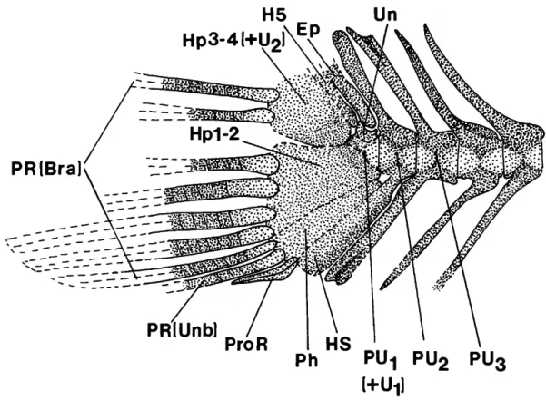
In the dorsal lobe of the caudal fin, only the lower two rays are preserved, borne on the upper hypural plate. The long, intact pelvic spine appears to be that of the left side when viewed in medial view.
PRlBra
A center ridge along the length of the impression represents a groove on the posterior surface of the spine. The base of the pelvic fin is surrounded by enlarged scales that form a protection or shield, the exposed surface of which is tuberculous.

Ver4
Ver1
LtPS
The anguloarticular articulates approximately in the middle of its upper surface with the head of the quadrate. The long posterior process of the pelvis is shaft-like, as in hollardiin triacanthodids and triacanthids (the posterior process is basin-like in triacanthodin triacanthodids).

PECar
The specimen in which the articulation of the maxilla is best preserved (USNM 481512, acid prepared) indicates that the maxilla is wide ventrally and dorsally tapering into a narrower head with a concavity for articulation with the premaxilla (Fig. 14), but this is somewhat speculative. Several large, bridged foramina are visible in the tooth groove for the sensory canal in some specimens, but the bone surface is relatively poorly preserved in other specimens. Small teeth (-0.1 mm in MCSNV 1374) are present in the region of the oral cavity of the vomer anterior to the lateral ethmoid and posterior to the dorsal head of the premaxilla, and are interpreted as from the vomer.
The prootic and parasphenoid are clearly exposed within the lower part of the orbital cavity. The vomer is only weakly indicated at the front of the parasphenoid where small teeth are present. The frontals and the bones of the occipital and opercular series are extensively ornamented with tubercles similar to those of the carapace.
PRiUnbl
Slightly larger (-0.4 mm in MCSNV 1374) and more elongated teeth are present in the region above the ceratohyal, probably yes. Among the epiotics, some specimens have evidence of a sphenotic anterior and a pterotic posterior, both articulating with the dorsal margin of the hyomandibular (Figure 15), which is a stout curved shaft supporting the relatively well-preserved opercle and preopercle.
PRlBra]
ProR
AECar
The parhipural is located between the lower hypopural plate and the haemal spine of the penultimate vertebra. No centra of caudal peduncle particularly compressed anteroposteriorly (vs. one to four compressed). The second barb is much shorter and placed very close to the base of the first barb.
The second spine is thick mainly where it is near the posterior end of the first spine. The posterior end of the third infraorbital has a weakly marked connection with the anterior end of the fourth infraorbital. A subocular shelf is visible in two specimens (IGPUB 1FDC29 (holotype) and MCSNV T913) below the middle of the orbit.

PR (Unb)
The parasphenoid is clearly preserved as a strong shaft without a ventral flange in the orbit region. The parasphenoid curves upward posteriorly and joins the prootic at the back of the orbit. The ectopterygoid is elongated and connects the anterodorsal region of the quadrate to the posterior region of the palatine.
Only the centra of the vertebrae below the carapace are visible (and only in one acid-prepared specimen, MCSNV T915), but the vertebrae posterior to the carapace are relatively well preserved in several specimens (especially in the holotype). The anterodorsal margin of the carapace is distinctly separated from the head by a scaleless area. The well-developed carapace and the posterior placement of the soft dorsal and anal fins of protriacanthids (as well as of plectocretacicides) are generally similar to those conditions in ostrasioids (except for the continuity of the carapace on the head in the latter), but it are represented as independent acquisitions (see "Analysis of Characters," characters 25, 28).

The fossil fishes of Comen are found in the Monte Coste Member of the dark gray to blackish, compact and sometimes bituminous limestone, in which there is some evidence of centrimetric-polydecimetric bedding (Cucchi, Pirini Radrizzani and Pugliese, 1987). . The general phylogeny of the recent Tetraodontiformes followed here is that established in the myologically based cladistic analysis of Winterbottom (1974), modified with the phylogeny of the balistoid + ostracioid clade by Winterbottom and Tyler (1983) and the phylogenies of a variety of fossil taxa by Tyler. and Gregorova (1991, for ostracioids), Tyler and Bannikov (1992a, for the balistoid + ostracioid clade; 1992b, for molids), and Tyler et al. Because the phylogenetic relationships of the five families of zeiforms are still undeciphered, use we include representatives of all five families in our comparisons with tetraodontiforms.
One of the taxa (Protriacanthus) that we include in the plectocretacicoids was previously tentatively referred to the gasterosteiforms, but this was at least partly on the basis of the somewhat misleading original description. Tyler agreed with Patterson's analysis of the original description of Protriacanthus and dismissed it as a possible tetraodontiform, and Medizza and Sorbini (1980) also followed Patterson in listing incertae sedis among the fishes of Comen. Based on the characters in our redescription of Plectocretacicus (especially as seen in the acid-prepared specimens), together with those of the new taxon Cretatriacan - thus the numerous specialized characters that Protriacanthus shares in combination with those taxa, and the synapomorphies that are common are for Protriacanthus and Plectocretacicus, we are convinced that Protriacanthus is a tetraodontiform.
I 111 gife
Length of First Dorsal-Fin Spine. In cretatriacanthids the first spine is rudimentary and the
In all other tetraodontiforms with a spiny dorsal fin, the first spine is well developed and at least as long as, and usually longer than, the second. One exception to this generalization occurs in triodontids, where the entire spiny dorsal fin is rudimentary, and although the first spine is usually longer than the second, in some specimens of the single extant species the first spine may be a buried nubbin. The single redundant dorsal spine of zeiforms is not comparable to the condition of two supernumerary spines in tetraodontiforms; in any case, the first spine in many families of sea forms is usually robust, but of such variable length (very short to elongate but never as rudimentary as in Cretatriacanthus) and relationship to the second spine (shorter than or equal to ) that this would not help establish the polarity of first spine length in tetraodontiforms.
However, the data indicate that a rudimentary first spine is not a primitive feature of zeiforms. Because the first spine is long in protriacanthids, triacanthoids, balistoids, and eoplectids, the rudimentary first dorsal spine of cretatriacanthids is considered an autapomorphy.
Length of Second Dorsal-Fin Spine. The second spine in the dorsal fin of cretatriacanthids is relatively
We think it more likely that the primitive pletocretacychoid condition is one of average spine length as found in its sister groups, with increases in length being ambiguous autapomorphies of cretriacanthids and protriacanthids. According to one of two equally nonsensical hypotheses for the increased length, the reduction in length is autapomorphic for pletocretacidids (see character 39). In cretatria-canthids and protriacanthids, but not in pletocretacycanthids, canthids and protriacanthids, but not in pletocretacanthids, the anterior edge of the quadrate is relatively vertically oriented or inclined anterodorsally and bears the main reinforcing ridge immediately posterior to its articulations.
In plectocretaccids and all other tetraodontiforms, the main supporting ridge of the quadrate is oriented more horizontally, more or less along its ventral edge, whereas the anterior edge of the quadrate, which articulates with the ectopterygoid, is obliquely oriented posterodorsally. Clearly, the vertical orientation of the quadrate buttress and its articulation with the ectopterygoid, as well as the anterodorsal orientation of the latter, must be considered derived in Cretatriacanthus and Protriacanthus. We believe that it is more likely that the primitive plectocretacicoid state is one of horizontal orientation as found in its sister groups, where the vertical orientations are ambiguous autapomorphies of cretatriacanthids and protriacanthids.
Premaxillary Serrations. The external surface of the angle of the premaxilla bears prominent deep
Nevertheless, because four synapomorphies link Protriacanthus to Plectocretacicus and there is only one other derived feature shared by Cretatriacanthus and Protriacanthus (long pelvic spine, see character 33), it is equally parsimonious to suggest that the ancestral state of plectocretacicoids was a horizontal orientation like triacanthoids, with the vertical orientation in cretatria-canthids and protriacanthids independently derived, or to assume that the vertical orientation occurred in the plectocretacicoid ancestor, with a secondary reversal to the horizontal orientation in plectocretacicids. As documented under character 8, the reduction in the documented under character 8, the reduction in the number of primary caudal fin rays to 12 is primitive for tetraodontiforms, and the reversal in Plectocretacicus to the higher number of 14 found in some zeiforms is autapomorphic.
Maxillary Articulation Dorsally. In plectocre- tacicids the dorsal end of the maxilla seems to articulate
Therefore, the unique grouping and articulation of the last pair of rays in protriacanthids is considered autapomorphic. In molids, all these elements are absent at the truncated rear of the body. The members of the plectocretacicoid clade share four derived features (presence of subocular shelf; absence of teeth in the jaws; crest of modified shield scales around base of pelvic fin spine; diminutive size), while the other three main clades of tetraodontiforms (triacanthoids), balistoids + ostrasioids and tetraodontoids), known from the Eocene to the present, are linked by five unambiguous synapomorphies (absence of grooves in dentary for sensory canal; absence of infraorbitals; small mouth, at least primitive; six or fewer tachyostegal rays; absence of vomerine teeth ) and two others.
All the major lineages of extant tetraodontiforms (triacanthoids, balistoids + ostracioids, tetraodontoids) are only known in the Eocene. On the systematic position of the family Caproidae with reference to the Eocene genus Acanthonemus. The skull of the Eocene Triodon antiquus (Triodontidae; Tetraodontiformes): Similar to that of the recent trident pufferfish, T.