The first step in any virus entry process is binding to the plasma membrane of the host cell. Six of the fifteen were shown by sequence analysis to react with overlapping regions of a protein identical in sequence to the mouse high-affinity laminin receptor. We also wanted to determine the domains of the virus envelope proteins responsible for attachment to the cell membrane.
We attempted to map neutralization epitopes on the surface of the virus, including epitopes implicated in virus binding to cells from the antiidiotype antibody results described above. We hypothesize that this E2 domain centered at residue 200 is part of the virus binding site to bind to the cell to initiate infection.
Possible mechanisms of virus entry
For example, vaccination must be administered before the host is exposed to the virus; the host immune response to a vaccine may not be consistent in all individuals; antibody titers in response to vaccination may decline over time; a vaccine against one serotype of a virus with multiple serotypes may not protect against the predominant serotype in a host population (eg, there are > 100 serotypes of rhinovirus that cause the common cold); and certain viruses such as HIV insert into host chromosomal DNA and escape vaccine-induced antibody interaction (Ginsberg et al., 1988; Haseltine et al., 1989). The uptake process of such viruses, recognized as opportunistic ligands such as toxins, hormones and growth factors, is a receptor-mediated form of endocytosis, i.e. constitutive pinocytosis (Steinmen, et al., 1983; Pastan and Willingham, 1983; Silverstein et al., 1977). During receptor-mediated endocytosis, ligands first bind to cell surface receptors to form ligand-receptor complexes, after which the region of the plasma membrane containing the ligand-receptor complexes is internalized by endocytosis at clathrin-coated pits, which are specialized depressions on the cell. surface.
In other words, entry can occur by fusion of the viral membrane with the cell membrane. The point of contact between the two membranes expands slightly for fusion processing allowing the hydrophobic regions of the extracytoplasmic sheets of the two membranes to contact each other.
Entry of Alphaviruses
Identification of cell surface receptors
Ambiguous results have also been reported in the identification of the C3d receptor for complement as the Epstein-Barr virus receptor (Jonsson et al., 1982; Simmons et al., 1983) and in the identification of a 140 kDa protein as the receptor. for Friend murine leukemia virus (Robinson et al., 1980; Bubber et al., 1978). The characterization of the host cell receptor for Sindbis virus and the interaction between the Sindbis virus enveloped spikes and its receptor will be discussed in my dissertation. It is noteworthy that among rhinoviruses, ICAM-1 is the receptor for 75 major serotypes, but there are other receptors for other serotypes (Greve et al., 1989).
The tip proteins and the viral membrane do not simply provide a protective coat for the nucleocapsid; they play a crucial role in binding the virus to receptors on host cells. The cellular receptor for poliovirus: Molecular cloning, nucleotide sequence, and expression of a new member of the immunoglobulin superfamily. Nucleotide sequence of the 26S mRNA of Sindbis virus and the deduced sequence of the structural proteins of the encoded virus.
Studies of the Epstein-Barr receptor found in Raji cells: Extraction of the receptor and preparation of anti-receptor antibodies.
Chapter 2
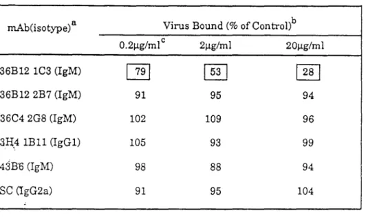
We show here that a high-affinity laminin receptor serves as a receptor for Sindbis virus in mammalian cells. The initial event in the interaction of a virus with the host cell is the binding of the virus to receptors on the plasma membrane (Lonberg-Holm and Philipson, 1974; Lonberg-Holm and Philipson, 1981; Tardieu et al., 1982). A hybridoma from one of the BHK whole-cell immunized mice (out of -1500 examined from this mouse) was obtained that secreted an IgM (mAb 36B 12 lC3, hereafter referred to as 1 C3) that partially blocked virus binding to cells BHK.
It therefore appears clear that mAb 1C3 is directed against the C-terminal domain of the high affinity laminin receptor. We wanted to determine whether the laminin receptor also serves as a receptor for Sindbis virus in cells of different origins. Either the virus uses a different receptor in chicken cells or the attachment of the virus to the chicken larnin receptor is not blocked by mAb 1 C3.
There are no potential sites for asparagine-linked carbohydrates in the amino acid sequence and the nature of the posttranslational modifications remains unclear. It is also of interest that a high affinity laminin receptor encoding a 50kDa protein has been identified in Staphylococcus aureus (Lopes et al., 1985). The high affinity laminin receptor binds to a YIGSR sequence in the central region of laminin.
This suggests that the sites in the laminin receptor to which laminin binds and to which Sindbis virus binds are distinct and spatially separated. From the overlap of the A.gt 11 clones to which mAb 1C3 binds, this mAb must bind a site in the C-terminal 48 amino acids of the laminin receptor. It is also possible that the differential susceptibility may arise from differential posttranslational modifications of the laminin receptor in different cells or from the availability of other (as yet unidentified) Sindbis receptors on these cells.
Processing of Sindbis virus nonstructural polyproteins: an in vivo kinetics study using monospecific antibodies. A positional marker for the dorsal embryonic retina is homologous to the high-affinity laminin receptor.
Fluorescent Intensity
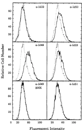
In each panel, the dashed line is the profile of BHK cells stained with secondary antibody FITC-conjugated goat anti-mouse IgM (~-chain) as a control. Monolayers of BHK cells were washed with PBS lacking divalent cations and removed from the culture dish by treatment with 4 mM EDTA in PBS for 15 min at 37°C. Six of the 15 clones were found to cross-hybridize and the sequences of five of these were detected; they were found to start at different points in the laminin receptor sequence and terminate with 3' poly(A).
Of these, 52 were analyzed by PCR for the 5' end of the laminin receptor coding region using the 17-mer oligonucleotide primer 5'-GCAAACTTCAGCACAGC-3' complementary to the 5' end of clone 7 and either the 24-mer Agt11 forward primer or the 24- mer Agt11 reverse primer. Amplified inserts were characterized by electrophoresis and DNA sequencing; clone 3 was found to encode the 5' end of the laminin receptor. The published amino acid sequence (Makrides et al., 1988) for the mouse high-affinity laminin receptor is identical to that for BHK shown here.
A second published sequence for the mouse laminin receptor (Rao et al., 1989) differs by two amino acids from the sequence published by Makrides et al. The expression of mRNA for the laminin receptor was analyzed using the 3' end of the eDNA of the BHK laminin receptor from A clone 26 to create a 32p_labeled probe. The EcoRI fragment encoding the 3' end of the BHK laminin receptor from clone A 26 was used to make 32P-dCTP-labeled probes by nick translation (Sambrook et al. 1989).
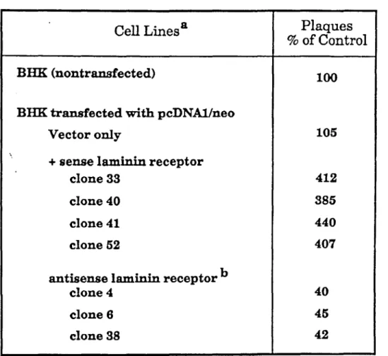
Efficiency of Sindbis virus plaque formation in BHK cells transfected with the laminin receptor gene or with the antisense gene. 7 Binding of mAb lC3 to BHK laminin receptor transfectant cells as determined by F ACS analysis. In each panel, the dashed line is the profile of BHK cells transfected with the pcDNA/neo vector alone and stained with rnAb lC3.
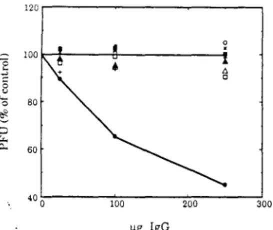
The number of plaques was normalized to the number of plaques on monolayers of the same cells without mAb treatment. Plaques were allowed to develop under 1% agarose in 12-well plates for 18 h in BHK cells, 24 h in Vero cells, 32 h in SW13 cells, and 13 h in chicken cells before staining. Cell lysates were prepared from 35S-labeled murine neuroblastoma cells, Vero cells, BHK cells, and chicken fibroblast cells as indicated and immunoprecipitated with mAb 1 C3.
Cells were washed, removed from the flask by treatment with 4 mM EDTA in PBS, and collected by centrifugation. Cell pellets were resuspended in 1 mM NaHC03 containing 10 mM EDTA and protease inhibitors (1 mM PMSF, 0.7 ~ g/ml pepstain A, 0.5 ~ g/mlleupeptin) and cells were broken by Dounce homogenization. Immunoprecipitations were performed as described ( Hardy and Strauss, 1988 ), using goat anti-mouse IgM together with Sepharose 4B to collect immunoprecipitates.
Antiidiotypic Antibodies as Probes for the Sindbis VInIS Receptor
Chapter 4
Mapping Neutralization Epitopes on Glycoproteins of Sindbis Virus
The neutralizing epitope consists of the sequence or structure to which such a neutralizing antibody binds. These include analysis of genetic variants resistant to an antibody (Diamond et al., 1985; Stec et al., 1986) and analysis of the interaction of the antibody with peptides, either short polypeptides produced synthetically or by fragmentation of the protein or expressed as fusion proteins in a convenient system (Lerner, 1882). A study of variants resistant to many of these neutralizing antibodies has shown that variants in a domain of E2 from 180 to 220 are selected by many of the E2-specific antibodies (Strauss et al., 1991).
We have used this system in an attempt to define neutralization epitopes of E2 and E1 in order to compare these epitopes with the results from antibody-resistant variants and to more precisely define the domain of the glycoproteins that appears to function in binding of virus to receptors on the host cell. An Agt 11 library containing short inserts of Sindbis cDNA was constructed by a modification of the method of Young and Davis (1983). With this procedure, we expected that immunoreactive phage would carry sufficiently small Sindbis virus cDNA inserts to allow precise mapping of the neutralizing epitopes on the Sindbis virus glycoprotein E1 and E2.
The Agtll system provides a rapid, specific and sensitive strategy for physically mapping on large viral genomes the genes encoding proteins for which antibody reagents are available. The limitation of the system is the fact that these protein domains are expressed as part of a fusion protein and therefore cannot be folded in the same way as the native protein, and only antibodies that interact with contiguous linear domains of the proteins of interest can be reactive with phage plaques. We examined six neutralizing monoclonal antibodies directed against Sindbis virus glycoproteins E 1 and E2 for their ability to recognize short segments of the glycoprotein sequence expressed in Agt11.
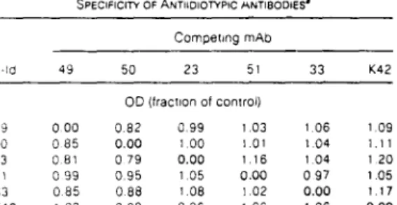
By sequencing the inserts in four clones immunoreactive with mAb 23, we determined that this antibody was able to react with a single continuous region of the Sindbis glycoprotein E2 and that the neutralization epitope must lie within the 48 residues between amino acids 173 and 220. In Chapter 3 results were reported that antiidiotypic antibodies to mAb 49, and to a lesser extent antiidiotypic antibodies to mAbs 23 and 50, functioned as an antibcxiy antireceptor in chicken cells, suggesting that this E2 domain may form part of antireceptor on the virus spike that binds to the cellular receptor. Identification of significant antigenic domains in Sindbis virus glycoproteins by analyzing antibody escape variants.