Anatomy, functional morphology, evolutionary ecology and systematics of the invasive gastropod Cipangopaludina japonica (Viviparidae: Bellamyinae)
Bert Van Bocxlaer1, 2, 3, 4, 5, Ellen E. Strong3
1 Museum für Naturkunde, Leibniz Institute for Evolution and Biodiversity Science, Invalidenstraße 43, D-10115 Berlin, Germany
2 Department of Animal Ecology and Systematics, Justus Liebig University, Heinrich-Buff-Ring 26-32 IFZ, D-35392 Giessen, Germany
3 Department of Invertebrate Zoology, National Museum of Natural History, Smithsonian Institution, PO Box 37012, MRC 163, Washington, DC 20013-7012
4 Limnology Unit, Department of Biology, Ghent University, K.L. Ledeganckstraat 35, B-9000 Ghent, Belgium
5 E-mail: [email protected]
Key words: basal Architaenioglossa, Bellamya, fecundity, geometric morphometrics, histology, reproductive anatomy, sexual dimorphism
Abstract
The anatomy, functional morphology and evolutionary ecology of the Viviparidae, and the subfamily Bellamyinae in particu- lar, are incompletely known. Partly as a result, genealogical re- lationships within the family remain poorly understood. Be- cause of this lack in knowledge, few informed hypotheses exist on ancestral states, how differences in body plans between the subfamilies evolved, and how the peculiar biogeographic distri- bution patterns of viviparids have arisen. Here we document the anatomy, morphology, life history and systematics of Cipango- paludina japonica, a Japanese species that has been introduced into North America, to resolve taxonomic confusion and to im- prove our understanding of how form and function are related in bellamyines. Anatomical and histological examinations demonstrate marked differences between C. japonica and other bellamyines in the radula, salivary gland, kidney, nerve ring and reproductive organs. Substantial differences also exist be- tween male and female body organization, but conchological differences between sexes in semi-landmark morphometric analyses are limited. The volume of the brood pouch of fe- males, and hence body and shell size, appear to be good predic- tors of reproductive success, and the species’ ecological versa- tility may relate to high fecundity and the ability to alternate between feeding modes. Comparing our observations on C. ja- ponica with other viviparids and basal Architaenioglossa, we identify several persistent misinterpretations in the literature on how form and function are related in viviparids, not in the least as to female reproductive anatomy. Our reinterpretations im- prove understanding of the evolution of Viviparidae and its sub- families, and hopefully will allow future workers to isolate key traits that shaped the evolution of viviparids at the taxonomic levels of their interest for more detailed studies.
Contents
Introduction ... 235
Material and methods ... 237
Material ... 237
Anatomy and histology ... 237
Morphometrics ... 238
Results ... 238
Systematics ... 238
Shell morphology ... 240
External anatomy ... 242
Mantle cavity ... 243
Alimentary system ... 244
Reno-pericardial system ... 247
Nervous system ... 248
Reproductive system ... 249
Fecundity ... 252
Morphometrics ... 252
Discussion ... 253
Shell morphology ... 253
Anatomy ... 254
Functional morphology and evolutionary ecology ... 259
Conclusions ... 260
Acknowledgements ... 260
References ... 260
Introduction
The anatomy and functional morphology (i.e. how form is related to function in its broadest sense) of viviparid gastropods (Viviparidae) of the subfamily
Bellamyinae are poorly documented, although sub- stantial differences have been observed in comparison to other viviparid subfamilies, i.e. Viviparinae and Lioplacinae (Bouchet and Rocroi, 2005), particularly in reproductive anatomy (Rohrbach, 1937; Vail, 1977;
Simone, 2004). For many of the approximately 125- 150 known viviparid species (Strong et al., 2008), lim- ited anatomical and molecular data are available, leav- ing their position within the family and genus-level relationships uncertain (but see Sengupta et al., 2009;
Du et al., 2013). Because of this lack of knowledge, no hypotheses exist as to how these differences in body plan among the subfamilies have arisen, how the sub- families are phylogenetically related to one another, what the ancestral states were in the earliest viviparids (but see Starobogatov, 1992 for some suggestions), or what the functional advantages of one body plan over the others may be for each region of occurrence.
The family Viviparidae has a temperate to tropical global distribution (e.g. Prashad, 1928), but is not na- tive to Antarctica or South America, although it has been introduced to the latter. The subfamilies had
largely separate distributions until introductions brought them into contact, with conspicuous absences from large regions, e.g. the Middle East (Prashad, 1928), where the family was present during the Plio- Pleistocene (Sivan et al., 2006). The Bellamyinae is largely confined to the Old World tropics and subtrop- ics, whereas the Lioplacinae has a Nearctic distribu- tion, and the Viviparinae is Holarctic. As mentioned above, however, some opportunistic Asian species were introduced elsewhere: Cipangopaludina chinen- sis (Gray in Griffith and Pidgeon, 1833) and C. japon- ica (von Martens, 1861) were first brought to Northern America in ~1892 and ~1911, respectively, as a human food source (Prashad, 1928; Clench and Fuller, 1965), and very recently bellamyines have been discovered in South America (Sinotaia quadrata [Benson, 1842]) (Ovando and Cuezzo, 2012) and in Europe (C. chinen- sis) (Soes et al., 2011). Some bellamyines, such as the introduced species, appear to be relatively opportunis- tic and ecological generalists, but others, notably some African Bellamya, are more stenotopic, although they have radiated in some of the East African Rift lakes
Table 1. Specimens examined in anatomical and histological studies. Most organ systems were subjected to comparative studies in males and females. Dissected specimens received individual museum numbers (those collected from the Potomac River in Alexandria in 2012: USNM 1296930-1296947; from Lake Barcroft in 2013: USNM 1296948; and from Gunston Cove in 2014: USNM 1296844, 1296846, 1296850, and 1296952; see material and methods). On the first row (gross morphology) specimen numbers are followed by M or F, indicating the sex of each specimen.
Organ system Specimens analyzed
Gross morphology all specimens (1296930M, 1296931F, 1296932F, 1296933F, 1296934M, 1296935M, 1296936M, 1296937M, 1296938M, 1296939F, 1296940F, 1296941F, 1296942M, 1296943M, 1296944M, 1296945M, 1296946M, 1296947M, 1296948F, 1296844F, 1296846F, 1296850F, 1296852F) Pallial organs (general) 1296930; 1296931; 1296933; 1296934; 1296942; 1296947; 1296844
Respiratory system 1296930 (gill); 1296931 (gill); 1296932 (gill + vascular system); 1296844 (gill + gill leaflet) Alimentary system 1296930 (gastric chamber, intestine, rectum); 1296931 (digestive gland); 1296932 (gastric chamber,
intestine, rectum; histology of digestive gland); 1296933 (buccal apparatus and haemocoel, SEM of radula); 1296935 (buccal mass, haemocoel); 1296937 (gastric chamber); 1296940 (histology of diges- tive gland); 1296944 (gastric chamber, digestive gland including histology); 1296946 (gastric chamber, digestive gland); 1296947 (gastric chamber); 1296948 (gastric chamber, digestive gland); 1296844 (buccal mass, gastric chamber, digestive gland); 1296846 (gastric chamber)
Reno-pericardial system 1296930 (pericardium); 1296931 (kidney); 1296932 (ureter, kidney); 1296933 (pericardium, vascular system); 1296934 (kidney, pericardium); 1296937 (pericardium, pallial vascular system); 1296941 (pericardium); 1296850 (pericardium)
Nervous system 1296933 (nerve ring, nerves in haemocoel); 1296935 (nerve ring); 1296844 (nerve ring)
Male reproductive system 1296930 (right tentacle, prostate, testis); 1296934 (testis); 1296935 (right tentacle, prostate); 1296942 (testis, including histology); 1296947 (testis)
Female reproductive system 1296932 (seminal receptacle, copulatory bursa, albumen gland, brood pouch); 1296933 (seminal receptacle, renal oviduct, copulatory bursa, albumen gland, brood pouch, SEM of juveniles); 1296939 (brood pouch); 1296941 (brood pouch); 1296948 (seminal receptacle including histology, copulatory bursa incl. histology, albumen gland incl. histology, brood pouch), 1296844 (ovary, visceral oviduct, seminal receptacle, copulatory bursa, albumen gland, brood pouch), 1296850 (ovary), 1296852 (anatomy and histology of the seminal receptacle, copulatory bursa and albumen gland)
and paleolakes (Brown, 1994; Van Bocxlaer and Hunt, 2013; Salzburger et al., 2014; Schultheiß et al., 2014).
Despite general uncertainty of genus-level affilia- tions and the limited availability of comparative data for several viviparid genera, the genera Bellamya Jous- seaume, 1886, Idiopoma Pilsbry, 1901, Cipangopaludi- na Hannibal, 1912 and presumably Neothauma Smith, 1880 show similar anatomical features that allow clas- sification in the Bellamyinae (e.g. Rohrbach, 1937).
Anatomical characters of the Australian viviparids No- topala Cotton, 1935 and Larina Adams, 1854 also war- rant classification as bellamyines (Simone, 2004), like the Asian genera Filopaludina Habe, 1964 (with its sub- genenera Filopaludina s.s. and Siamopaludina Brandt, 1968), Trochotaia Brandt, 1974, Eyriesia Fischer, 1885, Mekongia Crosse and Fischer, 1876, Sinotaia Haas, 1939, Anulotaia Brandt, 1968 (Brandt, 1974), Angulya- gra Rao, 1931 (Du et al., 2013), and also Margarya Nevill, 1877 and Taia Annandale, 1918 (Vail, 1977).
Hence, Bellamyinae is the most diverse viviparid sub- family as Lioplacinae and Viviparinae each contain two confirmed genera (Campeloma Rafinesque, 1819 and Lioplax Troschel, 1856, Viviparus de Montfort, 1810 and Tulotoma Haldeman, 1840, respectively) (Vail, 1977). Great diversity but limited systematic control and coverage in anatomical studies in comparison to Viviparinae and Lioplacinae (e.g. Leydig, 1850;
Baudelot, 1863; Auerbach, 1896; Crabb, 1929; Mattox, 1938; Vail, 1977) make the subfamily Bellamyinae an appropriate target for additional comparative anatomi- cal work. Moreover, an improved understanding of the intrinsic biological properties of the group is a key re- quirement to comprehend the evolutionary history, the ecology and the invasion biology of the Bellamyinae, notably those properties that may have played a key role in their diversification and evolutionary success.
Here we study the anatomy and life history of C.
japonica, a relatively familiar bellamyine with impor- tant implications for biological conservation of benthic ecosystems due to its introductions in the Neotropics.
The species remains poorly studied, and as a result many misconceptions exist in the literature on the sys- tematics, anatomy and functional morphology of C.
japonica (e.g. Smith, 2000). We perform morphologi- cal, anatomical and histological investigations of C.
japonica to understand how form and function relate to one another, and how they have affected the evolu- tionary ecology of the species. Subsequently we com- bine our findings with literature data to build a com- parative framework for life history studies of vivi- parids in general.
Material and methods Material
Living specimens and empty shells of C. japonica were collected in September 2012 from the Potomac River in Alexandria, VA (living specimens at 38.8103°
N, 77.0393° W and 38.8137° N, 77.0380° W; empty shells between 38.8154° N, 77.0381° W and 38.8101°
N, 77.0393° W). Other living specimens were collect- ed in October 2013 from Lake Barcroft, VA (38.8529°
N, 77.1552° W), and in June 2014 from Gunston Cove, VA (38.6817° N, 77.1533° W). Living specimens were heat-shocked (Fukuda et al., 2008), then preserved in 80% ethanol for use in studies of the external gross morphology and detailed functional anatomy. 23 spec- imens were used in anatomical studies; a total of 53 shells were used for the study of shell morphology and allometry. This material is deposited at the Smithso- nian Institution, National Museum of Natural History (Potomac River: USNM 1296930-1296947; Lake Barcroft: USNM 1296948; Gunston Cove: USNM 1296844, 1296846, 1296850, 1296952 and 1296902).
Anatomy and histology
Even after heat shocking the specimens, it was not possible to remove bodies from the shells without damaging the tissues, especially the more voluminous females. Hence, voucher images were made after which the shells were broken with a vice, soft tissues were removed, and individuals sexed. Anatomical dis- sections were made using a Wild M5 and Leica MZ16 microscope with camera lucida. Staining with tolui- dine blue was used to enhance contrast when neces- sary. Most organ systems were studied in multiple specimens to allow differentiation between individual variation and characteristics that define the popula- tion, and by extension the species (Table 1). Anatomi- cal renderings consist of a baseline drawing for a rep- resentative specimen, which was subsequently modi- fied into a more generalized representation.
Histological sections were prepared from the testis of males (not shown), and the albumen and capsule glands of females. Target organs were dissected, dehy- drated through a graded ethanol series, cleared in xy- lene, embedded in paraplast, and serially sectioned at 7-8 µm. Sections were stained with haematoxylin and eosin-phloxine following standard protocols (Hu- mason, 1967); cover slips were mounted with a solution of permount and 5 % terpenol, dissolved in toluene.
Histological sections were viewed and photographed with a Leica DMLS2 compound microscope and a Leica DFC320 digital camera.
Radulae were isolated, cleaned in diluted bleach, sonicated, mounted on SEM stubs with adhesive tabs, sputter coated with 25-30 nm gold/palladium (60/40) and studied with a Hitachi TM3000 tabletop scanning electron microscope (SEM).
Embryos and developing juveniles from the brood pouch of seven anatomically-studied females that were collected in September 2012 were removed and quan- tified to provide an insight into fecundity.
Morphometrics
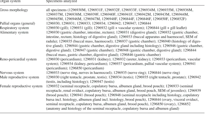
To explore sexual dimorphism and allometric varia- tion in shell morphology, we applied semi-landmark morphometrics with 10 landmarks and 4 open semi- landmark curves (Fig. 1) to 53 shells of C. japonica that represent a growth series, including 20 sexed adult specimens. For the allometric studies, five juvenile specimens obtained from the anteriormost part of the
brood pouch were used as a baseline. These specimens reflect juvenile shell morphology at birth. Juveniles were cleaned following the protocol used for radula cleaning (see methods for anatomy and histology), mounted on SEM stubs with carbon adhesive tabs, sputter coated and photographed in apertural view with the abovementioned SEM. Larger, more mature specimens were photographed in the same view with a Canon EOS 350D SLR camera equipped with an EFS 60 mm f/2.8 Macro USM lens.
For morphometric analyses, we used semi-land- mark morphometrics following the recommendations of Van Bocxlaer and Schultheiß (2010), using the mod- ified protocol of Van Bocxlaer and Hunt (2013). Digi- tal images were compiled in .tps format using TpsUtil 1.50 (Rohlf, 2012). Landmarks and semi-landmark curves were digitized in TpsDig 2.16 (Rohlf, 2010).
These curves were anchored between landmarks (LM7
& 2, LM8 & 6, LM5 & 6 and LM 7 & 9, respectively;
Fig. 1) and each consists of a fixed number of equidis- tant points that were sampled via the resample-curve- by-length option in TpsDig 2. Specimens were aligned using Procrustes superimpositioning in CoordGen6h (Sheets, 2008), after which the semi-landmarks were slid along their respective curve using the minimum Procrustes distance criterion in SemiLand6 (Sheets, 2008). The resulting Procrustes superimposition coor- dinates document shell shape, and shell height was measured in Klonk Image Measurement Light version 14.1.1.7 and logarithmically transformed in base e.
Morphometric shape data and size measurements were imported in R 3.0.1 (R Development Core Team, 2013) for further analysis using Vegan 2.0-8 (Oksanen et al., 2013) and Boot 1.3-9 (Ripley, 2013). The Procrustes superimposition coordinates were subjected to Princi- pal Component Analysis (PCA). Relevant principal components were plotted against shell height to assess allometric changes. For PC1 a linear relationship was obtained which was investigated with linear least squares regression.
Results Systematics
Family Viviparidae Gray, 1847
Subfamily Bellamyinae Rohrbach, 1937 Genus Cipangopaludina Hannibal, 1912
Type species: Paludina malleata Reeve, 1863, by orig- inal designation.
Fig. 1. Generalized viviparid shell in apertural view with land- marks (Arabic numerals), semi-landmark curves (Roman nu- merals with the number of points per curve indicated in paren- theses) and the direction of digitization (arrows) as used in this study.
Remarks. Adult shells of Cipangopaludina species are usually very similar to those of Idiopoma, but are larg- er and have a more inflated body whorl; unlike in many Idiopoma species, brownish zones above and below the periphery of the whorls are lacking; embry- onic and juvenile whorls of Cipangopaludina are ad- pressed, with a peripheral, basal angulation and often two spiral ridges, whereas the embryonic shells of Idi- opoma are less adpressed, more globose and never keeled; embryonic shells have numerous spiral lines, the three most prominent bear long, curved periostra- cal hairs, whereas embryonic shells of Idiopoma lack periostracal hairs entirely; mantle edge is thickened in Cipangopaludina compared to Idiopoma; other ana- tomical differences are largely unknown.
The relationships of Asian viviparids are poorly un- derstood and, hence, the literature contains much dis- cussion and speculation on the genus-group classifica- tion within the Bellamyinae, including the circum- scription and affinities of Cipangopaludina Hannibal, 1912. Material classified here as Cipangopaludina was earlier grouped in Viviparus (e.g. Pilsbry, 1902; Kuro- da, 1929; Thiele, 1929; Clench and Fuller, 1965), and has also been referred to Vivipara Sowerby, 1813 (e.g.
Hannibal, 1911), an incorrect subsequent spelling, and to Paludina Férussac, 1812 (e.g. von Martens, 1861;
Kobelt, 1879; Fischer, 1887), an invalid name placed on the Official Index of Rejected and Invalid Generic Names in Zoology in 1959 by ICZN Opinion 573.
Hannibal (1912) described Cipangopaludina, with the Japanese species Paludina malleata Reeve, 1863 as type species, as a subgenus of Idiopoma. The latter was proposed with Vivipara henzadensis Pilsbry, 1901 as type species, which is considered to be a form of the Indian species Nerita dissimilis Müller, 1774 (Brandt, 1974). However, ambiguity remained concerning the status of Idiopoma and other viviparid genera. Some authors raised Cipangopaludina to genus level (e.g.
Yen, 1943), whereas others (e.g. Prashad, 1928; Thiele, 1929) lumped it and many other genera that currently belong to the three subfamilies into a single genus, Viviparus, with several subgenera. Annandale (1920) proposed the genus Lecythoconcha with Paludina le- cythis Benson, 1836 as type species, to which the spe- cies Paludina japonica von Martens, 1861 would be- long based on the generic description and geographic coverage. This classification was maintained by some (Rao, 1925), but others (Prashad, 1928; Kuroda, 1929;
Brandt, 1974) synonymized Lecythoconcha with Cipangopaludina. Rohrbach (1937) listed only a sin- gle large genus in his subfamily Bellamyinae for Afri-
can and Asian viviparids, namely Bellamya Jous- seaume, 1886. Other authors (e.g. Yen, 1943; Brandt, 1974) assigned several genera to the Bellamyinae, and Bellamya and Cipangopaludina were hypothesized to be closely related but separate genera. Recently, Smith (2000) proposed to accommodate Cipangopaludina as a subgenus of Bellamya based on anatomical data, which was followed by others (Solomon et al., 2010).
However, the type species of Bellamya is Bellamya bellamya Jousseaume, 1886, by original designation, which is considered a synonym of Vivipara duponti De Rochebrune, 1881 (Germain, 1920), and represents a West African form of Bellamya unicolor (Olivier, 1804) (Brown, 1994). Bellamya unicolor is nested in a clade of African viviparids and a deep phylogenetic split exists between African and Asian viviparids (Sengupta et al., 2009; Du et al., 2013; Schultheiß et al., 2014). Based on fossil calibration, the base of the African clade would date to ~15 Ma or possibly older (Schultheiß et al., 2014), and a clade consisting of spe- cies of Angulyagra (= a replacement name for Dacty- lochlamys Rao, 1925), Filopaludina, Larina and Taia is more closely related to the African viviparids than the clade containing species of Cipangopaludina, Margarya, Mekongia, and Sinotaia (Sengupta et al., 2009; Du et al., 2013). Consequently, synonymizing Cipangopaludina with Bellamya does not accurately reflect evolutionary relationships. Other Asian species like Idiopoma dissimilis also appear deeply divergent from African Bellamya species, and we consider it likewise ill-founded to consider Idiopoma a junior synonym or subgenus of Bellamya. Further work is re- quired to assess the relationship of Asian viviparids and their genus-level classification.
Cipangopaludina japonica (von Martens, 1861) Paludina japonica von Martens, 1861: 44. Type local- ity: Japan.
Viviparus japonicus var. iwakawa Pilsbry, 1902, p. 117, pl. 9, fig. 3. Type locality: Furukawa, Rikuzen, Japan.
Other references:
Paludina japonica – Reeve 1863, pl. 3, figs 13a,b. – Kobelt 1879: 120, pl. 11, fig. 1.
Paludina oxytropis – Kobelt, 1879: 123, pl. 11, fig. 6.
– Iwakawa 1897: 88, figs 8-17.
Viviparus japonicus – Pilsbry 1902: 117, pl 9, fig. 1.
Remarks. Hannibal proposed Cipangopaludina as a subgenus of Idiopoma for Paludina malleata from Ja- pan, but considered japonica to belong to Idiopoma
s.s. Paludina malleata is regularly considered a syno- nym of P. chinensis (see e.g. Jokinen, 1982), a species illustrated by Gray without description. However, Reeve (1863) noted the native range of chinensis to be Chusan (Eastern China), whereas the type locality of malleata is Japan. Both taxa may hence belong to deeply divergent clades, and molecular analysis of to- potypic specimens of chinensis and malleata are re- quired before synonymizing these species. Cipango- paludina japonica also has been synonymized with C.
chinensis (e.g. Dundee, 1974; Clarke, 1978), however others have considered the morphological differences to be sufficiently large to retain them as separate spe- cies (Clench and Fuller, 1965; Stańczykowska et al., 1971; Jokinen, 1982; Smith, 2000; Solomon et al., 2010), which has been confirmed by molecular analy- ses (Hirano et al., 2015). Rigorous molecular studies are also required to elucidate the relationships of C.
malleata and C. japonica. Ideally, these would also
include specimens of C. stelmaphora (Kobelt, 1879) described from Japan, which may be a synonym of C.
japonica.
Shell morphology
Juvenile specimens from the anteriormost brood pouch of four whorls (Fig. 2A, B); embryonic shell el- evated above the teleoconch and comprising ~1.2 whorls, but variable and in some populations compris- ing up to 2 whorls (Fig. 2C); apical cap of the embry- onic shell bears an outer furrow or crease (Fig. 2D);
transition between the embryonic shell and the tele- oconch inconspicuous, marked by a sudden but slight increase in diameter of the whorl; ornament of fine spiral threads are present, but the apical part of the embryonic shell has no hairs on these threads; spiral rows of axial striae may be present (Fig. 2D, E) be- tween the spiral threads.
Fig. 2. Juvenile shell morphology of Cipangopaludina japonica. A, B) Juveniles extracted from the anteriormost pallial oviduct of specimen USNM 1296933 (population from the Potomac River in Alexandria, USA), and C) of a female from a Japanese population (USNM 424320) for comparison. Note the higher, more acute spire and the slightly more angular body whorl of the Japanese specimen compared to those from the USA. D) Apical cap of the embryonic shell, showing the apical outer furrow or crease. E, F) Apical view showing periostracal hairs and spiral threads.
Early teleoconch with a bluntly-angular basal cari- na and two prominent spiral cords (Fig. 2E) corre- sponding to periostracal ridges with long hairs; some variation exists in the translation of the first teleoconch whorls (hence, the apical angle), and in the angularity of the cords and the basal carina (compare Fig. 2A-C);
midway between the two cords is a third spiral thread with shorter hairs; shell does not have pits at the inser- tion of hairs; many other, finer spiral threads without hairs are present (Fig. 2E, F); axial sculpture consist- ing of abundant fine, prosocline growth lines that be- come more prominent with age; on the juvenile shell they intersect with the spiral elements to give the shell a finely reticulate texture.
Subadult shells (Fig. 3A-E) have a pronounced basal angulation that becomes less conspicuous with age;
early whorls usually display two conspicuous cords, and are adpressed, whereas later whorls are not; aper- ture of subadults somewhat angulate below; adult shells have a more smoothly rounded, inflated penultimate and body whorl than those of juveniles and subadults (compare Fig. 3A-E with 3F-H); adult shells typically lack the basal angulation and cords of juveniles and subadults but may display more or less conspicuous spiral lines in corresponding positions (Fig. 3F); su- tures become more deeply impressed, the shoulder more rounded, and the whorls more inflated as the shell grows; spire high, spiral angle ~84° in juveniles and
Fig. 3. Subadult and adult shell morphology of Cipangopaludina japonica. A, B) Apertural and adapertural views as illustrated by Reeve (1863). C) Post-birth juvenile showing strongly angulated early whorls. D, E) Subadults showing the characteristically angulated base of the body whorl. F-H) Large adults. F and G are females, with slightly more inflated body whorls than males (H). Scale bar for C-H; the size of Reeve’s specimen was unspecified.
~74° and ~68° in adult females and males, respectively (compare Fig. 3H with 3F, G); shape of the adult aper- ture large and ovate, narrower adapically, with a well- rounded base; parietal callus present, but not very pro- nounced; umbilicus open.
External anatomy
Operculum (Fig. 4C) corneous, sub-pyriform, thick, slightly translucent, brown in color, thicker and darker at the parietal and columellar margins than at the pala- tal one; growth concentric, but faster at the palatal margin, resulting in a subcentral nucleus (i.e. ~1/4-1/3 of the total width from the columellar margin).
Soft tissues of adult specimens comprising ~3.5
whorls (Fig. 4); foot broad, muscular with a long, shal- low pedal gland along the anterior margin of the pro- podium. Columellar muscle moderately long, extend- ing 0.5-1.0 whorl posteriorly. Cephalic tentacles short and thick in preserved specimens, up to roughly twice the length of the snout; right cephalic tentacle modi- fied in males to a penis with a terminal gonopore (Fig.
5). Eyes terminal on short ocular peduncles at outer bases of cephalic tentacles. Neck modified on the right into a tall, broad nuchal lobe at the level of the urinary pore, the anus and (in females) the female gonopore at the right side of the mantle cavity, just behind the thick, muscular mantle edge (Fig. 5). It can be extend- ed somewhat beyond the shell, whereas the smaller, shorter left nuchal lobe cannot.
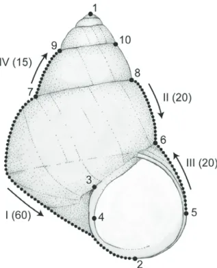
Fig. 4. Body morphology and anatomy.
A) Female in apertural view; B) Female in adapertural view; C) operculum mor- phology; D) Male in abapertural view.
Abbreviations: ag = albumen gland; av = afferent branchial vessel; bp = brood pouch; bv = blood vessel; cm = columel- lar muscle; ct = ctenidium; dg = diges- tive gland; eg = egg; em = embryo; es = endostyle; ev = efferent branchial vessel;
f = foot; gs = gastric chamber; it = intes- tine; kd = kidney; mc = mantle collar; mr
= mantle roof; pc = pericardium; re = rectum; sr = seminal receptacle; ss = style sac; te = testis; ur = ureter.
Mantle cavity
Mantle cavity (Fig. 5) long, usually somewhat shorter in males (~1.0 whorls) than in females (~1.5 whorls) and bounded posteriorly by the reno-pericardial sys- tem. On the mantle floor, slightly to the left of the mid- line, a conspicuous food groove extends from the pos- terior ctenidium to the head; a prominent ridge marks the base of the food groove and anteriorly fuses with the right nuchal lobe, forming a trough that is deflected around the cephalic tentacle and continues anteriorly towards the mouth. The monopectinate ctenidium is composed of elongate triangular leaflets (Fig. 5, inset) and extends along the left mantle roof from near the mantle collar to the posterior end of the cavity. At the
anteriormost third of the gill, overlying the large effer- ent branchial vein, lies the slender osphradium, which forms a ridge with distinct papillae along the side fac- ing the ctenidial axis, likely representing vestigial leaf- lets. The endostyle runs along the length of the afferent axis of the ctenidium (Fig. 4A, B, D, 5); posteriorly the endostyle wraps around the base of the ctenidium and is continuous with the posterior end of the food groove.
The hypobranchial gland is weakly developed. At or near the dorsal midline lies the rectum, and further to the right the ureter and pallial gonoduct (testis or
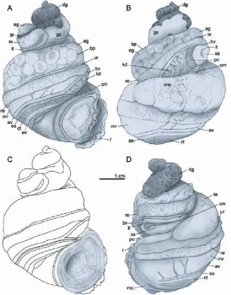
Fig. 5. Mantle cavity morphology of a male, with inset of a sin- gle gill filament. Orientation: A = anterior; L = left; P = poste- rior; R = right. Abbreviations: a = anus; av = afferent branchial vessel; bv = blood vessel; ce = cephalic tentacle; ct = ctenidium;
e = eye; en = endostyle; ev = efferent branchial vessel; f = foot;
fg = food groove; ki = kidney; mc = mantle collar; mf = mantle floor; mr = mantle roof; nl = nuchal lobe; os = osphradium; pe
= penis; pr = prostate; re = rectum; te = testis; up = urinary pore;
ur = ureter.
Fig. 6. Dorsal view of buccal apparatus and mid-oesophagus, inset with a schematic representation. Orientation: A = anterior;
L = left; P = posterior; R = right. Abbreviations: ba = buccal apparatus; bg = buccal ganglion; bp = buccal pouch; cg = cere- bral ganglion (largely concealed by salivary gland); df = dorsal fold; dfg = dorsal food groove; m = mouth; n = nerve; oe = oe- sophagus; sd = salivary gland duct; sg = salivary gland; sp = supra-oesophageal ganglion; t = tensor.
brood pouch) are present. The slit-like urinary pore opens slightly behind the papillate anus, which opens near the mantle edge.
Alimentary system
Buccal apparatus. Mouth forming transverse slit, opening ventrally at anterior end of the smooth oval snout; buccal mass long and slender (Fig. 6). Odonto- phore occupying ~4/5 of the length of the buccal mass, with narrowly triangular subradular organ projecting below the radula near the mouth. A small, thin paired jaw at the anterior end of the dorsal folds flanks the mouth dorsolaterally. Shallow, non-glandular buccal pouches are elaborated below the dorsal folds in the anteriormost oesophagus, just behind the buccal gan- glia. Two salivary gland ducts open dorsolaterally
alongside the dorsal food groove in the dorsal folds, shortly behind the middle of the buccal apparatus; the ducts pass posteriorly through the circumoesophageal nerve ring, to the large, massive salivary glands, which cover the nerve-ring dorsally and laterally, and parts of the foregut and the supra-oesophageal connective.
Thick buccal retractors extend laterally from the rear of the buccal mass and insert on the walls of the ce- phalic hemocoel just anterior to the nerve ring. The radular sac is short, projecting ventrally slightly past the end of the buccal mass and through the nerve ring.
Radula. The radula is small (~6 mm in length in adults), slender and delicate, taenioglossate with ~80 rows of developed radular teeth, and several more rows of developing teeth (Fig. 7). The rachidian (Fig. 7A, B, E) possesses a broad, angular central cusp flanked by
Fig. 7. Radular morphology. A) Section of anterior radular ribbon; B) Detail of rachidian and lateral teeth; C) Detail of inner and outer marginal teeth; D, F) Lateral teeth; E) Rachidian; G-H) Inner marginal teeth; H-I) Outer marginal teeth. Scale bars in µm.
one very small cusp and two larger ones that decrease outwardly in size, giving the functional surface of the rachidian an obtuse, isosceles triangular appearance, but with marked gaps adjacent to the central cusp. The lateral teeth (Fig. 7A, B, D, F) are similar in width but longer than the rachidian, and somewhat asymmetri- cal. They have a larger central cusp than the rachidian, which is flanked by one small and two larger inner cusps, and by one or two small and four larger outer cusps. The inner marginal teeth (Fig. 7C, G, H) are more asymmetrical than the lateral teeth, with long slender shafts and a cutting edge with one large, rounded median cusp flanked by two inner and three outer smaller cusps. The outer marginal teeth (Fig. 7C, H, I) have slightly broader shafts than the inner mar- ginals, and are slightly bowed outwardly toward the base, with a broadly rounded cutting edge bearing eight cusps of more or less similar size and shape, but decreasing somewhat in size toward the outer edges.
Oesophagus. The anterior oesophagus emerges from the dorso-posterior side of the buccal apparatus and
forms a simple tube bearing continuations of the dorsal folds and dorsal food groove (Fig. 6). In the mid-oe- sophagus, which starts just behind the nerve ring, the continuation of the dorsal folds and the dorsal food groove displays the effects of torsion. The walls of the oesophagus between the dorsal folds bear fine, irregu- lar longitudinal ridges. The mid-oesophagus continues posteriorly as a simple tube in a central position, slight- ly to the right of the food groove, with no outpocketings or glandular elaborations, and deflects slightly towards the right at the posterior part of the mantle cavity to continue posteriorly along the columellar aspect, pass- ing below the reno-pericardial complex. The posterior oesophagus is finely longitudinally ridged and widens substantially just before opening ventrally to the poste- rior gastric chamber.
Gastric chamber. The gastric chamber (Fig. 8) occu- pies most of the body just behind the elongate pericar- dium. It lies embedded in the lobes of the digestive gland and has non-muscular, thin walls. A prominent T-shaped ridge partially compartmentalizes the gas-
Fig. 8. Gastric chamber and proximal style sac, inset with a schematic repre- sentation. Evenly stippled area indi- cates cuticular lining; dashed line indi- cates the position of the gastric shield;
T-shaped ridge indicated in grey in in- set; continuation of structures is indi- cated by small arrows in inset. Orien- tation: A = anterior; L = left; P = poste- rior; R = right. Abbreviations: c = gas- tric pouch; cf = ciliated (or marginal) fold; dd = opening of digestive gland duct; et = expanded tip; gp = glandular pad; ig = intestinal groove; ilf = inner- most left longitudinal fold; lr = longitu- dinal ridge (continuation of finger-like ridges); oe = oesophagus; olf = outer- most left longitudinal fold; p1 = longi- tudinal part of the prominent T-shaped ridge (homologous to glandular pad);
p2 = transverse part of the T-shaped ridge; pss = posterior edge of the style sac region; sa = sorting area; ssa = sec- ond sorting area with textured pad pos- teriorly; sf = smaller (outermost right longitudinal) fold; t1 = major typhlo- sole; t2 = minor typhlosole.
tric chamber into three regions. The longitudinal limb (= glandular pad) emerges from the right, posterior gastric chamber wall and forms a ridge along the floor to the right of the oesophageal opening. Roughly mid- way along the left side of the glandular pad, a single digestive gland duct opens to the gastric chamber floor.
At the anterior end of the longitudinal limb, the trans- verse limb crosses the gastric chamber floor just in front of the oesophageal aperture and bears the gastric shield at right. A prominent sorting area is developed in the left gastric chamber floor and roof anterior to the oesophageal opening, and extends anteriorly to the in- testinal groove. Two curving longitudinal folds in the gastric chamber roof border the left side of the sorting area, the outer left longitudinal fold more prominent than the inner. The inner left longitudinal fold emerges anteriorly in the vicinity of the proximal end of the minor typhlosole and has a prominently expanded tip;
the outer fold has its anterior tip much farther back, at the level of the gastric shield. The two folds merge near the posterior end of the sorting area, forming a single fold that crosses from left to right just in front of the oesophageal opening, and continues posteriorly along the base of the glandular pad, passing above and to the right of the digestive gland duct. Two additional longitudinal folds border the sorting area at right (the
ciliated or marginal fold and a smaller right longitudi- nal fold), and extend posteriorly from the vicinity of the proximal end of the major typhlosole. They are themselves bordered at the right by a smaller, narrower sorting area to the left of the gastric shield. This sec- ond sorting area persists posteriorly on the left side of the glandular pad to the rear wall of the gastric cham- ber. At the level of the digestive gland duct, another longitudinal fold emerges to the left of the duct in the gastric chamber floor. At the rear of the gastric cham- ber, this longitudinal fold and those on the left side of the glandular pad curve around the base of the glandu- lar pad into the roof, merging and terminating at the right posterior end of the glandular pad, behind the gastric shield. Within the gastric chamber roof, from the prominent outer longitudinal fold to the left side of the main sorting area, a series of flattened finger-like ridges are elaborated. The ridges begin as small undu- lations at the anterior tip of the longitudinal fold, grad- ually elongating and broadening, spanning the roof and extending onto the gastric chamber floor behind the gastric shield at the right side of the glandular pad.
The ridges stain lightly purple with toluidine blue, in- dicating they are glandular. Anteriorly, the transverse limb of the T-shaped ridge separates a deep, concave, cuticularized style sac pocket at the right (stippled re-
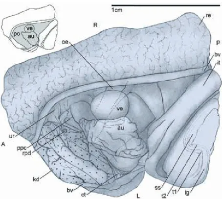
Fig. 9. Dorsal view of the pericardium, inset with a schematic representation.
Orientation: A = anterior; L = left; P = posterior; R = right. Abbreviations: au = auricle; bv = blood vessel ; ct = ctenidi- um; ig = intestinal groove; it = intestine;
kd = kidney; oe = oesophagus; pc = peri- cardium; rpd = reno-pericardial duct;
ppc = posterior margin of the pallial cav- ity; re = rectum; ss = style sac; t1 = major typhlosole; t2 = minor typhlosole; ur = ureter; ve = ventricle.
gion, Fig. 8) that is interrupted ventrally at the left only by the sorting area and receives two ducts of the diges- tive gland below the lip of the style sac. At the left, anterior end of the main sorting area, the expanded tip of the weak inner left longitudinal fold delimits a shal- low gastric pouch. The division between the gastric chamber and the style sac is demarcated by a constric- tion.
Digestive gland. The digestive gland is a large yellow, brown or greenish organ located in the visceral hump, usually occupying the ~2.0-2.5 most apical whorls, with its anterior ~0.5 whorl supporting the right aspect of the gastric chamber (Fig. 4). The digestive gland is incompletely divided into two lobes: a smaller anterior one, which borders the right wall of the gastric cham- ber, and a larger posterior one. The gland is drained by tubules that join the digestive gland ducts, which run along the columellar aspect of the visceral whorls.
These ducts increase in size anteriorly, and open to the gastric chamber; one posteriorly, two anteriorly.
Style sac. The style sac is roughly 1.5 times the length of the gastric chamber and forms a voluminous undi- vided sac. It bears two ventral typhlosoles that traverse its length, bounding the intestinal groove. At the distal end of the style sac, the typhlosoles turn sharply to the left before continuing anteriorly through a 90° angle (Fig. 9), with two shallow pyloric caecae elaborated between these curves. A crystalline style is absent.
Intestine. The intestine (Figs 4, 9) emerges from the anterior end of the style sac and curves ~180° to con- tinue posteriorly; it overlies the pericardium and the right side of the style sac (Fig. 4). The typhlosoles of the style sac continue laterally into the intestine, with the major typholosole present for half the length of the intestine. The intestine continues posteriorly and its posterior limit coincides with the posterior wall of the pericardium; there the intestine turns back on itself to widen into the much larger and more thin-walled rec- tum.
Rectum. The rectum is thin-walled and semi-transpar- ent so that the contents are easily visible externally.
Posteriorly the rectum borders the right dorsal side of the pericardium (Fig. 9), and at the anterior end of the pericardium it enters the pallial cavity (= mantle cavi- ty) to continue dorsally at the right side of the mantle roof, to the left of the ureter and the testis/brood pouch (Fig. 5). The rectum is surrounded by a layer of con-
nective tissue that thickens ventrally and is penetrated by blood vessels, including a prominent vessel dorsally at the left side of the rectum, which comprise the rectal sinus. Anteriorly, at the mantle collar, the rectum be- comes more muscular, and terminates in a free anal papilla (Fig. 5). In the anterior part of the rectum the feces are compacted into small, oval pellets.
Reno-pericardial system
Excretory system. The kidney is a compact, glandular mass of tubules with a small lumen that bounds the rear of the mantle cavity and encloses the region where the ctenidium and endostyle curve into the food groove (Figs 4, 9). The kidney typically has a curved tetrahe- dral shape, but may be deformed somewhat by con- traction at fixation. It is bordered at its dorso-lateral edges by two prominent blood vessels that converge at its pointed anterior tip; the vessel at right emerges from the rectal sinus (see the section on the rectum) and that at the left is the afferent branchial vessel (Fig.
4A, B). Another smaller vessel (the efferent vein of the nephridial gland sensu Andrews, 1979) along the pos- terior border of the kidney also connects to the poste- rior end of the afferent branchial vessel. The efferent branchial vein lies in close proximity to the afferent branchial vein at the posterior end of the kidney, and it is also connected to the auricle. A small nephropore opens dorsally at the right side, roughly at the posterior third of the kidney. It connects the main kidney cham- ber to a proximal expansion of the ureter lying be- tween the kidney and the rectum. A small reno-peri- cardial duct at the right posterior side of the kidney (Fig. 9) lies in close proximity to the nephropore. The ureter continues anteriorly as a tall and narrow unin- terrupted chamber between the pallial gonoduct and the rectum. As described above, the ureter opens ante- riorly via the urinary pore (Fig. 5).
Heart and circulation. The heart lies antero-ventrally in the large, elongate pericardium, which is bordered by the rectum dorsally at the right, and by the intestine and the style sac dorsally at the left. The large auricle is somewhat anterior and to the left of the smaller, more muscular ventricle (Fig. 9). From the left, posterior as- pect of the ventricle a short tube branches ventrally into two slender arteries, roughly equal in diameter, the ce- phalic aorta which runs anteriorly, and the visceral aorta. The visceral aorta branches almost immediately into two main branches; the left branch penetrates the viscera shortly behind the ventricle below the style sac,
whereas the right branch continues posteriorly along the floor of the pericardium alongside the posterior oe- sophagus.
Nervous system
The broad, epiathroid circum-oesophageal nerve ring lies obliquely just behind the buccal mass; it is rather symmetric and has widely separated ganglia (Fig. 10).
The cerebral ganglia are especially well-developed and situated dorsally, just below the salivary glands on ei- ther side of the anterior oesophagus, behind the buccal mass; they possess well-defined labial lobes and are connected via a short, thick commissure. The buccal ganglia, are strongly rounded, well-defined in compari- son to the other ganglia, and are situated on the poste- ro-lateral sides of the buccal mass at the emergence of the anterior oesophagus (Fig. 6). The pedal ganglia are
situated within the floor of the hemocoel, below the buccal apparatus; they are elongated (ventrally trun- cated in Fig. 10) and connected via a short commissure.
Statocysts bearing numerous statoconia are laterally slightly separated from the pedal ganglia and slightly dorsal to the level defined by the pedal commissure.
The pedal and cerebral ganglia are connected by long, slender cerebro-pedal connectives. The pleural ganglia lie posteriorly and ventrally to the cerebral ganglia;
they are connected with the cerebral ganglia via short, thick connectives, and also to the pedal ganglia, via pleuro-pedal connectives that are shorter and thicker than the cerebro-pedal connectives; there is no pleural commissure, nor are there zygoses or a fusion of the pleural and the more posteriorly positioned sub- and supra-oesophageal ganglia. Long connectives emerge posteriorly from the pleural ganglia to join the supra- and sub-oesophageal ganglia. The oesophageal ganglia
Fig. 10. Nerve ring in left lateral (above) and ventral view (below). A = anterior;
D = dorsal; L = left; P = posterior; R = right; V = ventral. Abbreviations: cg = cerebral ganglion; pg = pedal ganglion (ventrally truncated); plg = pleural gan- glion; sb = sub-oesophageal ganglion; sp
= supra-oesophageal ganglion.
lie alongside but slightly separated from the mid-oe- sophagus with the sub-oesophageal ganglion in a more anterior position than the supra-oesophageal ganglion.
Long connectives from the supra-oesophageal and sub- oesophageal ganglia continue posteriorly towards the single visceral ganglion at the posterior end of the man- tle cavity dorsal to the oesophagus.
The cerebral ganglia innervate the snout, eyes, ten- tacles, the buccal ganglia the buccal apparatus, the pedal ganglia the foot, the pleural ganglia the mantle, its edge and the mantle floor; the sub-oesophageal and supra-oesophageal ganglia innervate the columellar muscle and right mantle organs, and the ctenidium and osphradium, respectively; the single visceral ganglion innervates the viscera, including the kidney, the heart and the stomach.
Reproductive system
Bodies of females are usually more voluminous than those of males (Fig. 4), owing largely to the presence of the voluminous brood pouch, which occupies the same position as the more compact testis in males.
Male reproductive system. The male reproductive or- gans all lie within the pallial cavity (Fig. 11), although in some specimens the testis may extend somewhat posteriorly into the viscera. The testis is a large organ situated at the right side of the pallial cavity, bordered ventrally and on the right by the columellar muscle and by the ureter on the left. It is semi-lunar in shape with a wider, rounded dorsal side and a more pointed ventral one. The left side is more convex, whereas the right side is concave. The organ is compartmentalized and follicles drain to small tubules which converge to- wards canals which connect to the central, ventral re- gion, where the vas deferens emerges. The vas defer- ens crosses just below the mantle floor towards the el- evated area underneath the food groove at the left of the mantle cavity. There it opens to the base of the prostate, which continues anteriorly below the food groove for more than half the length of the mantle cav- ity, and deflects toward the right tentacle before reach- ing the head. In the right tentacle, the glandular penile duct continues below the central blood vessel and the tentacle nerve, both of which are more conspicuous and better developed in males than in females. Male gonopore at the tip of the tentacle.
Fig. 11. Male reproductive anatomy. Tes- tis is deflected and seen from the left; A
= anterior; D = dorsal; P = posterior; V = ventral. Abbreviations: dc = central drainage channel; e = eye; fg = food groove; h = head; mf = mantle floor; nl = nuchal lobe; oe = oesophagus; oc = ocu- lar peduncle; ped = penile duct; pe = pe- nis; pr = prostate; te = testis; vd = vas deferens.
Female reproductive system. The reproductive organs of females span the pallial cavity and part of the vis- ceral mass to the posterior end of the pericardium, and the junction of the intestine with the rectum (Fig. 12A).
The ovary is very small, consisting of an elongate, lo- bate mass ventral to the anterior lobe of the digestive gland and along the columellar aspect of the pericar- dium (Fig. 12A). It borders the posterior oesophagus at the right, and extends posteriorly to the anterior ducts of the digestive gland. It constricts somewhat at its an- terior end where it connects to the very short visceral oviduct. The visceral oviduct continues anteriorly as the renal oviduct, which forms a narrow, muscular, ciliated U-shaped tube at the ventral margin of the proximal brood pouch (Fig. 12B); a gono-pericardial
duct is lacking. The narrower, proximal, descending limb of the renal oviduct is connected with a duct to a complexly branched seminal receptacle. The duct emerges at the antero-ventral margin of the seminal receptacle and continues posteriorly along the right ventral aspect of the receptacle (Fig. 12C, 13A). The small, darkly staining acini of the receptacle bear ori- entated sperm (Fig. 13F). The distal, ascending limb of the renal oviduct has a slightly wider diameter than the proximal limb, and it opens to the bulbous tip of the copulatory bursa, roughly at the level of the connec- tion between the proximal limb and the seminal recep- tacle (Fig. 12B). The bursa is an elongate tube extend- ing posteriorly flanked by the albumen gland and the seminal receptacle their entire length (Fig. 13B). The
Fig. 12. Female reproductive anatomy. A) Overview; orientation: A = apical; B = basal; B) Oblique left-ventral view of albumen gland and copulatory bursa; position of seminal receptacle indicated by dashed line; A = anterior; P = posterior; C) Seminal receptacle; A = anterior; P = posterior. Abbreviations: ag = albumen gland; bp = brood pouch; cb = copulatory bursa; d = duct of seminal receptacle;
dg = digestive gland; fg = female gonopore; gg = sperm groove; lf = longitudinal fold; mc = mantle collar; o = ovary; ot = U-turn of renal oviduct; ro = renal oviduct; sr = seminal receptacle; vo = visceral oviduct; tf = transverse fold. * indicates the part of the seminal receptacle that borders the copulatory bursa.
Fig. 13. Histology of female reproductive organs. A) Schematic drawing of the proximal reproductive organs with indications of the illustrated sections; the cross section is taken too far posteriorly to illustrate the position of section D; B) albumen gland (left), copula- tory bursa with muscular wall embedded in connective tissue (center), and seminal receptacle (right); C) detail of ventral channel of the albumen gland with unorientated sperm; D) detail of ventral channel of albumen gland (left) and seminal receptacle (right) with recep- tacle duct at the lower right; E) copulatory bursa with unorientated sperm (left) and seminal receptacle with orientated sperm (right);
F) orientated sperm in acini of the seminal receptacle. Scale bar = 50 µm in B-E, 200 µm in F.
bursa is surrounded by a conspicuous layer of circular muscles, and its lumen, roughly double the width of the distal renal oviduct, is lined by conspicuous longi- tudinal folds and contains copious quantities of unori- ented eusperm. The distal bursa opens broadly to the posterior end of the voluminous albumen gland. The albumen gland is lined interiorly by tall, pendulous, glandular, longitudinal folds (Fig. 13C). A longitudi- nally grooved ventral channel, lined with abundant goblet cells (Fig. 13C), extends forward along the left floor of the albumen gland, and deepens anteriorly.
Histological sections revealed the presence of unorien- tated eu- and parasperm in the vicinity of the channel.
The large lumen of the albumen gland was observed to contain large quantities of flocculent material within which small, unencapsulated developing embryos were embedded. The albumen gland opens broadly at its anterior end to the voluminous, thin-walled brood pouch, which spans ~1.5 whorls, or ~5 × the length of the albumen gland. A tall, longitudinal fold extends the length of the brood pouch along its ventral floor; it is bilobed in cross section, with a tall, free-standing left arm and a smaller, thicker right arm under which passes the sperm groove, the anterior extension of the albumen gland ventral channel. Embryos become en- capsulated in the posterior brood pouch, which is ho- mologous to the capsule gland. Strings of capsule gland material form at the angular intersections of the freshly encapsulated eggs, but disappear anteriorly as the eggs mature. In smaller females the eggs in the brood pouch usually maintain a tightly packed forma- tion (with angular margins), but these angulations dis- appear as the eggs mature, and in large females the eggs usually obtain a more chaotic arrangement. In large females the brood pouch usually contains many eggs, embryos and juveniles in advancing stages of de- velopment towards the anterior end (Fig. 4). Develop- ing juveniles usually rupture the egg membrane before birth. Anteriorly, the pallial oviduct narrows and bears
a set of transverse epithelial folds, before narrowing further and terminating near the mantle collar in the vagina. The vagina is a free hanging, muscular tube with a round terminal gonopore.
Fecundity
After examination of the contents of the brood pouch we developed the following fecundity index: F = 0.5 * Ea + Ed + 2 * Ju (with Ea, Ed and Ju the number of freshly encapsulated, angulated eggs, more rounded developing eggs and juveniles, respectively). This in- dex accounts for the viability of specimens at different stages of development should they be released to the environment at their current stage, and also for the vol- ume one specimen of each stage occupies in the brood pouch. The results indicate that fecundity increases markedly with female shell size and hence body vol- ume (Table 2). For example, a female with a shell height of ~5.75 cm has a fecundity index of ~50, but this increases to ~140 in a female with a shell height of
~6.75 cm.
Morphometrics
Upon subjecting the semi-landmark data to PCA, a scree plot indicates that the first 2 PCs explain signifi- cant proportions of the variation in the dataset (~77.8
%; Fig. 14) and should be retained. Two trends are clear from the morphometric data: first, small juve- niles, subadults and adults occupy different portions of morphospace suggesting that important allometric changes occur during the growth of C. japonica, as was expected also from qualitative evaluation of the morphological variation displayed in Fig. 3. Second, males and females generally occupy the same region of morphospace. Most of the substantial allometric variation observed in C. japonica follows a linear function (Fig. 15A), indicating that allometric changes
Specimen Ln (height) Ea Ed Ju Total Fecundity
1296931 1.88 2 81 28 111 138.0 1296932 1.74 14 28 12 54 59.0 1296933 1.90 15 42 22 79 93.5 1296939 1.93 0 115 22 137 159.0 1296940 1.93 106 103 11 220 178.0 1296941 1.76 42 55 9 106 94.0 1296948 1.75 34 5 7 46 36.0 Table 2. Fecundity information for the
seven female specimens that were sub- jected to anatomical study (indicated with their USNM numbers). Ea, Ed and Ju represent the number of freshly en- capsulated, angulated eggs, more round- ed developing eggs and juveniles, re- spectively.
accumulate constantly through time. We observed an extremely good linear fit of PC1 with size (r = -0.963;
p < 0.001). No net allometric changes between juve- niles and adults occur along PC2 (Fig. 15B), but some intermediate life stages display allometric shape vari- ation along PC2, i.e. mainly in specimens with a shell height of 2.7-4.5 cm.
Discussion
In the discussion we relate our anatomical and mor- phological observations on C. japonica to others made for this taxon, for other viviparids and for other basal Architaenioglossa (Ampullarioidea Gray, 1824 and Cyclophoroidea Gray, 1847), to improve insights into the evolutionary ecology of the species and to establish a comparative framework for the study of viviparid natural history.
Shell morphology
Shells of C. japonica display several diagnostic fea- tures, for example, the adpressed whorls of the juvenile shells have a basal angulation and two spiral cords above the basal angulation (Figs 2-3), whereas the body whorls of adult specimens are much more inflat- ed (Fig. 3). Juvenile shells obtained from the anterior- most brood pouch consistently display 4 whorls, with
the protoconch elevated above the early teleoconch (von Martens, 1861; Reeve, 1863; Smith, 2000). De- spite Smith’s (2000) suggestion that C. chinensis has more spiral ridges than C. japonica, numerous ridges may be present in some C. japonica juveniles as well (e.g. Fig. 2B, C, E, F). Moreover, despite some variabil- ity in shell shape, as described earlier, the embryonic shells of unborn juveniles that we examined never dis- played the high-spired morphology illustrated by Smith (2000; Fig. 5), neither in our samples from Vir- ginia, nor in other records of C. japonica at the USNM
Fig. 14. PCA showing morphospace occupation (PC1 vs. PC2) of shells of Cipangopaludina japonica for male, female and un- sexed specimens. The plot gives a first-order indication of allo- metric changes and sexual dimorphism in shell shape.
Fig. 15. Allometry in Cipangopaludina japonica. A) Shape change along PC1 versus shell height (regression line: y = -0.044979 x + 0.093648); B) Shape change along PC2 versus shell height. Central dotted line and shaded area indicate 95%
probability intervals of no allometric change. Some specimens plot well outside this probability interval in both A and B, indi- cating multiple components of allometric change.
from North America or Japan. The specimens most resembling Smith’s are hatchlings from lot USNM 424320 (Fig. 2C).
Anatomy
Mantle cavity. The organization of organs in the man- tle cavity of C. japonica (Fig. 5) compares well to that of other bellamyines (Chang, 1929), such as Filopalu- dina bengalensis (Lamarck, 1882) (Annandale and Sewell, 1921), Filopaludina sumatrensis (Dunker, 1852), Filopaludina (Siamopaludina) martensi (Frau- enfeld, 1865) (Berry, 1974), Bellamya unicolor (Rohr- bach, 1937), and also with that of other viviparids like Viviparus (Leydig, 1850; Simone, 2004) and Campelo- ma (Mattox, 1938). Ctenidial leaflets of viviparids ap- pear to show consistent morphological differences that are possibly taxonomically diagnostic (Fig. 5) (Rao, 1925). One conspicuous difference between males and females of C. japonica is that the food groove is raised considerably more above the mantle floor in males, which relates to the presence of the large prostate be- low the mantle floor; the elevation of the food groove above the mantle floor in both sexes may assist some- what in the functional separation of the inhalant (left) and exhalant (right) currents. Overall the mantle cavity of viviparids is organized somewhat differently than that of other basal Architaenioglossa. Unlike ampul- lariids (Berthold, 1991; Hayes et al., 2012), viviparids have no lung sac and respiration is performed primar- ily with the comparatively larger ctenidium. The main difference with the terrestrial cyclophoroids is that species of this superfamily lack a ctenidium and os- phradium but have a highly vascularized mantle roof that acts as a lung (Andrews and Little, 1972; Haszpru- nar, 1988; Strong, 2003; Simone, 2004). The osphra- dium of viviparids is simple (Fig. 5) and differs con- siderably from that of ampullariids, which has an el- liptical shape and bipectinate morphology (Berthold, 1991; Simone, 2004; Hayes et al., 2012). Additionally, unlike in other basal Architaenioglossa, the viviparid ctenidium is used for suspension feeding, and associ- ated structures for such feeding, i.e. the endostyle and food groove (Fig. 5), which entrap filtered particles and deliver them to the mouth, are absent in other ba- sal Architaenioglossa (Berthold, 1991; Simone, 2004;
Hayes et al., 2012). Several authors have interpreted the endostyle of viviparids to be homologous to the hypobranchial gland of other gastropods (Annandale and Sewell, 1921; Rohrbach, 1937; Fretter and Gra- ham, 1994), but the endostyle is bordered between the
gill on the left and the afferent blood vessel on the right, whereas the hypobranchial gland is situated more to the right, along the midline of the mantle roof.
Consequently, they do not share positional homology and therefore we consider this interpretation to be like- ly erroneous. Other basal architaenioglossans (ampul- lariids, but especially cyclophoroids) have better devel- oped hypobranchial glands, which relates to the am- phibious lifestyle of ampullariids and the terrestrial one of cyclophorids, as the gland would be involved in secretory and excretory functions during aestivation (Fretter and Graham, 1994). Viviparids, with their poorly developed hypobranchial glands, do not have such capabilities.
The mantle cavity of viviparids is comparatively longer than that of other basal architaenioglossans, where it usually occupies only ~0.5-1.0 whorl (Simone, 2004). Several functional reasons exist for an elongat- ed mantle cavity: the ctenidium is the sole respiratory organ in viviparids and it also serves for suspension- feeding; moreover, the ovoviviparous mode of repro- duction, with juveniles developing in a brood pouch may have caused selective pressures on the volume of the brood pouch and, hence, the dimensions of the mantle cavity.
Alimentary system. Overall the alimentary tract of C.
japonica is relatively similar to that of other vivi- parids. To our knowledge in the first comparative study of bellamyine taxa, Rao (1925) claimed that no appre- ciable variation exists between bellamyines in alimen- tary organs, the kidney and the central nervous system, and this has been re-iterated by a number of other au- thors for specific parts of these organ systems. How- ever, in what follows we will illustrate that this view cannot be maintained after in-depth study of these or- gans in C. japonica and comparison of our findings to published information for other viviparids.
According to some previous authors (Annandale and Sewell, 1921; Rohrbach, 1937) the radula is con- sistently shaped among Viviparidae, and is of limited systematic utility, but others have used it to delimit genera (Clench, 1962) or even to trace species relation- ships (Falniowski et al., 1996). The radula of C. ja- ponica is similar to that of F. bengalensis (Annandale and Sewell, 1921; Starmühlner, 1983) but it bears few- er denticles on each tooth type, there is a greater dif- ferentiation in denticle size in C. japonica, and addi- tionally the rachidian has conspicuous gaps adjacent to the central cusp (Fig. 7). It also resembles the radula of Trochotaia (Du et al., 2011), but the very small cusps