A SIMPLE VERTEBRATE CAPABLE OF
PROTEIN SYNTHESIS-DEPENDENT MEMORY FORMATION.
Thesis by Flora Irma Hinz
In Partial Fulfillment of the Requirements for the Degree of
Doctor of Philosophy
California Institute of Technology Pasadena, California
2012
(Defended April 5, 2012)
© 2012 Flora Irma Hinz All Rights Reserved
ACKNOWLEDGEMENTS
First and foremost, I would like to thank my advisor Erin Schuman for her unwavering support over the past five years and two continents. She has inspired me with her infectious enthusiasm for science, and I am extremely grateful for her willingness to support my ambitious project (new model organism and all) and her critical guidance of my thesis work. I couldn’t have asked for a better advisor.
Next, I would like to thank the current and past members of my thesis committee, Gilles Laurent, Scott Fraser, David Prober and Michael Dickinson. My meetings with them helped focus my thesis work and I very much appreciate their extra effort to advise me throughout my time on ‘detached duty’. Furthermore, I would like to thank David Tirrell and John Ngo for their advice regarding all things ‘click chemistry’, Sean Megason and Le Trinh for their patience while teaching me the basics of zebrafish genetics and Julian Langer for his efforts to explain the fundamentals of mass spectroscopy and his help running preliminary samples.
The guidance, advice and friendship of Schuman lab members both in Pasadena and in Frankfurt have been absolutely indispensible to my thesis work and my happiness during my graduate career. In particular, I would like to thank Daniela Dieterich and Jennifer Hodas for guiding me through my first BONCAT and FUNCAT experiments, their patience with my repeated questions concerning the details of protocols even from afar and their mentoring during my time at Caltech. Mark Aizenberg, with his dry sense of humor, his creativity and his indefatigable efforts to get those darn larvae to learn something (anything!), is an absolute inspiration without whom Chapter IV of this thesis
would not exist. Furthermore, I am indebted to Georgi Tushev for writing most of the Matlab scripts used to acquire and analyze the behavioral data, usually at a moments’
notice, to Iván Cajigas for cloning advice, to Sakshi Garg for software advice and to Susanne tom Dieck for her optimism and for ensuring that the lab keeps running smoothly on a daily basis. The fact that Stefanie Bunse can tell you how transient transgenesis using a UAS::EGFP responder construct injected into a Gal4 driver line embryo can lead to mosaic fluorescence in larval zebrafish without ever having touched a fish indicates just how essential she has been as a sounding board, cheerleader and, most importantly, great friend.
Last, but certainly not least, I would like to thank my family, who have been unbelievably supportive, each in their own way, for as long as I can remember. The unquestioning faith they instilled in me that I can achieve anything I set my mind to gave me the confidence necessary to make it through graduate school. So I would like to conclude by thanking my parents, Henriette and Volker, for gently pushing me by asking every week how my experiments were coming along and James Karnesky, for understanding and not inquiring once.
ABSTRACT
Determining which neural circuits and proteins are involved in encoding memories is a central goal in neuroscience. Protein expression in the nervous system is known to undergo regulated changes in response to changes in behavioral states, in particular long- term memory formation. In this study we developed tools to investigate protein synthesis in an intact organism, the larval zebrafish, capable of simple learning behavior. Methods have recently been developed (BONCAT and FUNCAT), which introduce noncanonical amino acids bearing small bioorthogonal functional groups into proteins using the cells’
own translational machinery. Using the selective ‘click reaction’, this allows for the identification and visualization of newly synthesized proteins in vitro.
Here we demonstrate that noncanonical amino acid labeling can be achieved in vivo in the larval zebrafish. We show that azidohomoalanine is metabolically incorporated into newly synthesized proteins, in a time- and concentration-dependent manner, but has no apparent toxic effect and does not influence simple behaviors such as spontaneous swimming and escape responses. This enables fluorescent labeling of newly synthesized proteins in whole mount larval zebrafish. Furthermore, we demonstrate that genetically restricted expression of a mutant methionyl-tRNA synthetase permits cell- specific metabolic labeling with the larger noncanonical amino acid, azidonorleucine, both in vitro and in vivo. Finally, we present an associative conditioning paradigm for larval zebrafish. During a three-hour training period, 6-8dpf larvae learn to associate the social reward of visual access to a group of conspecifics with a dark environment. The memory formed during this place-conditioning paradigm undergoes rapid extinction, but
is extremely stable, lasting for up to 36h. Furthermore, memory formation is both protein synthesis- and partially NMDAR-dependent. Together, the techniques developed in this study will enable the investigation of protein synthesis during long-term memory formation in the larval zebrafish.
TABLE OF CONTENTS
ACKNOWLEDGEMENTS ... iii
ABSTRACT ...v
TABLE OF CONTENTS ...vii
LIST OF ILLUSTRATIONS ...x
ABBREVIATIONS USED ...xii
Chapter I...1
INTRODUCTION...1
The role of protein synthesis in long-term memory formation...2
Bioorthogonal chemistry...9
The larval zebrafish as a model organism...15
Chapter II...24
NONCANONICAL AMINO ACID LABELING IN VIVO TO VISUALIZE AND AFFINITY PURIFY NEWLY SYNTHESIZED PROTEINS IN LARVAL ZEBRAFISH..24
Introduction...25
Application of BONCAT and FUNCAT techniques to larval zebrafish ...27
Incubation with AHA is not toxic to larval zebrafish and does not alter simple behaviors ...29
AHA is metabolically incorporated in larval zebrafish ...33
Newly synthesized proteins can be visualized in whole-mount larval zebrafish...38
FUNCAT and BONCAT can be used to detect changes in protein synthesis with chemical stimulation in larval zebrafish ...43
Discussion ...45
Methods...47
Chapter III...52
LABELING NEWLY SYNTHESIZED PROTEINS IN GENETICALLY SPECIFIED LARVAL ZEBRAFISH CELL POPULATIONS MEDIATED BY SELECTIVE EXPRESSION OF A MUTANT MetRS ...52
Introduction...53
Cell-specific metabolic labeling of a multicellular organism, the larval zebrafish ...56
Zebrafish L13G-MetRS mutant enables metabolic labeling with ANL in vitro...59
Zebrafish L13G-MetRS mutant enables metabolic labeling with ANL in vivo...61
NLL and PLL mutations of the zebrafish MetRS sequence enable ANL incorporation neither in vitro nor in vivo...69
Discussion ...72
Methods...73
Chapter IV...81
PROTEIN SYNTHESIS-DEPENDENT PLACE-CONDITIONING IN LARVAL ZEBRAFISH ...81
Introduction...82
Associative place-conditioning paradigm for 6-8dpf larval zebrafish...84
Memory extinction occurs rapidly, whereas memory retention lasts up to 36h ...91
Memory formation is protein synthesis- and NMDAR-dependent...95
Exposure to social environment sustains exploratory behavior...99
Discussion ...102
Methods...104
Chapter V...109
DISCUSSION AND FUTURE DIRECTIONS ...109
Discussion ...110
Future directions ...113
Chemical screening ...113
Proteomics ...114
Live labeling...115
Visualizing memory formation ...117
Work cited ...121
APPENDIX ...134
A: Vector maps ...134
B: Drawings of behavioral chambers...178
C: Matlab scripts ...190
D: Protein identification list...193
E: Publications ...204
Hinz et al., 2012 ...205
tom Dieck et al., 2012 ...215
LIST OF ILLUSTRATIONS
Figure 1.1. Chemical structures and ‘click chemistry’ reaction scheme ...11
Figure 1.2. The twelve amino acid residues of MetRS found within 4Å of bound methionine are predicted to be part of the catalytic binding pocket...14
Figure 1.3. The binary Gal4-UAS gene expression system ...18
Figure 2.1. Labeling of newly synthesized proteins for quantification, affinity purification (BONCAT) and visualization (FUNCAT) in larval zebrafish...28
Figure 2.2. At low concentrations, AHA exposure is not toxic and does not significantly alter simple behaviors...30
Figure 2.3. Tracks of spontaneous swimming behavior of 7-day-old larval zebrafish ...32
Figure 2.4. AHA is metabolically incorporated into larval zebrafish proteins in vivo...34
Figure 2.5. Metabolic labeling is AHA concentration dependent ...36
Figure 2.6. AHA incorporation occurs throughout the proteome ...37
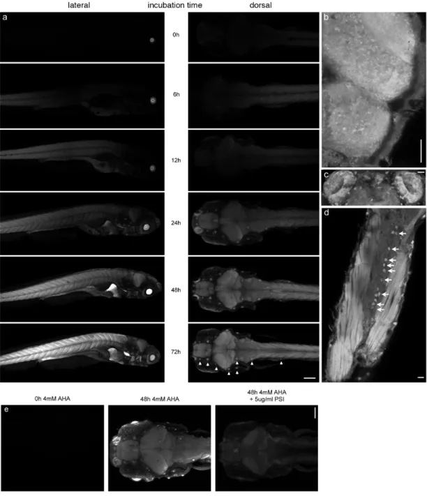
Figure 2.7. Newly synthesized proteins can be visualized in whole-mount larval zebrafish after in vivo labeling ...39
Figure 2.8. FUNCAT can be combined with antibody staining to identify specific cell populations in the whole-mount larval zebrafish ...42
Figure 2.9. The GABA antagonist PTZ induces increased protein synthesis in larval zebrafish ...44
Figure 3.1. Genetically restricted metabolic labeling...57
Figure 3.2. MetRS protein sequence alignment of E. coli, Danio rerio, Mus musculus and Homo sapiens...58
Figure 3.3. Cell-selective labeling with ANL in vitro...61
Figure 3.4. Cell-selective labeling with ANL in transiently L13G-MetRS-expressing larval zebrafish ...64
Figure 3.5. Cell-selective labeling with ANL in F1 transgenic larval zebrafish expressing L13G-MetRS...67
Figure 3.6. Restricted mutant MetRS expression in the telencephalon of larval
zebrafish via FlipTrap gene trapping...68
Figure 3.7. NLL and PLL mutations of zebrafish MetRS do not enable metabolic labeling with ANL in vitro or in vivo...71
Figure 4.1. Place-conditioning apparatus and experimental set up ...85
Figure 4.2. 6-8pdf larval zebrafish show unconditioned preference for light and social environment ...87
Figure 4.3. Associative place-conditioning paradigm for 6-8dpf larval zebrafish...89
Figure 4.4. Rapid extinction of the conditioned association ...92
Figure 4.5. Memory of association persists for at least 36h...94
Figure 4.6. Memory formation is protein-synthesis dependent...96
Figure 4.7. 3h incubation with puromycin, cycloheximide or MK-801 does not significantly alter unconditioned light and social environment preference ...98
Figure 4.8. Exposure to social environment sustains exploratory behavior...100
Figure 5.1. Physical mapping of preliminary proteomic data ...116
ABBREVIATIONS USED
4E-BP2: eIF4E binding protein aaRS: Aminoacyl-tRNA synthetase AHA: Azidohomoalanine
AMPA: α-amino-3-hydroxy-5-methyl-4-isoxazolepropionic acid ANL: Azidonorleucine
ARC: Activity-regulated cytoskeleton-associated protein ATF4: Activation transcription factor 4
BDNF: Brain-derived neurotrophic factor
BONCAT: Bioorthogonal noncanonical amino acid tagging CaMKII: Ca2+/calmodulin-dependent protein kinase II cAMP: Cyclic adenosine monophosphate
CREB: Cyclic-AMP-response-element-binding protein dpf: Days post fertilization
eIF2a: Eukaryotic initiation factor 2a E-LTP: Early LTP
ERK: Extracellular signal-regulated kinase
FUNCAT: Fluorescent noncanonical amino acid tagging GABA: γ-Aminobutyric acid
GFP: Green fluorescent protein GCN-2: General control norepressor 2 HEK: Human embryonic kidney hpf: Hours post fertilization HPG: Homopropargylglycine IEG: Immediate early gene KO: Knock-out
L13G: E. coli MetRS leucine 13 mutated to glycine LED: Light-emitting diode
L-LTP: Late LTP
LTP: Long-term potentiation
MAPK: Mitogen-activated protein kinase MetRS: Methionyl-tRNA synthetase
MK-801: Dizocilpine, non-competitive antagonist of the NMDA receptor NGF: Nerve growth factor
NLL: E. coli MetRS mutation NMDA: N-methyl-D-aspartate OKR: Optokinetic response OMR: Optomotor response
PBDTT: Phosphate-buffered solution containing DMSO, Triton X-100 and Tween-20 PBS: Phosphate-buffered solution
PLL: E. coli MetRS mutation PKMζ: Protein kinase Mζ PSI: Protein synthesis inhibitor PTZ: Pentylenetetrazol
RT: Room temperature
SILAC: Stable isotope labeling with amino acids in cell culture TCEP: Tris(2-carboxyethyl)phosphine
t-PA: Tissue plasminogen activator UAS: Upstream activating sequence
Chapter I
INTRODUCTION
The role of protein synthesis in long-term memory formation
Changes in behavior, specifically memory formation, are thought to depend on synaptic plasticity in specific circuits of the nervous system. A central goal of neuroscience is to characterize these physical changes that underlie learning and memory. Learning, in the most general sense, is defined as the process by which new information about the environment is acquired. Memory formation is considered to be the process by which that knowledge is stored. Over the last hundred years, researchers have developed countless training paradigms to investigate these processes in a variety of different model organisms.
Generally, these paradigms can be classified into two main groups, those that induce non-associative learning and those that induce associative learning. Non- associative learning, such as habituation and sensitization, refers to a behavioral change that occurs in response to a single stimulus or to two stimuli not temporally related, while associative learning, such as entrained during place-conditioning, refers to the formation of an association either between two stimuli (classical conditioning) or between a behavior and a stimulus (operant conditioning).
Both non-associative and associative learning can have different time constants.
While short-term memory is produced immediately after information is acquired and lasts minutes to hours, long-term memory is formed during a distinct second phase, lasting from hours to days or longer depending on the organism and the type of memory.
Furthermore, short-term memory is thought to depend on post-translational modification at the synapse, such as residue-specific phosphorylation or proteolytic cleavage of key
proteins, which can alter enzymatic activity of target molecules and regulated trafficking of receptors. In contrast, long-term memory has been shown to require regulated changes in gene transcription and protein synthesis (reviewed in Abel and Lattal, 2001 and Goelet et al., 1986). The connection between long-term memory formation and protein synthesis has been extensively studied using both protein synthesis inhibitors (PSI) and genetic manipulation of key players of translational control.
Studies using protein synthesis inhibitors, such as the antibiotics puromycin, anisomycin and cycloheximide, in many different model organisms have shown that protein synthesis, during or shortly after learning, is an essential step in the formation of long-term memory (Davis and Squire, 1984). In a seminal experiment in 1964, Agranoff et al. showed that the PSI puromycin injected intracranially into the goldfish produced impairment of memory for a shock avoidance task and that this impairment was time- and PSI concentration-dependent (Agranoff and Klinger, 1964; Agranoff et al., 1966). Since then, protein synthesis has been shown to be necessary for long-term memory formation in a variety of learning paradigms, including appetitively and shock-motivated discrimination learning, passive and active avoidance learning, shuttle box learning, and long-term habituation (reviewed in Davis and Squire, 1984). These studies demonstrating the necessity of protein synthesis for long-term memory formation paved the way for the idea that the physical basis of memory lies in the learning-related growth or remodeling of synaptic connections in a protein synthesis-dependent manner.
In 1973, Bliss and Lømo found that a high frequency train of action potentials resulting from stimulation of the perforant path in the rabbit hippocampus lead to a long- term potentiation (LTP) of synaptic transmission in the dentate gyrus (Bliss and Lømo,
1973). This phenomenon, which can also be induced in vitro in cultured slices (Alger and Teyler, 1976; Lynch et al., 1977; Schwartzkroin and Wester, 1975), has been widely regarded as a potential cellular mechanism underlying information storage, both because of its occurrence in the hippocampus, a structure known to be involved in memory formation and because of its relative stability. Since then, the appeal of LTP has widened through accumulated evidence that LTP exhibits additional features that have been shown to reflect important characteristics of memory formation in vivo. For one, correlates of short-term and long-term memory have been identified in LTP, termed early (E-LTP) and late (L-LTP) LTP, respectively. Furthermore, L-LTP specifically has been shown to be both transcription- and translation-dependent using chemical stimulation with drugs such as PSI in vitro (Abraham and Williams, 2003).
Although extremely important in elucidating the connection between long-term memory formation and protein synthesis, PSI, which are thought to block ~90% of all cellular protein synthesis (Klann and Sweatt, 2008), are relatively blunt tools. The most frequently used PSI are antibiotics that interfere with the elongation step of translation.
Puromycin causes premature chain termination as it can mimic the 3’-end of an aminoacylated tRNA, producing abnormal peptidyl-puromycin fragments (Flexner and Flexner, 1968; Nathans, 1964), while anisomycin binds to the 60S ribosomal subunit, blocking peptide bond formation (Pestka, 1971; Vasquez, 1979). Cycloheximide, another frequently used PSI, is specific to eukaryotic cells and also binds to the 60S ribosomal subunit, interfering with both initiation and the translocation step of elongation (Gale et al., 1981). Beyond being indiscriminant blockers of protein synthesis, most PSI have non-specific effects, such as activating the mitogen-activated protein kinase (MAPK)
superfamily pathways (Rudy et al., 2006), altering catecholamine function (Weiner and Rabadjija, 1968), impairing DNA and RNA synthesis (Gale et al., 1981), or causing toxic side effects such as seizures, lethargy and gustatory aversions when administered at high concentrations in vivo (Davis and Squire, 1984).
To investigate the role of protein synthesis in long-term memory formation while avoiding the use of PSI, researchers have recently started to genetically manipulate key players of translational control using knock-out (KO) mice models. Costa-Mattioli et al., for instance, examined plasticity in mice lacking general control norepressor 2 (GCN2), a protein kinase that inhibits translation initiation by phosphorylating eukaryotic initiation factor 2a (eIF2a). Phosphorylation of eIF2a stimulates translation of activating transcription factor 4 (ATF4), an antagonist of cyclic-AMP-response-element-binding protein (CREB). Thus, in the hippocampus of GCN2 KO mice expression of ATF4 is reduced and CREB activity is increased. In these animals, stimuli that normally lead to early LTP resulted in long-lasting LTP, whereas stimuli that normally lead to late LTP led to reduced LTP. Mirroring this phenotype, researchers observed an enhancement of learning in the Morris water maze following weak training, but a reduction in learning after intense training (Costa-Mattioli et al., 2005), indicating that tight translational control is necessary for normal memory formation. Furthermore, Banko et al. examined both LTP and spatial learning in mice lacking eIF4E binding protein 2 (4E-BP2), which normally inhibits translation by binding to eIF4E and observed the same plasticity phenotype as above. In these animals, disinhibition of protein translation results in impaired spatial learning and long-term contextual fear conditioning (Banko et al., 2005).
In contrast, mice with conditional expression of a dominant-negative regulator of MAPK
in the forebrain exhibit inhibition of protein translation, which results in inhibition of L- LTP, as well as deficits in spatial learning and contextual fear conditioning (Kelleher et al., 2004). Although these elegant experiments provide further evidence that long-term memory formation is protein-synthesis dependent, they also have their disadvantages. As most of the gene deletions described above were not conditional, some or all of the deficits observed could be due to long-term loss of these molecules, changes in development that compensate for lack of these molecules or lack of these molecules in other cell types. Furthermore, it is possible that the deficits are due to other translation- independent functions of the genetically manipulated molecules.
Both in vitro studies of LTP and in vivo experiments investigating behavioral correlates of learning and memory have helped form our current understanding of the molecular mechanisms underlying memory formation. It is now known that once activated by coincident pre-synaptic release of glutamate and post-synaptic depolarization, N-methyl-D-aspartate (NMDA) receptors allow calcium entry into the cell, thereby triggering a range of intracellular signaling cascades (Collingridge et al., 1983; Cotman et al., 1989; reviewed in Sweatt, 2009). Among others, the influx of calcium stimulates calcium-binding protein Ca2+/calmodulin and increases the production of cyclic adenosine monophosphate (cAMP) by adenylyl cyclases. cAMP, in turn, activates protein kinase A (PKA), which activates CREB, resulting in plasticity-related gene transcription and translation. CREB can also be activated via calcium signaling through the MAPK/extracellular signal-regulated kinase (ERK) pathway by a number of important kinases, including Ca2+/calmodulin-dependent protein kinases II (CaMKII) and protein kinase C (PKC) (reviewed in Sweatt, 2009). CREB, in concert with other
activity-regulated transcription factors, then controls the expression of a variety of plasticity-related genes with CRE response elements in their promoters (Hagiwara et al., 1996). These include a number of immediate-early genes (IEG), such as C/EBP, zif/268, and krox 20, which in turn act as transcription factors regulating the expression of
‘effector’ genes (Sweatt, 2009). It is the protein products of these effector genes that are needed for growth or stabilization of synapses, the physical change thought to underlie memory formation. Effector proteins that show increased translation with memory formation include: neurotrophic factors such as brain-derived neurotrophic factor (BDNF) and neurotrophin-3; signaling molecules such as CaMKII and the atypical PKC protein kinase Mζ (PKMζ) (Saktor, 2011); secreted proteases such as tissue plasminogen activator (t-PA); α-amino-3-hydroxy-5-methyl-4-isoxazolepropionic acid (AMPA) receptor subunits; the metabotropic receptor scaffolding protein homer; and activity- regulated cytoskeleton-associated protein (arc), a cytoskeleton-associated protein that may be involve in stabilizing structural changes at potentiated synapses (reviewed in Sweatt, 2009; Hernandez and Abel, 2008). In particular, the increased translation of secreted proteases is interesting, as degradation of proteins is not intuitively associated with synaptic growth or increased stability. But degradation of structural proteins may be integral to allow for the structural rearrangement that is synaptic plasticity (Mysore et al., 2008; Tai et al., 2008).
Most of the effector proteins that show activity-dependent changes in translation have so far been identified in vitro using targeted genetic manipulation of individual candidate proteins. Aakalu et al., for example, using GFP flanked by the 5’
and 3’ untranslated regions of CaMKIIα, demonstrated that this reporter construct shows
increased local translation when neurons are stimulated with BDNF (Aakalu et al., 2001).
More recently, Chen et al., using a photoconvertible fluorescent protein kaede to report new protein synthesis, have visualized CREB-dependent transcriptional activation of CaMKII and of period in the dorsal-anterior-lateral neurons of Drosophila after training that induces long-term memory formation (Chen et al., 2012). Using these techniques the researchers were both able to confirm that these proteins are translationally regulated with memory formation and identify the neural circuits in which these particular translational changes occur.
However, such studies rely on fluorescent protein reporter systems and a candidate- based approach, possibly perturbing endogenous localization of newly synthesized proteins and severely limited in their potential to identify unknown effector proteins and the circuits underlying memory formation. According to conservative estimates, vertebrate genomes may have about 30,000 genes and recent studies have shown that at least 50% are expressed in the brain (Pan et al., 2011). A large number of the protein products of these genes may play a role in signaling cascades and structural changes associated with memory and learning, which are unlikely to be identified using a candidate based approach. Furthermore, monitoring translation of one or maximally a few candidate proteins is unlikely to lead to a complete understanding of which neuronal circuits show changes in translation with memory formation. Instead, we hypothesize that by developing new techniques utilizing bioorthogonal chemistry to tag newly synthesized proteins specifically, we may circumvent these problems, enabling unbiased visualization and identification of proteins underlying memory formation.
Bioorthogonal chemistry
Recently, new techniques for labeling a variety of molecules based on the principle of bioorthogonal metabolic labeling have been developed (Best, 2009). Here, small functional groups that are commonly absent in the cellular environment, most prominently ketones and azides or alkynes, are introduced using the cells’ own synthetic machinery. Using this approach, sugars (Laughlin and Bertozzi, 2009), lipids (Kho et al., 2004), virus particles (Bruckman et al., 2008), DNA and RNA (Weisbrod et al., 2008) have been labeled and subsequently visualized using fluorescent dyes or enriched and identified using affinity reagents. Bertozzi and coworkers, in particular, have demonstrated in vivo labeling of glycans in living organisms ranging from rodents (Prescher et al., 2004; Chang et al., 2010), to larval zebrafish (Laughlin et al., 2008;
Baskin et al., 2010; Dehnert et al., 2011) and C. elegans (Laughlin and Bertozzi, 2009).
In the case of larval zebrafish, embryos were treated with an unnatural azide-bearing sugar to metabolically label cell-surface glycans, which were subsequently reacted to fluorescent alkyne conjugates at different time points. This enabled spatiotemporal visualization of expression and trafficking of cell-surface glycans in vivo during development (Laughlin et al., 2008).
Using a similar method, proteins can be labeled with noncanonical amino acids bearing novel side chains. Noncanonical amino acids are amino acids that are not part of the canonical set of twenty used in translation by all living systems. Some of these noncanonical amino acids can act as surrogates for naturally occurring amino acids, be charged onto wild-type tRNAs by endogenous aminoacyl-tRNA synthetases (aaRS) and
therefore be metabolically incorporated into newly synthesized proteins. However, this is not a new area of research. In the 1950s, Cowie and Cohen demonstrated that selenomethionine serves as an effective surrogate of methionine (Cowie and Cohen, 1957). Since then a number of different amino acids, including methionine, leucine, phenylalanine, and tryptophan, have been replaced by noncanonical amino acid analogs bearing bromo-, iodo-, cyano- and ethynyl- substituents and thereby allowing for investigation of how these novel side chains effect structure and function of labeled proteins (reviewed in Link et al., 2003).
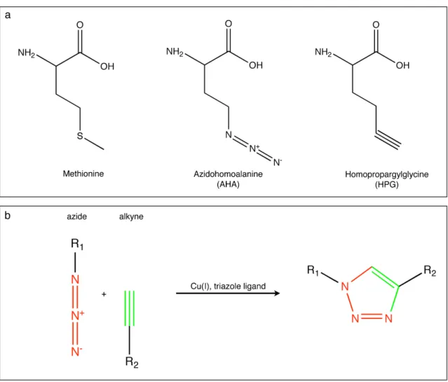
More recently, Tirrell and coworkers have established the use of the azide-bearing noncanonical amino acid azidohomoalanine (AHA) and the alkyne-bearing noncanonical amino acid homopropargylglycine (HPG) as surrogates for methionine in bacterial cells (Figure 1.1a) (Kiick et al., 2002; Link et al., 2004; Beatty et al., 2005). Azides and alkynes are stable under biological conditions, essentially absent from cellular environments and can be covalently linked via selective Cu(I)-catalyzed [3+2] azide- alkyne cycloaddition (Figure 1.1b) (Rostovtsev et al., 2002; Tornøe et al., 2002), making them ideal candidates to label proteins. Using this approach, Dieterich et al. developed the sister techniques bioorthogonal noncanonical amino acid tagging (BONCAT), and fluorescent noncanonical amino acid tagging (FUNCAT). During BONCAT, proteins labeled with noncanonical amino acids are tagged using affinity tags to enable affinity purification, while FUNCAT utilizes fluorescent tags to enable visualization, and thereby localization, of newly synthesized proteins in mammalian cells (Dieterich et al., 2006, 2007 and 2010).
Figure 1.1. Chemical structures and ‘click chemistry’ reaction scheme
(a) Chemical structures of methionine, azidohomoalanine (AHA) and homopropargylglycine (HPG). (b) Scheme of Cu(I)-catalyzed of [3+2] azide-alkyne cycloaddition.
Affinity-tagged proteins can be quantified using immunoblot analysis or separated from the preexisting proteome by affinity purification and identified by tandem mass spectrometry. BONCAT has already been successfully applied to study the proteome of HEK293 cells during a two hour time window, allowing the identification of 195 newly synthesized proteins (Dieterich et al., 2006). Fluorescent tags can be used to visualize
newly synthesized proteins, including those proteins of interest whose identities may not be known. In this manner, FUNCAT has been used to investigate temporally defined protein populations in Rat-1 fibroblast (Beatty et al., 2006; Beatty and Tirrell, 2008) and local protein synthesis in dissociated hippocampal neurons and hippocampal slices (Dieterich et al., 2010). Furthermore, metabolic AHA incorporation has been used to identify regions of the Drosophila genome that show high levels of histone turnover (Deal et al., 2010), to show that Chlamydia co-opt the functions of the lysosomes of their host cells to acquire essential amino acids (Ouellette et al., 2011), as well as to demonstrate that treatment of primary sensory neurons with the cytokine interleukin-6 or the neurotrophin nerve growth factor (NGF) increases nascent protein synthesis in axons (Melemedjian et al., 2010). Recently, these techniques have also been used to show that the transmembrane receptor DCC may regulate protein synthesis in a localized manner within the cells, as DCC enrichment was found to mark areas of new protein synthesis at the tips of filopodia in commissural neurons (Tcherkezian et al., 2010).
AHA and HPG are able to penetrate cell membranes, bind to methionyl-tRNA synthetase (MetRS) and be charged onto met-tRNAs in wild-type cells. BONCAT and FUNCAT depend on this promiscuous nature of MetRS that enables the charging of these structurally similar methionine analogs and thereby their incorporation into newly synthesized proteins. AaRS specificity is the most critical proofreading mechanism to ensure accurate translation of proteins from their respective mRNAs, as the ribosome lacks proofreading capabilities.
AaRS catalyze the aminoacylation of their cognate tRNAs by activation of the amino acid by ATP, followed by transfer onto the 3’ end of the tRNA molecule. The
recognition of the cognate amino acid by the aaRS is a multistep process. First, amino acids and ATP physically bind to aaRS to induce a conformational change in the aaRS that leads to the formation of the aminoacyl-adenylate complex. Next, misactivated noncognate animoacyl-adenylate complexes are eliminated, followed by transfer of the aminoacyl group to the tRNA and pretransfer proofreading. Finally some aaRS have sieve-type post-transfer proofreading capabilities to eliminate mischarged tRNAs. Each of these steps leads to increased specificity for the cognate amino acid, while discriminating against the noncognate amino acid.
MetRS is a member of the class I aaRS. Crystal structures are available of E. coli MetRS both with and without bound methionine (Mechulam et al., 1999 [PDB:1QQT];
Serre et al., 2001 [PDB:1F4L]). From these structures, it has been determined that MetRS undergoes a significant conformational change upon binding its substrate, but apparently lacks a sieve-type proofreading mechanism. This structural change is thought to be associated with the main proofreading step in the selective recognition of methionine (Datta et al., 2004). Twelve amino acids are found within 4Å of bound methionine and are therefore predicted to be part of the catalytic binding pocket (Figure 1.2). These residues include L13, Y260 and H301. Both the NH2 moiety and the sulfur atom of the side chain of methionine form hydrogen bonds with the L13 carbonyl oxygen atom and the L13 backbone amide, respectively. The sulfur atom of the side chain of methionine forms another hydrogen bond with Y260, while the backbone of methionine makes electrostatic interactions with H301 (Fourmy et al., 1991; Ghosh et al., 1991).
Most of the residues that are in close contact with the methionine are strictly conserved among MetRS of different bacterial organisms. This is particularly the case for L13,
Y260, D52, W253, Y15, A256 and H301 (Serre et al., 2001). Although, MetRS has been observed to be slightly promiscuous and has been seen to incorporate a variety of different noncanonical amino acid analogs, such as AHA (Kiick et al., 2001), the significant conformational change of the catalytic pocket after binding of methionine increases the specificity of MetRS for methionine. This severely limits the chemical functionalities that can be introduced into newly synthesized proteins.
Figure 1.2. The twelve amino acid residues of MetRS found within 4Å of bound methionine are predicted to be part of the catalytic binding pocket.
(a) and (b) show two different orientations of MetRS (top) and its catalytic binding pocket (bottom). L13 is highlighted in red, Y260 and H301 are shown in orange and A12, P14, Y15, D52, V252, W253, A256, P257 and F300 are shown in yellow. The structure was first published by Serre et al., 2001; PDB:1F4L.
BONCAT and FUNCAT may be ideal techniques to visualize and identify effector proteins synthesized during memory formation. As opposed to genetically encoded fluorescent proteins, azides and alkynes are small, so labeling with AHA or HPG is likely to only cause modest, perhaps even insignificant, perturbations of protein folding and localization (Dieterich et al., 2006) and therefore function of the labeled proteins in vivo. Furthermore, introduction of a chemical handle allows for affinity purification of newly synthesized proteins specifically. As the nervous system proteome is extremely complex, reducing the complexity of the sample may facilitate the identification of proteins of low abundance. However, so far, these techniques have only been applied in vitro. Given the role of protein synthesis in learning and memory, described earlier, developing BONCAT and FUNCAT for use in an intact organism in which simple forms of learning may be investigated, such as the larval zebrafish, is the essential next step.
The larval zebrafish as a model organism
The zebrafish is a tropical sweet-water cyprinid found mainly on the Indian subcontinent, its range extending from Pakistan in the west to Myanmar in the east and Nepal in the north (Engeszer et al., 2007). Adults live in shallow vegetated areas in rivers and small streams and are thought to feed mainly on insects and zooplankton, while they themselves are hunted by a variety of fishes including the snakehead (Channa) (Spence et al., 2008). During the monsoon season, adults move to shallow flooded ponds, often
connected with rice cultivation, to spawn. Adult zebrafish are about 4cm long, become sexually mature after about 3 months and females lay clutches of several hundred eggs in a single spawning. They have been reported to survive temperature ranges from 6˚C to 38˚C (Spence et al., 2008) in the wild, and are usually found in environments with a pH range of pH 7.9-8.2 (Engeszer et al., 2007).
Due to their high fecundity, rapid development, relatively fast generation time, external fertilization and environmental robustness, the zebrafish has emerged as an important model organism for developmental genetics and biomedical research. In the laboratory, a large number of zebrafish can be housed in a small area as a result of their social nature. A number of companies sell customizable aquatic habitats that can self- regulate temperature, pH, conductivity and water quality, greatly reducing maintenance time. In captivity, females can spawn up to twice a week, laying large, optically transparent embryos. Eggs are fertilized externally and adult zebrafish provide no parental care, enabling researchers to collect single cell embryos for genetic manipulation and developmental study. Embryos develop rapidly; the first neurons can be identified approximately 24 hours post-fertilization (hpf) (Kimmel et al., 1995). After 3 days post- fertilization (dpf) larvae hatch; by 5dpf larvae are estimated to have 100,000 neurons and by 7dpf larvae, now approximately 5mm long, are capable of a diverse set of simple behaviors. Interestingly, in zebrafish, all gonads originally develop as ovaries, which in males start maturing around 6-7 weeks post-fertilization and reach maturity after approximately three months (Devlin and Nagahama, 2002; Maak and Segner, 2003).
Although the genetic mechanism of sex determination is unknown, evidence suggests a role for food availability and water temperature (Lawrence et al., 2007). Furthermore,
larval zebrafish can absorb a variety of small chemical compounds directly from their surrounding environment, making them amenable to chemical screens (Zhong and Lin, 2011) and possibly noncanonical amino acid labeling techniques.
Over the last 15 years a number of genetic tools have been developed, mainly to study larval zebrafish development, but also extremely useful in investigating neuronal circuit morphology and function. The discovery and development of the Tol2 transposable element, originally described in medaka fish, which has a very high rate of genomic integration in the germline, immensely facilitates the construction of stable transgenics (Kawakami, 2005; Suster et al., 2009). DNA fragments of up to 10kb can be flanked with Tol2 sequences and co-injected with transposase mRNA into single-cell embryos to enable germline integration rates up to 50-70% (Suster et al., 2009). Using this system, a large number of gene- and enhancer-trap constructs have been generated to study the expression, function and localization of a number of genes.
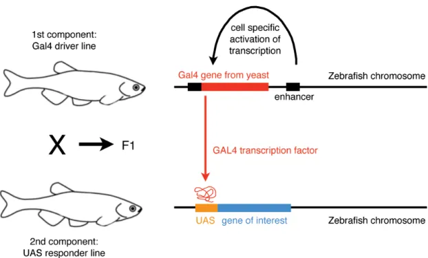
More recently, Tol2 was used to create transgenic zebrafish for targeted gene expression in specific tissues and cells using the binary Gal4-UAS system (Figure 1.2.).
Gal4 is a yeast transcriptional activator which can bind to its cognate upstream activating sequence (UAS) to activate transcription of target genes. The Gal4-UAS system can be used as a two-component gene expression system in a number of different model animals, including the zebrafish (Sheer and Campos-Ortega, 1999; Köster and Fraser, 2001). Two transgenic lines are created: one expressing the Gal4 sequence under the control of a cell- type-specific promoter (termed driver line), the other expressing a gene of interest, such as GFP, under the control of the UAS promoter (termed the responder line). Crossing driver lines with responder lines allows for expression of a variety of genes of interest
(determined by the responder line) in a variety of specific cells or tissues (determined by the driver line).
Figure 1.3. The binary Gal4-UAS gene expression system
Crossing Gal4 driver lines with UAS responder lines allows for expression of a variety of genes of interest (determined by the UAS responder line) in a variety of specific cells or tissues (determined by the Gal4 driver line). [Adult zebrafish schematic adapted from Smith and Croll, 2011.]
Recently, large enhancer-trapping screens have led to the creation of hundreds of nervous system-specific Gal4 driver lines with different, sometimes cell-type-specific expression patterns (Davison et al., 2007; Scott et al., 2007; Asakawa et al., 2008).
Furthermore, a variety of different responder lines now allow for the visualization of expression using fluorescent proteins (Scott et al., 2007; Asakawa et al., 2008), targeted cell ablation using NTR system or KillerRed (Davidson et al., 2007; Del Bene et al., 2010), light-gated control of neuronal activity using engineered ion channels (Szobota et
al., 2007; Janovjak et al., 2010) and inhibition of neurotransmitter release by tetanus toxin light chain (Asakawa et al., 2008), making this binary system extremely useful for studying nervous system development and function (reviewed in Asakawa and Kawakami, 2009). Furthermore, pigment mutants lacking melanophores, such as the nacre line, have been identified in mutant screens, enabling direct imaging of the larval zebrafish nervous system in intact animals (Lister et al., 1999).
Despite some obvious differences in size and complexity of certain structures of the zebrafish brain, the overall organization of the major brain components is comparable to that of the mammalian brain (Tropepe and Sive, 2003). Furthermore, as in other vertebrates, zebrafish possess all of the classical senses (vision, olfaction, taste, tactile, balance and hearing) and their sensory pathways share an overall homology with mammals. However, in mammals the telencephalon undergoes evagination during development, while in teleost fish such as the zebrafish, the telencephalon is everted. As a result, the hippocampus, which in mammals is structurally derived from the medial part of the dorsal telencephalon, is thought to be structurally homologous to the dorsal lateral telencephalon in zebrafish. In contrast, the amygdala, which is a lateral structure in mammals, is thought to be structurally homologous to the dorsal medial telencephalon in zebrafish (Broglio et al., 2005).
Despite differences in location, a number of lesion studies in the closely related goldfish have demonstrated that lesions of the dorsal lateral telencephalon result in deficits in tasks that, in mammals, rely on the hippocampus, such as spatial learning and trace classical conditioning, but do not affect hippocampus-independent delay conditioning and heart-rate conditioning. In contrast, lesions of the dorsal medial
telencephalon disrupt amygdala-dependent emotional, heart-rate conditioning and avoidance conditioning, but spare spatial memory and temporal stimulus processing (Vargas et al., 2006; Saito and Watanabe, 2006; Salas et al., 1996; Overmier and Papini, 1986; Portavella et al., 2004a; Portabella et al., 2004b; Portavella et al., 2002; reviewed in Broglio et al., 2005). Gross similarity in brain structure and identification of homologous areas involved in memory storage indicate that this simple vertebrate, the zebrafish, is more comparable to humans than invertebrate models such as Drosophila and C. elegans and therefore a preferable model organism to investigate neuronal circuits underlying behavior.
Not only are zebrafish easily maintained, genetically tractable, simple vertebrates, they also have an extensive behavioral repertoire. Adults show a range of complex and well-described social behaviors including courtship (Darrow and Harris, 2004), shoaling, aggression and dominance (Larson et al., 2006), escape and avoidance (reviewed in Colwill and Creton, 2011) and exploratory behaviors (reviewed in Spence et al., 2008).
Some simple behaviors develop early and can be observed during the first week of development. The spontaneous locomotor repertoire of the larval zebrafish includes routine turns and slow scoots, while they produce a well-characterized C-bend escape response to escape from threatening stimuli (Budick and O’Malley, 2000). Even before hatching from the chorion, larvae begin to show startle responses when exposed to abrupt stimuli. By 4dpf larvae will induce rapid escape responses to tactile stimuli (Granato et al., 1996; McLean and Fetcho, 2009), water flow (Froehlicher et al., 2009; Kohashi and Oda, 2008) and visual stimuli (Emran et al., 2008); by 5-6dpf larvae will respond to acoustic stimuli (Burgess and Granato, 2007). Between 4-5dpf larvae will begin to
follow moving objects with their eyes, a behavior that is referred to as the optokinetic response (OKR) and by 5dpf larvae are actively hunting for food (Neuhauss, 2003).
Furthermore, larvae have been shown to swim in the same direction as a pattern of moving stripes, a behavior that is called the optomotor response (OMR) (Fleisch and Neuhauss, 2006) and display diurnal rhythms in activity (Prober et al., 2006).
Memory and learning capabilities of the zebrafish have been extensively explored in the adult. In the last decade, a number of conditioning paradigms have been developed, including avoidance-conditioning (Ng et al., 2012; Blank et al., 2009; Xu et al., 2006; Pradel et al., 2000; Pradel et al., 1999), place-conditioning (Mathur et al., 2011), plus maze learning (Sison and Gerlai, 2010) and shuttle box conditioning (Williams et al., 2002). These paradigms use food and social rewards (Al-Imari and Gerlai, 2007), as well as mild electroshock and exposure to alarm signal as unconditioned stimuli and visual, olfactory (Braubach et al., 2009) and acoustic stimuli as conditioned stimuli. However, very few conditioning paradigms exist for the larval zebrafish to date.
The first study investigating the learning capabilities of larval zebrafish showed that larvae can learn to habituate to an acoustic stimulus (Best et al., 2008). In this non- associative conditioning paradigm, 7dpf larvae individually placed in 96-well plates and repeatedly exposed to an acoustic stimulus exhibited an iterative reduction in startle response, which spontaneously recovered and showed dishabituation when exposed to a visual stimulus. This work was extended upon by the Wolman and colleagues, who showed that spaced training blocks of repetitive visual stimuli elicit protein synthesis- dependent long-term habituation in larval zebrafish, lasting up to 24h (Wolman et al., 2011). Finally, previous studies from our laboratory demonstrated that 6-8dpf larval
zebrafish can be associatively conditioned (Aizenberg and Schuman, 2011). Trained using an hour-long classical conditioning paradigm, larvae rapidly developed an enhanced motor response to a visual stimulus when it was paired with a tactile stimulus.
Memory retention, in this very labor-intensive paradigm, is unfortunately not long term and decays to baseline within 1hr of acquisition. To enable visualization and identification of proteins newly synthesized with memory formation using bioorthogonal protein labeling techniques in the larval zebrafish, a high throughput, protein synthesis- dependent conditioning paradigm needs to be established.
To conclude, the larval zebrafish is an excellent model organism, as it is a genetically tractable, simple vertebrate which is transparent and therefore ideal for imaging. Furthermore, adult zebrafish, as well as larval zebrafish, have a well-defined behavioral repertoire (Colwill and Creton, 2012), and the range of experimental paradigms to test this has recently been expanded to include associative conditioning (Aizenberg and Schuman, 2011). Larval zebrafish can absorb small chemical compounds directly from their surrounding environment, all of which makes them not only amenable to chemical screens and an emerging human disease model, but also an excellent system in which to study the applicability of bioorthogonal metabolic labeling of newly synthesized proteins underlying memory formation in vivo.
Memory formation, thought to depend on physical changes at specific synapses, has been conclusively shown to be protein synthesis-dependent. However, a majority of proteins regulated with memory formation to bring about these changes in signaling cascades and
synaptic structure have most likely not yet been identified. Furthermore, although it is well established that the mammalian hippocampus is necessary for memory formation, precisely which neuronal circuits and how many neurons are involved has not been investigated using an unbiased approach. In this study we show that the bioorthogonal metabolic labeling techniques BONCAT and FUNCAT can be applied in vivo to visualize and affinity purify newly synthesized proteins of the larval zebrafish. We explore the possibility of genetically restricting metabolic labeling via selective expression of a mutant MetRS and demonstrate that larval zebrafish can undergo protein synthesis-dependent place-conditioning. Thus, we have developed the tools necessary to monitor changes in protein synthesis in the larval zebrafish nervous system and therefore possibly identify neuronal circuits involved in long-term memory formation.
Furthermore, the techniques described here could be paired to facilitate the identification of effector proteins that are necessary for the physical changes underlying memory formation.
Chapter II
NONCANONICAL AMINO ACID LABELING IN VIVO TO VISUALIZE AND AFFINITY PURIFY NEWLY SYNTHESIZED PROTEINS IN LARVAL ZEBRAFISH
Introduction
Long-term memory formation requires new protein synthesis. Understanding what physical changes within the nervous system underlie learning and memory, specifically what neuronal circuits are involved and what proteins are newly synthesized during memory formation, are major goals in modern neuroscience. However, the identification of newly synthesized proteins has been sparse and limited to individually identified candidate proteins. Advances in mass spectrometry-based approaches now permit the characterization and quantification of proteins, especially when paired with approaches such as stable isotope labeling with amino acids in cell culture (SILAC) (Ong et al., 2002), which allow for comparative quantification between proteomes of differentially stimulated cell populations. However, the proteome of the nervous system is complex and without a chemical handle to enable affinity purification of the newly synthesized proteins specifically, proteins of low abundance will likely be missed.
In addition, the identification of cells or neural circuits that show increased protein synthesis in response to memory formation would allow us to understand the components of memory circuits that undergo long-term modifications after learning.
Genetically encoded fluorescent tags, such as GFP, have revolutionized cell biology by permitting visualization of fusion proteins of interest in vivo (Tsien, 1998). However, the size of GFP and the requirement for genetic manipulation of the target protein may interfere with its endogenous function, while at the same time only permitting investigation of a small number of candidates at once.
Recently, new techniques for labeling a variety of molecules based on the principle of bioorthogonal metabolic labeling have been developed (Best, 2009). Here, small functional groups that are commonly absent from the cellular environment, most prominently ketones and azides or alkynes, are introduced using the cells’ own synthetic machinery. BONCAT (Dieterich et al., 2006; Dieterich et al., 2007) and FUNCAT (Dieterich et al., 2010), two such techniques, have been used to tag and identify or visualize newly synthesized proteins, respectively. BONCAT and FUNCAT utilize noncanonical methionine derivatives, such as the azide-bearing AHA, to bioorthogonally label newly synthesized proteins. AHA can cross cell membranes and be charged onto methionine tRNAs by the endogenous MetRS. During protein synthesis, AHA is introduced in place of methionine, resulting in the introduction of azide groups into the newly synthesized proteins. These azide groups can be used to tag proteins with either an alkyne affinity tag (BONCAT) or an alkyne fluorescent tag (FUNCAT) via selective Cu(I)-catalyzed or strain-promoted [3+2] azide-alkyne cycloaddition (Rostovtsev et al., 2002; Tornøe et al., 2002; Agard et al., 2004). Affinity-tagged proteins can be quantified using immunoblot analysis or separated from the pre-existing proteome by affinity purification and identified by tandem mass spectrometry. Fluorescent tags can be used to visualize newly synthesized proteins, including those proteins of interest whose identities may not be known. Alternatively, the alkyne moiety may also be introduced into newly synthesized proteins by replacing methionine with the noncanonical amino acid homopropargylglycine (HPG) and subsequently labeled using azide-bearing affinity or fluorescent tags. Azides and alkynes are small, so labeling with AHA or HPG is likely to only cause modest, perhaps even insignificant, perturbations of protein folding,
localization (Dieterich et al., 2006) and therefore function of the labeled protein in vivo.
Furthermore, azides and alkynes are stable under biological conditions and essentially absent from vertebrate cells, which makes the azide-alkyne ligation (‘click chemistry’) very selective.
BONCAT and FUNCAT techniques have already successfully been applied to study changes in protein synthesis in a variety of different in vitro systems in order to investigate a diverse set of biological questions, as described in the introduction.
However, given the role of protein synthesis in learning and memory, developing BONCAT and FUNCAT for use in an intact organism in which simple forms of learning may be investigated is the essential next step.
In this chapter we describe the application of these techniques in vivo, in the 7- day-old larval zebrafish. We show that AHA is metabolically incorporated into newly synthesized proteins, in a time- and concentration-dependent manner, but has no apparent toxic effects and does not influence simple behaviors. This enables fluorescent labeling of newly synthesized proteins in whole-mount larval zebrafish. Furthermore, we find that stimulation with the GABA antagonist, pentylenetetrazole (PTZ), causes an increase in protein synthesis throughout the proteome, which can also be visualized in intact larvae.
Application of BONCAT and FUNCAT techniques to larval zebrafish
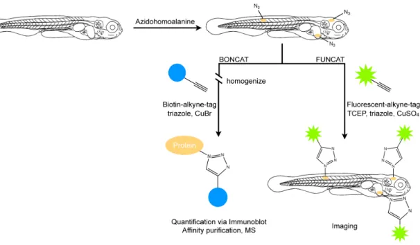
The BONCAT and FUNCAT protocols were adapted to larval zebrafish (Figure 2.1a).
All larvae, unless otherwise noted, were analyzed at 7dpf. We incubated larvae in E3
Figure 2.1. Labeling of newly synthesized proteins for quantification, affinity purification (BONCAT) and visualization (FUNCAT) in larval zebrafish
Scheme depicting metabolic labeling of newly synthesized proteins in 7-day-old larval zebrafish using AHA incorporation and Cu(I)-catalyzed [3+2] azide-alkyne cycloaddition. TCEP, tris(2-carboxyethyl)phosphine.
embryo medium supplemented with the methionine surrogate AHA (Figure 2.1b) for a period of 0-72h immediately prior to harvesting, with the aim of incorporating the azide group into newly synthesized proteins throughout the zebrafish proteome. To quantify successful incorporation of AHA into protein, larvae were washed, anesthetized, homogenized and the resulting lysate was reacted with biotin-alkyne in the presence of CuBr and the triazole ligand (see Methods). This allowed for detection and quantification of newly synthesized biotin-labeled proteins using immunoblot analysis or for affinity purification of the newly synthesized proteins (BONCAT). To visualize newly synthesized proteins following AHA exposure, larvae were washed, anesthetized,
fixed and permeabilized. Whole mounted larval zebrafish were reacted with AlexaFluor- 488-alkyne in the presence of CuSO4, the reducing agent tris(2-carboxyethyl)phosphine (TCEP) and the triazole ligand, before being imaged using a confocal microscope (FUNCAT). This allowed for visualization of new protein synthesis in the intact larval zebrafish.
Incubation with AHA is not toxic to larval zebrafish and does not alter simple behaviors
Previously, Dieterich et al. showed that metabolic labeling of mammalian cell culture with AHA does not alter global protein synthesis rates or promote ubiquitin-mediated degradation, indicating that AHA incorporation does not cause severe protein misfolding or degradation (Dieterich et al., 2006). To ensure that incubation and incorporation of AHA into newly synthesized proteins is not toxic to the living animal, larvae were exposed to E3 embryo medium supplemented with 0 to 20mM AHA, or 10mM methionine, for 6 to 72h. Larvae were scored as healthy if after incubation they were still responsive to light touch. No significant toxic effects were observed when larvae were incubated with 1-10mM AHA, even after 72h incubations (Figure 2.2a). Only incubations with extremely high (20mM) concentrations of AHA were toxic, beginning around 24h after onset of incubation. This indicates that incubation with low-to- moderate concentrations of AHA, even over extended periods of time, is not toxic to the living animal. In the remainder of the studies reported here concentrations < 4mM AHA were used.
Figure 2.2. At low concentrations, AHA exposure is not toxic and does not significantly alter simple behaviors.
(a) Survival rate of 7-day-old larval zebrafish when incubated with AHA (0 to 20mM, 6 to 72h) or methionine (10mM, 6 to 72h), n=20. (b) Quantification of spontaneous swimming behavior of larval zebrafish after AHA incubation (4mM, 0 to 48h).
Percentage of larvae that show no spontaneous swimming behavior per 15 minute interval. Mean swimming bursts per 15 minute interval, n = 10-12. Differences are not
statistically significant. (c) Traces depicting the angle of eye rotation during a typical optokinetic response after AHA incubation (4mM, 0h to 48h). (d) Sample startle response upon light flash after AHA incubation (4mM, 24h). (e) Mean response percentage to light or dark flash after AHA incubation (4mM, 0 to 48h), n=5 larvae, flashed three times each. Error bars represent standard deviation of response percentage. Differences are not statistically significant. (f) Mean delay in response to light or dark flash after AHA incubation (4mM, 0 to 48h), n=5 larvae, flashed three times each. Error bars represent standard deviation of response time. Differences are not statistically significant.
Next, we explored whether incorporation of AHA causes changes in simple behaviors. We conducted a series of behavioral tests after incubation in E3 medium supplemented with 4mM AHA, for 0-48h. First we investigated spontaneous swimming behavior. 7-day-old larval zebrafish were incubated in 4mM AHA for 0-48h prior to observation, and then placed individually into a 1-cm-by-7.5 cm swimming chamber (Figure 2.3) and their spontaneous swimming bouts were recorded for a period of 15 min.
Sample traces of spontaneous swimming behavior are depicted in Figure 2.3. There was no significant difference in the number of individual spontaneous swimming bouts initiated between 48h AHA-incubated, 24h AHA-incubated and control larvae, although there was a small, not significant decrease in the 48h and 24h AHA groups as compared to the control group (Figure 2.2b). There was also no difference in the number of AHA incubated and control larvae that failed to exhibit spontaneous swimming bouts during the 15 minute trial period (Figure 2.2b).
To study whether AHA incubation causes deficits in visual tracking, 7-day-old larvae were tested for the optokinetic response (Huang and Neuhauss, 2008) after incubation in 4mM AHA for 24-48h. Larvae were immobilized in 0.4% low-melting- point agarose in a circular array of LEDs, which delivered a spot of white light that moved in a horizontal plane around the immobilized larvae. Similar to control larvae,
Figure 2.3. Tracks of spontaneous swimming behavior of 7-day-old larval zebrafish with AHA incubation (4mM, 0 to 48h), indicating that spontaneous swimming behavior is not altered by prolonged AHA incubation. 15min interval; frame captured every 10s.
AHA-incubated larvae were able to track the light stimulus, producing smooth tracking eye movements and rapid saccades (Figure 2.2c), indicating that neither visual acuity nor neural circuits underlying visual tracking behavior seem to be affected by prolonged incubation with 4mM AHA. To further test whether AHA incubation altered visual acuity and simple reflexive behaviors, we tested the animal’s startle response to light flash and dark flash. Larvae were placed in a circular array of LEDs, which delivered either a light flash or a dark flash while the response of the larva was monitored. Figure 2.2d shows a representative startle response to a light flash in an animal following a 24h incubation with 4mM AHA. The larva is clearly exhibiting a stereotypical C-bend escape response (Kimmel et al., 1974) indicating that AHA has no effect on the motor function associated with escape behavior. Furthermore, incubation with 4mM AHA for 24-48h did not alter the percentage of larval zebrafish that responded to either light or dark flash (Figure 2.2e) nor did it affect the delay in response to either light or dark flash (Figure 2.2f). Therefore, we conclude that AHA incorporation is not toxic and has no effects on
simple behaviors at low concentrations (4mM), even over prolonged incubation periods, making it suitable for labeling newly synthesized proteins in vivo.
AHA is metabolically incorporated in larval zebrafish
To determine whether AHA is metabolically incorporated into newly synthesized proteins, we tagged lysates prepared from larval zebrafish incubated for 0-72h with 4mM AHA with biotin-alkyne in the presence of the Cu(I) catalyst. Subsequent dot blot analysis with a biotin antibody revealed successful incorporation of AHA into proteins in an incubation-time dependent manner. A sample dot blot is shown in Figure 2.4a, along with quantification of several experiments. After only a 6h incubation period with E3 embryo medium supplemented with 4mM AHA, statistically significant (p<0.005) AHA incorporation could be detected. After 24h, 48h and 72h incubations, approximately 140ng (±8ng), 375ng (±34ng) and 699ng (±72ng) of biotinylated protein were detected per homogenized larva, respectively. The total soluble protein per larva under the experimental conditions we used was 6.38µg (±0.53µg). From this we can estimate that 24h, 48h or 72h incubation with 4mM AHA leads to labeling and tagging of approximately 2.2%, 5.9% and 10.9%, respectively, of the total soluble protein per larval zebrafish. However, as different proteins may show different levels of AHA incorporation, and therefore different biotin signal strength, the analysis given here should be regarded as semi-quantitative.
Figure 2.4. AHA is metabolically incorporated into larval zebrafish proteins in vivo.
Labeling is both incubation time- and protein synthesis-dependent. Sample immunoblot and quantification of immunoblots of lysates from AHA-treated 7-day-old larval zebrafish reacted with biotin-alkyne (10µM) for 12h, probed with antibody against biotin.
(a) Larval zebrafish were incubated with 4mM AHA for 0 to 72h, n=4 (b) Larval zebrafish were incubated with AHA (0 or 4mM) or 4mM AHA in the presence of puromycin (2.5µg/ml to 10µg/ml) for 48 h, n=3. ***p<0.001.
To verify the specificity of AHA incorporation into newly synthesized proteins, we incubated larval zebrafish in E3 embryo medium supplemented with AHA along with low concentrations of the protein synthesis inhibitor puromycin. These very low concentrations of PSI did not have a toxic effect on larval zebrafish (data not shown).
Although abundant biotin signal was detected in lysates of larval zebrafish incubated with AHA only, no signal was detected when larval zebrafish were incubated without AHA, and a significantly lower signal was detected when larval zebrafish were incubated in AHA in the presence of puromycin (Figure 2.4b). Furthermore, when the concentration of PSI in the incubation medium was increased from 2.5µg/ml to 5µg/ml, a significant decrease in AHA-labeled and biotinylated proteins was observed. However, no further decrease was observed when the PSI concentration was further increased to 10µg/ml.
The above results confirm that BONCAT labels newly synthesized proteins with high specificity in the larval zebrafish. In addition, we observed that AHA incorporation in larval zebrafish scales non-linearly with incubation time (Figure 2.4a) and we assume that an incorporation plateau would be reached after even longer incubation periods.
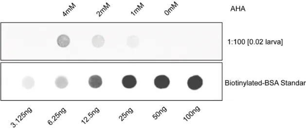
Also, labeling was AHA concentration-dependent (Figure 2.5). While no signal was detected when 4-day-old larval zebrafish were incubated with 0mM AHA, increasing the concentration of AHA in the incubation medium from 1mM to 4mM resulted in a detectable signal increase. Furthermore, AHA was incorporated not only into a few
select proteins, but into a large variety of newly synthesized proteins throughout the proteome over time, as is shown by the abundance of protein bands on the western blot of affinity purified biotinylated proteins from whole larval zebrafish lysates reacted with the biotin-alkyne and probed against biotin (Figure 2.6a). Biotin signal detected in the samples not incubated with AHA are likely a result of endogenous biotinylation.
Figure 2.5. Metabolic labeling is AHA concentration-dependent.
Immunoblot of lysates from 4-day-old larval zebrafish reacted with biotin-alkyne (10µM) for 12h, probed with an antibody against biotin. Larval zebrafish were incubated with 0 to 4mM AHA for 48h.
To examine whether AHA is also incorporated into newly synthesized proteins in deeper structures such as the nervous system, we incubated 4-day-old transgenic HuC::GFP larval zebrafish with 4mM AHA for 48h. HuC encodes an RNA-binding protein that serves as an excellent early marker for differentiating neurons and the HuC::GFP line is a stable zebrafish transgenic line in which GFP is expressed
specifically in neurons (Park et al., 2000). With the exception of a few cells in the olfactory pit and the lateral line, the majority of these neurons are not surface structu