G lobal M edical & H ealth C ommunication
Editor in Chief Herry Garna
Editors Arief Budi Yulianti Badrul Hisham Yahaya
Ike Rahmawaty Alie Jerico Franciscus Pardosi
Lisa Adhia Garina Listya Hanum Mirasari Putri Roy Rillera Marzo
Winni Maharani Yuktiana Kharisma
Layout Editor Yudi Feriandi Administrative Staff
Agus Chalid Deni Irawan Evi Apriani Editorial Address
Jalan Hariangbanga No. 2, Tamansari, Bandung 40132, West Java, Indonesia Phone/fax: (022) 4321213
E-mail: [email protected]
Website: https://ejournal.unisba.ac.id/index.php/gmhc Accredited by:
Ministry of Research, Technology and Higher Education of the Republic of Indonesia (Kemenristekdikti) Number: 30/E/KPT/2019, 11th November 2019
This journal is indexed on:
Published by:
Faculty of Medicine Universitas Islam Bandung;
Pusat Penerbitan Universitas-Lembaga Penelitian dan Pengabdian kepada Masyarakat (P2U-LPPM) Universitas Islam Bandung
Editorial Team
Publish Every 4 Months April, August, December
G lobal M edical & H ealth C ommunication
TABLE OF CONTENTS RESEARCH ARTICLES
Combination of Gabapentin and Vitamin B12 Compared with Gabapentin Monotherapy on Pain Improvement of Diabetic Neuropathy Patients
Mochamad Firdaus Bhuanaputra, Alya Tursina, Yuktiana Kharisma Comparative Study Gallbladder Contractility Index Using Ultrasound in Patients with and without Liver Cirrhosis
Hari Soekersi, Leni Santiana, Fetty Fatmawaty
Lumbar Radiculopathy: a Descriptive Study on Red Flag and Neurologic Symptoms in Dr. Hasan Sadikin General Hospital Bandung
Astrid Feinisa Khairani, Kuheinderan Radha Krishnan, Umar Islami, Siti Aminah Sobana
Differences in Expulsion on Post-placenta Intrauterine Contraceptive Device between Mother with Vaginal and Cesarean Delivery
Atika Zahria Arisanti, Tono Djuwantono, Sri Endah Rahayuningsih Eel Cookies Supplement and Incidence of Diarrhea in Children Aged 12–24 Months
Nur Eva Aristina, Dedi Rachmadi, Dewi Marhaeni Diah Herawati, Hadi Susiarno, Dida Akhmad Gurnida, Deni Kurniadi Sunjaya
Implementation of Importance-Performance Analysis (IPA) for Improving Medical Students’
Quality of Service in Teaching Hospital
Siska Nia Irasanti, Ieva Baniasih Akbar, Yani Dewi Suryani
Relationship of Soil-transmitted Helminth and Enterobius vermicularis Infection with Anemic in Students in Aceh Besar
Faisal Heri, A.A. Depari, Merina Panggabean
Dermatoglyphics Pattern on Breast Cancer Patients in Dharmais Cancer Hospital Faras Qodriyyah Sani, Mirfat, Iskandar
The Quality of Life on Asthmatic Adolescent and Its Correlation with the Severity and Control of Asthma
Lisa Adhia Garina, Muhammad Ridho Grahadinta, Ferry Achmad Firdaus Mansoer, Intan Puspitasari
The Effect of Health Education with Flashcard Media on Improvement of Knowledge and Reduction of Anxiety Degree in Adolescents Primigravida
Dwie Yunita Baska, Tita Husnitawati Madjid, Ponpon S. Idjradinata Effect of Phaleria macrocarpa (Scheff.) Boerl Dry Extract to the Level of Malondialdehyde
Meiyanti, Eveline Margo, Juni Chudri
The Effect of Low Impact Aerobic Exercise on Elderly with Dementia Cognitive Function Raden Ayu Tanzila, Sheilla Yonaka Lindri, Nindia Rahma
A Comparative Evaluation of Community Periodontal Index (CPI) and the Presence of Nicotine Stomatitis among Smokers after Oral Hygiene Instruction
Meta Maulida Damayanti, Yuktiana Kharisma, Fajar Awalia Yulianto, Santun Bhekti Rahimah, Winni Maharani, Meike Rachmawati, Herri S.
Sastramihardja, Muhammad Alief Abdul ‘Aziiz, Muhammad Ilham Halim
1
7
13
21
27
34
42
47 53
59
67 73 78 pISSN 2301-9123 │ eISSN 2460-5441
Volume 8 Number 1, April 2020
AUTHOR GUIDELINES
Global Medical & Health Communication (GMHC) is a journal that publishes medical and health scientific articles published every 4 (four) months. Articles are original research that needs to be disseminated and written in English.
The submitted manuscript must be the article that has never been published, and the author must ensure that all co-authors have agreed by signing a statement on the seal. Download template of the ethical statement (free plagiarism) here. The manuscript is an original article free from plagiarism. When the article published in another journal then in the next journal, the article will be disallowed.
All articles will be discussed by experts in the field of scholarly concerned (peer reviewer) and will be edited by the editor. The editor reserves the right to add or subtract sentences, both abstracts, and scripts without changing the meaning. Manuscripts that accepted for publication will become the property of the publisher.
It is not allowed to be published in other media. The needed revised manuscripts will be returned to the author. Research articles must be approved by the health research ethics committee or consider the ethical aspects of research accounted for
Article Writing
Typed the article on an 80 gsm A4 (21.0 × 29.7 cm) white HVS paper with 4 cm left and top margin, 3 cm down and right, not back and forth. The maximum script length is 20 pages (including images, tables, and photos). Each page is numbered typed in the bottom right page, sequentially starting from the title page to the last page. The font is black Georgia with 12 pt size, typed justified except for a title with a spacing of 2 spaces in Microsoft Word 2007 format. Typing a new paragraph 6 taps from the left edge of the line, unless the first paragraph is not typed indented.
In one manuscript only in English. Typed italic the untranslatable terms in a foreign language or regional language.
Table title is the typed center, font size 10 pt, bold, initial letter of each word written with capital letter, except conjunctions. The titles are numbered and written on top of the table. Example: Table 3 Neisseria gonorrhoeae Resistance to 8 Types of Antimicrobials in 20 Specimens. Table, no vertical dividing line, and there are only three horizontal borderlines. Created tables in sequence two spaces from the text. Table descriptions and abbreviations are placed in the table description, not on the table title.
Typed center figure title with 10 pt font size, bold, numbered according to the appearance in the text and typed under the image. The source of the cited image and or table should be added to references if it is not the author's work.
Pictures (graphs, diagrams, and photos) and tables besides written in its place, also created separately on other pages of texts with sufficient sharpness and
blackness. A maximum number of tables and or images are six pieces. Photos are sent in black and white glossy, or colored format when required, minimum size 3R (9×13.5 cm). Images and photos can also be sent on CD.
Write correspondence as the footnote on the first page containing the full name of the author with degrees/academic degrees, institution, address, phone number, fax, mobile, and e-mail.
Content and Format Articles
The article contains results of original research in the field of basic medical or applied, and health. The article format consists of Title & Abstract (English) and Judul & Abstrak (Indonesian), Introduction, Methods, Results, Discussion, Conclusion(s), Conflict of Interest, Acknowledgments, and References.
Articles Title
Maximum article title consists of 12 words (choose words and terms that are dense meaning and able to characterize the entire contents of the script). Typed with bold fonts, size 12 pt, one space, the initial letter of each word is written in capital letters (except the conjunctive), and center. The ownership row consists of 2 elements, the author name and origin institution.
Author's name written with the initial fonts are capital and bold, size 11 pt, one space, and center. Institution name written with the initial fonts are capital, size 10 pt, one space, and center.
Abstract
The abstract is typed using 12 pt font size and one spaces. The abstract is written in one paragraph, one space, maximum 250 words, and should describes the entire contents of the article. The abstract should be suitable for the format of introduction, methods (contain method, place, and time of study), results, and discussion. Abstract be equipped with key words consisting of 3–5 words.
Introduction
The introduction is written succinctly to stimulate the reader's interest include all the necessary information.
At the end of the introduction was written the purpose of the study.
Methods
Methods contains the material under study, and the way described briefly by the order of operation as well as the location and time of the study. Explain statistical methods in detail. Consideration of ethical issues is included. If the protocol has been approved then the ethical clearance/approval letter number and the health research ethics committee must be written.
Results
The result is the core of scientific writing. This section
presents data and information that will be used as the basis of the conclusion and even expected to get a new theory. In results, listed the tables and or images, graphics, photos to explain and abbreviate the description should be given; numbered according to their appearance in the text. Results of the study and discussion should be written separately.
Discussion
Discussion of the article reveals, explains, and discusses the results of the study with an analysis by the research design, interpretation, and explanation of its synthesis. Also, the results obtained are compared with the results of previous research of others.
Conclusion(s)
The conclusion is submitted by the results obtained by the researcher and written briefly and clearly in two or three sentences.
Conflict of Interest
All authors must make a formal statement at the time of submission indicating any potential conflict of interest that might constitute an embarrassment to any of the authors if it were not to be declared and were to emerge after publication. Such conflicts might include, but are not limited to, shareholding in or receipt of a grant or consultancy fee from a company whose product features in the submitted manuscript or which manufactures a competing product.
Acknowledgment
Acknowledgments should be provided to research contributors without writing a degree.
References
References are written by the Vancouver system's writing rules, given the sequence number corresponding to appearing in the article. List all author names if no more than six people; when more than six authors write the first six authors followed by et al. The references cited in the article are the most important references. The minimum referral number of 25 (twenty-five) copies of the most recent 10 (ten) years of journal article/book publishing. Reference should be sought from 80% primary literature and 20% secondary literature. Avoid referral in the form of personal communication except for information that is not possible from a public source. Include source name, date of communication, written permission, and confirmation of the accuracy of the source of communication.
Example How to Write References Journals
Theodoridou K, Vasilopoulou VA, Katsiaflaka A, Theodoridou MN, Roka V, Rachiotis G, et al.
Association of treatment for bacterial meningitis with the development of sequelae. Intern J Infect Dis.
2013;17(9):e707–13.
Zhang B, Kunde D, Tristram S. Haemophilus haemolyticus is infrequently misidentified as Haemophilus influenzae in diagnostic specimens in Australia. Diagn Microbiol Infect Dis. 2014;80(4):272–
3.
Books and Other Monographs Editor as Author
Nriagu J, editor. Encyclopedia of enviromental health.
Michigan: Elsevier BV; 2011.
Organization as Author
World Health Organization (WHO). Guideline:
neonatal vitamin A supplementation. Geneva: WHO Press; 2011.
Chapter in Book
Miller LG. Community-associated methicillin resistant Staphylococcus aureus. In: Weber JT, editor.
Antimicrobial resistance. Beyond the breakpoint.
Basel: Karger; 2010. p. 1–20.
Conference Proceeding
Nicholai T. Homeopathy. Proceedings of the Workshop Alternative Medicines; 2011 November 30; Brussels Belgium. Belgium: ENVI; 2011.
Journal Article from Internet
King P. Haemophilus influenzae and the lung (Haemophilus and the lung). Clin Transl Med.
2012;1:10 [cited 2015 August 15]. Available from:
https://clintransmed.springeropen.com/articl es/10.1186/2001-1326-1-10.
Authors
Written equipped in the covering letter, containing the full name (with degrees/academic degrees), the area of expertise, institution, address, phone number, fax, mobile, and e-mail.
Article Submission
Submit article and correspondence with the editorial board online. Register at http://ejournal.unisba.ac.id/
index.php/gmhc and follow the guidelines.
Editorial Board of
Global Medical and Health Communication Faculty of Medicine, Universitas Islam Bandung
Jalan Hariangbanga No. 2, Tamansari, Bandung 40132, West Java, Indonesia
RESEARCH ARTICLE
1
Global Medical and Health Communication
Online submission: http://ejournal.unisba.ac.id/index.php/gmhc
DOI: pISSN 2301-9123 │ eISSN 2460-5441
Correspondence: Mochamad Firdaus Bhuanaputra. Medical Undergraduate Study Program, Faculty of Medicine, Universitas Islam Bandung. Jln. Tamansari No. 22, Bandung 40116, West Java, Indonesia. E-mail: [email protected]
Combination of Gabapentin and Vitamin B12 Compared with Gabapentin Monotherapy on Pain Improvement of Diabetic Neuropathy Patients
Mochamad Firdaus Bhuanaputra,1 Alya Tursina,2 Yuktiana Kharisma3
1Medical Undergraduate Study Program, Faculty of Medicine, Universitas Islam Bandung, Bandung, Indonesia,
2Department of Neurology, Faculty of Medicine, Universitas Islam Bandung, Bandung, Indonesia, 3Department of Pharmacology, Faculty of Medicine, Universitas Islam Bandung, Bandung, Indonesia
Abstract
Diabetic neuropathy is the most common microvascular complication of diabetes mellitus (DM) occurring in 60–
70% of the world's DM population, 40% of the DM population in Asia, and 41% of the DM population in Indonesia.
The primary treatment of diabetic neuropathy pain in Indonesia is gabapentin and vitamin B12. The study aimed to compare pain improvements in diabetic neuropathy patients. The drug used was a combination of gabapentin and vitamin B12 and gabapentin monotherapy. For the pain degree measurement, we used the visual analogue scale (VAS). This experimental study was a pretest-posttest randomized control trial using a single-blind method at Dr.
M. Salamun Air Force Hospital Bandung from March to May 2017. Samples were 44, type two diabetic neuropathy patients. The Mann-Whitney test to compare pain improvement between 2 groups applied. The results indicated there were differences in pain improvement between diabetic neuropathy patients with gabapentin and vitamin B12 combination compare to gabapentin monotherapy (p=0.002). This result showed a synergistic effect of gabapentin as an inhibitor of neurotransmitter and vitamin B12 expenditure as an improvement in peripheral nerve cells. This study concluded that gabapentin and vitamin B12 combination is better in improving pain in diabetic neuropathy patients compared to gabapentin monotherapy.
Key words: Diabetes mellitus, diabetic neuropathy, gabapentin, pain repair, vitamin B12
Kombinasi Gabapentin dan Vitamin B12 Dibanding dengan Monoterapi Gabapentin terhadap Perbaikan Nyeri Pasien Neuropati Diabetik
Abstrak
Neuropati diabetik merupakan komplikasi mikrovaskular terbanyak diabetes melitus (DM) yang terjadi pada 60–
70% populasi DM di dunia, 40% populasi DM di Asia, dan 41% populasi DM di Indonesia. Pengobatan utama nyeri neuropati diabetik di Indonesia adalah gabapentin dan vitamin B12. Tujuan penelitian ini membandingkan perbaikan rasa nyeri pada pasien neuropati diabetik. Obat yang diberikan adalah kombinasi gabapentin dan vitamin B12 serta monoterapi gabapentin. Pengukuran tingkat nyeri menggunakan visual analogue scale (VAS). Penelitian eksperimental ini adalah pretest-posttest randomized control trial dengan menggunakan metode single-blind yang dilakukan di RSAU Dr. M. Salamun Bandung dari bulan Maret hingga Mei 2017. Sampel berjumlah 44 jenis, dua pasien neuropati diabetik. Sampel berjumlah 44, pasien neuropati diabetik tipe dua. Uji Mann-Whitney dilakukan untuk membandingkan perbaikan nyeri antara 2 kelompok perlakuan. Hasil penelitian menunjukkan terdapat perbedaan perbaikan rasa nyeri pasien neuropati diabetik yang diberi pengobatan kombinasi gabapentin dan vitamin B12 dibanding dengan monoterapi gabapentin (p=0,002). Hasil ini menunjukkan efek sinergis gabapentin sebagai inhibitor neurotransmiter dan vitamin B12 yang berfungsi memperbaiki sel saraf tepi. Simpulan penelitian ini adalah pengobatan kombinasi gabapentin dan vitamin B12 lebih baik dalam memperbaiki rasa nyeri pada pasien neuropati diabetik dibanding dengan gabapentin saja.
Kata kunci: Diabetes melitus, gabapentin, neuropati diabetik, perbaikan nyeri, vitamin B12
https://doi.org/10.29313/gmhc.v8i1.3676 GMHC. 2020;8(1):1–6
Received: 12 April 2018; Revised: 5 April 2020; Accepted: 22 April 2020; Published: 30 April 2020
2
Introduction
Diabetic neuropathy is a common complication of diabetes mellitus that happens to 60–70% of total patients of diabetes mellitus in the world.1 The prevalence of neuropathy and complications in the legs is counted high in the patients in Asia, which is about 40% of the total population of DM.2,3 Around 41% of DM’s patients have neuropathy complications in Indonesia.4
The symptoms caused are severe and acute pain like burnt, sore, allodynia, and electric shock. This pain has a significant effect on the patient’s life quality.5–8 Currently, there is still no curative diabetic neuropathy treatment. The treatment based on four pillars, such as blood glucose regulation approaching normal, therapy based on pathogenesis, symptomatic treatment, and avoiding risk factors and complications.5,9,10
The patient needs treatment pharmacologically to relieve symptoms, especially great pain.11 Gabapentin is an anti-seizure medication that affects the treatment of neuropathic pain.12–14 Gabapentin gives the effect as a substance that can increase gamma-aminobutyric acid (GABA) synthesis, non-N-methyl-D-aspartate (NMDA) receptor antagonist, and α2δ voltage-dependent calcium channels subunit bond that inhibits the release of excitatory neurotransmitters.15,16 In most patients, it needs 1.8 gram/day to relieve pain symptoms.
Besides symptomatic treatment, neuropathy patients need a supplement. Vitamin B12 has an essential role in the metabolism of essential fatty acids as the preservation of nerve myelin.
Prolonged vitamin B12 deficiency causes nerve cell degeneration and irreversible nerve damage.
Diabetic neuropathy with or without vitamin B12 deficiency often treated with neuropathic vitamin for decades.9,17
The description above about the effect of both
medicines on diabetic neuropathic, the researcher will compare gabapentin combination treatment with or without vitamin B12 to the pain relief in diabetic neuropathic’s patients.
Methods
This experimental study was a pretest-posttest randomized control trial using a single blinding method. The 44 study respondents divided into two groups—the first group consumed gabapentin and vitamin B12 combination, and the second group had gabapentin monotherapy.
The patients asked to take the drugs respective to the groups for eight weeks. The pain checks monitored using the monofilament and visual analogue scale (VAS) at the beginning and end of week 8. Subjects are diabetic neuropathic patients who seek treatment at Dr. M. Salamun Air Force Hospital Bandung from March to May 2017. Statistical analyses used were the Friedman test and Wilcoxon test. The study instrument used is monofilament.18–20
This study has been through ethical studies by the Health Research Ethics Committee of the Faculty of Medicine of Universitas Islam Bandung with letter number: 045/Komite Etik.
FK/III/2017.
Results
Table 1 shows that gabapentin and vitamin B12 combination group have pain relief marked by average VAS score reduction in the 0 weeks until the 8th week.
Table 2 shows that the gabapentin monotherapy treatment group has pain relief marked by average VAS score reduction in the 0 weeks until the 8th week.
Based on Table 3 with the Friedman test, the results obtained on the gabapentin
Mochamad Firdaus Bhuanaputra et al.: Combination of Gabapentin and Vitamin B12 Compared with Gabapentin
Table 1 Average Result of VAS Score Check in the Gabapentin and Vitamin B12 Combination Group
VAS Score Gabapentin and Vitamin B12 Combination Group Average (SD) Median (Min–Max)
VAS 0 week 7.45 (0.50) 7 (7–8)
VAS 4th week 7.36 (0.49) 7 (7–8)
VAS 8th week 6.55 (0.59) 6.5 (6–8)
Note: VAS=visual analogue scale
Global Medical and Health Communication, Volume 8 Number 1, April 2020 3
monotherapy test and gabapentin and also vitamin B12 combination, which shows that each has a significant different VAS score on two measurements.
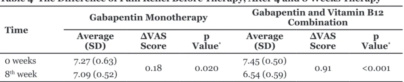
Table 4 shows that there is a significant average difference in pain relief between 0 week and 8th week in both groups. The gabapentin and vitamin B12 combination group has better average pain relief compared to the gabapentin monotherapy group.
Mochamad Firdaus Bhuanaputra et al.: Combination of Gabapentin and Vitamin B12 Compared with Gabapentin
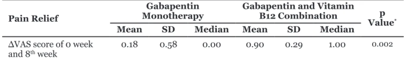
Table 5 shows that there is a significant difference in pain relief between gabapentin and vitamin B12 combination group and gabapentin monotherapy (p value<0.002).
Discussion
In the gabapentin and vitamin B12 combination group, there is a significant pain relief difference (p value<0.001) between 0 week and eighth Table 2 Average Result of VAS Score Check in the Gabapentin Monotherapy Group
VAS Score Gabapentin Group
Average (SD) Median (Min–Max)
VAS 0 week 7.27 (0.63) 7 (6–8)
VAS 4th week 6.77 (0.61) 7 (6–8)
VAS 8th week 7.09 (0.52) 7 (6–8)
Note: VAS=visual analogue scale
Table 3 VAS Score Different Test Before Therapy, After 4, and 8 Weeks Therapy
VAS Score p Value*
Gabapentin <0.001
Gabapentin and vitamin B12 combination <0.001
Note: * Statistic analysis with Friedman test, significant if p value≤0.05
Table 4 The Difference of Pain Relief Before Therapy, After 4 and 8 Weeks Therapy
Time
Gabapentin Monotherapy Gabapentin and Vitamin B12 Combination
Average
(SD) ∆VAS
Score p
Value* Average
(SD) ∆VAS
Score p
Value* 0 weeks 7.27 (0.63)
0.18 0.020 7.45 (0.50)
0.91 <0.001
8th week 7.09 (0.52) 6.54 (0.59)
Note: *Data analysis uses Wilcoxon test, significant if p value≤0.05; ∆=difference between weeks
Table 5 Pain Relief Comparison between Gabapentin and Vitamin B Combination and Gabapentin Monotherapy
Pain Relief
Gabapentin
Monotherapy Gabapentin and Vitamin B12 Combination p
Value*
Mean SD Median Mean SD Median
∆VAS score of 0 week
and 8th week 0.18 0.58 0.00 0.90 0.29 1.00 0.002
Note: *Mann-Whitney test, ∆=difference between weeks
4
week. This result was consistent with the study conducted by Mimenza Alvarado and Aguilar Navarro.21 They stated that there is a significant pain relief difference (p value<0.001).
Gabapentin and vitamin B12 have synergistic workability. The gabapentin increases gamma- aminobutyric acid (GABA) synthesis, the receptor antagonist of N-methyl-D-aspartate (NMDA), and subunit bond of α2δ voltage-dependent calcium channels that inhibits the release of excitatory neurotransmitters. That mechanism causes stimulation inhibition and pain reduction in neuropathic patients who consume gabapentin.
The role of vitamin B12 is to repair peripheral nerve cells by becoming a cofactor that facilitates homocysteine methylation for methionine, which activated to S-adenosyl-methionine, which donates the methyl group for methyl acceptors like myelin, neurotransmitter, and phospholipid membrane. The usage of gabapentin combined with vitamin B12 has a synergic effect to relieve the pain of diabetic neuropathic patients.15,16,22
In the gabapentin treatment groups, a significant pain relief difference is observed (p value=0.02) between 0 week and eighth week.
This study is following the study conducted by Surcheva et al.,23 which shows that there is a significant pain relief difference (p value=001).
The difference in improvement observed during 0 week and eighth week.
Gabapentin is a medicine of choice which mitigates the pain that works on the central nervous system but has a side effect that is classified small. Gabapentin affects neurotransmitter inhibitors. Gabapentin has a modification effect of releasing GABA. The release of GABA happened either presynaptic or postsynaptic on the central or even the arrangement of peripheral nerves.
Gabapentin increases GABA synthesis from glutamate and increases the release of GABA from astrocytes. Some researches show that there is a concentration increase of GABA in some regions of the brain after the administration of gabapentin so that glutamic acid decarboxylase increases and also decarboxylase glutamic acid enzyme destruction decreases which eventually increases the production of GABA.15,16,23
The pain relief observed in the gabapentin and vitamin B12 combination group and gabapentin monotherapy group. The pain relief is significantly better in the gabapentin and vitamin B12 combination group compared to the gabapentin monotherapy group with p value=0.002.
The finding is consistent with the study conducted by Mimenza Alvarado and Aguilar Navarro.21 The gabapentin and vitamin B complex administration compare to pregabalin shows better pain relief. The significant difference in pain relief between the treatment group and the control group observed in the 0 week and eighth week (p value<0.001).
Gabapentin and vitamin B12 combination groups have better pain relief improvement than the gabapentin monotherapy group. The results caused by better and more optimal treatment mix in gabapentin and vitamin B12 combination group. The administration of gabapentin will give an inhibiting effect to release neurotransmitters so that it will reduce the pain in diabetic neuropathic patients. The usage combined with the administration of vitamin B12, which serves to maintain and repair peripheral nerve cells. The combination treatment effect will give better pain relief improvement.23–25
Conclusion
A combination of gabapentin and vitamin B12 showed better pain relief compared to gabapentin monotherapy in diabetic neuropathic patients.
Conflict of Interest
The authors declare that no conflict of interest in this study.
Acknowledgment
We appreciate the support from the Head of Dr.
M. Salamun Air Force Hospital Bandung during the study.
References
1. Bansal D, Gudala K, Muthyala H, Esam HP, Nayakallu R, Bhansali A. Prevalence and risk factors of development of peripheral diabetic neuropathy in type 2 diabetes mellitus in a tertiary care setting. J Diabetes Investig.
2014;5(6):714–21.
2. Ramachandran A, Snehalatha C, Shetty AS, Nanditha A. Trends in prevalence of diabetes in Asian countries. World J Diabetes.
2012;3(6):110–7.
3. Guariguata L, Whiting DR, Hambleton I, Beagley J, Linnenkamp U, Shaw JE. Global
Mochamad Firdaus Bhuanaputra et al.: Combination of Gabapentin and Vitamin B12 Compared with Gabapentin
Global Medical and Health Communication, Volume 8 Number 1, April 2020 5
estimates of diabetes prevalence for 2013 and projections for 2035. Diabetes Res Clin Pract.
2014;103(2):137–49.
4. Indonesia Agency of Health Research and Development, Ministry of Health of Republic of Indonesia. Basic health research (Riskesdas) 2013 [Internet]. Jakarta:
Indonesia Agency of Health Research and Development, Ministry of Health of Republic of Indonesia; 2013 [cited 2017 June 15].
Available from: http://labdata.litbang.
kemkes.go.id/ccount/click.php?id=10.
5. Malik VS, Popkin BM, Bray GA, Després JP, Willett WC, Hu FB. Sugar-sweetened beverages and risk of metabolic syndrome and type 2 diabetes: a meta-analysis. Diabetes Care. 2010;33(11):2477–83.
6. Jin W, Patti ME. Genetic determinants and molecular pathways in the pathogenesis of type 2 diabetes. Clin Sci (Lond).
2009;116(2):99–111.
7. VA Center for Integrated Healthcare. Diabetic neuropathies: the nerve damage of diabetes [Internet]. July 2013 [cited 2017 June 16].
Available from: https://www.mirecc.va.gov/
cih-visn2/Documents/Provider_Education_
H a n d o u t s / D i a b e t i c _ N e u r o p a t h i e s _ Version_3.pdf.
8. Chawla R. Complications of diabetes. New Delhi: Jaypee Brothers Medical Publishers;
2012.
9. Schreiber AK, Nones CF, Reis RC, Chichorro JG, Cunha JM. Diabetic neuropathic pain:
physiopathology and treatment. World J Diabetes. 2015 Apr 15;6(3):432–44.
10. Yagihashi S, Mizukami H, Sugimoto K.
Mechanism of diabetic neuropathy: where are we now and where to go? J Diabetes Investig. 2011;2(1):18–32.
11. Emerging Risk Factors Collaboration, Sarwar N, Gao P, Seshasai SR, Gobin R, Kaptoge S, Di Angelantonio E, et al. Diabetes mellitus, fasting blood glucose concentration, and risk of vascular disease: a collaborative meta- analysis of 102 prospective studies. Lancet.
2010;375(9733):2215–22.
12. Wiffen PJ, Derry S, Moore RA, Aldington D, Cole P, Rice AS, et al. Antiepileptic drugs for neuropathic pain and fibromyalgia - an overview of Cochrane reviews. Cochrane Database Syst Rev. 2013;(11):CD010567.
13. Wiffen PJ, Derry S, Bell RF, Rice AS, Tölle TR, Phillips T, et al. Gabapentin for chronic
neuropathic pain in adults. Cochrane Database Syst Rev. 2017;6:CD007938.
14. Murnion BP. Neuropathic pain: current definition and review of drug treatment. Aust Prescr. 2018 Jun;41(3):60–3.
15. Attal N, Cruccu G, Baron R, Haanpää M, Hansson P, Jensen TS, et al. EFNS guidelines on the pharmacological treatment of neuropathic pain: 2010 revision. Eur J Neurol. 2010;17(9):1113–23.
16. Rosenquist RW, Vrooman BM. Chronic pain managament. In: Butterworth JF, Mackey DC, Wasnick JD, editors. Morgan and Mikhail's Clinical Anesthesiology. 5th Edition.
New York: McGraw-Hill Companies; 2013. p.
1023–86.
17. Agency for Healthcare Research and Quality, U.S. Department of Health & Human Services.
Effectiveness of treatments for diabetic peripheral neuropathy [Internet]. May 13, 2016 [cited 2017 June 17]. Available from:
https://effectivehealthcare.ahrq.gov/sites/
default/files/pdf/diabetic-neuropathy_
research-protocol.pdf.
18. Baraz S, Zarea K, Shahbazian HB, Latifi SM.
Comparison of the accuracy of monofilament testing at various points of feet in peripheral diabetic neuropathy screening. J Diabetes Metab Disord. 2014;13(1):19.
19. Wang F, Zhang J, Yu J, Liu S, Zhang R, Ma X, et al. Diagnostic accuracy of monofilament tests for detecting diabetic peripheral neuropathy: a systematic review and meta- analysis. J Diabetes Res. 2017;2017:8787261.
20. Dros J, Wewerinke A, Bindels PJ, van Weert HC. Accuracy of monofilament testing to diagnose peripheral neuropathy: a systematic review. Ann Fam Med. 2009 Nov- Dec;7(6):555–8.
21. Mimenza Alvarado A, Aguilar Navarro S. Clinical trial assessing the efficacy of gabapentin plus B complex (B1/B12) versus pregabalin for treating painful diabetic neuropathy. J Diabetes Res.
2016;2016:4078695.
22. Jayabalan B, Low LL. Vitamin B supplementation for diabetic peripheral neuropathy. Singapore Med J. 2016;57(2):55–
23. Surcheva S, Todorova L, Maslarov 9.
D, Vlaskovska M. Preclinic and clinic effectiveness of gabapentin and pregabalin for treatment of neuropathic pain in rats
Mochamad Firdaus Bhuanaputra et al.: Combination of Gabapentin and Vitamin B12 Compared with Gabapentin
6
and diabetic patients. Biotechnol Biotechnol Equip. 2017;31(3):568–73.
24. Vinik AI, Casellini CM. Guidelines in the management of diabetic nerve pain: clinical utility of pregabalin. Diabetes Metab Syndr Obes. 2013;6:57–78.
25. Lal R, Sukbuntherng J, Luo W, Tovera J, Lassauzet ML, Cundy KC. Population pharmacokinetics and pharmacodynamics of gabapentin after administration of gabapentin enacarbil. J Clin Pharmacol.
2013;53(1):29–40.
Mochamad Firdaus Bhuanaputra et al.: Combination of Gabapentin and Vitamin B12 Compared with Gabapentin
7 RESEARCH ARTICLE
Global Medical and Health Communication
Online submission: http://ejournal.unisba.ac.id/index.php/gmhc
DOI: pISSN 2301-9123 │ eISSN 2460-5441
Correspondence: Fetty Fatmawaty, dr., Sp.Rad. R. Syamsudin, S.H. Regional General Hospital. Jln. Rumah Sakit No. 1, Sukabumi 43113, West Java, Indonesia. E-mail: [email protected]
Received: 7 May 2018; Revised: 26 January 2020; Accepted: 2 March 2020; Published: 30 April 2020
https://doi.org/10.29313/gmhc.v8i1.3744 GMHC. 2020;8(1):7–12
Comparative Study Gallbladder Contractility Index Using Ultrasound in Patients with and without Liver Cirrhosis
Hari Soekersi,1 Leni Santiana,1 Fetty Fatmawaty2,3
1Department of Radiology, Faculty of Medicine, Universitas Padjadjaran/Dr. Hasan Sadikin General Hospital, Bandung, Indonesia, 2Radiology Study Program, Faculty of Medicine, Universitas Padjadjaran/Dr. Hasan Sadikin
General Hospital, Bandung, Indonesia, 3R. Syamsudin, S.H. Regional General Hospital, Sukabumi, Indonesia
Abstract
Liver cirrhosis leads to impairment of gallbladder contractility resulting in bile stasis and facilitate the development of gallstones that will aggravate the clinical symptoms of the patients. The gallbladder contractility index is an indicator of gallbladder motility measured using ultrasound as the radiological choice of modality. This study aims to determine differences in the gallbladder contractility index using ultrasound in patients with and without liver cirrhosis. This study was an observational study of comparative analytic with cross-sectional design with sampling conducted by consecutive admissions sampling at Dr. Hasan Sadikin General Hospital Bandung from December 2017 to February 2018. Statistical analysis than performed by using an independent t test to find out the difference of gallbladder contractility index in patients with and without liver cirrhosis. A total of 22 subjects, 12 men, 10 women, with the youngest 37 years old and the oldest 70 years old. The result of the study was obtained mean fasting gallbladder volume (35.56±22.16 mL) and postprandial (21.25±16.08 mL) in patients with liver cirrhosis higher than without liver cirrhosis with mean fasting gallbladder volume (16.50±4.14 mL) and postprandial (5.44±2.10 mL). The average gallbladder contractility index on patients with liver cirrhosis (41.64±24.52%) smaller than without liver cirrhosis (66.73±9.19%). The result of the statistical test showed that there was a significant difference in the gallbladder contractility index on patients with liver cirrhosis than without liver cirrhosis (p=0.007, p≤0.05).
In conclusion, there was a significant difference in the gallbladder contractility index that measured by using ultrasound between the patients with and without liver cirrhosis.
Key words: Contractility index, gallbladder, liver cirrhosis, ultrasound
Perbedaan Indeks Kontraktilitas Kandung Empedu Menggunakan Ultrasonografi pada Penderita Sirosis Hati dan tanpa Sirosis Hati
Abstrak
Sirosis hati menyebabkan gangguan indeks kontraktilitas kandung empedu yang mengakibatkan stasis cairan empedu dan memudahkan kejadian batu empedu yang akan memperberat gejala klinis pasien. Indeks kontraktilitas kandung empedu merupakan indikator motilitas kandung empedu yang diukur menggunakan ultrasonografi (USG) sebagai modalitas pilihan radiologi. Penelitian ini bertujuan mengetahui perbedaan indeks kontraktilitas kandung empedu menggunakan ultrasonografi pada pasien sirosis hati dan tanpa sirosis. Penelitian ini menggunakan studi observasional analitik komparatif dengan rancangan cross-sectional dan pengambilan sampel dilakukan secara consecutive admissions sampling di RSUP Dr. Hasan Sadikin Bandung dari bulan Desember 2017 hingga Februari 2018. Uji statistik menggunakan independent t test. Subjek penelitian berjumlah 22, laki-laki 12 dan perempuan 10, serta usia termuda 37 tahun dan tertua 70 tahun. Hasil penelitian didapatkan volume rerata kandung empedu puasa (35,56±22,16 mL) dan pascaprandial (21,25±16,08 mL) pada pasien sirosis hati lebih besar daripada tanpa sirosis hati dengan volume rerata kandung empedu puasa (16,50±4,14 mL) dan pascaprandial (5,44±2,10 mL).
Indeks kontraktilitas rerata kandung empedu penderita sirosis hati (41,64±24,52%) lebih rendah dibanding dengan tanpa sirosis hati (66,73±9,19%). Hasil uji statistik menunjukkan terdapat perbedaan bermakna antara indeks kontraktilitas kandung empedu penderita sirosis hati dan tanpa sirosis hati (p=0,007; p≤0,05). Simpulan, terdapat perbedaan bermakna antara indeks kontraktilitas kandung empedu menggunakan USG pada penderita sirosis hati dan tanpa sirosis hati.
Kata kunci: Indeks kontraktilitas, kandung empedu, sirosis hati, ultrasonografi
8
Introduction
Liver cirrhosis is the third leading cause of death in patients who are 45–46 years old (after cardiovascular and cancer). Liver cirrhosis is a disease characterized by diffuse and chronic inflammation of the liver progressively with distorted images of the hepatic architecture and the formation of regenerative nodules.1,2 Worldwide, liver cirrhosis ranks seventh as the cause of death. There are more male patients with liver cirrhosis compared with women with a ratio of about 1.6:1. The average age group is 30–59 years old, with a peak of around the age of 40–49 years old.3,4 In 2015, the World Health Organization (WHO) reported that 720,000 deaths caused by liver cirrhosis in the world.5
In 2012, liver cirrhosis in Indonesia is the sixth cause of death, with 48.9 thousand deaths.
The incidence of liver cirrhosis in Indonesia due to hepatitis B ranged between 21.2–46.9%
and hepatitis C ranged 38.7–73.9%.6,7 In Dr.
Hasan Sadikin General Hospital Bandung hospitalized patients with liver cirrhosis in 2012 until September 2017 were about 365 people, and about 1,716 people went through internal medicine polyclinic.8
Patients with liver cirrhosis have a higher incidence and prevalence of gallstones than the general population, with a prevalence of 25–30%.
Liver cirrhosis is one of the significant risk factors for the formation of gallstones.9 Gallstones in patients with liver cirrhosis are asymptomatic and symptomatic to cause complications. In patients with liver cirrhosis, Child-Pugh A and B with symptomatic gallstones recommended doing cholecystectomy before the complication that will aggravate the patient clinical condition and before the severe condition of the liver symptoms.10 The formation of gallstones in patients with liver cirrhosis is due to several factors; one of them is due to bile stasis caused by the decrease in gallbladder contractility index.11
The decrease in the gallbladder contractility index is due to the humoral, hypoalbuminemia, and also neurological disorders. Neurological disorders in patients with liver cirrhosis are in the form of neuropathy. The severity of this neuropathy is proportional to an increase in liver damage. The patients with liver cirrhosis, especially decompensated liver cirrhosis, usually have autonomic dysfunction directly affect the contractility of the gallbladder through the
neural path.12 Gallbladder contractility disorder in cirrhotic patients is also affected by the thickening of the gallbladder wall. This thickening is due to edema and structural changes in the gallbladder wall due to hypoalbuminemia and portal hypertension. In addition to the above, the disorder of gallbladder contractility in patients with liver cirrhosis also influenced by humoral changes.13–17
Humoral changes that occur in liver cirrhosis are due to cholecystokinin (CCK) receptor resistance in the gallbladder. Thus, the contraction of the gallbladder disrupted. Besides, decreased hepatic clearance also occurs in patients with liver cirrhosis, increasing the level of intestinal peptides that affect the relaxation of the gallbladder wall. Gallbladder contractility disorders in patients with liver cirrhosis assessed by evaluating the gallbladder contractility index using ultrasound.13
Ultrasound is the preferred modality for assessing the gallbladder contractility index by measuring the volume of the gallbladder, and postprandial. Ultrasound is a radiological modality that is accurate, noninvasive, cheap, easy to obtain, no radiation and repeated to assess the gallbladder contractility index.12,14,18
A research conducted by Buzaş et al.19 suggested that the volume of gallbladder fasting and postprandial volume using ultrasound was higher in patients with liver cirrhosis. Loreno et al.14 also suggest that there was a difference in the gallbladder contractility index in patients with liver cirrhosis compared with no liver cirrhosis.
Acalovschi13 suggests that there was a decreasing index of gallbladder contractility in patients with liver cirrhosis following the severity of cirrhosis.
It is contrary to research conducted by Kul et al.,12 suggesting that there was an increase in gallbladder contractility index in patients with liver cirrhosis compared with healthy control. Shirole et al.11 suggest that patients with liver cirrhosis have a higher prevalence of the occurrence of gallstones than in a healthy population. One of the causes is a decrease in the gallbladder contractility index.
Because of differences in the above studies, the researchers will determine differences in the gallbladder contractility index using ultrasound in patients with and without liver cirrhosis.
Methods
This study was an analytic observational study
Hari Soekersi et al.: Comparative Study Gallbladder Contractility Index Using Ultrasound in Patients with and without
Global Medical and Health Communication, Volume 8 Number 1, April 2020 9
with a cross-sectional design conducted from December 2017 to February 2018. Sampling was patients with liver cirrhosis and without liver cirrhosis in Dr. Hasan Sadikin General Hospital Bandung, who matched the inclusion criteria with consecutive admissions method (based on the order of the registered patient). The inclusion criteria were patients with adult liver cirrhosis diagnosed from the Gastroentero-hepatology clinic of Dr. Hasan Sadikin General Hospital Bandung and undiagnosed liver cirrhosis that had gone through ultrasound examination in the Department of Radiology Dr. General Hospital Hasan Sadikin and willing to follow the research.
Exclusion criteria for this study were patients with a history of bile disease, gallbladder hydrops, diabetes mellitus, post gastric resection surgery, and patients who were pregnant.
Statistical analysis used to describe the difference of gallbladder contractility index using ultrasound in patients with and without liver cirrhosis. A statistical test using the parametric method, independent t test performed using SPSS version 22.0 for Windows at a 95% confidence level.
Ethical approval for this study has obtained from the Health Research Ethics Committee of Dr. Hasan Sadikin General Hospital Bandung with the letter number: LB.04.01/A05/EC/379/
XII/2017.
Results
Table 1 shows that by gender, there were more male patients than females with and without liver cirrhosis who came to Dr. Hasan Sadikin General Hospital as many as 12 of 22.
The differences in gallbladder contractility index using ultrasound in patients with and without liver cirrhosis in Dr. Hasan Sadikin General Hospital can be seen in Table 2.
Based on Table 2, it can be seen that the average volume of fasting gallbladder using ultrasound on the liver cirrhosis patients is 35.56 mL, with a standard deviation of 22.16 mL. Patients without liver cirrhosis was 16.50 mL, with a standard deviation of 4.14 mL. The mean postprandial gallbladder volume using ultrasound in patients with liver cirrhosis was 21.25 mL, with a standard deviation of 16.08 mL,
Hari Soekersi et al.: Comparative Study Gallbladder Contractility Index Using Ultrasound in Patients with and without
Table 1 Characteristics of Study Subjects by Age and Gender in Patients with and without Liver Cirrhosis
Age and Gender n=22 Average (SD) Median (Min–Max)
Age (years) 30–40 41–50
>50
85 9
49.45 (9.53) 48 (37–70)
Gender
MaleFemale 12
10
Table 2 The Differences in Gallbladder Contractility Index Using Ultrasound in Patients with and without Liver Cirrhosis
Gallbladder
Liver Cirrhosis Incident
Valuep * Liver Cirrhosis Without Liver
Cirrhosis
Average SD Average SD
Fasting gallbladder volume 35.56 22.16 16.50 4.14
0.007 Postpartum gallbladder volume 21.25 16.08 5.44 2.10
Gallbladder contractility index 41.64 24.52 66.73 9.19
Note: *Independent t test
10
whereas in patients without liver cirrhosis was 5.44 mL, with a standard deviation of 2.10 mL.
Based on Table 2, it can be seen that the average gallbladder contractility index using ultrasound in patients with liver cirrhosis was 41.64%, with a standard deviation of 24.52%.
Patients without liver cirrhosis were 66.73%, with a standard deviation of 9.19%. The independent t test result on a 95% confidence degree shows that there was a significant difference in gallbladder contractility index using ultrasound in patients with and without liver cirrhosis (p=0.007, p≤0.05).
The differences in gallbladder contractility index using ultrasound in patients with and without liver cirrhosis is in Figure.
Discussion
The results of age-based, the age distribution of most patients with liver cirrhosis in this study were in the age group >50 years. It is by the research conducted by Hussain et al.,10 which states that patients with liver cirrhosis are more common in middle age and older age due to disease progression of liver cirrhosis that usually takes 5–10 years. Patients with liver cirrhosis
in this study were mostly male. The results are consistent with Basic Health Research 2013 that patients affected by hepatitis are more likely to be men even when it is less significant and mostly work-related that often occurs in patients who work as farmers/laborers or fishermen.20,21
The results based on fasting gallbladder volume show that the average fasting gallbladder volume in liver cirrhosis patients was 35.56 mL, with a standard deviation of 22.16 mL. The results are higher than those without liver cirrhosis with an average fasting gallbladder volume of 16.5 mL and a standard deviation of 4.14 mL. It suggests that most of the volume of the fasting gallbladder in patients with liver cirrhosis is more significant than without liver cirrhosis. It is under research conducted by Buzaş et al.19 and Loreno et al.14 state that the average gallbladder volume of fasting patients with liver cirrhosis is higher than without liver cirrhosis. The mechanism of fasting gallbladder volume of patients with liver cirrhosis larger than without liver cirrhosis is suspected because increased intestinal peptides lead to the relaxation of gallbladder muscles and autonomic nervous disorders resulting in lower gallbladder contraction in the patients with liver cirrhosis.
The average postprandial gallbladder volume in liver cirrhosis patients is 21.25 mL with a standard deviation of 16.08 mL, while in the group without liver cirrhosis, the mean postprandial gallbladder volume is 5.44 mL and a standard deviation of 2.1 mL. It appears that the average of postprandial gallbladder volume in most liver cirrhosis patients tends to be higher than the average of postprandial gallbladder volume in patients without liver cirrhosis.22
Based on the average gallbladder contractility index, the index of biliary tactility in liver cirrhosis patients was 41.64%, with a standard deviation of 24.52%. In comparison, the average of gall gallbladder contractility in the group without liver cirrhosis was 66.73%, with a standard deviation of 9.19%. It is under research conducted by Loreno et al.14 and Acalovschi,13 who suggest that the gallbladder contractility index in patients with liver cirrhosis is lower than without liver cirrhosis.
Independent t test analysis at 95% confidence degree showed that there is a statistically significant difference between gallbladder contractility index using ultrasound in patients with and without liver cirrhosis (p=0.007, p≤0.05). It is consistent with the research Figure The Differences in Gallbladder
Contractility Index Using
Ultrasound in Patients with and without Liver
Hari Soekersi et al.: Comparative Study Gallbladder Contractility Index Using Ultrasound in Patients with and without
Global Medical and Health Communication, Volume 8 Number 1, April 2020 11
conducted by Loreno et al.14 and Acalovschi,13 who suggest that there is a significant difference in the gallbladder contractility index of patients with and without liver cirrhosis.
The causes of gallbladder contractility index differences in patients with liver cirrhosis may due to a decrease in CCK receptor sensitivity in the gallbladder, autonomic neuropathy, hypoalbuminemia, and increased concentrations of intestinal peptides that affect gallbladder muscle relaxation.13
Conclusion
There was a significant difference in gallbladder contractility index using ultrasound in patients with and without liver cirrhosis in Dr. Hasan Sadikin General Hospital Bandung.
Conflict of Interest
There is no ethical/legal conflict involved in the article. All authors have no relevant financial interests related to the material.
References
1. Siregar GA, Tampubolon SE. Comparison of lipid profile between degrees of severity of hepatic cirrhosis in Haji Adam Malik General Hospital Medan. IOP Conf Ser Earth Environ Sci. 2018:125:012216.
2. Yeom SK, Lee CH, Cha SH, Park CM.
Prediction of liver cirrhosis, using diagnostic imaging tools. World J Hepatol.
2015;7(17):2069–79.
3. Zatoński WA, Sulkowska U, Mańczuk M, Rehm J, Boffetta P, Lowenfels AB, et al. Liver cirrhosis mortality in Europe, with special attention to central and eastern Europe. Eur Addict Res. 2010;16(4):193–201.
4. Blachier M, Leleu H, Peck-Radosavljevic M, Valla DC, Roudot-Thoraval F. The burden of liver disease in Europe: a review of available epidemiological data. J Hepatol.
2013;58(3):593–608.
5. World Health Organization (WHO). Global hepatitis report 2017. Geneva: WHO; 2017.
6. Widjaja FF, Karjadi T. Pencegahan perdarahan berulang pada pasien sirosis hati.
J Indon Med Assoc. 2011;61(10):417–24.
7. Nurdjanah S. Sirosis hati. In: Sudoyo AW, Setiyohadi B, Alwi I, Simadibrata KM, Setiati
S, editors. Buku ajar ilmu penyakit dalam. 6th Edition. Jakarta: Interna Publishing; 2014. p.
1978–83.
8. Sistem Informasi Rumah Sakit (SIRS) RSUP Dr. Hasan Sadikin (RSHS). Pasien sirosis hati 2012–2017. Bandung: SIRS RSHS; 2017.
9. Li X, Guo X, Ji H, Yu G, Gao P. Gallstones in patients with chronic liver diseases. Biomed Res Int. 2017;2017:9749802.
10. Hussain A, Nadeem MA, Nisar S, Tauseef H. Frequency of gallstones in patients with liver cirrhosis. J Ayub Med Coll Abbottabad.
2014;26(3):341–3.
11. Shirole NU, Gupta SJ, Shah DK, Gaikwad NR, Sankalecha TH, Kothari HG. Cirrhosis of liver is a risk factor for gallstone disease. Int J Res Med Sci. 2017;5(5):2053–6.
12. Kul K, Serin E, Yakar T, Coşar AM, Özer B. Autonomic neuropathy and gallbladder motility in patients with liver cirrhosis. Turk J Gastroenterol. 2015;26(3):254–8.
13. Acalovschi M. Gallstones in patients with liver cirrhosis: incidence, etiology, clinical and therapeutical aspects. World J Gastroenterol.
2014;20(23):7277–85.
14. Loreno M, Travali S, Bucceri AM, Scalisi G, Virgilio C, Brogna A. Ultrasonographic Study of Gallbladder Wall Thickness and Emptying in Cirrhotic Patients without Gallstones.
Gastroenterol Res Pract. 2009;2009:683040.
15. Butt Z, Hyder Q. Cholelithiasis in hepatic cirrhosis: evaluating the role of risk factors. J Pak Med Assoc. 2010;60(8):641–4.
16. Son JY, Kim YJ, Park HS, Yu NC, Ko SM, Jung SI, et al. Diffuse gallbladder wall thickening on computed tomography in patients with liver cirrhosis: correlation with clinical and laboratory variables. J Comput Assist Tomogr. 2011;35(5):535–8.
17. Mohammadi A, Ghasemi-Rad M, Mohammadifar M. Differentiation of benign from malignant induced ascites by measuring gallbladder wall thickness. Maedica (Buchar).
2011;6(4):282–6.
18. Popescu A, Sporea I. Ultrasound examination of normal gall bladder and biliary system.
Med Ultrason. 2010;12(2):150–2.
19. Buzaş C, Chira O, Mocan T, Acalovschi M.
Comparative study of gallbladder motility in patients with chronic HCV hepatitis and with HCV cirrhosis. Rom J Intern Med.
2011;49(1):37–44.
20. Indonesia Agency of Health Research
Hari Soekersi et al.: Comparative Study Gallbladder Contractility Index Using Ultrasound in Patients with and without
12
and Development, Ministry of Health of Republic of Indonesia. Basic health research (Riskesdas) 2013 [Internet]. Jakarta:
Indonesia Agency of Health Research and Development, Ministry of Health of Republic of Indonesia; 2013 [cited 2017 June 15].
Available from: http://labdata.litbang.
kemkes.go.id/ccount/click.php?id=10.
21. Pusat Data dan Informasi, Kementerian Kesehatan Republik Indonesia. Situasi dan
analisis hepatitis [Internet]. 2014 September [cited 2018 February 20]. Available from:
https://www.kemkes.go.id/resources/
download/pusdatin/infodatin/infodatin- hepatitis.pdf.
22. Abdelmaksoud MA, El-Shamy MH, Hussein HIM, Bihery AS, Ahmed H, El Hady HA.
Frequency of cholelithiasis in patients with chronic liver disease: a hospital-based study.
AJIED. 2016;6(3):134–41.
Hari Soekersi et al.: Comparative Study Gallbladder Contractility Index Using Ultrasound in Patients with and without
13 RESEARCH ARTICLE
Global Medical and Health Communication
Online submission: http://ejournal.unisba.ac.id/index.php/gmhc
DOI: pISSN 2301-9123 │ eISSN 2460-5441
Correspondence: Astrid Feinisa Khairani. Department of Biomedical Sciences, Faculty of Medicine, Universitas Padjadjaran. Jln.
Raya Bandung-Sumedang km 21, Jatinangor, Sumedang 45363, West Java, Indonesia. E-mail: [email protected] Received: 21 October 2019; Revised: 24 April 2020; Accepted: 25 April 2020; Published: 30 April 2020
https://doi.org/10.29313/gmhc.v8i1.5253 GMHC. 2020;8(1):13–20
Lumbar Radiculopathy: a Descriptive Study on Red Flag and Neurologic Symptoms in Dr. Hasan Sadikin General Hospital Bandung
Astrid Feinisa Khairani,1 Kuheinderan Radha Krishnan,2 Umar Islami,3,4 Siti Aminah Sobana5
1Department of Biomedical Sciences, Faculty of Medicine, Universitas Padjadjaran, Sumedang, Indonesia,
2Medical Undergraduate Study Program, Faculty of Medicine, Universitas Padjadjaran, Sumedang, Indonesia,
3Graduate School of Biomedical Sciences Master Program, Faculty of Medicine, Universitas Padjadjaran, Bandung, Indonesia, 4Department of Histology and Medical Biology, Faculty of Medicine, Universitas Islam
Bandung, Bandung, Indonesia, 5Department of Neurology, Faculty of Medicine, Universitas Padjadjaran/
Dr. Hasan Sadikin General Hospital, Bandung, Indonesia
Abstract
Over 80% of the adult population will experience an episode of low back pain (LBP). Low back pain is a pain in the lumbosacral region. When it progresses, which may be identified earlier with signs of a red flag, the manifestation might become radiculopathy. Radiculopathies are nerve root disease which may show signs of neurologic symptoms from the sensory, motoric, or autonomic origin. This study to help identify the clinical characteristics of a red flag in low back pain patients, which became lumbar radiculopathy for prognostic and diagnostic use. It is a descriptive quantitative cross-sectional study of medical records from patients hospitalized with complaints of low back pain with lumbar radiculopathy between January 2013–December 2015 in the Department of Neurology, Dr. Hasan Sadikin General Hospital, Bandung. It identifies a specific red flag and neurogenic symptoms. Patients most affected were housewives (26%), females (60%), and middle-aged adults (31%). The highest recorded symptom being sensory (76%), the highest progression was sensory to motoric (59%), affected by all three neurologic symptoms (39%), and trauma was the highest red flag recorded (48%). Low back pain patients who have signs of red flag show a high tendency to develop radiculopathy, which shows neurologic symptoms. If left untreated earlier, it may become a permanent disability.
Key words: Low back pain, radiculopathy, red flag
Radikulopati Lumbar: Studi Deskriptif Gejala Klinis Red Flag dan Gejala Neurologis di RSUP Dr. Hasan Sadikin Bandung
Abstrak
Lebih dari 80% populasi penduduk dewasa akan mengalami episode low back pain (LBP). Low back pain merupakan nyeri pada bagian tulang belakang regio lumbo-sakral. Pada saat rasa sakitnya bertambah berat, gejala berbahaya dapat menjadi awal perkembangan LBP menjadi radikulopati. Radikulopati merupakan penyakit saraf pada daerah radiks neuron berupa gejala sensorik, motorik, dan otonomik. Penelitian ini bertujuan mengidentifikasi karakteristik gejala klinis tanda berbahaya (red flag) pada pasien LBP yang berlanjut menjadi radikulopati lumbar sebagai alat bantu diagnostik dan prognostik. Penelitian ini menggunakan metode penelitian cross-sectional kuantitatif deskriptif dari rekam medis pasien yang dirawat dengan diagnosis LBP dan radikulopati lumbar antara bulan Januari 2013–Desember 2015 di Departemen Ilmu Penyakit Saraf, RSUP Dr. Hasan Sadikin, Bandung.
Penelitian ini mengidentifikasi gejala klinis red flag spesifik dan neurologis. Pasien yang terkena terutama ibu rumah tangga (26%), wanita (60%), usia dewasa pertengahan (31%). Gejala yang tercatat paling banyak adalah sensorik (76%), perkembangan progresif sensorik ke motorik (59%), mengalami gangguan ketiganya (39%), dan trauma menjadi penyebab red flag yang paling tinggi (48%). Pasien LBP yang memiliki gejala tanda berbahaya (red flag) memiliki kecenderungan tinggi berkembang menjadi radikulopati yang menunjukkan gejala neurologis. Jika tidak diobati lebih awal, ini dapat mengakibatkan kecacatan permanen.
Kata kunci: Low back pain, radikulopati, red flag
14
Introduction
Sometime during life, over 80 percent of the population will experience an episode of low back pain (LBP).1 LBP defined as the complaint of pain in the area of spinal or paraspinal structures in the lumbosacral region.2 According to the Global Burden of Disease Study 2015 (GBD 2015), the expert group stated that among the top ten high burden diseases and injuries, LBP is one of them with an average number of DALYs (disability-adjusted life years). It is higher than HIV, tuberculosis, road injuries, lung cancer, chronic obstructive pulmonary disease, and preterm birth complications.3 Low back pain, if left untreated with the red flag, may cause a more severe medical condition. Low back pain remains a common condition among primary care patients with an estimated lifetime prevalence of 13.8% for chronic pain and 80% for any episode of pain.4–6
Low back pain affecting both men and women, with age, is the primary risk factor, as it occurs secondary to the degenerative process within the spinal column.7 Symptoms typically begin in midlife, with men often affected in the 40s while women are affected in the 50s and 60s.8,9 Females have a higher risk in specific populations, with physically demanding careers such as service in the military. Patients commonly present with back pain that is associated with their radiculopathy.
By definition, radiculopathy describes pain that radiates down the legs and is often described by patients as electric, burning, or sharp. The most common underlying cause of radiculopathy is irritation of a particular nerve, which can occur at any point along the nerve itself and is most often a result of a compressive force. The diagnosis of the causative agent and subsequent treatment starts with a thorough physical exam.
The term red flags are symptoms and signs that show a possible manifestation of more serious medical conditions, which may cause permanent disability and, if not managed correctly, may even lead to death.10 These red flags seen in LBP patients, when neglected without treatment, may become radiculopathy. Radiculopathies are a disease of nerve roots from inflammation or impingement from a tumor or a bony spur.11 One form of radiculopathy is one that affects the lumbar region. The prevalence of lumbar radiculopathy varies from about 2.2–8%, and the incidence ranges from 0.7–9.6%. About 76.1%
of lumbar radiculopathies involve the L5 and S1 nerve roots.12 Lumbar radiculopathy refers to a pathologic process involving the lumbar roots causing radicular symptoms in a lower extremity.
The nerve root pathology arises primarily from direct neural compression irrespective of whether the etiology is an acute herniated or displaced disc, bony spurs, foraminal stenosis, central stenosis, or hypermobility of a vertebral segment.13
While a substantial burden on our health-care system, low back pain is typically self-limited, with most cases resolving after conservative management in 6 to 8 weeks.14 Most low back pain is nonspecific; patients with low back pain may have neurologic impairments or a severe underlying pathology that requires timely and accurate diagnosis. Low back pain that is due to severe pathology occurs between 1% and 4% <