These protein extensions are highly enriched in phenylalanine - glycine (FG) repeats and constitute the bulk of the permeability barrier of the NPC (Terry et al. 2007). The release of mRNP into the cytoplasm is thought to be mediated by Dbp5, a member of the conserved family of DEAD-box helicases ( Cordin et al. 2006 ; Linder 2006 ). Additionally, hGle1 contains a movement domain that is not conserved in yGle1 that plays a role for mRNA export ( Kendirgi et al. 2003 ).
Genetic studies in yeast have identified components of the inositol signaling pathway that give rise to inositol hexakisphosphate (IP6) production as key players in the Gle1-driven step in mRNA export (York et al. 1999). The yeast Ipk1 enzyme, responsible for the formation of IP6, is localized to the nuclear periphery (York et al. 1999); For Tir1, IP6 interacts with several structurally important elements of the tertiary structure (Tan et al. 2007); however, a.
IP6 can also be converted to diphosphorylated inositols (such as diphosphorylinositol 1,3,4,5,6 pentakis phosphate; PP-IP5) by the enzyme Kcs1 (York et al. 2005).

The localization of poly(A)+ RNA was analyzed by growing strains in rich or synthetic medium at 23°C before the shift to 36°C for one hour. In situ hybridizations were performed using a digoxigenin oligo (dT)30 probe as previously described (Iovine et al. 1995). Cells were observed using a fluorescent microscope (model BX50; Olympus, Lake Success, NY) using an Uplan 100x/1.3 objective.
IP6 and not IP7, is specifically required for proper mRNA export in vivo Genetic interactions between gle1 or dbp5 mutants and the kcs1∆ mutant, which. Indeed, bulk poly(A)+ mRNA is retained in the nucleus through an unknown mechanism during heat shock (Saavedra et al. 1997) and the mRNA export factor Dbp5 undergoes reversible changes in its localization during ethanol stress (Takemura et al. 2004) ), a condition that where cells also retain mRNA in the nucleus. Rollenhagen et al. 2004; Strahm et al. 1999 ), we predicted that this strain was compromised in two specific ways and could be used to isolate mRNA export factors that required both IP6 production and efficient Gle1 localization at NPC cytoplasmic fibrils.
A 2µ yeast genomic library was transformed into ipk1∆ nup42∆ cells and colonies that grew at the non-permissive temperature were selected. Two of the identified genes, PDE2 and SSD1, were independently tested for their ability to genetically interact with other mRNA export factors (Figure 10). Additionally, PDE2 has been found to have genetic interactions with SSD1 (Matsuura and Anraku 1994), also isolated in this screen, and GLC7 (Uesono et al. 1997), a protein phosphatase with roles in many processes including mRNA export .
dhh1 mutants have been reported to increase the lethality of dbp5 mutants, suggesting functional overlap between Dhh1 and Dbp5 (Tseng-Rogenski et al. 2003). In direct assays, DBP5 overexpression was found to suppress the ipk1∆ nup42∆ growth defect (Figure 9 C). Interestingly, DBP5 overexpression also rescued the mRNA export defects (Figure 9 B), whereas DHH1 did not (data not shown).
This suggested that bypassing the mRNA export defect in the ipk1∆ nup42∆ mutant specifically required Dbp5. We concluded that the partial rescue of the ipk1∆ nup42∆ mutant by overexpressing DHH1 was indirect and due to the partial functional overlap between Dbp5 and Dhh1 (Tseng-Rogenski et al. 2003), or to an altered flux in global mRNA biosynthesis. pathway and potential connections between mRNA export and turnover (Moore 2005). Based on these results and published genetic data closely linking GLE1, IPK1, and DBP5 function (Hodge et al.
Surprisingly, addition of both Gle1 (250 nM) and IP6 (100 nM) stimulated the Dbp5 ATPase activity almost fivefold (Figure 11 B). This correlated with previous observations that the elevated IP5 level in ipk1∆ cells did not rescue dbp5 or gle1 mutants, and indicated that specific stimulation of the ATPase activity of Dbp5 by IP6 is the physiologically relevant mechanism. The Michaelis-Menten saturation kinetics were determined for Dbp5-driven ATP hydrolysis in the presence and absence of Gle1 and/or IP6.
Under the same conditions plus 1 mM ATP, the RNA concentration for half-maximal activity was also significantly affected (Figure 11 E). Gle1 and IP6 work together to increase the catalytic efficiency of Dbp5 and lower the RNA concentration threshold required for Dbp5 activity.
To date, two members of the DEAD-box protein family have been shown to remove or displace proteins from RNA (Fairman et al. 2004; Jankowsky et al. 2001). It has also been proposed that Dbp5 functions by reshaping mRNP protein composition during export (Schmitt et al. Dbp5 shuttles between the nucleus and cytoplasm and interacts with transcription factor components of the elongation complex TFIIH (Estruch and Cole 2003) and Yra1, a nuclear mRNP protein , involved in mRNA processing prior to export (Schmitt et al. 1999).
There is evidence that human Gle1 also shuttles between the nucleus and cytoplasm and interacts with both the vertebrate ortholog Nup42 (hCG1) and another Nup, human Nup155 (Kendirgi et al. Suntharalingam et al. 2004), as well as IP6 and soluble inositols. capable of diffusion throughout the cell (Miller 2004). A complete lack of IP6 production, in addition to the reduced levels of IP6 in the catalytically compromised ipk1-5 mutant, is insufficient for the function of the gle1-2 mutant (Ives et al. 2000; York et al. 1999).
This is consistent with a recent report showing that IP6 is required for the activity and stability of adenosine deaminases acting on RNA (Macbeth et al. 2005). Members of the conserved Mex67/Mtr2 heterodimer family (also known as TAP/p15 or NXF1/NXT1) are believed to be the major mRNA export receptors that directly bridge mRNA to FG domains within the aqueous NPC channel (Gruter et al. e.g., intron-containing pre-mRNAs are filled with exon-exon junctions during splicing, a mark absent in intronless mRNAs (Le Hir et al. 2001).
The physiological relevance of mRNP protein signatures is illustrated by transcript-specific mRNA export defects in yra1-1 and mex67-5 temperature-sensitive mutants (Hieronymus and Silver 2003) and the lack of an SSA4 (hsp70) mRNA export defect during general mRNA export. block caused by the heat shock response (Saavedra et al. 1997). Importantly, transcript-specific regulation has been observed in the splicing machinery by inhibiting the splicing of a subset of messages during different environmental conditions in yeast (Pleiss et al. 2007a, b). Dbp5, a member of the DEAD-box helicase family of RNA-dependent ATPases, is thought to mediate mRNP remodeling through the displacement of proteins from mRNAs (Snay-Hodge et al.
RESPONSE TO HIGH OSMOLARITY IMPACTS mRNA EXPORT AND GROWTH OF mRNA EXPORT MUTANTS
In yeast, the HOG pathway controls a cellular response to high osmolarity by activating a MAP kinase pathway that ends in the phosphorylation and subsequent nuclear import of the Hog1 MAP kinase. Since osmotic changes lead to accumulation of IP6, and PLC1 is required to cope with high osmotic stress, we tested whether high osmolarity would impact mRNA export from wild type and cells carrying mutations in genes encoding mRNA export factors. have. We tested the impact on growth and mRNA export efficiency of mRNA export mutants in high osmolarity.
We found that high osmolarity rescues the growth defect of strains lacking IP6 production and containing mutations in NUP159, NUP42, NUP116, or GLE2. Further tests revealed that high osmolarity rescues both the growth and mRNA export defects of nup159-1 , gle1-4 , and mex67-5 . Moreover, these effects do not require the production of IP6 and affect different steps of the mRNA export process, such as targeting by Mex67 and release by Dbp5.
We can use a hog1Δ mex67-5 mutant and test for mRNA export defects at 37˚C in high osmolarity. It is possible that activation of the HOG pathway alters the mRNA export pathway through the NPC by modifying mRNPs. If high osmolarity rescues the defects of xpo1-1 mutants, a general study of NPC permeability and composition should follow.
The arginine at position 417 appears to be required for the solubility of Gle1 (see above). The B motif (also called RLK/R motif for residues found in sc518-519) is called B because of its presence in hGle1B and not in hGle1A. In addition, the positively charged residues of the RLK/R sequence are only present in Gle1 from groups 2 and 3 (see below).
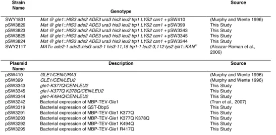
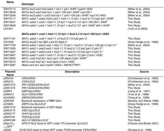
Yeast strains used in this study
Plasmids used in this study
Methods
Coordination of mitogen-activated protein kinase mating and cellular integrity pathways in Saccharomyces cerevisiae. Visualization of inositol phosphate-dependent mobility of Ku: depletion of the DNA-PK cofactor InsP6 inhibits Ku mobility. Chemical inhibition of Pho85 cyclin-dependent kinase reveals a role in environmental stress response.
Overexpression of the inositol phosphatase SopB in human 293 cells stimulates cellular chloride flux and inhibits nuclear mRNA export. Suppressors of a Saccharomyces cerevisiae pkc1 mutation identify alleles of the PTC1 phosphatase gene and of a novel gene encoding a putative leucine basic chain protein. The GLFG repeat region of the nucleoporin Nup116p interacts with Kap95p, an essential yeast nuclear import factor.
Characterization of poly(A)+ mRNA export in Saccharomyces cerevisiae during the winemaking process. Lack of expression of three isoenzymes of inositol 1,4,5-trisphosphate 3-kinase does not inhibit inositol pentakisphosphate and hexakisphosphate formation in mouse embryonic fibroblasts. Lethal congenital contracture syndrome type 2 (LCCS2) is caused by a mutation in ERBB3 (Her3), a modulator of the phosphatidylinositol-3-kinase/Akt pathway.
Nuclear export of the yeast mRNA-binding protein Nab2 is linked to a direct interaction with Gfd1 and to Gle1 function. Activation of the DExD/H box protein Dbp5 by the nuclear pore protein Gle1 and its coactivator InsP6 is required for mRNA export. The FG repeat asymmetry of the nuclear pore complex is dispensable for bulk nucleocytoplasmic transport in vivo.