E3 The Cellular Basis of the Antibody Response 108 E4 Antibody Responses in Different Tissues 112 Section F – The T Cell Response – Cell Mediated Immunity. Diseases and Deficiencies of the Immune System (sections J-L) - allergy, autoimmunity, and congenital and acquired immune deficiency.
P REFACE
To test your understanding of the subject, we have included 125 multiple choice questions with answers at the back of the book. Finally, we would like to thank our wives, Meriel, Annette and Sharon for their support and understanding during the preparation of the book.
K EY TO CELL SYMBOLS
A1 T HE NEED
Key Note
A2 E XTERNAL DEFENSES
Key Notes
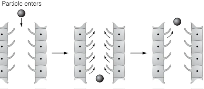
They are part of the body's innate defense mechanisms and are highly conserved across species, perhaps representing one of the most primitive defense mechanisms against microbes. When a particle is inhaled, it comes into contact with the cilia of the bronchial or nasal epithelium, which beat in an upward direction to a position where the particle can be coughed or sneezed out.
A3 I MMUNE DEFENSE
This regulation is maintained by cells and molecules of the immune system and externally by non-immune cells, tissues and their products (Section G). Having penetrated the external defenses, microbes come into contact with cells and products of the immune system and the battle begins.
A4 A NTIGENS
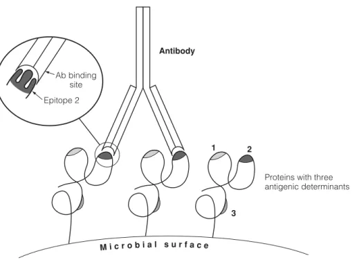
Although the linear sequence of residues in a molecule is equated with an antigenic determinant, the physical structures to which antibodies bind are primarily a consequence of the conformation of the molecule. Due to folding, residues on different parts of the molecule can be close together and can be recognized by the B cell receptor or antibody as part of the same determinant (Figure 1).
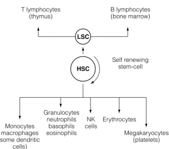
A5 H EMOPOIESIS – DEVELOPMENT OF BLOOD CELLS
CSFM-CSF
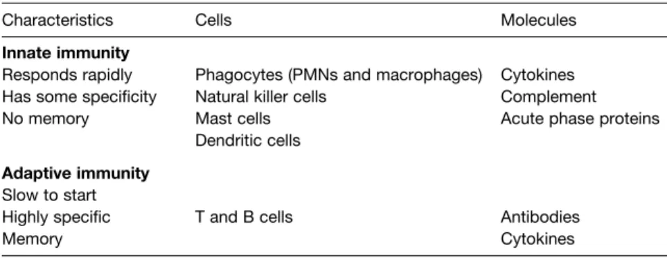
B1 C ELLS OF THE INNATE IMMUNE SYSTEM
They help in the attachment of the microbe to the phagocyte and also trigger the activation of phagocytosis. This release is almost immediate and is essential in the development of an acute inflammatory response (Topic B4).
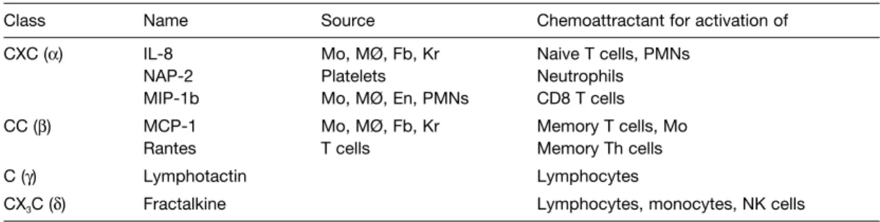
B2 M OLECULES OF THE INNATE IMMUNE SYSTEM
Most of the molecules that play a role in the innate immune system are also associated with adaptive immunity. Although the role each plays in immune defense and pathology is still being elucidated, it is evident 30 Section B – Cells and Molecules of the Innate Immune System.
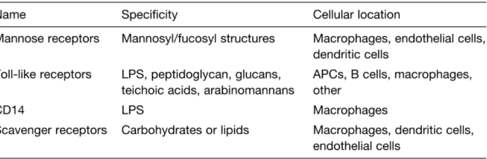
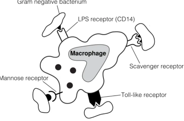
B3 R ECOGNITION OF MICROBES BY THE INNATE IMMUNE SYSTEM
Because the mannose receptor is expressed throughout the body on macrophages, it is probably one of the first of the innate receptors to interact with microbes (Fig. 1). Of particular interest is that these germline-encoded molecules of the innate immune system can not only signal the presence of a pathogen, but can also induce expression of co-stimulatory molecules and effector cytokines, thus preparing the cell for its involvement in development of the adaptive immune response.
B4 I NNATE IMMUNITY AND INFLAMMATION
The expression of these adhesion molecules is induced by proinflammatory cytokines released from macrophages. Source of inflammatory mediators due to microbial infection Source of initiating factors Mechanism of induction.

C1 L YMPHOCYTES
Most of the thymocytes generated in the thymus each day die by apoptosis with only 5-10% surviving. Each of the T cells produced in the thymus has only one specificity for which its antigen receptor is coded.
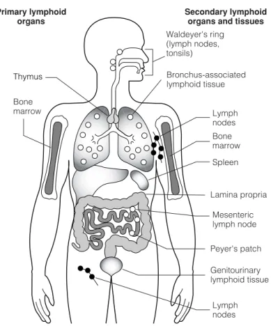
C2 L YMPHOID ORGANS AND TISSUES
The mucosa-associated lymphoid tissue (MALT), together with other lymphoid cells in sub-epithelial sites (lamina propria) of the respiratory, gastrointestinal and genitourinary tracts, constitutes the majority of lymphoid tissue in the body. These 'accessory' cells are important in the differentiation of the immigrating T-cell precursors and their 'education' (positive and negative selection) prior to their migration into the secondary lymphoid tissue (Topic F3).
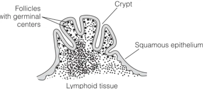
C3 M UCOSA - ASSOCIATED
LYMPHOID TISSUES
Inset: Here, B cells develop into IgA-secreting plasma cells, and IgA is transported across the epithelium into the intestinal lumen. Lymph complexes along the gastrointestinal tract; the volume of the rings indicates the relative amount of lymphoid tissue.
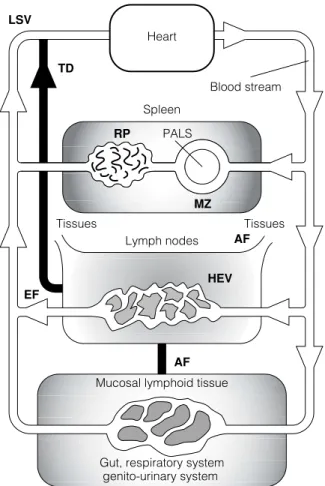
C4 L YMPHOCYTE TRAFFIC AND RECIRCULATION
These cell surface adhesion molecules attach to molecules (addressins) on specialized endothelial cells of the HEV. Movement of lymphocytes from the bloodstream via HEV. a) Lymphocytes (L) attach to the endothelial cells (EC) in the HEV through adhesion molecules.
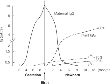
C5 A DAPTIVE IMMUNITY AT BIRTH
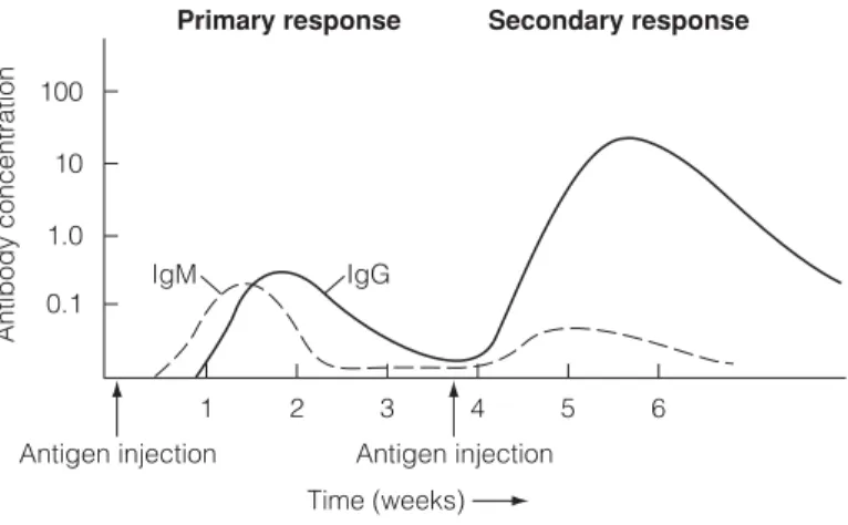
This protective IgG then decreases due to catabolism and disappears completely at about 6–8 months of age. De novo synthesis of IgM by the baby first occurs after 6-8 months of pregnancy and this is followed around birth by IgG and later IgA.
D1 A NTIBODY STRUCTURE
Most of the antibody molecules (the C-terminal three-quarters of the H-chain and the C-terminal half of the L-chain) are. This combined effect, avidity, results from synergy of the binding strengths of each binding site.
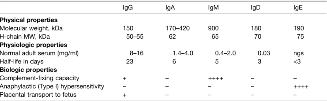
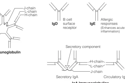
D2 A NTIBODY CLASSES
IgA This immunoglobulin is present in the serum as a 170 kDa protein with four polypeptide chains (two L and two H). In the blood, IgM consists of five four-chain units held together by disulphide bridges at the carboxy terminus of the µ chains (Fig. 1).
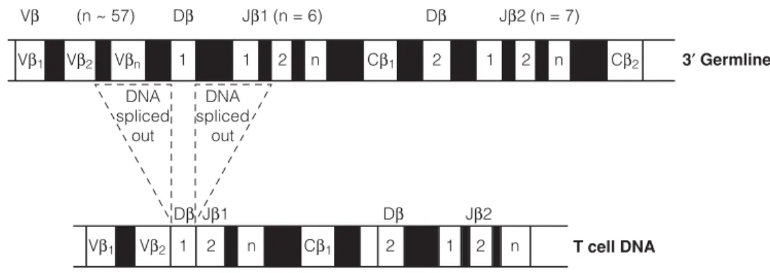
D3 G ENERATION OF DIVERSITY
Gene segments encoding the different parts of the V regions of antibodies rearrange during the pro-B cell stage. The exons encoding the V region of the H chain are: V segment (encodes approximately the first 102 amino acids), D segment (encodes 2-4 amino acids), and J segment (encodes the remainder approximately 14 amino acids in the H chain). V region).
D4 A LLOTYPES AND IDIOTYPES
Jerne (who shared the Nobel Prize with Kohler and Milstein in 1984) described a network theory which proposes that a series of idiotype-anti-idiotype reactions are partly responsible for regulation of the immune response (Topic G4).
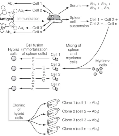
D5 M ONOCLONAL ANTIBODIES
Chimeric mAbs are created by replacing the murine light (L) and heavy (H) chain constant region genes with the corresponding human constant region genes. Humanized mAbs are created by inserting the gene sequences for each of the hypervariable (Hv) regions of the mouse antibody into the corresponding site in the genes for the L and H chains of a human antibody.
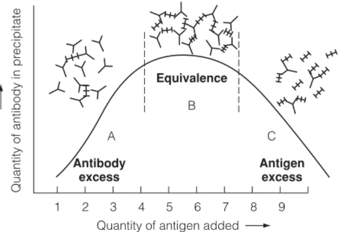
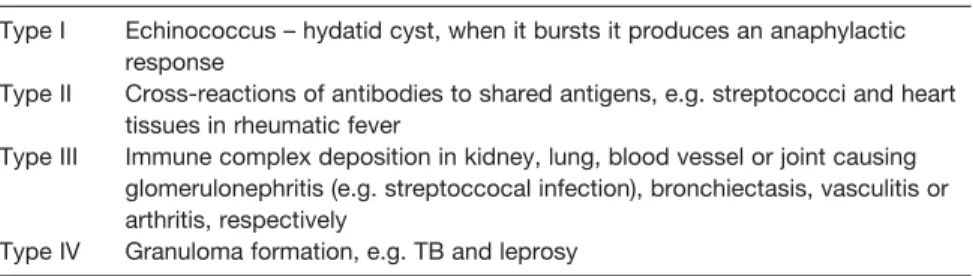
D6 A NTIGEN / ANTIBODY COMPLEXES
IMMUNE COMPLEXES )
Ag diffuses radially from the well into the gel and interacts with the Ab, forming a ring of precipitate, the diameter of which is related to the concentration of the Ag (Fig. 2). Holes are cut in the gel and filled with Ag which diffuses radially out of the well and interacts with the Ab in the gel.
D7 I MMUNOASSAY
Add Ab bound enzyme to different Ag determinants and wash Add substrate and read. An enzyme-linked Ab with a different determinant to the Ag is then added, followed by washing.
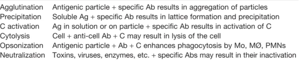
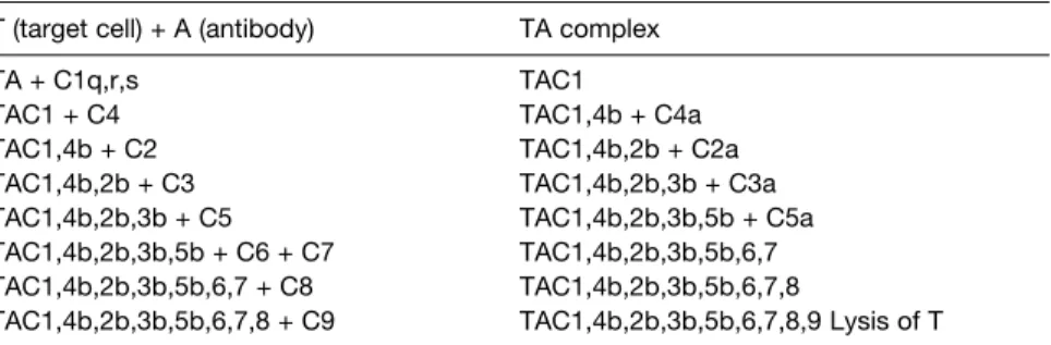
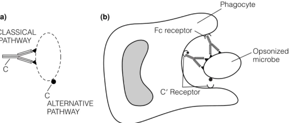
D8 A NTIBODY FUNCTIONS
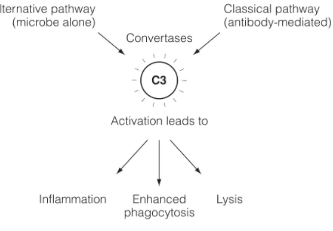
The Clq component of the Cl complex (C1q, C1r, C1s) then binds to the Fc regions of the cell-bound antibodies (Fig. 1). The cleavage of C3 into C3a and C3b is the single most important event in the activation of the complement system.
CO - RECEPTORS AND SIGNALING
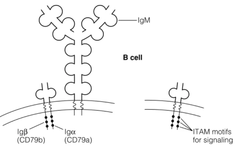
Molecules associated with the B cell receptor complex are expressed early in development to allow assembly of a functional antigen receptor on the surface of the B cell. This coreceptor complex includes CD21 (complement receptor 2, CR2), CD32 (receptor for the Fc region of IgG, FcγRIIB), and CD19 (signaling molecule).
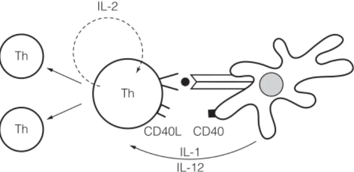
E2 B CELL ACTIVATION
This Th cell now triggers the activation of the B cell via the CD40 surface receptor (Fig. 4). Therefore, other molecules associated with these receptors with involvement of the B cell antigen receptor mediate the signaling.
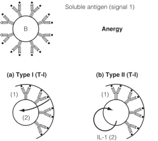
E3 T HE CELLULAR BASIS OF THE ANTIBODY RESPONSE
Natural or innate antibody to a wide variety of molecules is likely the result of the same phenomenon. Group A β-hemolytic streptococcal infections can lead to rheumatic fever due to the development of antibodies against the streptococcal determinants.
E4 A NTIBODY RESPONSES IN DIFFERENT TISSUES
LymphocytePlasma cell
Antigen is also collected by dendritic cells (Langerhans cells) in the dermis, processed and transported via the lymphatic system to the draining lymph nodes, where it is presented to T helper cells. B and T cells are concentrated in different parts of the lymph nodes, the B cells in the follicles and the T cells in the paracortical areas.
BlastFollicular
Some of these altered receptors cannot bind the same antigen that triggered them, so B cells with these receptors will not be able to be re-stimulated by that antigen. However, some receptors will be able to bind more strongly to this same antigen, which is often found bound to the surface of FDCs in the form of antibody/antigen complexes.
F1 T HE ROLE OF T CELLS IN
IMMUNE RESPONSES
Th2 cells are mainly involved in helping B cells develop into memory cells and antibody-producing plasma cells. Recognition of a peptide antigen by the TCR is not sufficient to activate cells, as accessory molecules are also required together with co-receptors involved in signaling events.
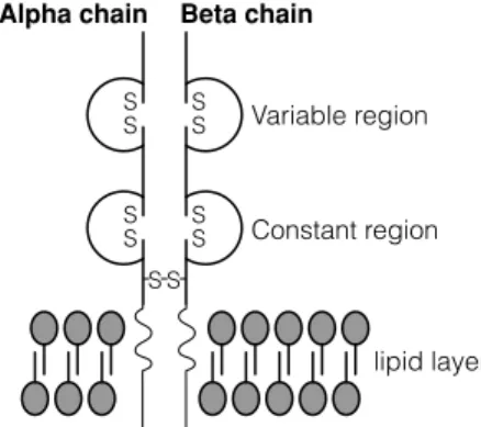
F2 T CELL RECOGNITION OF ANTIGEN
The peptide-binding domains of MHC class I and class II molecules are different for each allelic form of the MHC molecules. Comparison of the pathways used to generate peptides that bind to MHC class I and class II molecules.
F3 S HAPING THE T CELL REPERTOIRE
As in the case of immunoglobulin rearrangements, the expression of a complete α chain and a complete β chain by the T cell precludes further rearrangement (allelic exclusion). Similarly, in some developing T cells, γ and δ gene rearrangements occur, resulting in the T cell expressing γδ TCRs.
F4 T CELL ACTIVATION
Upon binding of the TCR, CD45 (an endogenous phosphatase) activates (by removing phosphates) two enzymes lck and Fyn that phosphorylate the ITAMs of the ζ-chains. Activation of phospholipase C-γ via the phosphatidylinositol pathway leads to activation of the transcription factors NFAT (nuclear factor of activated T cells) and NFκB and their translocation to the nucleus.
F5 C LONAL EXPANSION AND DEVELOPMENT OF EFFECTOR
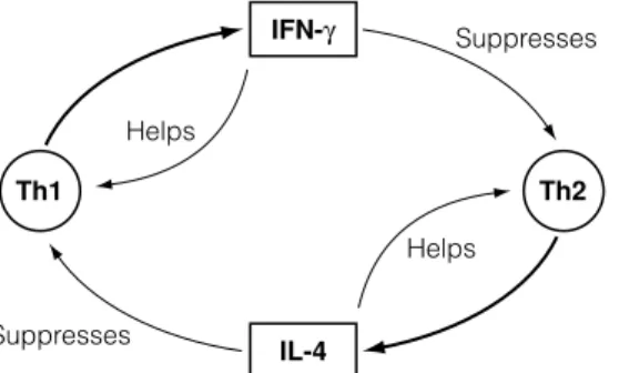
In contrast, Th0 cells are stimulated to become Th2 cells under the influence of IL-4 released by B cells and other cells (eg, mast cells). When a CTL interacts with a virus-infected cell, the granules move toward the part of the membrane near the point of contact with the target cell.
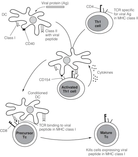
F6 C ELL - MEDIATED IMMUNITY IN CONTEXT
Dendritic cells (DC) conditioned (licensed) by Th1 cells present antigen to Tc via MHC class I. Interaction of specific Th1 cells with peptide (eg from a virus or tumor cell protein) presented in MHC class II on DC involves adhesion molecules such as. as well as binding of B7 to CD28.
G1 O VERVIEW
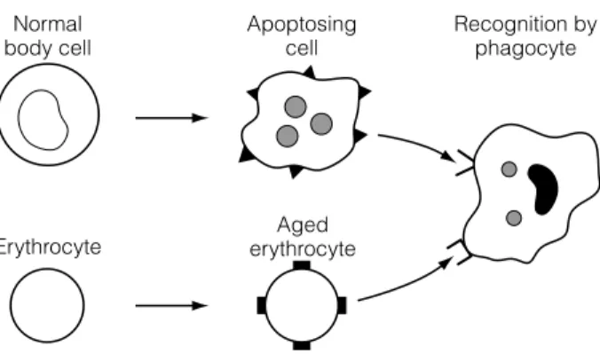
Phagocytic cells of the innate system, including macrophages and neutrophils, do not normally 'recognise' or phagocytize living self cells. They are prevented from killing the body's non-infected nuclear cells by a balance of signaling that includes killer activating receptors (KAR) and killer inhibitory receptors (KIR) that recognize molecules on self cells.
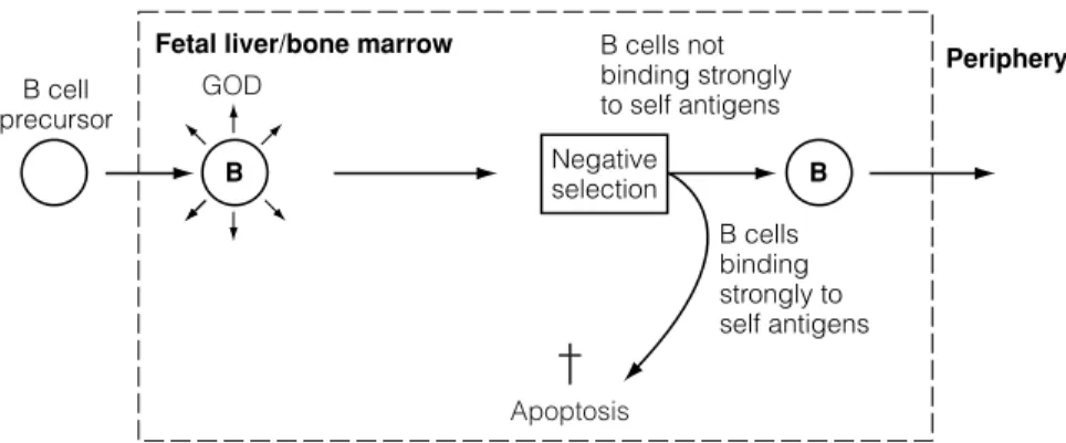
G2 C ENTRAL AND PERIPHERAL TOLERANCE
Most self-reactive lymphocytes cannot be removed in primary lymphoid organs for two reasons. First, many self-antigens are neither present in primary lymphoid organs nor delivered to them via the bloodstream.
G3 A CQUIRED TOLERANCE
The genetic background of the host can also influence the development of tolerance as the immune response is under the control of immune response genes (IR genes, Topics F2 and G5). The closer the composition and structure of the antigen is to self-antigens, the easier it is to induce tolerance.
G4 R EGULATION BY ANTIGEN AND ANTIBODY
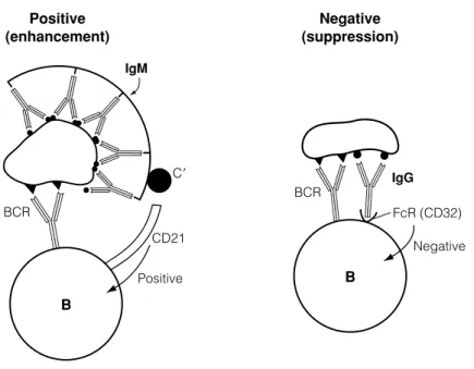
Similarly, the newborn's failure to respond to certain antigens may be related to the passive immunity acquired from the mother (Topic C5). This is probably the result of simultaneous interaction of the C3b component of complement with the CD21 molecule in the antigen receptor complex, which then transduces a positive signal to the B cell.
G5 G ENES , T HELPER CELLS ,
CYTOKINES AND THE
NEUROENDOCRINE SYSTEM
The activity of the immune system is influenced by other systems and perhaps most importantly by the neuroendocrine axis. Growth hormone and prolactin produced by the pituitary gland are also apparently capable of modulating the activity of the immune system.
H1 T HE MICROBIAL COSMOS
The immune responses to bacteria, viruses, fungi, protozoa and worms differ in the variety of defense mechanisms used. On the other hand, the immune response to certain infectious microbes can be more destructive than the offending pathogen itself, especially in persistent conditions (Section K).
H2 I MMUNITY TO DIFFERENT ORGANISMS
These intracellular bacteria evade immune system surveillance by surviving in host cells such as monocytes and macrophages. Since viruses require binding to host cells before they can replicate and cause infection, antibodies against the virus that prevent binding represent an important mechanism that protects against viral infection.
PEPTIDE
Diseases such as those caused by Schistosoma mansoni (schistosomiasis) and Wuchereria bancrofti (lymphatic filariasis, elephantiasis) thus represent major problems, especially in the developing world. When this happens, key phagocytic cells and immunity are compromised.

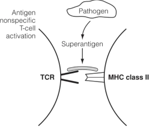
H3 P ATHOGEN DEFENSE STRATEGIES
Although this seems like an excellent strategy, it can lead to the development of autoimmune disease (Topic L3). Streptococcus pneumoniae and Candida, release large amounts of soluble antigens that bind to antibodies and block their binding to the microbe.
Vaccination
I1 P RINCIPLES OF VACCINATION
This passive transfer can be a disadvantage in that the presence of the maternal antibody inhibits effective immunization. The cell being targeted is determined by the presence of the foreign protein in association with MHC class I molecules.
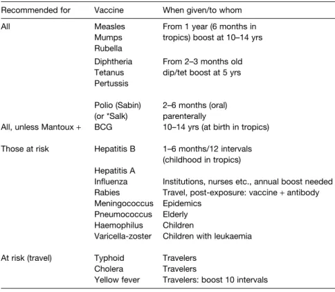
I2 I MMUNIZATION
The carbohydrate vaccines for pneumococcal, meningococcal and haemophilus infections are usually given at about 2 years of age because they respond poorly to polysaccharides before this age unless they are associated with protein components that can act to recruit T cell help to the development of anti-polysaccharide antibody, e.g. Adjuvant vaccines and live vectors have been used with some success to target the mucosal immune system.
I3 A NTIGEN PREPARATIONS
The last 20 years of studies of viral and bacterial pathogenesis have identified the components of the immune system that are protective against many infectious agents. The use of such effector cytokines is considered a useful adjunct to vaccination, as polarization of the immune system to a Th1 or Th2 response may be preferable in some cases, e.g.
I4 V ACCINES TO PATHOGENS AND TUMORS
Viral vaccines Vaccines against viruses that infect the respiratory tract (influenza, adenovirus), that infect the gastrointestinal tract (polio, rota), that infect the skin (yellow fever, La Crosse fever), and some that infect the reproductive tract (herpes) have been developed. In fact, some viral vaccines have been developed for viruses that are in the same genetic classification group as HIV.
J1 D EFICIENCIES IN THE IMMUNE SYSTEM
In other words, infections with certain microbes are a reflection of which components of the immune system are defective. A large number of specific congenital or acquired immune system abnormalities have been identified that contribute to the patient's susceptibility to recurrent infections.
J2 P RIMARY / CONGENITAL
INHERITED ) IMMUNODEFICIENCY
Deficiencies in the later complement components and in the regulatory molecules of the complement system also result in increased susceptibility to certain infections (by meningococci, e.g. Neisseria) or to inflammation, respectively. CD18, with CD11, forms the C3bi receptor (CR3) required to bind C3b and thus to bind opsonized microbes, a critical step in the cell's attempt to engulf a bacterium.