Introduction to retinal diseases and risuteganib
Leading causes of blindness
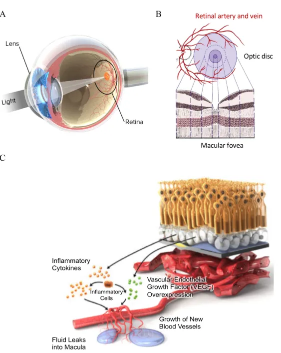
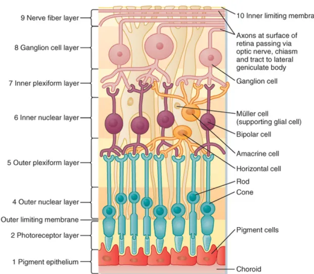
Both the blinding eye diseases, AMD and DME, affect the function of a fine structure in the retina, the macular region. The retina is a light-sensitive layer of tissue that lines the inner surface of the eye (Figure 1.1A).
Vascular endothelial growth factor (VEGF) and current therapeutics
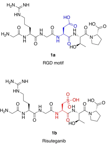
Risuteganib design principle and clinical observations
The clinical studies showed promising visual acuity gains and reduction in central macular thickness (Figure 1.3A) that were non-inferior to bevacizumab monotherapy. Optical coherence tomography (OCT) images showed that some of the subjects produce sub-normal final macular thicknesses consistent with the long-standing nature of their disease and the accompanying atrophy of the underlying retina (Figure 1.3B).
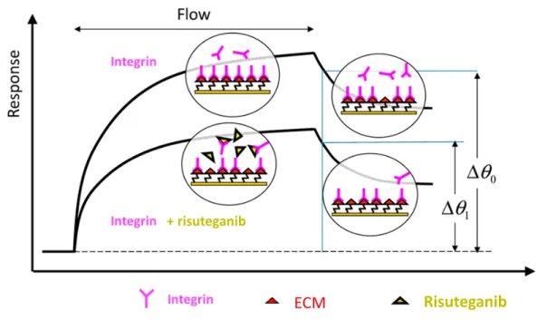
Integrin-binding mechanism of action overturned
This study became more challenging but also more exciting to uncover the MOA of risuteganib.
A journey of a thousand miles begins with a single step
In the early phase of the study, we only looked at blood vessel cells (human umbilical vein endothelial cell model to be more specific) based on the originally proposed MOA. The first step we took was to find the target cell type(s) in the retina. Higher resolution confocal micrographs further confirmed the staining of risuteganib-Cy5 on the RPE monolayer (Figure 2.3B). These results suggest that the risuteganib binding loci are relatively concentrated in the RPE layer of the retina.
Clinical pathology studies show that oxidative damage to the RPE layer is observed early in the development of the disease59. Based on the results in the stressed cell model, we began to form a hypothesis about the mechanism of action of risuteganib in RPE cells. The RPE layer has one of the highest ATP requirements in the retina67, consuming a significant portion of the energy needed to support photoreceptor functions.
Its function in the inhibition of PDK leads to increased oxidative phosphorylation with more ATP production. However, increase in growth factors in the tissue is only the symptom of the diseases. Induction of Differentiation by Pyruvate and DMEM in the Human Retinal Pigment Epithelial Cell Line ARPE-19.
Finding binding loci in the retina
Approach to find the binding loci
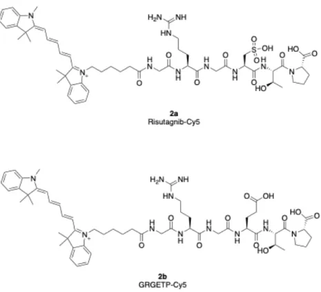
Synthesis of peptide-fluorophore conjugate
To visualize ligand-receptor binding sites in retinal tissue, peptide-fluorophore conjugates were designed and synthesized using a standard solid-phase peptide synthesis (SPPS) method. After solid-phase peptide synthesis (SPPS) of Risuteganib-Cy5 and GRGETP-Cy5 conjugates, the crude products were purified by preparative-scale high-performance liquid chromatography (HPLC).
Peptide-directed fluorescent staining in retinal tissue
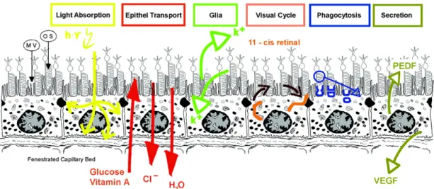
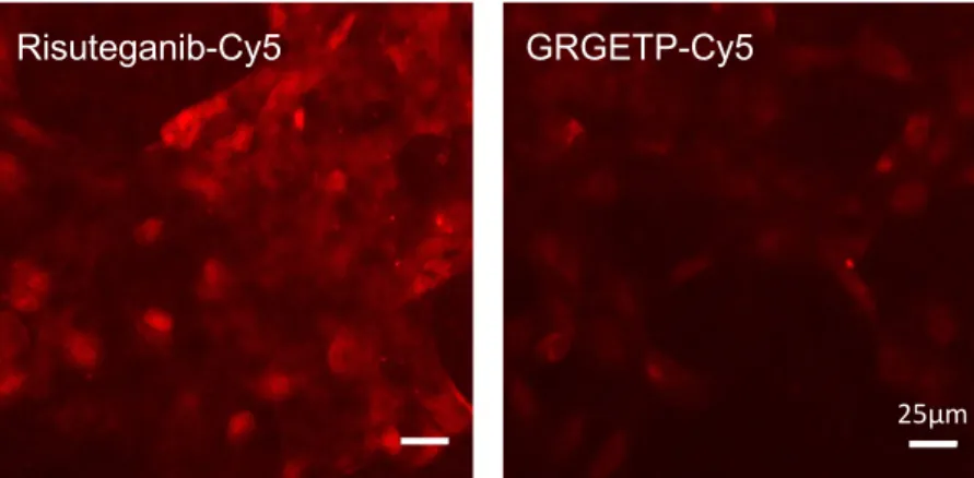
At concentrations below 5 μM, specific staining by Risuteganib-Cy5 relative to control peptide is observed in the RPE cell layer (B). Higher magnification confocal micrographs of mouse retinal sections confirm the preferential staining of risuteganib-Cy5 over the monolayer of RPE cells. The RPE was never thought to be a potential target because it is a far cry from risuteganib's original design. The RPE regulates the transport of ions, water, metabolic end products and nutrients between the choroidal circulation and the photoreceptor layer.
Finally, the RPE maintains a polarized environment where the retinal side of the RPE cell layer has appropriate complement growth factors and immunosuppressive factors, excl. proinflammatory cytokines and immune cells present in the choroid (Figure 2.4)45. Since the RPE layer interacts closely with photoreceptors on its retinal side and with endothelial cells and cells of the immune system on its choroidal side, it serves an important role as the blood-retinal barrier (BRB)46, which separates the inner space of the eye from the bloodstream.
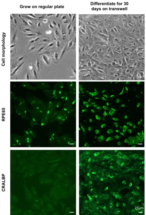
ARPE19 as a cell model for retinal pigment epithelium (RPE)
In comparison, 30-day differentiated ARPE19 cells grown in laminin-coated transwells exhibit roughly hexagonal shapes and express high levels of both protein markers. Based on the result of §2.3, we tested the properly differentiated ARPE19 cells in vitro for specific binding of risuteganib-Cy5 compared to control GRGETP-Cy5 (Figure 2.6). The result indicates that ARPE19 cells with proper differentiation can provide an in vitro model to study effects of risuteganib on RPE cells.
ARPE19 cells were immunostained with either mouse anti-human RPE65 mAb (Thermo Fisher Scientific) or mouse anti-human CRALBP mAb (Thermo Fisher Scientific), followed by alexa fluorine goat anti-mouse IgG (H+L) 488. Day -differentiated ARPE03 cells were washed with cold PBS three times and fixed with cold 4% paraformaldehyde for 10 minutes.
Risuteganib protects ARPE19 cells against oxidative stress
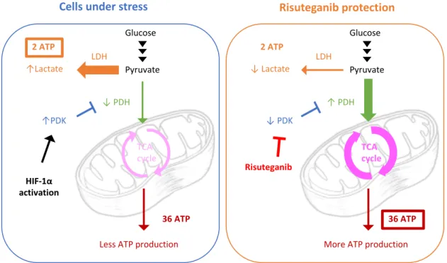
In the stressed or apoptotic cells, the mitochondrial membrane potential collapses and the cationic dye fluoresces green. The current study provides evidence that an increase in mitochondrial bioenergetics may be involved in the effects of the drug risuteganib. Nevertheless, PDK, the mitochondrial metabolic switch, has been suggested to represent a potential therapeutic target to protect against macular degeneration in the retina84.
Cells were then harvested, lysed and centrifuged to collect supernatant containing PDH enzyme in the buffer. PDH enzyme, the gatekeeper of oxidative phosphorylation in mitochondria, was activated by risuteganib in the ARPE19 cell model. Mitochondrial bioenergetic regulation improves mitochondrial function and protects mitochondria from oxidative stress, resulting in the protective effect on cells under disease conditions (§2.5).
Cells were treated with 300 μM risuteganib, 300 μM GRGETP peptide or no peptide as control in the culture medium for 24 h at 37oC. How the retina works: Much of the construction of an image takes place in the retina itself through the use of specialized neural circuits. The role of glycolysis and mitochondrial respiration in the formation and function of endothelial tip cells during angiogenesis.

Potential connection with RPE dysfunction in retinal disease
Risuteganib protects mitochondria through regulation of oxidative
Energetic requirements of the retina pigment epithelium (RPE)
Drug effect on mitochondrial respiration in RPE cells
The OCR value measured in the Seahorse XF cell mitostress test (Figure 3.2A) prior to the introduction of an active agent represents basal respiration, which showed the amount of oxygen consumption related to ATP production after oligomycin injection, and which after injection of FCCP indicates the amount of oxygen consumption related to the production of ATP. maximum mitochondrial respiration capacity of the cells. The measured ECAR value reflects lactate production and is used as an index for glycolysis (Figure 3.3A). We found that treatment with risuteganib significantly increased the rate of oxygen consumption in a dose-dependent manner, both in basal and maximal respiratory capacities (in a dose-dependent manner), and most interestingly, ATP-coupled respiration in the mitochondria, while there was no statistically significant effect on glycolysis.
The increase in ATP-related respiration indicates that risuteganib treatment increases ATP production in RPE mitochondria, which may be associated with the protective effect of risuteganib on RPE cells under oxidative stress. Measurements of oxygen consumption rate (OCR, measured by change in oxygen concentration) and extracellular acidification rate (ECAR, measured by change in pH) were obtained.

Potential inhibition of pyruvate dehydrogenase kinase
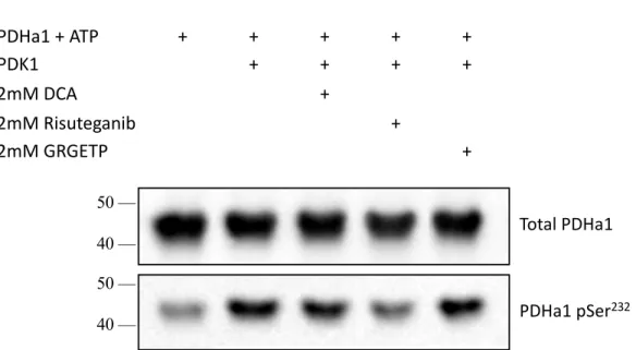
To test the hypothesis that risuteganib inhibits pyruvate dehydrogenase kinase (PDK), we measured the levels of phosphorylation on its substrate using recombinant proteins. PDK1, known to be inhibited by DCA in the mM range, was incubated with its substrate, pyruvate dehydrogenase E1 subunit alpha 1 (PDHa1), with or without 2mM inhibitor (DCA, risuteganib, GRGETP). Since DCA and risuteganib both have strong acid moieties (Figure 3.4A), we used assay buffer (25mM Tris or 25mM HEPES in the universal kinase kit) that provides sufficient buffering capacity to maintain pH between 7-8.
This study revealed that risuteganib can inhibit PDK, which induces more PDH in its active form. The membrane was first immunostained by anti-pyruvate dehydrogenase rabbit mAb (Cell Signaling Technology) and secondary antibody anti-rabbit IgG HRP (Thermo Fisher Scientific) to reveal total PDHa1.
Enzymatic effect in RPE cell model
PDH enzymatic activity in vitro is examined in 30-day-differentiated ARPE19 cells, by 24-h preincubation with 300μM risuteganib or 300μM control peptide, GRGETP, in cell culture medium before measurements. Inhibition of PDK by risuteganib results in increased PDH activity, which leads to an increase in oxidative phosphorylation metabolism and allows more ATP turnover (§3.2). Pyruvate dehydrogenase activity is determined by the assay kit from MilliporeSigma, using a coupled enzyme reaction, which results in a colorimetric product (450 nm).
Each sample was mixed 1:1 with the reaction mixture and the absorbance at 450nm was measured every 5min at 37oC. The PDH activity was calculated by the amount of NADH generated in the sample well, divided by reaction time and sample volume.

Current hypothesis on mechanism of action of risuteganib
Zach Shao, a former graduate student in our group, studied gene regulation by risuteganib in a mouse model that mimics the state of angiogenesis disease in the retina. Cancer cells rely heavily on glycolysis even when enough oxygen is present, known as the Warburg effect. If enough ATP is present in the mitochondria, it is possible to transform cancer cells into resting cells in the tissue.
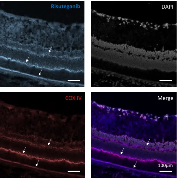
1μM risuteganib-Cy5 colocalizes with the mitochondria distribution in the retina, which showed a purple color when merged. Characteristics of patients losing vision after 2 years of monthly dosing in the phase III clinical trials of ranibizumab.
Summary and future work
Retracing our path along this journey
Looking back at the steps we took, I realized that our approach was unconventional for drug discovery. We're exploring where the drug effects are coming from by digging from the tissue level, to the cellular level, to the cellular organelle level, and finally to the molecular level, which was very exciting every time we went from one level to the next. It was a brave decision we made not to limit ourselves to the originally proposed MOA, as we found that the experimental evidence did not support it.
Now they are targeting the treatment of retinal diseases in a much larger patient population, dry AMD to be more specific, which was not even relevant to the originally proposed MOA of integrin targeting and angiogenesis inhibition. In my suggestions for future work, it would be recommended that the next step in colocalization experiments use immunostaining for PDK1 (more specific than COX IV) and use risuteganib-Cy5 (and GRGETP-Cy5 as a control) to examine further co-localization.
Research and clinical observations agree with the current hypothesis
It is interesting to note that there are some other layers of the retina that are rich in mitochondria90. Among more than 200 patients treated with risuteganib in Phase I and Phase II clinical trials, no drug toxicity directly related to the drug itself has been reported (adverse effects reported are all from the injections). Since there are several metabolic pathways in the cells, regulation of PDK will not leave cells to deal with only one pathway, but will provide more energy compared to others.
In the clinical trials, the drug efficacy of visual acuity improvement and macular thickness reduction lasted for at least three months after drug administration, much longer than typical small peptides. This can be explained by the drug effect of risuteganib that targets the mitochondrial metabolism, protects cells from stress conditions and restores the normal RPE and photoreceptor function, which maintains the homeostasis of the entire retina tissue.
Broader context of inhibitors of pyruvate dehydrogenase kinase
Recommendation for future work
Projected global prevalence of age-related macular degeneration and burden of disease for 2020 and 2040: A systematic review and meta-analysis. Retinal energetics requires control of retinal vascular supply in development and disease: Role of neuronal lipid and glucose metabolism. Distinct structural mechanisms for the inhibition of pyruvate dehydrogenase kinase isoforms by AZD7545, dichloroacetate, and radicicol.
HIF-1-mediated expression of pyruvate dehydrogenase kinase: a metabolic switch required for cellular adaptation to hypoxia. Pyruvate dehydrogenase kinase/lactate: a therapeutic target for neovascular age-related macular degeneration identified by metabolomics.