Mechanistic insights into anchoring the cytokinetic ring
By
Chloe Elizabeth Snider
Dissertation
Submitted to the Faculty of the Graduate School of Vanderbilt University
in partial fulfillment of the requirements for the degree of
DOCTOR OF PHILOSOPHY in
Cell and Developmental Biology February 28th, 2021 Nashville, Tennessee
Approved:
Matthew J. Tyska, Ph.D.
Ian G. Macara, Ph.D.
Alissa M. Weaver, M.D., Ph.D.
Matthew J. Lang, Ph.D.
Kathleen L. Gould, Ph.D.
ACKNOWLEDGEMENTS
The work described herein would not have been possible without the support of my mentors, peers, family, and friends. To the following I offer my sincerest thanks.
First, I would like to express my immense gratitude to my mentor, Dr. Kathy Gould, for providing the environment that fostered my success. Thank you for the lessons, the
encouragement, and the many celebrations. I acknowledge my collaborators and informal mentors: Dr. Lauren Jackson, Dr. Marjia Zanic, Dr. Melanie Ohi, Dr. Mintu Chandra, Dr. Göker Arpağ, and Dr. Scott Collier. Thank you for your support and for allowing me to try and think about new things. Thank you to my committee members, Dr. Matthew Tyska, Dr. Ian Macara, Dr. Alissa Weaver, and Dr. Matthew Lang for your input and encouragement. I would also like to thank Dr. Kerry Bloom, Dr. Elaine Yeh, and Dr. Andrew Stephens for providing my first
opportunity to conduct research at the University of North Carolina and for continuing to support my career.
I would like to thank the members of the Gould lab, past and present, for offering training, support, a sounding board, criticism when necessary, and a fun environment for
science. I also thank the Vanderbilt Department of Cell and Developmental Biology for excellent scientific and administrative support.
I acknowledge the financial support received from the predoctoral fellowship awarded by the American Heart Association and support from the Cellular, Biochemical, and Molecular Sciences training grant. Thank you to the Vanderbilt Cell Imaging Shared Resource for the access to fancy microscopes and resources.
Lastly, I thank my family and friends for their support and encouragement. Thank you to my parents, siblings, grandparents, and the extended Snider bunch for the words of
encouragement. Thank you to Michael Tackenberg, for your partnership in life and in science. I
iii
am also grateful to the friends I’ve made throughout my graduate school journey for all of the happy hours, the laughs, the cries, and the lasting friendships.
TABLE OF CONTENTS
ACKNOWLEDGEMENTS ... ii
LIST OF TABLES ... vi
LIST OF FIGURES ... vii
CHAPTER I. Introduction ... 1
Introduction to cytokinesis in Schizosaccharomyces pombe ... 1
Evidence for roles of plasma membrane lipid composition in cytokinesis ... 3
F-BAR domains: membrane anchors for actin-based structures ... 5
Summary ... 13
II. Phosphoinositide-mediated ring anchoring resists perpendicular forces to promote medial cytokinesis ... 15
Introduction ... 15
Results and discussion ... 16
Materials and methods ... 30
III. Analysis of the contribution of phosphoinositides to medial septation in fission yeast highlights the importance of PI(4,5)P2 for medial contractile ring anchoring ... 35
Introduction ... 35
Results and discussion ... 36
Materials and methods ... 49
v
IV. Fission yeast Opy1 is an endogenous PI(4,5)P2 sensor that binds to the
phosphatidylinositol 4-phosphate 5-kinase Its3 ... 52
Introduction ... 52
Results and discussion ... 54
Materials and methods ... 66
V. Opposite surfaces of the Cdc15 F-BAR domain create a membrane platform that coordinates cytoskeletal and signaling components for cytokinesis ... 72
Introduction ... 72
Results ... 75
Discussion ... 95
Materials and methods ... 97
VI. Conclusions and future directions ... 108
Conclusions ... 108
Future directions ... 108
REFERENCES ... 118
LIST OF TABLES
Table Page
5.1. Summary of crystallographic structure determination statistics ... 79
5.2. ITC Summary. ... 83
5.3. Protein localizations examined in cdc15-3A. ... 90
5.4. fPALM summary. ... 94
vii
LIST OF FIGURES
Figure Page
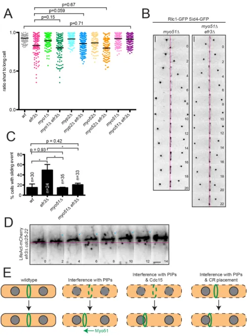
2.1. efr3∆ display off-center septa. ... 17
2.2. stt4 and ypp1 are essential in S. pombe. ... 18
2.3. CRs slide in efr3∆. ... 20
2.4. Cytokinesis kinetics are unperturbed in efr3Δ. ... 21
2.5. Localization of membrane-binding proteins in efr3∆. ... 24
2.6. The Stt4-complex and CR positioning machinery cooperate in septum positioning. ... 25
2.7. efr3∆ CR sliding events depend on Myo51. ... 28
2.8. N and C termini of Myo51 are necessary for efr3Δ CR sliding. ... 29
3.1. Analysis of septum placement in gene deletions of PIP regulators. ... 37
3.2. PIP kinases are important for medial division. ... 39
3.3. CRs form off-center in mutants of PI(3,5)P2 regulators. ... 41
3.4. Cells with large vacuoles form the CR off center. ... 42
3.5. Mutants of PI(4,5)P2 regulators display CR sliding. ... 44
3.6. Localization of membrane-binding proteins in its3-1. ... 45
3.7. Mutants with CR anchoring defects have altered PIP composition. ... 47
3.8. Mutants of PIP regulators have disrupted PIP composition. ... 48
4.1. Opy1 binds PI(4,5)P2-containing membranes. ... 55
4.2. Localization of Opy1 N- and C-terminal PH domains. ... 57
4.3. Opy1 and Its3 directly interact. ... 58
4.4. Opy1 does not regulate Its3 localization or kinase activity. ... 60
4.5. Opy1 does not influence localization of Stt4 or Its3, nor does it affect growth of mutants compromised for PI5-phosphatase activity. ... 61
4.6. Opy1 over-production results in cytokinesis defects. ... 63
4.7. Over-expression of Opy1 results in negative genetic interaction with efr3∆. ... 64
5.1. The Cdc15 F-BAR domain binds membrane and Cdc12 simultaneously. ... 78
5.2. Comparison of Cdc15 F-BAR domain with other F-BAR domain structures. ... 80
5.3. Cdc12 binds the cytosolic face of the Cdc15 F-BAR domain. ... 82
5.4. The Cdc15 F-BAR domain cytosolic face has functions in addition to scaffolding Cdc12. .. 84
5.5. Cdc15-3A is competent for membrane-binding, oligomerization, expression, and localization. ... 86
5.6. The Cdc15 F-BAR domain coordinates other binding partners required for proper CR architecture. ... 93
6.1. rgf1∆ and scd1∆ do not phenocopy efr3∆. ... 111
6.2. spcc594.01∆, which encodes a candidate Its3 interactor, has an off-center septation phenotype. ... 113
6.3. Like Cdc12, Pxl1 contains an N-terminal F-BAR binding motif. ... 116
1 Chapter I
Introduction
Introduction to cytokinesis in Schizosaccharomyces pombe
Cytokinesis is the last step in cell division in which two daughter cells physically separate. This process must be spatially and temporally coordinated with the division of the segregating DNA in order to avoid damage to the dividing genome. Failure during cytokinesis can lead to cell death, aneuploidy, or the formation of tetraploid intermediates, which can promote carcinogenesis (Fujiwara et al., 2005; Ganem et al., 2009; Hayashi and Karlseder, 2013; Krajcovic et al., 2011). However, differential regulation of cytokinesis is employed during development; for instance, cardiomyocytes purposefully inhibit cytokinesis to become
multinucleate (Clubb, 1984; Lacroix and Maddox, 2012; Li et al., 1996; Li et al., 1997).
Therefore, mechanistic studies of cytokinesis are informative for understanding both normal development and disease pathologies.
In eukaryotic cells, cytokinesis takes place in four general steps (reviewed in (Guertin et al., 2002)). The first is the selection of the division plane. Although different organisms employ a variety of mechanisms to specify the site of cytokinesis, the general strategy is that the
cytokinetic plane is established in a spatially distinct area from that of the dividing genome. The second step is the assembly of an actin- and myosin-based cytokinetic ring (CR). The CR is a contractile structure that must be built in close proximity to the plasma membrane (PM), so that during the third step, CR constriction, opposing membranes are brought together as the CR decreases in diameter. In the last step, abscission, the two daughter cells physically separate.
Cell division in Schizosaccharomyces pombe, or fission yeast, has been studied in detail for over two decades. S. pombe are rod-shaped unicellular yeast that grow by elongating their tips and divide by medial fission. Like many other eukaryotes, S. pombe assemble and constrict an actin- and myosin-based CR on the PM to facilitate cytokinesis (Pollard and Wu, 2010).
Because of the technical advantages of working with S. pombe, including short generation times and amenability to facile and precise genetic manipulation and large-scale biochemical assays, studies utilizing this organism have provided detailed insights into the structure and function of the CR (Cheffings et al., 2016; Goyal et al., 2011; Mangione and Gould, 2019; Pollard, 2010;
Rincon and Paoletti, 2016). As a result, S. pombe is perhaps the premier model for understanding the mechanistic basis of eukaryotic cytokinesis.
In S. pombe, the cytokinetic machinery typically clusters in precursor nodes. The medial assembly of nodes is promoted by the shuttling of the anillin-like protein Mid1 out of the nucleus in late G2 phase, promoting its accumulation on the cortex proximal to the central nuclear position (Sohrmann et al., 1996). Mid1 then recruits additional components such as myosin II, IQGAP Rng2, F-BAR protein Cdc15, and formin Cdc12 that assemble in punctae on the medial cortex of the cell (Wu et al., 2003). The nodes are then assembled into a coherent CR in the cell center using a “search-capture-pull-release” mechanism dependent upon actin polymerization and myosin activity (Vavylonis et al., 2008; Wu et al., 2006). The next step is CR maturation, during which the CR must remain stably anchored in the cell center while nuclear division occurs. Once the daughter nuclei have segregated to opposite ends of the cell, the CR then constricts and disassembles (Pelham and Chang, 2002; Pollard and Wu, 2010; Stachowiak et al., 2014). Concomitantly, cell wall material is synthesized and deposited behind the CR to form the division septum, or the new cell wall between the two daughter cells, providing the force necessary for CR constriction (Muñoz et al., 2013; Proctor et al., 2012).
Despite decades of work detailing how the division plane is selected and how the CR is assembled, our understanding of how the CR is attached to the PM is incomplete. S. pombe
3
build a medial CR early in mitosis (Kitayama et al., 1997; Marks and Hyams, 1985). How the CR remains anchored until constriction is not yet clear although several players have been
implicated. One is the paxillin-like protein Pxl1 that plays a role in CR anchoring and integrity evidenced by CR sliding and splitting during anaphase in pxl1∆ (Cortés et al., 2015; Ge and Balasubramanian, 2008). Another factor is the cell wall: loss of β(1,3)glucan (Muñoz et al., 2013) or loss of the integral membrane protein Sbg1 (Davidson et al., 2016; Sethi et al., 2016) result in CR sliding and instability, suggesting that cell wall-PM linkage is important for CR maintenance. Additionally, the microtubule post-anaphase array ensures a medial CR during a cytokinesis arrest (Pardo and Nurse, 2003). Another major factor is the essential F-BAR protein Cdc15: when cdc15 expression is repressed or Cdc15 oligomerization disrupted, the CR can slide along the PM and disassemble (Arasada and Pollard, 2014; McDonald et al., 2015). In each of these situations, CR sliding is observed in only a fraction of cells (Arasada and Pollard, 2014; Cortés et al., 2015; McDonald et al., 2015; Pardo and Nurse, 2003), indicating that multiple mechanisms contribute to CR anchoring. Consistent with this, combined repression of pxl1 with a hypomorphic cdc15 allele results in exacerbated CR sliding (Cortés et al., 2015). In addition to protein factors, the lipid composition of the PM has been implicated in promoting the integrity of cytokinesis as efr3∆, which encodes a scaffolding protein implicated in
phosphoinositide metabolism, displays defects in division site placement (Baird et al., 2008;
Chen et al., 2015). Although there is evidence that the lipid composition of the PM influences cytokinesis in other eukaryotic systems, this was an unexplored subject area in S. pombe, and part of my dissertation research investigated this as described in Chapters II-IV.
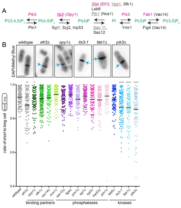
Evidence for roles of plasma membrane lipid composition in cytokinesis
Though phosphoinositides (PIPs) comprise ~5-10% of PM lipid species in mammalian cells (Wenk et al., 2003) and are important for cytokinesis (Echard, 2012), the mechanisms by which PIPs promote accurate cytokinesis are not fully defined.
Of the 7 PIP species found in the cell, only a subset have been implicated in cell division, including phosphatidylinositol-4-phosphate (PI4P) and phosphatidylinositol-4,5- bisphosphate (PI(4,5)P2). A role for PI4P in cytokinesis was revealed through studies focused on PI4-kinases, the enzymes that generate PI4P from phosphatidylinositol (PI). In Drosophila melanogaster spermatocytes, absence of the type IIIβ PI4-kinase (PI4KIIIβ) encoded by four wheel drive results in cytokinesis failure and multinucleate spermatids (Brill et al., 2000).
Similarly, the catalytic activity of S. pombe PI4KIIIβ Pik1 is essential for normal septation and abscission (Park et al., 2009). The role of PI4KIIIβ in human cells is less clear although one study showed that elevated levels of PI4KIIIβ activity and thus higher PI4P levels inhibit
cytokinesis, resulting in multinucleate cells (Rajamanoharan et al., 2015). Type IIIα PI4-kinases (PI4KIIIα) have also been implicated in cytokinesis. D. melanogaster cells lacking PI4KIIIα display cytokinesis defects that result in binucleate cells (Eggert et al., 2004). Additionally, deletion of the gene encoding Efr3, a non-essential PM scaffolding protein of the S. pombe PI4KIIIα Stt4, results in off-center septation, indicating a defect in cytokinesis (Chen et al., 2015). A role of PI4KIIIα in human cytokinesis has not been reported though the kinase and scaffolding machinery are conserved from yeast to humans (Baird et al., 2008; Chung et al., 2015).
Another PIP species implicated in modulating cell division is PI(4,5)P2, the most
abundant PIP species in the PM. PI(4,5)P2 is enriched at the division site of mammalian (Abe et al., 2012; Dambournet et al., 2011; Emoto et al., 2005; Field et al., 2005; Kouranti et al., 2006), S. pombe (Snider et al., 2017; Zhang et al., 2000) and D. melanogaster S2 (El Kadhi et al., 2011; Roubinet et al., 2011) cells. In addition, PI5-kinases that generate PI(4,5)P2 from PI4P localize to the division site in human (Emoto et al., 2005), D. melanogaster S2 (Roubinet et al., 2011) and S. pombe (Zhang et al., 2000) cells. PI(4,5)P2 can also be generated by PI3-
phosphatases acting on PI(3,4,5)P3 and PI3-phosphatases localize to the division site in
Dictyostelium discoideum and S. pombe (Janetopoulos et al., 2005; Mitra et al., 2004). In HeLa
5
and Chinese hamster ovary cells, depletion of PM PI(4,5)P2 results in separation of the PM from the actin cytoskeleton and cytokinesis failure (Field et al., 2005). It is hypothesized that proteins that mediate actin-PM adhesion may require PI(4,5)P2 for this function; candidates relevant to the process of cytokinesis include anillin (Liu et al., 2012; Sun et al., 2015) and other regulators of F-actin dynamics such as N-WASP and profilin (Higgs and Pollard, 2000; Machesky et al., 1990). However, the exact combination of molecules involved in CR detachment when PI(4,5)P2
isdepleted remains to be determined.
F-BAR domains: membrane anchors for actin-based structures
BAR domains: oligomeric bridges between membrane and actin networks
Bin/Amphiphysin/Rvs (BAR) superfamily proteins are critical components of membrane- linked processes in eukaryotic cells, including endocytosis, cytokinesis, and motility. These proteins are defined by the namesake BAR domain, which dimerizes to form a crescent-shaped bundle of six alpha-helices that directly binds membrane. BAR proteins are usually modular and contain additional domains that generally link components of the actin cytoskeleton to
membrane via the BAR domain (Carman and Dominguez, 2018; Nishimura et al., 2018;
Suetsugu et al., 2010).
BAR domains associate with membrane through positively charged residues on their concave faces that interact with negatively charged lipids, and they have the propensity to self- associate into oligomeric assemblies (Carman and Dominguez, 2018). The membrane-binding properties, tendency to oligomerize, banana-shape of the dimers, and localization of some BAR domains at sites of membrane curvature in cells led to the view that BAR domains are curvature generating and/or sensing modules. Indeed, the earliest in vitro studies of BAR domains
demonstrated their ability to tubulate when concentrated on membrane (Itoh and De Camilli, 2006). However, later studies on the Fes/Cip4 homology BAR (F-BAR) subfamily revealed that this description of BAR domain function may be over-generalized. The F-BAR subfamily is
distinguished from classical BAR domains based on sequence and structure; F-BAR dimers form a more extended and less curved shape compared to BAR dimers (Frost et al., 2007).
Although some F-BAR domains tubulate membrane in vitro, a subset of the family cannot, suggesting that membrane curvature generation is not a unifying function of the family (McDonald et al., 2015).
Like classical BARs, F-BAR proteins typically have additional domains. Many have at least one SH3 domain that recruits factors to link actin networks to membrane and/or promote actin polymerization locally (McDonald and Gould, 2016b). Though most known partners bind to the SH3 domains of F-BAR proteins, there is growing evidence that F-BAR domains themselves function as protein assembly platforms.
Here, I review the structures and oligomerization strategies of F-BAR domains and how these properties confer biological functions. Additionally, I discuss the emerging evidence that oligomerization allows F-BAR domains to polymerize protein interaction networks to promote the structure and function of actin-based structures, including the CR.
All F-BAR domains bind, but not all bend membrane
BAR domains have been described as generators, stabilizers, and/or sensors of membrane curvature. Membrane deformation activity was first reported for the BAR domain of human amphiphysin (reviewed in (Itoh and De Camilli, 2006)). Purified amphiphysin BAR
domain binds and deforms liposomes in vitro and electron microscopy (EM) analyses suggested that it self-associates to drive vesicle tubulation. Further evidence for amphiphysin’s ability to deform membrane was obtained by overexpressing its BAR domain in cultured cells, which resulted in plasma membrane tubulation. These two assays – adding purified BAR domains to liposomes and over-expressing BAR domains in cultured cells – became standard for assaying tubulation activity.
7
Further studies utilizing these assays revealed that multiple members of the BAR superfamily can generate membrane curvature at high concentrations. These include some members of the F-BAR subfamily (Frost et al., 2007; Henne et al., 2007; Shimada et al., 2007), leading to the idea that F-BAR domains may generate different degrees of membrane curvature than classical BARs. There is supporting evidence for this hypothesis, as F-BARs cluster in spatially separate areas when overexpressed simultaneously with classical BAR domains in cultured cells, and F-BARs generate wider tubules than classical BARs (Frost et al., 2008).
However, comprehensive examination of human F-BAR family members showed that some do not tubulate membranes at all, but simply bind it (McDonald et al., 2015). These F-BAR domains are also incapable of tubulating membranes when overexpressed in cultured cells.
If membrane deformation is a critical function of BAR domains, then why do some members, specifically a subset of the F-BAR family, lack this ability? Here, I discuss the developing understanding of the diversity of oligomerization strategies employed by F-BAR domains, and how this relates to in vitro tubulation activity, and more importantly, to in vivo function.
F-BAR oligomerization strategies inform function
F-BAR domains use varying modalities to oligomerize. For example, crystal packing contacts implicate residues at the tips of the F-BARs FBP17 and CIP4 in mediating
oligomerization (Shimada et al., 2007). Cryo-EM of CIP4 F-BAR assembled on membrane allowed for single-particle reconstruction, which provided further evidence of tip-to-tip assembly of dimers in vitro (Frost et al., 2008). This analysis also revealed that lateral contacts can occur between neighboring F-BAR dimers, facilitating membrane tubulation in vitro and resulting in spiral filaments with the concave faces of the assembled F-BAR domains contacting membrane.
Disrupting CIP4 F-BAR domain tip-to-tip or lateral oligomerization reduces tubule formation in standard assays (Frost et al., 2008; Shimada et al., 2007).
The Schizosaccharomyces pombe Imp2 F-BAR domain crystalized as a dimer of dimers, with residues at the tip of one dimer contacting the core of a neighboring dimer, resulting in a helical assembly when extended mathematically (McDonald et al., 2016). Similar to the orientation of CIP4 F-BAR dimers on membrane tubules, the concave membrane-binding surfaces of the Imp2 F-BAR oligomer face the interior of the helical assembly. This
oligomerization strategy is consistent with Imp2 F-BAR domain’s ability to tubulate vesicles in vitro, and mutation of oligomerization residues renders Imp2 tubulation-deficient (McDonald et al., 2016). It is logical that F-BAR domains like CIP4 and Imp2 that can assemble into spiral structures are able to tubulate membrane in vitro.
However, F-BAR domains also assemble on flat membranes, and this requires different oligomerization strategies. FBP17 F-BAR dimers make tip-to-tip contacts when assembled on a flat membrane in vitro and imaged by EM (Frost et al., 2008). Intriguingly, in this instance, F- BAR domains contact the membrane with the lateral side of the crescent, rather than the canonical membrane-binding surface, occluding lateral oligomeric contact sites observed on membrane tubules. Although this flat assembly has not been detected in vivo, it may be relevant to FBP17 action at lamellipodia membranes (Tsujita et al., 2015). In another example of sheet- like assembly, the mammalian GAS7 F-BAR domain forms flat filamentous oligomers (FFO) through lateral interactions of the F-BAR dimers on monolayers in vitro (Hanawa-Suetsugu et al., 2019). Similar to the orientation of FBP17 F-BAR domains observed on a flat membrane, the configuration of GAS7 dimers in the FFO suggests that residues on the lateral side of the GAS7 F-BAR contact the membrane. This oligomerization strategy of the GAS7 F-BAR domain into a flat sheet is consistent with its lack of tubulation activity (Hanawa-Suetsugu et al., 2019;
McDonald et al., 2015).
The S. pombe Cdc15 F-BAR domain forms tip-to-tip linear oligomers as visualized by EM, mediated by electrostatic interactions between reciprocally charged residues at dimer tips (McDonald et al., 2015; Roberts-Galbraith et al., 2010). A strictly linear assembly of Cdc15 F-
9
BAR domains is consistent with their inability to deform membrane in vitro and also with the function of Cdc15 at a flat membrane along the cell wall of rod-shaped S. pombe (Fankhauser et al., 1995). D. melanogaster Nervous wreck (Nwk) F-BAR domain is also non-tubulating and assembles tip-to-tip on monolayer membranes in vitro. Nwk dimers come together in a V-shape, resulting in zig-zag assemblies as visualized by EM (Becalska et al., 2013). This arrangement may explain the scalloped shapes of Nwk-bound membranes in vitro and when Nwk is
overexpressed in S2 cells (Becalska et al., 2013). In the Cdc15 and Nwk F-BAR examples, the evidence suggests that the concave faces are utilized for membrane-binding when assembled in an oligomer.
Mammalian GAS7 and S. pombe Cdc15 exemplify how F-BAR domain structure and oligomerization strategy are connected to biological function. Consistent with the shallow curvature and sheet-like assembly of the GAS7 F-BAR domain, GAS7 localizes to the flat membrane at the base of the phagocytic cup in macrophages as revealed by super-resolution microscopy (Hanawa-Suetsugu et al., 2019). Furthermore, mutations that disrupt GAS7 F-BAR domain oligomerization inhibit phagocytosis, indicating the functional requirement for this GAS7 oligomerization mechanism. The mammalian FCHSD2 F-BAR dimer also has shallow curvature and localizes to the flat membrane at the base of clathrin-coated pits during endocytosis
(Almeida-Souza et al., 2018b). It is unknown what oligomeric FCHSD2 looks like in this context, but it will be interesting to see if FCHSD2 also assembles into flat sheets like the GAS7 F-BAR domain or in zig-zags like its orthologue Nwk (Becalska et al., 2013). S. pombe Cdc15 is a scaffolding protein of the cytokinetic ring, linking protein partners to the plasma membrane. In vitro studies demonstrated that oligomerization allows high avidity Cdc15 membrane-binding, suggesting that oligomerization allows efficient scaffolding of the cytokinetic ring in cells (McDonald et al., 2015). In support of this, Cdc15 oligomerization mutants are more dynamic and less stably associated with the membrane compared to wildtype and display cytokinetic ring instability. The shallow curvature of the Cdc15 F-BAR dimer, the tip-to-tip oligomerization
strategy, and the inability to tubulate membrane in vitro are all consistent with function at a flat membrane in vivo.
At flat membranes, instead of driving membrane curvature, F-BAR domain
oligomerization may function to generate high avidity membrane-binding and concentrate binding partners at the membrane. For instance, Cdc15 scaffolds a network of proteins via its F- BAR and SH3 domains that promote completion of cytokinesis (Ren et al., 2015; Roberts-
Galbraith et al., 2009). In mammalian cells, oligomerized F-BAR domains likely function similarly to create a high-density of binding modules for partners that promote actin polymerization for endocytosis, as well as podosome and phagocytic cup formation (Almeida-Souza et al., 2018b;
Ho et al., 2004; Takano et al., 2008; Tsuboi et al., 2009).
While it is now appreciated that F-BAR domains oligomerize in a variety of modes, the next frontier is linking the structure of F-BAR dimers and their higher-order oligomeric
assemblies to functions in vivo. For example, it is unclear how helical structures formed by CIP4 and FBP17 F-BAR domains in vitro relate to their functions at flat membranes, such as those of lamellipodia (Tsujita et al., 2015). While the F-BAR domain of PACSIN/Syndapin can generate membrane tubules in vitro, crystal lattice contacts suggest that PACSIN1 assembles into sheets with contacts between the tip of one dimer and the core of another, with the membrane binding face of each dimer facing the same direction toward the membrane (Wang et al., 2009). It is unclear how such an assembly could generate membrane tubulation, and it is unknown if this flat assembly is utilized in vivo (Wang et al., 2009). In the case of S. pombe Imp2, although the F-BAR domain forms helical assemblies and tubulates membrane in vitro, structure-function analysis showed that mutations that disrupt oligomerization have little consequence for Imp2’s function in cytokinesis in vivo (McDonald et al., 2016). Interestingly in this regard, it has
emerged in recent years that membrane crowding of any protein on a membrane can generate curvature (Snead et al., 2017), and membrane-anchored GFP is sufficient to drive GUV fission (Steinkühler et al., 2020). These biophysical studies illustrate that caution is warranted when
11
drawing conclusions about the physiological relevance of membrane deformation from in vitro assays performed with supra-physiological concentrations of F-BAR domains.
F-BAR domains as protein interaction modules
F-BAR domains are typically linked to other domains that have protein interaction and/or enzymatic activities that in turn link F-BAR proteins to the actin cytoskeleton (reviewed in
(McDonald and Gould, 2016b; Suetsugu and Gautreau, 2012)). Hence, F-BAR domains might be viewed simply as membrane-targeting modules, which merely localize other domains to their site(s) of action. Indeed, substitutions of an essential F-BAR domain in yeast with various yeast and human F-BAR domains support cell viability, suggesting that F-BAR domains are
remarkably interchangeable membrane-binding modules (Mangione et al., 2019). However, there is mounting evidence that a key F-BAR domain function is mediating protein-protein interactions in addition to protein-membrane interactions, and these dual properties can allow F- BAR domains to form interaction platforms that link complex actin-based structures to
membrane.
The list of reported F-BAR domain binding partners is expanding, as is insight into the functional consequences of such interactions in different biological contexts. For some F-BAR domains, a partner is required for robust localization to a site of action within the cell. An example of this type of relationship is the cooperation between S. pombe cytokinetic F-BAR protein Rga7 and coiled-coil protein Rng10 (Liu et al., 2019). Although the Rga7 F-BAR domain binds membrane in vitro, it does so with low affinity unless in complex with Rng10, and in cells Rga7 does not localize to the cell division site without Rng10, or vice-versa.
In platelets and megakaryocytes, the F-BAR domain of PACSIN2 binds the actin-binding protein Filamin A, and deletion or mutation of Filamin A disrupts PACSIN2 localization (Begonja et al., 2015). In epithelial cells, PACSIN2 F-BAR domain binds to polycystin-1 to facilitate its localization at lamellipodia membranes (Yao et al., 2014). Similarly, the cytoskeleton-associated
protein pyrin binds the PSTPIP1 F-BAR domain (Shoham et al., 2003). When expressed together in COS cells, pyrin directs PSTPIP1 to inflammasomes, while PSTPIP1 does not localize there on its own (Waite et al., 2009). In these cases, testing if F-BAR affinity for membrane is modulated by interacting partners would provide important mechanistic insight.
Binding partners also regulate F-BAR protein properties other than membrane binding.
The D. melanogaster Nwk promotes membrane remodeling at synapses by activating WASp and directing actin assembly. Nwk’s membrane and actin remodeling activities are autoinhibited by interactions between its N-terminal F-BAR and C-terminal SH3 domains (Stanishneva- Konovalova et al., 2016). Full activation of Nwk requires association with membrane, WASp, and Dap160/intersectin, a partner that binds both the F-BAR domain and a C-terminal SH3 domain of Nwk (Del Signore et al., 2020). Multi-step activation of Nwk may ensure it promotes actin assembly only at the appropriate place and time. It will be interesting to determine if other F-BAR proteins are regulated in such a multi-factor manner.
In a reciprocal mechanism to that described for Nwk, some F-BAR domains influence the activity or localization of their partners. For example, during cytokinesis in Saccharomyces cerevisiae, the Hof1 F-BAR domain binds and inhibits Chs4, an chitin synthase III activator, which synthesizes the secondary septum at the division site (Oh et al., 2017). Because Chs4 arrives at the bud neck during the early stages of cytokinesis, inhibition by the Hof1 F-BAR domain prevents premature septum synthesis (Oh et al., 2017). The Hof1 F-BAR domain also inhibits actin cable formation by binding the formin Bnr1 to block actin nucleation (Garabedian et al., 2018). This regulation ensures normal actin cable morphology and secretion (Graziano et al., 2014).
F-BAR domains also serve to anchor proteins directly to membrane. In S. pombe, the Cdc15 F-BAR domain binds the cytokinetic formin Cdc12, promoting its recruitment to the division site where it nucleates the F-actin of the cytokinetic ring (Willet et al., 2015a). Acting as a bridge between the cytoskeleton and the PM through F-BAR domain binding to Cdc12 is likely
13
a key mechanism by which Cdc15 anchors the cytokinetic ring. The human F-BAR protein PSTPIP1 similarly localizes to the plasma membrane of the cytokinetic cleavage furrow (Spencer et al., 1997), but scaffolding roles for the PSTPIP1 F-BAR domain are yet to be explored in this context.
In some cases, the F-BAR domain residues that mediate protein-protein interactions have been identified, but there does not appear to be a unifying mechanism of F-BAR domain–
protein engagement. The PACSIN2 F-BAR domain associates with F-actin in vitro, but the residues implicated in this interaction are on the concave side of the F-BAR domain, which is also the membrane-binding face (Kostan et al., 2014). This suggests that the PACSIN2 F-BAR domain would be unable to bind membrane and F-actin simultaneously, and the physiological function of this interaction is unclear. For the Filamin A–PACSIN2 association, the residues important for binding are at the tips of the F-BAR dimer (Begonja et al., 2015). Because membrane binding is generally coincident with F-BAR dimer tip-to-tip oligomerization, determining if PACSIN2 can oligomerize on membranes in vivo and bind Filamin A
simultaneously will be critical to understanding its physiological function. Therefore additional studies are required in order to understand the molecular basis of F-BAR domain interaction with their partners and how this promotes linkage of actin-based structures to membrane.
Summary
In this work, I have provided new insights into two aspects of CR anchoring in S. pombe:
the PIP composition of the PM and the scaffolding roles of the Cdc15 F-BAR domain. In
Chapter II, I discovered that the PIP composition of the PM promotes medial CR anchoring after its formed, representing a distinct mechanism for CR anchoring from those previously
described. In Chapter III, through a combination of genetics, live-cell imaging, and development of lipid biosensors for S. pombe, I determined that PI(4,5)P2 is the specific PIP species that promotes CR anchoring. In Chapter IV, I challenged the field’s view of the function of the dual
PH domain-containing protein family by demonstrating that a family member in S. pombe, Opy1, does not regulate the kinase activity of the PI5-kinase Its3. However, Opy1 specifically binds PI(4,5)P2 and can sequester it and cause CR anchoring defects when over-expressed. In Chapter V, I explored how the Cdc15 F-BAR domain serves as a CR anchor by examining the structure and mechanism of its interaction with the formin Cdc12. Together these studies advance our understanding of how the CR is anchored to promote the fidelity of cell division.
15 Chapter II
Phosphoinositide-mediated ring anchoring resists perpendicular forces to promote medial cytokinesis1
Introduction
To divide, many eukaryotes assemble and constrict an actin- and myosin-based contractile ring (CR) (Cheffings et al., 2016) that is anchored to the plasma membrane (PM) (Gould, 2016). Despite decades of work on how the division plane is selected and the CR assembles (Bohnert and Gould, 2011; Goyal et al., 2011; Lee et al., 2012; Pollard and Wu, 2010; Rincon and Paoletti, 2016), mechanisms of CR-PM anchoring remain incompletely understood.
Here we define a mechanism distinct from those previously reported which anchors CRs during anaphase, explaining why cells lacking S. pombe efr3 divide asymmetrically (Chen et al., 2015). In S. cerevisiae, Efr3 and its partner Ypp1 form a platform at the PM for Stt4, a PI4- kinase, which regulates PIP composition and supports endocytosis (Baird et al., 2008).
Similarly, human homologues of Efr3 and Ypp1 (EFR3A/B and TTC7) scaffold a PI4-kinase type-IIIα at the PM (Nakatsu et al., 2012).
We find that S. pombe lacking properly positioned Stt4 have altered PM PIPs. These cells form CRs in the cell middle that can then slide towards one end in a directed manner. CR
1 Adapted from Chloe E. Snider*, Alaina H. Willet*, Jun-Song Chen, Göker Arpağ, Marija Zanic, Kathleen L. Gould (2017). Phosphoinositide-mediated ring anchoring resists perpendicular forces to promote medial cytokinesis. Journal of Cell Biology 216(10):3041-3050.
sliding in efr3∆ requires the type V myosin Myo51, indicating for the first time that the CR is subject to perpendicular forces in addition to being under constrictive tension (Proctor et al., 2012), and that these forces can dislodge the CR from the cell center. Thus, PM PIP
composition contributes to CR anchoring, promoting proper septum positioning and ensuring accurate genome segregation.
Results and discussion
We previously observed that a high percentage of efr3∆ divide asymmetrically (Figure 2.1A), sometimes resulting in a “cut” phenotype (Chen et al., 2015). To determine if this is due to altered PM PIP composition, we first determined if Efr3 co-localizes with Stt4 and Ypp1 in S.
pombe by analyzing the localization of three distinct pairs of these proteins tagged with mCherry and mNeonGreen (mNG) or GFP. Each pair co-localized on the PM in a punctate pattern
(Figure 2.1B), resembling the PI kinase patch localization of the S. cerevisiae Stt4 complex (Baird et al., 2008). The PM enrichment of S. pombe Stt4 and Ypp1, but not their levels,
depended on Efr3 (Figure 2.1C). Efr3 co-immunoprecipitated with Ypp1 (Figure 2.2A) and both Ypp1 and Stt4 were identified in an Efr3-TAP by LC-MS/MS analysis (Figure 2.2B) indicating that these proteins associate in S. pombe. To further study the influence of Stt4 on septa positioning, we attempted to construct stt4∆ and ypp1∆, but found that these genes are
essential (Figure 2.2C-D). However endogenous GFP-stt4 displayed off-center septa indicating that although GFP-Stt4 localizes correctly to the PM, it is likely to be a hypomorphic allele (Figure 2.1A-B). These data establish that proteins of the Stt4 complex are important for medial division.
17
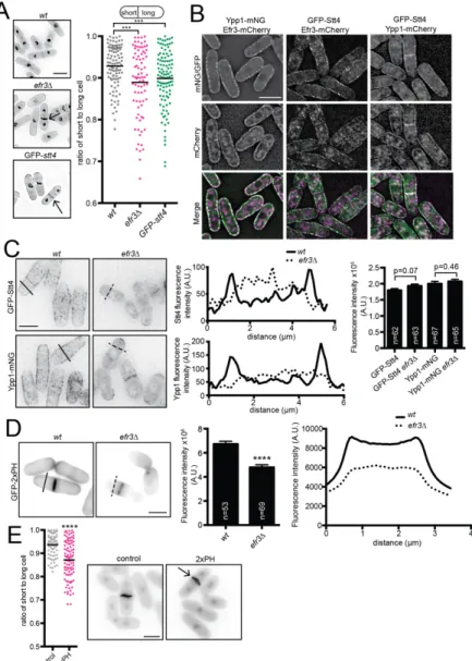
Figure 2.1. efr3∆ display off-center septa. (A, left) Representative images of indicated strains stained for cell wall and nuclei. Arrows indicate off-center septa. (A, right) Schematic of centered and off-centered septa classification and quantification. Individual points
represent the ratio of the length of the short to long daughter cell at septation and black bars denote mean. (B) Efr3-mCherry, Ypp1-mNeonGreen (mNG), Ypp1-mCherry and GFP-Stt4.
Images are of a single medial Z-slice. (C, left) GFP-Stt4 or Ypp1-mNG in either (wt) or efr3∆.
Images are Z-projections and are not identically scaled. (C, middle) Line scans of the
fluorescence intensity of the solid (wt) and dotted (efr3∆) black lines shown in left panels. (C, right) Whole-cell fluorescence intensity of the indicated strains. (D, left) Representative images of GFP-2xPH(PLCδ) localization in either wt or efr3∆. (D, middle) Quantification of the fluorescence intensity at the division septum. (D, right) Line scans of the fluorescence intensity of the solid (wt) and dotted (efr3∆) black lines. Data in graphs are from three biological replicates. (E) Representative images (left) and quantification (right) of cells stained for cell wall and nuclei. Arrow indicates off-center septum. ***p ≤ 0.001 ****p ≤ 0.0001; (A) one-way ANOVA or (C,D,E) Student’s t-test. Error bars represent SEM. Scale bars = 5 µm.
Figure 2.1. efr3∆ display off-center septa. (A, left) Representative images of indicated strains stained for cell wall and nuclei. Arrows indicate off-center septa. (A, right) Schematic
Stt4 phosphorylates PM PI to produce PI4P, which can be further modified to PI(4,5)P2. Therefore, disruption of PI4-kinases results in a reduction of both PI4P and PI(4,5)P2 (Audhya and Emr, 2002; Baird et al., 2008; Nakatsu et al., 2012). The PI(4,5)P2 sensor GFP-2xPH(PLCδ) (Stefan et al., 2002) was reduced at the cell cortex and the division site in efr3∆ compared to (Figure 2.1D), indicating that PIP PM abundance is reduced in efr3∆. In accord, overexpression of GFP-2xPH(PLCδ), expected to sequester PI(4,5)P2, resulted in off-center septa (Figure 2.1E).
We next addressed how off-center septa arise in efr3∆. Because septum position is dictated by CR position (Marks et al., 1986; Marks and Hyams, 1985), we reasoned that either
Figure 2.2. stt4 and ypp1 are essential in S. pombe. (A) Anti-HA immunoblot (IB; top) and anti-Flag immunoblot (middle) of anti-HA and anti-Flag immunoprecipitations (IPs) from the indicated strains. Anti-CDK was used as a loading control (bottom). (B) Proteins affinity purified with efr3-TAP csp1–191 detected by mass spectrometry. TSC, total spectral count. (C and D) Diploids heterozygous for ypp1::ura+ (C) or stt4::ura+ (D) were sporulated on glutamate plates, and tetrads were picked and allowed to germinate on YE at 29°C. Pictures of plates were taken after 5 d of growth at 29°C. All viable colonies were susceptible to growth media without uracil.
Figure 2.2. stt4 and ypp1 are essential in S. pombe. (A) Anti-HA immunoblot (IB; top) and anti-Flag immunoblot (middle) of anti-HA and anti-Flag immunoprecipitations (IPs) from the indicated strains. Anti-CDK was used as a loading control (bottom). (B) Proteins affinity purified with efr3-TAP csp1–191 detected by mass spectrometry. TSC, total spectral count. (C and D) Diploids heterozygous for ypp1::ura+ (C) or stt4::ura+ (D) were sporulated on glutamate plates, and tetrads were picked and allowed to germinate on YE at 29°C. Pictures of plates were taken after 5 d of growth at 29°C. All viable colonies were susceptible to growth media without uracil.
19
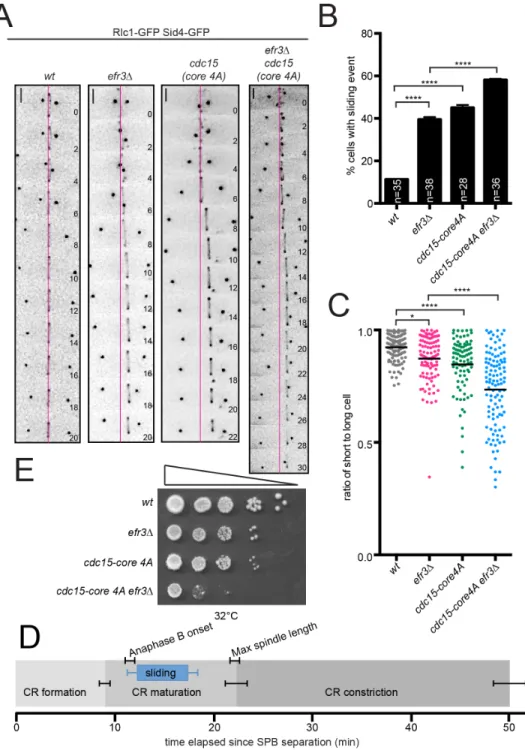
the CR forms off-center or it slides from its original medial position. To distinguish between these possibilities, we imaged wildtype and efr3∆ expressing CR (Rlc1-GFP) and spindle pole body (Sid4-GFP) markers. In wildtype the CR formed in the cell center and maintained this position during cytokinesis (Figure 2.3A). In efr3∆, the CR formed in the cell center, but slid from its original position while remaining perpendicular to the cell’s long axis (Figure 2.3A-C).
Temporal progression through cytokinesis was unchanged in efr3∆ compared to wildtype (Figure 2.4A-B). CR sliding occurred during anaphase B, after the CR formed in early mitosis, but stopped before or coincidently with CR constriction (Figure 2.3A and D). This indicates that the CR cannot slide in efr3∆ once septation begins, likely because septum formation locks the CR in position (Muñoz et al., 2013; Willet et al., 2015b).
20
Figure 2.3. CRs slide in efr3∆. (A) Live-cell imaging of indicated strains expressing Rlc1- GFP and Sid4-GFP. Magenta lines mark the cell center. Scale bars = 2 µm. (B)
Quantification of the frequency of CR sliding events from (A). (C) Quantification of off-center septa. Individual points represent the ratio of the length of the short to long daughter cell at septation and black bars denote mean. (D) Average timing of CR events in efr3∆ determined from (A). (E) Growth assay of serial 10-fold dilutions of the indicated strains at 32°C on YE media. Error bars represent SEM. *p ≤ 0.05 ****p≤ 0.0001, one-way ANOVA.
Figure 2.3. CRs slide in efr3∆. (A) Live-cell imaging of indicated strains expressing Rlc1- GFP and Sid4-GFP. Magenta lines mark the cell center. Scale bars = 2 µm. (B)
21
Figure 2.4. Cytokinesis kinetics are unperturbed in efr3Δ. (A) Timing of cytokinesis events was monitored by Rlc1-GFP and Sid4-GFP in live cells. (B) Quantification of
cytokinesis kinetics in WT and efr3Δ from A. (C) Off-center septa were measured in indicated strains. Individual points represent the ratio of the length of short to long cells at septation, and black bars denote means. **, P < 0.01; one-way ANOVA. (D) Tetrads resulting from cross of pxl1Δ and efr3Δ. (E) Representative live-cell images (left) and quantification (right) of Pob1-GFP in WT and efr3Δ. Error bars represent SEM. Bars, 2 µm.
Figure 2.4. Cytokinesis kinetics are unperturbed in efr3Δ. (A) Timing of cytokinesis events was monitored by Rlc1-GFP and Sid4-GFP in live cells. (B) Quantification of
cytokinesis kinetics in WT and efr3Δ from A. (C) Off-center septa were measured in indicated strains. Individual points represent the ratio of the length of short to long cells at septation, and black bars denote means. **, P < 0.01; one-way ANOVA. (D) Tetrads resulting from
CR sliding, indicative of a CR anchoring defect, was observed when oligomerization of Cdc15’s F-BAR domain was prevented or when cdc15 expression was repressed (Arasada and Pollard, 2014; McDonald et al., 2015). To determine if Cdc15-mediated CR anchoring involves Efr3, we compared CR sliding events in efr3∆ and cdc15-core 4A, a mutation that specifically impairs membrane binding (McDonald et al., 2015). Alone, cdc15-core 4A displayed CR sliding.
When combined with efr3∆, the frequency of CR sliding events was increased compared to each single mutant (Figure 2.3A-B). Also, CRs slid farther in the double mutant, indicated by the lower average ratio of short to long cell at septation (Figure 2.3C), ultimately leading to growth defects (Figure 2.3E). Therefore Cdc15- and Efr3-mediated CR anchoring are independent mechanisms that maintain central CR positioning. Mutants of pxl1 also display CR sliding (Cortés et al., 2015), however pxl1∆ efr3∆ was inviable (Figure 2.4D), suggesting that Pxl1 contributes to CR anchoring independently of Efr3. Because efr3∆ does not change the kinetics of cytokinesis (Figure 2.4A-B), as do defects in ß-glucan enzymes Bgs1 and Bgs4 (Davidson et al., 2016; Muñoz et al., 2013; Sethi et al., 2016), Efr3-dependent anchoring appears to be an independent mechanism from cell wall anchoring as well.
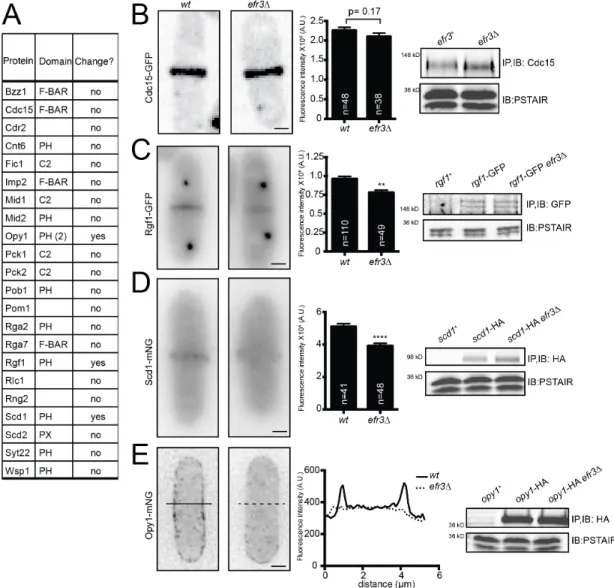
Because efr3∆ have reduced levels of PM PI(4,5)P2, we hypothesized that the cortical enrichment of proteins with membrane-binding domains (F-BAR, PH, PX or C2) would be diminished in efr3∆ compared to wildtype. Consistent with Cdc15 acting independently of Efr3 and interacting with a wide range of anionic phospholipids (McDonald et al., 2015), we found no difference in Cdc15 CR intensity (Figure 2.5A,B). The localizations of many other membrane- binding proteins, such as Pob1 (Figure 2.4E), were also unaltered in efr3∆ (Figure 2.5A), suggesting that they do not rely upon PI4P or PI(4,5)P2. However, we identified three PH domain-containing proteins with reduced PM localization in efr3∆ compared to wildtype. The RhoGEF Rgf1 and Cdc42 GEF Scd1 were reduced at the division site without any reduction in total protein levels (Figure 2.5C-D). Opy1, encoded by the ORF SPCPB16A4.02c, is normally enriched at the PM, but was diffusely localized in efr3∆ (Figure 2.5E). Opy1 contains two PH
23
domains and the S. cerevisiae ortholog, Opy1, is implicated in sensing PI4P at the PM and inhibiting the PI5-kinase Mss4 (Its3 in S. pombe) (Ling et al., 2012). Thus, it may be a collective reduction of several proteins at the cortex that compromises CR-PM attachment in efr3∆.
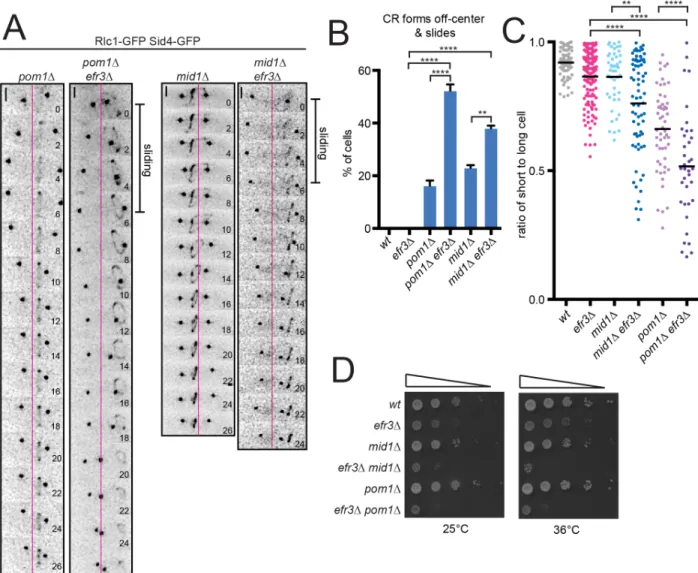
Off-center septa in S. pombe are observed when CRs slide and also when they assemble off-center. Mid1 and Pom1 dictate CR positioning (Rincon and Paoletti, 2016); cells lacking either divide asymmetrically due to misplaced CRs (Bähler and Pringle, 1998; Bähler et al., 1998a; Chang et al., 1996; Sohrmann et al., 1996). In mid1∆, CRs assemble at random positions and angles along the cortex but eventually coalesce into an orthogonal CR (Huang et al., 2008) although if CRs form within the curved cell pole of mid1-18, they can slide towards the tip, decreasing in diameter (Mishra et al., 2012). To test whether the Mid1 and Pom1 cues are influenced by PM composition, given that both proteins bind membrane PIPs (Celton-Morizur et al., 2004; Hachet et al., 2011), we combined mid1∆ or pom1∆ with efr3∆ and analyzed CR dynamics. We scored if CRs formed off-center and if fully formed CRs slid during anaphase. In our experiments, mid1∆ rarely formed CRs at the extreme cell tip that slid. As previously observed, mid1∆ and pom1∆ form off-center CRs but we did not detect a significant number of subsequent sliding events. Consistent with our finding that initial CR placement is not altered in efr3∆, the localizations of PM-binding proteins Mid1 and Pom1 were not influenced by deletion of efr3 (Figure 2.5A). In combination with efr3∆, mid1∆ and pom1∆ CRs formed off-center and slid during anaphase, resulting in septa that were farther off-center than single mutants (Figure 2.6A-C). These combinations also led to significant growth defects (Figure 2.6D), most likely due to cutting of chromosomes by off-center septation.
24
Figure 2.5. Localization of membrane-binding proteins in efr3∆. (A) List of proteins tagged with GFP or mNG and screened for differences in protein localization in efr3∆
compared to wt. Fluorescence intensity at the division site was measured and statistically significant differences are noted. (B-E, Left) Live-cell imaging of Cdc15-GFP (B), Rgf1-GFP Sid4-GFP (C), Scd1-mNG (D), or Opy1-mNG (E) in either wtor efr3∆. (B-D, Middle)
Quantification of fluorescence intensity at the cell division site. (E, Middle) Line scan of fluorescence intensity. (A-E, Right) Western blots of protein levels in wt and efr3∆.
Measurements in (B-D) represent three biological replicates. **p ≤ 0.01 ****p≤ 0.0001, Student’s t-test. Error bars represent SEM. Scale bar = 2 µm.
Figure 2.5. Localization of membrane-binding proteins in efr3∆. (A) List of proteins tagged with GFP or mNG and screened for differences in protein localization in efr3∆
compared to wt. Fluorescence intensity at the division site was measured and statistically significant differences are noted. (B-E, Left) Live-cell imaging of Cdc15-GFP (B), Rgf1-GFP Sid4-GFP (C), Scd1-mNG (D), or Opy1-mNG (E) in either wtor efr3∆. (B-D, Middle)
Quantification of fluorescence intensity at the cell division site. (E, Middle) Line scan of fluorescence intensity. (A-E, Right) Western blots of protein levels in wt and efr3∆.
25
Figure 2.6. The Stt4-complex and CR positioning machinery cooperate in septum positioning. (A) Live-cell imaging of indicated strains expressing Rlc1-GFP and Sid4-GFP.
Magenta lines mark the cell center. Scale bars = 2 µm. (B) Quantification of the frequency of events shown in (A) where the CR forms off center and slides. (C) Quantification of CR sliding. Individual points represent the ratio of the length of the short to long cell at septation and black bars denote mean. (D) Growth assay of serial 10-fold dilutions of the indicated strains at the indicated temperatures. **p ≤ 0.01 ****p≤ 0.0001, one-way ANOVA. Error bars represent SEM.
We next considered whether CR sliding events in efr3∆ are enabled by diminishing cell circumference from the cell middle towards one end similar to CRs in spheroplasts that move along the cortex while constricting (Mishra et al., 2013). This seemed unlikely, however, because efr3∆ have normal morphology (Chen et al., 2015) and CRs slide only 1-2 µm (Figure 2.3A), not approaching the region of curvature at the hemispherical cell ends (Atilgan et al., 2015). Also pos5∆, which is tapered at one cell end (Hayles et al., 2013), does not have off- center septa, indicating that a CR does not automatically slide toward a tapered end (Figure 2.4C). The CR width, as a proxy of cell diameter, also does not decrease during a sliding event in efr3∆, which would be expected if the circumference of the cell changed (∆ width = 0.045 µm +/- 0.039 µm (SEM, n = 19)). Further, in pos5∆ efr3∆, there is no worsening of the off-center septa phenotype compared to efr3∆ (Figure 2.4C) and sliding CRs have no bias toward the tapered end of the cell (54% to tapered end vs 46% to non-tapered end) similar to efr3∆ where the CR is equally likely to slide to the old or new end of the cell (48% to old end vs 52% to new end). We conclude that CR sliding events in efr3∆ are not dictated by cell geometry.
Because CR sliding in spheroplasts and cdc15 defective cells depends on type II myosins Myo2 and Myp2, respectively (Arasada and Pollard, 2014; Mishra et al., 2013), we tested if myosin-generated force is required for CR sliding in efr3∆. Neither Myp2, the type I myosin Myo1, nor the type V myosin Myo52 were necessary for CR sliding in efr3∆ (Figure 2.7A). To test if CR sliding depends on Myo2, we attempted to combine efr3∆ with the temperature sensitive myo2-E1 allele (Balasubramanian et al., 1998) but these alleles were synthetically lethal (Figure 2.8A). Instead, we used a myo2-E1 GFP-stt4 combination. GFP-stt4 (GFP-stt4 is hypomorphic) and myo2-E1 each had off-centered septa at 32°C, and the
combination resulted in more cells with off-center septa that were even farther away from center (Figure 2.8B). Live-cell imaging revealed that GFP-stt4 CRs slid during anaphase while myo2- E1 CRs formed off-center but did not slide (Figure 2.8C). GFP-stt4 myo2-E1 formed CRs off-
27
center that then also slid along the cortex (Figure 2.8C). Thus, CR sliding in efr3∆ does not depend on Myo2.
In contrast, deletion of the type V myosin Myo51 eliminated the efr3∆ off-center septa phenotype (Figure 2.7A). CR sliding events no longer occurred in myo51∆ efr3∆ (Figure 2.7B, C) and the average ratio of short to long cell was significantly higher than in efr3∆ (Figure 2.7A).
As expected given that Stt4 binds Efr3, the GFP-stt4 off-center septa phenotype is also Myo51- dependent (Figure 2.8B). Interestingly, none of the myosins were necessary for CR sliding in cdc15-core 4A or pxl1∆ mutants (Figure 2.8D-E) consistent with the genetic evidence that Cdc15-, Pxl1-, and Efr3-dependent CR sliding events occur through independent mechanisms (Cortés et al., 2015). CR sliding in cdc15 and pxl1∆ mutants, as well as in ß-glucan synthase mutations, may occur due to structural instability of the CR rather than a directed movement of the CR along the cortex (Arai and Mabuchi, 2002; Arasada and Pollard, 2014; Balasubramanian et al., 1998; Davidson et al., 2016; Ge and Balasubramanian, 2008; Hachet and Simanis, 2008;
Laporte et al., 2011; McDonald et al., 2015; Muñoz et al., 2013; Roberts-Galbraith et al., 2009;
Sethi et al., 2016; Wachtler et al., 2006).
Myo51 contains an N-terminal motor head domain and a C-terminal tail domain necessary for CR localization (Wang et al., 2014). By testing Myo51 N- and C-terminal truncations in efr3∆, we found that both the N-terminal head and C-terminal tail of Myo51 are necessary for CR sliding (Figure 2.8F), suggesting that Myo51 tail binding to CR components and the ability to walk along actin filaments (Wang et al., 2014) are both required to move the CR.
28
Figure 2.7. efr3∆ CR sliding events depend on Myo51. (A) Quantification of off-center septa. Individual points represent the ratio of the length of the short to long daughter cell at septation and black bars denote mean. (B) Live-cell imaging of indicated strains expressing Rlc1-GFP and Sid4-GFP. Magenta lines mark the cell center. (C) Quantification of the frequency of CR sliding events from (B). Error bars represent SEM. (D) Montage of time- lapse imaging of LifeAct-mCherry in efr3∆ cdc25-22. Montage is of a single Z slice and arrows indicate an actin cable in close proximity to a sliding CR. Scale bars = 2 µm. (E) Model for CR anchoring in cytokinesis. Proper PIP composition, dependent on Efr3, promotes CR anchoring. When PM lipid and CR protein composition is altered, CRs can slide. Cdc15 is an independent mechanism for CR anchoring as cdc15 mutants combined with efr3∆ results in exacerbated CR sliding defects. Disruption of CR positioning machinery in combination with efr3∆ leads to exacerbation of off-center septa. *p ≤ 0.05, one-way ANOVA.
29
Figure 2.8. N and C termini of Myo51 are necessary for efr3Δ CR sliding. (A) Analysis of resulting tetrads from a cross of efr3Δ and myo2-E1. (B) Off-center septa were measured in the indicated strains. Individual points represent ratios of the length of short to long cells at septation, and black bars denote means. (C) Live-cell imaging of indicated strains expressing mCherry-Cdc15 and Sid4-RFP. Magenta lines represent the center of the cell measured in a differential interference contrast image. Bars, 2 μm. (D and E) Off-center septa were
measured in WT, cdc15–core 4A (D), cdc15–core 4A with myosin mutants (D), pxl1Δ (E), and pxl1Δ with myosin mutants (E). (F) Off-center septa were measured in the indicated strains. ****, P ≤ 0.0001; one-way ANOVA. (G) MSD of cytokinetic ring position trajectories for WT (n = 34 trajectories from 17 cells) and efr3Δ (n = 36 trajectories from 18 cells). All trajectories are assumed to be independent realizations. The red line is the quadratic best fit to the MSD data obtained over the first 300 s (see the MSD analysis section of Materials and methods). Error bars represent SEM.
Figure 2.8. N and C termini of Myo51 are necessary for efr3Δ CR sliding. (A) Analysis of resulting tetrads from a cross of efr3Δ and myo2-E1. (B) Off-center septa were measured in the indicated strains. Individual points represent ratios of the length of short to long cells at
The necessity of Myo51 force generation for CR movements in efr3∆ provides strong evidence for the existence of forces on the CR perpendicular to the cell axis. Such forces could be involved in the splitting of CRs observed in some mutants, e.g. pxl1∆ (Ge and
Balasubramanian, 2008) and sbg1-3 (Sethi et al., 2016). We hypothesize that perpendicular forces are balanced in a wildtype cell and/or that CR-PM attachments are sufficient to resist these forces so that the CR remains in its central position. Further, in efr3∆ force imbalances may arise that cannot be stabilized, resulting in Myo51-dependent CR sliding along the cell axis.
In support of this hypothesis, mean squared displacement analysis of sliding efr3∆ CRs shows a statistically significant drift velocity term, suggesting directed transport of the CR (Figure 2.8G, v
= 1.35 nm/s, 95% confidence interval: 1.32-1.37 nm/s). No such transport term was measured for wildtype rings, which exhibit minimal changes in position over the imaging period (Figure 2.8G and Methods). A possible explanation for this behavior is that Myo51 in the CR associates with longitudinal actin cables as well as F-actin within the CR, pulling the CR along cables when PM anchoring is weakened. In support of this, Myo51 has been shown to play a role in the medial accumulation of actin cables during cytokinesis (Huang et al., 2012) and actin cables can be seen in proximity to sliding CRs in efr3∆ (Figure 2.7D).
Altogether, our data reveal a novel CR anchoring mechanism that depends on a
conserved PM-localized PI4-kinase complex (Figure 2.7E). An ensemble of proteins sensitive to correct PM PIP composition synergize with Cdc15-, Pxl1- and cell wall-dependent anchoring to promote stable CR placement and faithful segregation of the genetic material during cell division. Given the large number of lipid-binding proteins at the cell division site of many eukaryotes, and the importance of a PI4-kinase for cytokinesis (Brill et al., 2000; Eggert et al., 2004), it is likely to be a broadly relevant factor for CR-PM adhesion.
Materials and methods
Yeast Methods
31
S. pombe strains were grown in yeast extract (YE). efr3+, ypp1+, opy1+, scd1+ and rgf1+ were tagged at the 3’ end of their ORFs with TAP: kanR, Flag3:kanR, HA3:hygR, mCherry:kanR, mNeonGreen:kanR or mNeonGreen:hygR using pFA6 cassettes as previously described (Bähler et al., 1998b; Wach et al., 1994). A lithium acetate method (Keeney and Boeke, 1994a) was used in S. pombe tagging transformations, and integration of tags was verified using whole-cell PCR and/or microscopy. Introduction of tagged loci into other genetic backgrounds was
accomplished using standard S. pombe mating, sporulation, and tetrad dissection techniques.
Fusion proteins were expressed from their native promoters at their normal chromosomal loci unless otherwise indicated.
For growth assays, cells were grown to log phase at 25˚C in YE, 10 million cells were resuspended in 1 mL of water and 10-fold serial dilutions were made. 2.5 μl of each dilution was spotted on YE plates and the plates were incubated at the indicated temperatures.
Stt4 was N-terminally tagged with GFP at the endogenous locus using a Cre-loxP method as described (Werler et al., 2003). A cassette with the sequence that encodes GFP, the sup3-5 selection marker and a temporary promoter (nmt1) was integrated at the N terminus of the stt4 gene. Next the selection marker and temporary promoter were removed with Cre recombinase resulting in the insertion of sequences encoding GFP at the 5’end of stt4 under the normal promoter.
In order to express GFP-2xPH(PLCδ) in cells, the medium strength cdc2 promoter (Carpy et al., 2014) was PCR amplified from S. pombe genomic DNA and GFP-2xPH(PLCδ) was PCR amplified from plasmid pRS426 (Stefan et al., 2002). The two fragments were cloned into S. pombe integration vector pJK148 using Gibson Assembly. This construct was linearized and inserted into the S. pombe leu1 locus by a lithium acetate method (Keeney and Boeke, 1994a).
To overexpress GFP-2xPH(PLCδ), this sequence was PCR amplified from plasmid PRS426 and cloned into pREP81 under the nmt81 promoter. This construct was introduced into
cells by sorbitol transformation. Cells were fixed in 70% ethanol after induction of expression for 24 hours at 32°C.
Microscopy
Live-cell images of S. pombe cells were acquired using a personal DeltaVision
microscope system (Applied Precision) that includes an Olympus IX71 microscope, 60x NA 1.42 planApo and 100X NA 1.40 UPlanSApo objectives, a Photometrics Coolsnap HQ2 camera and softWoRx imaging software. Images were acquired at 25-29˚C and cells were imaged in YE media. Images in figures are either single slices or maximum-intensity projections of z sections spaced at 0.2-0.5 μm. Images used for quantification were not deconvolved. Other images not used for fluorescence quantification were deconvolved with 10 iterations. Time-lapse imaging was performed on cells in log phase on a YE agar pad at 32-36°C with the exception of LifeAct- mCherry cdc25-22 efr3∆, where cells were shifted to 36°C for four hours and then imaged at 25°C.
Intensity measurements were made with ImageJ software (http://rsweb.nih.gov/ij/). For all intensity measurements, the background was subtracted by creating a region of interest (ROI) in the same image where there were no cells (Waters, 2009). The raw intensity of the background was divided by the area of the background, which was multiplied by the area of the ROI. This number was subtracted from the raw integrated intensity of that ROI (Waters, 2009).
For CR intensity quantification, a ROI was drawn around the CR and measured for raw integrated density.
All cells were grown to log phase at 32°C before fixation. For nuclei and cell wall imaging, cells were fixed in 70% ethanol for 30 minutes before DAPI and methyl blue staining.
For quantification of ring sliding, a line was drawn through the fully formed contractile ring marked by rlc1-GFP using ImageJ software. Any movement of the CR away from the original line placement during the entire length of imaging was scored as a ring sliding event.
33
For ring sliding distances, fixed cells stained for nuclei and cell wall were imaged. The
coordinates of the cell tips and septum were logged. Length of the shorter and longer cell were calculated from these coordinates and reported as a ratio.
Mean squared displacement analysis
Time-lapse images of a medial Z slice of a strain expressing a CR and spindle pole body marker were acquired every 10 seconds, registered for both DIC and fluorescent channels using ImageJ plugin Image Stabilizer (Li) and merged together to determine the position of the ring relative to the cell boundary. Cytokinetic ring positions were recorded for 17 wildtype and 18 efr3∆ mutant cells beginning when the CR was fully formed. Individual trajectories were obtained by reslicing the timelapse images to a 3-pixel wide line along the cell boundary and tracking the position of the ring with subpixel resolution using the ImageJ plugin TrackMate (Tinevez et al., 2017). Total absolute displacements for wildtype trajectories over a 300-second period were measured to be 83±66 nm (SD, N=34 tracks from 17 cells), which is within the spatial resolution of the imaging (pixel size 106 nm). For efr3∆ mutant cells, 1-dimensional position over time data were used to perform Mean-squared displacement (MSD) analysis for the first 300s of the data using MATLAB® (R2016b, The MathWorks, Natick, MA) and
@msdanalyzer tool as previously described(Tarantino et al., 2014) . MSD data for efr3∆ mutant cells were fit by the 2nd degree polynomial function:
𝑀𝑆𝐷(𝜏) = 2𝐷𝜏 + 𝑣!𝜏!+ 𝜎
where 𝐷 is the diffusion coefficient, 𝑣 is the drift velocity, and 𝜎 is the noise term. The fitting procedure yielded:
𝑀𝑆𝐷(𝜏) = -7.42 × 10"# µ𝑚!
𝑠 7 𝜏 + -1.82 × 10"$ µ𝑚!
𝑠! 7 𝜏!+ (1.24 × 10"% µ𝑚!) r2=99.90%
Protein Methods and Mass Spectrometry
Cell pellets were snap-frozen in dry ice–ethanol baths. Lysates were prepared using a Fastprep cell homogenizer (MP Biomedicals). Immunoprecipitations were performed as previously described (Gould et al., 1991) in NP40 buffer for native lysates. Protein samples were resolved by SDS-PAGE and transferred to PVDF membrane (Immobilon P, EMD Millipore). Anti-HA (12CA5), anti-Flag (M2, Sigma) or anti-Cdc2 (Sigma) was used in
immunoprecipitations and/or as primary antibodies in immunoblotting. Secondary antibodies were conjugated to IRDye680LT or IRDye800 (LI-COR Biosciences). Blotted proteins were detected via Odyssey (LI-COR Biosciences).
Purification of Efr3-TAP and subsequent identification of interacting proteins by mass spectrometry were performed as previously described (Chen et al., 2013; Elmore et al., 2014;
Gould et al., 2004) with the following changes: a newer version of Scaffold (Scaffold v4.4.1.1) was used and the minimum peptide identification probability was changed to 95.0%.
All ANOVA statistical analyses used Tukey’s post-hoc analysis.