Thesis by Tara Adele Gomez
In Partial Fulfillment of the Requirements for the Degree of
Doctor of Philosophy
California Institute of Technology Pasadena California
2011
(Defended May 20, 2011)
© 2011 Tara Adele Gomez All Rights Reserved
ACKNOWLEDGMENTS
“Only love has no limits. In contrast, our predictions can fail, our communication can fail, and our knowledge can fail. For our knowledge is patchwork, and our predictive power is limited. But when perfection comes, all patchwork will disappear.”
(1 Cor. 13:8–10)
I am extremely grateful to have had the chance to work in the lab of Dr. Raymond Deshaies. Ray is one of the smartest people I have ever met. His scientific brilliance is however, matched by his master negotiation skills. I am thankful that I learned not only, how to be a better scientist from Ray, but also, some business savvy! I am grateful for all of the support and help Ray offered me. I feel truly privileged to have received said support and encouragement.
I would also like to thank my thesis committee members: Dr. Judy Campbell, Dr. David Chan, Dr. William Dunphy, and Dr. Angelike Stathopoulos. My committee was
immensely supportive and helpful in giving me both excellent suggestions and encouragement.
I am also very indebted to my colleagues in the Deshaies lab. I would like to thank the Deshaies’ lab graduate students for continued advice, community, and support. I will miss our monthly “Grad Student Lunches”. I would like to especially thank KJ Chang and Natalie Kolowa for being great colleagues, work-out companions, and friends. In addition, Nathan Pierce, Michael Rome, and Ruzbeh Mosadeg were great inspirations.
I am also very grateful to Rati Verma for sharing with me her 26S knowledge, which helped propel my project forward during its darkest days. Gary Kleiger was also
extremely helpful in helping my project during dark days, and helped teach me how to be a better scientific writer. The technical support of Heenam Park and Rob Oania
undoubtedly deserve my accolades as well.
I would be remiss to not thank the summer students I worked with during my time in the Deshaies lab. Marvin Gee, a Caltech SURF student, performed an experiment that
contributed to my publication. Caitlin Rugani did many experiments that never made it to the paper, but were essential and for which I will be eternally grateful. My 2008 summer high school student volunteer, Derek Tu, would open my centrifuge tubes for me and kept me company during my RY2H screen!
I am thankful for the amazing undergraduate and postbach research opportunities I was provided with in the lab of Dr. Steven G. Clarke, my undergraduate thesis advisor. His continued support throughout my graduate career allowed us to publish several papers during my tenure here at Caltech, although unfortunately, they were not allowed in my Caltech thesis!
I would also like to acknowledge the following programs, offices and people at Caltech and in the Pasadena area that made what could have become a very mundane five years, a much more balanced five years: Liz Ayala, Gwen Murdock, Denise Nelson-Nash, the YESS program, Caltech Classroom Connection (CCC), Huntington Library and Botanical Gardens, Eye Dreams, Biology Graduate Student Invited Speaker Series, Caltech Public Events Office for letting me host Reel Science and Science Saturdays, MSE/CCD office, Grad Chats, Women’s Center, GSC and the Caltech Y.
To my closest friends, and graduate school mentors: Julianne Lyons, Dr. Chris Lyons, Sindhuja Kadambi, Michelle Fontes, Nicole Tetreault, Brianna Williams, Schetema Stevens, Chinney Idigio, Dr. Princess Imoukhuede, Dr. Kelle Cruz, Nneka Williams, Dr.
Jennifer Keefe, Dr. Cecilia Zurita-Lopez, Dr. Tanya Porras-Yakushi, and Dr. Melissa Gulmezian, I would like to thank you for advice, companionship, and mentorship throughout the PhD process. I would especially like to thank the Lyons who walked this path with me the closest, and are the absolute best friends one could ever wish for.
I would like to thank my entire family and extended family. To my in-laws, the Hampton clan, you are extraordinary people that have enriched my life and remind me to laugh as much as I can! To my brother Jason Gomez, I think 2011 was a big year for the both of us. I am proud of you. To my sister, Felicia Gomez, I am very blessed to have you as a sister. Your love and support are unwavering and just what I need. Furthermore, I love that we have a shared passion for seeing the world and a similar sense of humor that allows us to share the world together.
And to my biggest supporters in this world, who are undoubtedly my parents and my husband, I realize I am where I am today because of you! This thesis is dedicated to my mother, who swears that I was the “best dancer” in the 2011 Caltech Dance Show, not because this is a fact (it isn’t), but because she believes in me more than anyone I have ever met. This thesis is also dedicated to my father who loves me deeply and cultivated my knowledge as a child. When I was a child, he even exchanged landscaping work for tuition fees at my private elementary school to ensure I got an amazing education. This thesis is also dedicated to my husband Tyron Hampton, who honestly thinks that I am capable of anything and puts up with all of my temper tantrums. I don’t know how he does it!
Finally, I am most thankful for the grace and guidance of God. Despite sin and
imperfection, God offers His grace. I believe I am here today because of the silent and merciful hand that He put out for me to reach hold to. “The LORD is the stronghold of my life…” Psalm 27:1.
ABSTRACT
Protein degradation is essential for many basic cellular functions. Most intracellular protein degradation occurs via the ubiquitin proteasome system. Cellular proteins are marked for degradation by the appendage of an ubiquitin chain. Ubiquitin receptor proteins recognize the ubiquitin chains and play a “garbage man” function in ensuring delivery of the protein trash to the cell’s degradation machinery, the proteasome.
One such class of ubiquitin receptor proteins, known as UBA-UBL proteins, recognizes ubiquitylated substrates and shuttles them to the proteasome. These shuttle receptors include Rad23, Dsk2, and Ddi1. The goal of this dissertation research has been to understand how these UBA-UBL proteins interact with the proteasome. In budding yeast, Saccharomyces cerevisiae, Rpn1 has been proposed to be the major docking site for UBL-containing proteins. More recent studies suggested that proteasome subunits Rpn10 and Rpn13, may also bind UBA-UBL proteins. However, no cis proteasome mutants existed to address these plausible redundant modes of delivery to the proteasome.
The specific aims of this proposal were to: identify the sites on the proteasome that are necessary for specific UBA-UBL receptor docking, to study the consequence of the deletion of these sites (such as the effects on protein turnover), and to assess if the elimination of these sites is the same as elimination of the receptor proteins themselves.
Here, I describe a two-pronged genetic screen I conducted to identify a specific docking site within Rpn1 for UBA-UBL proteins. I uncover a highly conserved residue, D517A that appears to impinge on the ability for both Ddi1 and–in the absence of Rpn13 or the dual absence of the ubiquitin interaction motifs of Rpn10 and Rpn13–Dsk2 to
interact with the proteasome. However, under no set of genetic conditions does a mutation at Rpn1-D517 have any effect on Rad23 or the UBL-containing ubiquitin isopeptidase Ubp6. Taken together, my observations point to unanticipated diversity and complexity in the mechanisms underlying the recruitment of UBA-UBL proteins to the proteasome.
Hence, I show that docking sites on the proteasome are not completely exclusive, both Ddi1 and Dsk2 share the D517 residue of Rpn1. However, there may be exclusive sites for docking Rad23 and Ubp6. There are also appears to be a layer of complexity and redundancy in docking UBA-UBL proteins to the proteasome. Follow-up studies on these proteasome cis mutatnts have also uncovered roles of the proteasome in regulating mitochondrial protein import and the methyl cycle.
TABLE OF CONTENTS
Title Page i Copyright Page ii Acknowledgements iii Abstract v
Table of Contents vii List of Tables and Figures x
CHAPTER 1: INTRODUCTION TO SUBSTRATE RECOGNITION BY THE 26S
PROTEASOME 1
The 26S proteasome 2
The ubiquitin proteasome system 3
Substrate delivery to the proteasome 4
Rad23, the best-characterized shuttle receptor 5
Dsk2, the next best-characterized UBA-UBL protein 6
Ddi1, the most controversial shuttle receptor 7
Overlapping and distinct roles for the UBA-UBL shuttle receptors 9
Proteins implicated in binding UBA-UBL proteins 11
Research objectives 12
CHAPTER 2: REVERSE-YEAST TWO-HYBRID SCREENING FOR PUTATIVE
INTERACTION-DEFECTIVE ALLELES OF RPN1 17
Introduction 18
Methods 20
Cloning RY2H bait and prey proteins 20
Results 25
Optimizing the Rpn1 reverse-yeast two-hybrid interaction 25
Expression of baits on a 2µ plasmid enhance positive and false positive signals 26
Isolation of putative Rpn1 IDAs 27
Rpn1 IDAs appear universal in their ability to disrupt binding to UBL proteins 28 Putative Rpn1 IDA alleles have wild-type phenotypes when introduced into Rpn1 null cells 28 Rpn1 mutant proteins assemble into fully intact and active 26S proteasomes 29 RY2H-derived Rpn1 IDA alleles do not impair the interaction between the proteasome and the
UBA/UBL proteins in vivo 30
Discussion 31
Figure legends 34
CHAPTER 3: IDENTIFICATION OF A FUNCTIONAL DOCKING SITE IN THE RPN1 LRR DOMAIN FOR THE UBA–UBL DOMAIN PROTEIN DDI1 44
Abstract 45
Methods 50
Results 55
Rpn1391-642 interacts with UBL domain proteins 55
Identification of mutations in Rpn1391-642 that block binding to UBL domain proteins 55 Mutant rpn1 alleles display synthetic growth defects in combination with ubiquitin receptor mutants
56 Recruitment of Ddi1, Dsk2, and ubiquitin conjugates to proteasomes is compromised in rpn1-D517A
and rpn1-K484A mutants 58
In vitro confirmation of a Ddi1 binding defect of Rpn13-deficient Rpn1-D517A mutant proteasomes 60 rpn1-D517A mutants exhibit a selective defect in protein degradation 61
Discussion 62
Authors' contributions 65
Acknowledgements 65
Figure legends 66
Supplementary data 75
CHAPTER 4: COMPARATIVE ANALYSIS OF PROTEASOME CIS MUTANTS
TO UBA-UBL NULL STRAINS 87
Introduction 88
Methods 90
Results 91
One surface of Rpn1 may be necessary for binding UBA-UBL proteins 91 The rpn1-VKD mutant shows only slightly more dramatic synthetic lethality than rpn1-STN in
combination with mutations of intrinsic ubiquitin receptors 92
Only Rpn1 VKD surface mutations stabilize the Ddi1 substrate Ufo1 93
Rpn1-D517A and Ddi1 interact genetically 93
The rpn1-D517A does not mimic a ddi1∆ mutant in steady-state accumulation of HO endonuclease 94 The ddi1∆ dsk2∆ mutant does not have diminished Ub conjugate binding to the proteasome 95
Discussion 96
Figure legends 99
CHAPTER 5: INSIGHTS INTO THE BINDING OF RAD23 AND UBP6 TO THE
26S PROTEASOME 104
Introduction 105
Methods 106
Results 106
Rpn10 and Rpn13 contribute to docking of UBA-UBL receptor proteins 106 A sole deletion of Rpn13 is not sufficient to interupt binding of any ubiquitin receptor protein to the
proteasome 107
Proteasomes from ufd2∆ are less abundant for Rad23 108
Discussion 108
Figure legends 110
CHAPTER 6: MITOCHONDRIAL IMPORT AND METHIONINE SYNTHESIS PATHWAYS ARE PERTURBED BY INTERFERENCE OF UBA-UBL BINDING
TO THE PROTEASOME 119
Introduction 120
Methods 121
Results 124
Rpn1-D517A proteasomes show diminished amounts of specific PIPs 124 Galactose-inducible Aac2 appears to be stabilized in rpn1-D517A 125 Cellular concentrations of Sam2, Cys3, and Fsr1 are shifted in rpn1-D517A mutants 126 Methionine and cysteine biosynthesis pathways appear perturbed in rpn1-D517A cells 127 rpn1-D517A mutants have an abundance of mitochondrial proteins at their proteasomes and
respiration defects 128
rpn13∆ rpn1-D517A mutants accumulate precursor forms of Hsp60 128
Discussion 129
Figure legends 133
CHAPTER 7: FINDINGS, IMPLICATIONS, AND FUTURE DIRECTIONS 154
Summary 155
Future directions 158
BIBLIOGRAPHY 159
List of Tables and Figures
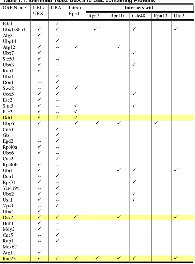
TABLE 1.1. IDENTIFIED YEAST UBA AND UBL CONTAINING PROTEINS... 14
TABLE 1.2. UBA-UBL RECEPTORS HAVE VARYING AFFINITIES FOR THE DIFFERENT FORMS OF UBIQUITIN... 15
FIGURE 1.1. THE EUKARYOTIC PROTEASOME ... 16
TABLE 2.1. RY2H STATISTICS... 36
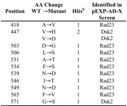
TABLE 2.2. POINT MUTATIONS IN RPN1 THAT DISRUPT BINDING TO EITHER RAD23 OR DSK2 IN A RY2H SYSTEM. ... 37
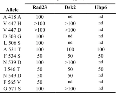
TABLE 2.3. 3-AT RESISTANCE OF BAIT/PREY PAIRS... 38
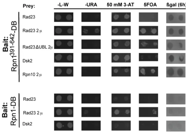
FIGURE 2.1. FORWARD-YEAST TWO-HYBRID INTERACTIONS BETWEEN RPN1-DB AND PREY PROTEINS... 39
FIGURE 2.2. MOST PUTATIVE RPN1 IDAS PRODUCE VIABLE YEAST STRAINS. ... 40
FIGURE 2.3. MUTANT RPN1 ALLELES DO NOT DISPLAY TYPICAL PROTEASOME MUTANT PHENOTYPES... 41
FIGURE 2.4. MUTANT RPN1 ALLELES DO NOT INTERFERE WITH PROTEASOME ASSEMBLY42 FIGURE 2.5. SELECT RY2H SCREEN RPN1 ALLELES REDUCE PROTEASOME STABILITY BUT DO NOT IMPINGE ON UBA-UBL BINDING TO THE PROTEASOME... 43
FIGURE 3.1. A TWO-PRONGED STRATEGY IDENTIFIES 18 RPN1 RESIDUES THAT MAY BE IMPORTANT FOR BINDING UBL DOMAIN PROTEINS... 69
FIGURE 3.2. MUTANT RPN1 ALLELES DISPLAY GENETIC INTERACTIONS WITH MUTATIONS IN GENES THAT ENCODE UBIQUITIN RECEPTORS INTRINSIC TO THE PROTEASOME. . 70
FIGURE 3.3. RECRUITMENT OF DDI1, DSK2, AND UB CONJUGATES TO THE PROTEASOME IS COMPROMISED IN RPN1-D517A AND RPN1-K484A MUTANTS. ... 71
FIGURE 3.4. RPN1-D517A REDUCES BINDING OF DDI1 IN VITRO.... 72
FIGURE 3.5. RPN1-D517A MUTANTS EXHIBIT A SELECTIVE DEFECT IN PROTEIN DEGRADATION. ... 73
FIGURE 3.6. MODEL FOR UBL PROTEIN INTERFACING WITH THE PROTEASOME ... 74
SUPPLEMENTAL TABLE 3.1. ADDITIONAL RATIONAL RPN1 MUTANTS USED IN THIS STUDY ... 77
SUPPLEMENTAL TABLE 3.2. YEAST STRAINS USED IN THIS STUDY... 79
SUPPLEMENTAL TABLE 3.3. PLASMIDS USED IN THIS STUDY ... 81
SUPPLEMENTAL TABLE 3.4. ANTIBODIES USED IN THIS STUDY... 82
FIGURE 3.S1. MUTANT RPN1 ALLELES DERIVED FROM BOTH THE RY2H SCREEN AND RATIONAL MUTATIONS DISPLAY GENETIC INTERACTIONS WITH MUTATIONS IN GENES THAT ENCODE UBIQUITIN RECEPTORS INTRINSIC TO THE PROTEASOME.... 83
FIGURE 3.S2. RPN1-D517A , RPN10-UIM RPN1-D571A, AND RPN10-UIM RPN13-KKD RPN1-D571A LIMIT BINDING OF UBA-UBL PROTEINS DDI1 AND DSK2. ... 84
FIGURE 3.S3. MUTATIONS AT RPN1 RESIDUES A418, N549, F565, AND G571 RENDER UNSTABLE PROTEASOMES... 85
FIGURE 3.S4. RPN1-D517A MUTANTS EXHIBIT A SELECTIVE DEFECT IN PROTEIN DEGRADATION. ... 86
TABLE 4.1 STRAINS USED IN THIS CHAPTER... 101
FIGURE 4.1. THE ‘VKD’ SURFACE OF RPN1 MAY BE RESPONSIBLE FOR BINDING UBA-UBL PROTEINS... 102
FIGURE 4.2. RPN1-D517A AND DDI1∆ MUTANTS MAY HAVE SOME PARALLEL FUNCTIONS IN PROTEIN TURNOVER. ... 103
TABLE 5.1 STRAINS USED IN THIS CHAPTER... 111
TABLE 5.2. PIPS THAT ARE LESS ABUNDANT IN RPN13∆ PROTEASOMES ... 112
TABLE 5.3. PIPS THAT ARE MORE ABUNDANT IN RPN13∆ PROTEASOMES ... 114
TABLE 5.4. UBIQUITIN RECEPTOR LEVELS ARE UNCHANGED IN RPN13∆ PROTEASOMES. 116 FIGURE 5.1. RPN10 AND RPN13 CONTRIBUTE TO DOCKING OF UBA-UBL RECEPTOR PROTEINS... 117
FIGURE 5.2. UFD2 CONTRIBUTES TO RECRUITMENT OF RAD23 TO THE 26S PROTEASOMES. ... 118
TABLE 6.2. PLASMIDS USED IN THIS CHAPTER ... 140 TABLE 6.3. PIPS THAT ARE REDUCED IN RPN13∆ RPN1-D517A 26S PROTEASOME
PREPARATIONS ... 141 TABLE 6.4. PIPS THAT ARE INCREASED IN RPN13∆ RPN1-D517A 26S PROTEASOME
PREPARATIONS ... 143 FIGURE 6.1. SCREENING FOR PUTATIVE RPN1-D517A SUBSTRATES FROM THE SILAC DATA
SET ... 145 FIGURE 6.2. GALACTOSE-INDUCIBLE AAC2 IS SLIGHTLY STABILIZED IN AN RPN1-D517A
STRAIN. ... 146 FIGURE 6.3. STEADY-STATE CONCENTRATIONS OF MULTIPLE GFP TAGGED ORFS CHANGE
IN AN RPN1-D517A MUTANT. ... 147 FIGURE 6.4. STEADY-STATE CONCENTRATIONS OF ORFS FOUND LESS ABUNDANTLY IN
RPN13∆ RPN1-D517A PROTEASOMES... 148 FIGURE 6.5. STEADY-STATE CONCENTRATIONS OF ORFS FOUND MORE ABUNDANT IN
RPN13∆ RPN1-D517A PROTEASOMES... 149 FIGURE 6.6. METHIONINE AND CYSTEINE BIOSYNTHETIC PATHWAYS CONTAIN MULTIPLE
ENZYMES THAT ARE PERTURBED IN RPN1-D517A.... 150 FIGURE 6.7. RPN13∆ RPN1-D517A MUTANTS HAVE MITOCHONDRIAL DEFECTS. ... 151 FIGURE 6.8. RPN13∆ RPN1-D517A MUTANTS ACCUMULATE HSP60 PRECURSOR PROTEINS.152 FIGURE 6.9 TWO HYPOTHESES DESCRIBE HSP60 ACCUMULATION IN PROTEASOME
MUTANTS. ... 153
Chapter 1:
Introduction to substrate recognition
by the 26S proteasome
Individual proteins, pairs of proteins, or small protein complexes, carry out many important cell processes. However, in some cases, very large assemblies of proteins form complicated quaternary structures, resembling nanomolecular machines, and they carry out pivotal jobs in the cell as well. These large protein complexes are enigmatic. Why is such a large complex required to complete the job? Such large protein complexes include the nuclear pore complex (~ 120 mDA), ribosomes (2–10 mDA), the largest known eukaryotic protease, tripeptidyl peptidase II (~ 6 mDa), and the 26S proteasome (~ 2.5 mDa). While we may never truly understand why evolution selected for such complex molecular machines, it appears to be factual that the complexity of the machine itself represents only a small fraction of the complexity found in the biological system it regulates. The 26S proteasome is no exception to this rule.
The 26S proteasome
The 26S proteasome is analogous to a recycling center. Cellular protein trash is delivered to the proteasome and the proteasome proteolyzes the target into small peptides that can be recycled in the cell for the creation of new proteins. The proteasome has ~ 33 unique protein subunits making up a ~ 66-protein subunit complex (Figure 1.1). The proteasome is composed of two main components: A 19S regulatory particle (RP) and the 20S core. The 19S caps one or both ends of the 20S catalytic cylindrical core (CP). The 19S regulatory particle is itself composed of two subcomplexes, the lid that is composed of ~ 11 non-ATPase subunits, and the base that is made up of two non-ATPase subunits, Rpn1 and Rpn2, that act as scaffolding subunits, and six ATPase subunits, Rpt1–6, that
require energy for the unfolding of substrates. The 19S RP recognizes an ubiquitin tag found on protein substrates, unfolds the substrate, and sends it into the channel of the 20S CP. Using three different proteolytic activities, the 20S degrades the substrate into small peptides (Pickart and Cohen, 2004).
The ubiquitin proteasome system
Degradation of cellular proteins is a selective process, that in many cases, is even controlled at a spatial and temporal level (Grabbe et al., 2011). Ubiquitin (Ub) is a small 76-amino acid protein whose conjugation to target proteins precedes degradation, and is the crux of the ubiquitin protesome system (UPS). Ubiquitin is added to protein
substrates through a series of three reactions involving enzymes known as E1s, E2s, and E3s. An E1 enzyme activates a free mono-Ub molecule by forming a covalent thioster bond. This activated ubiquitin is then transferred to an E2 conjugating enzyme and finally to an E3 ubiquitin ligase enzyme. Each E3 enzyme acts only on specific substrates and adds one of many levels of selectivity to the UPS (Hershko and Ciechanover, 1998).
Polyubiquitin (polyUb) chains can be built onto substrates through the sequential addition of ubiquitin molecules to one another via the E1, E2, E3 enzymatic steps (Pierce et al., 2009). Ubiquitin molecules form isopeptide linkages with one another through their lysine resides. Ubiquitin has multiple lysine residues through which multiple types of chain linkages can be produced.
Polyubiquitin tags of different linkages play diverse cellular roles. Lys-48
polyubiquitin chain appendages target proteins to the proteasome for degradation. A Lys- 48 tetraubiquitin chain has been shown to be the minimal signal necessary for
proteasomal recognition (Thrower et al., 2000). Alternatively, polyubiquitin chains made from Lys-63 or Lys-29 linkages generally have non-proteasomal signaling roles in the cell (Ikeda and Dikic, 2008).
Substrate delivery to the proteasome
In 1995, the discovery that a human proteasome subunit, S5a, bound
polyubiquitin, led to the first insights into how ubiquitylated substrates are recognized by the proteasome (Deveraux et al., 1995a). Homologs of S5a, including the yeast homolog, Rpn10, were also found to bind polyubiquitin and to be necessary for the proper turnover of artificial substrates (van Nocker et al., 1996). Rpn10 is believed to create a hinge between the 19S and 20S (Glickman et al., 1998) and contains an ubiquitin interacting motif (UIM) that was found to be responsible for this recognition of Ub chains (Young et al., 1998) and necessary for the efficient delivery of ubiquitinated targets to the
proteasome (Lambertson et al., 1999). However, deletion of RPN10 did not lead to phenotypes suggestive of large-scale deficits in cellular protein degradation (Saeki et al., 2002a), and only 27% of all UPS substrates are influenced by RPN10 (Mayor et al., 2007). This all leads to the belief that there are multiple cellular proteins involved in recognizing substrates for degradation at the proteasome.
Such a belief was confirmed shortly. After the discovery of Rpn10 as an intrinsic ubiquitin receptor, receptors known as UBA-UBL (ubiqitin association-ubiquitin-like) proteins–namely, Rad23, Dsk2, and Ddi1–were discovered. These proteins are sub- stoichiometric interactors with the proteasome. They act as shuttles that bind and unbind the proteasome rapidly and transiently (Wang and Huang, 2008). They contain an amino
terminal ubiquitin like domain (UBL) that is responsible for docking to the proteasome and a carboxyl terminal ubiquitin association domain (UBA) that binds ubiquitin chains (Chen et al., 2001; Wilkinson et al., 2001). Binding to ubiquitin chains via the C- terminal and the proteasome via the N-terminal presented a novel mode of translocation for ubiquitylated targets to the proteasome. These UBA-UBL proteins are conserved from yeast to man (Mueller and Feigon, 2002; Zhu et al., 2007), although they are not essential (Díaz-MartÌnez et al., 2006). The yeast proteaome has an array of proteins containing UBA and/or UBL domains (Table 1.1). However, Rad23, Dsk2, and Ddi1 represent a very small group of proteins that actually contain both domains (Table 1.1).
The UBA-UBL proteins have been the focus of my dissertation research and each of them has unique attributes that appear to play a role in their function in substrate delivery to the proteasome.
Rad23, the best-characterized shuttle receptor
Rad23 is a highly conserved from yeast to man (Bertolaet et al., 2001a). It was originally identified as a gene important for cellular resistance to UV stress; specifically it plays a role in nucleotide excision repair (Miller et al., 1982). It was later discovered that the UBL domain of Rad23, which shares 23% identity with ubiquitin (Bertolaet et al., 2001a), binds the proteasome. Rad23 was the first UBA-UBL domain protein of its kind to be invoked in the UPS system, and the discovery of this role made it plausible that other similar proteins behave in the same way (Schauber et al., 1998).
To date, Rad23 is the best-characterized UBA-UBL protein. Distinguishing it from other yeast proteins in its family, Rad23 has two UBA domains, each of which
appears to have different affinities for Lys-48 and Lys-63 chains (Raasi et al., 2005).
Nonetheless, as a whole, Rad23 has a greater affinity for binding Lys-48 linked chains than for any other, suggesting that its major cellular role is recognition of proteasome substrates (Raasi et al., 2005).
The roles Rad23 plays in UV stress response and in proteasome substrate delivery are not necessarily individual separable tasks. For instance, rad23ΔUBL mutants are sensitive to UV irradiation and other DNA damage responses (Watkins et al., 1993).
Additionally, only partial complementation of a RAD23 mutant can be achieved by transformation with rad23ΔUBL (Lambertson et al., 2003). These results may indicate that Rad23 needs to bind the proteasome for full activity in DNA damage responses.
Dsk2, the next best-characterized UBA-UBL protein
Budding yeast Dsk2 was first described as a receptor by Funakoshi and colleagues in 2002, who showed that it preferentially bound Lys-48 chains in comparison to Lys-63 chains (Funakoshi et al., 2002). Like Rad23, Dsk2 also plays multiple roles in the cell.
Specifically, it is important for spindle pole body duplication (Biggins et al., 1996).
Overexpression of DSK2 causes accumulation of cellular Ub conjugates and is a lethal event (Funakoshi et al., 2002). Multiple papers show that Dsk2 and its homologs bind the proteasome (Chandra et al., 2010; Fatimababy et al., 2010; Saeki et al., 2002b; Wilkinson et al., 2001). The UBA domain of Dsk2 has been proposed to bind monoUb and Lys-48, Lys-63, and Lys-29 chains with similarly strong affinities (Raasi et al., 2005). Another recent study also suggests that Dsk2 binds Lys-63 linkages more tightly than any of the other UBA-UBL proteins (Fatimababy et al., 2010). However, structural models of the
Dsk2 UBA domain agree with the original Funakoshi paper in that it would actually selectively bind Lys-48 chains over any other linkage (Lowe et al., 2006). Nonetheless, it has been found to be necessary for the proper proteolytic turnover of several endogenous cellular proteins and model substrates (Barbin et al., 2010; Liu et al., 2009; Medicherla et al., 2004; Richly et al., 2005).
Ddi1, the most controversial shuttle receptor
Before its role in the UPS was uncovered, Ddi1 was identified as a DNA damage- inducible gene, and was studied for its role in binding v-SNARE proteins (Liu and Xiao, 1997; Lustgarten and Gerst, 1999). Ddi1 is expressed in the nucleus and the cytoplasm and has a high level of sequence conservation and representation throughout all
eukaryotic genomes (Gabriely et al., 2008; Krylov and Koonin, 2001). Interestingly, Ddi1 appears to be the most divergent of the UBA-UBL protein family. Its UBL domain only shares 16% identity to ubiquitin (Bertolaet et al., 2001a) and such a distinction is thekey element that differentiates the proteolytic functionsof Rad23 and Ddi1 (Kim et al., 2004).
The S. pombe homolog of Ddi1, Mud1, was first linked to the UPS when it was identified as a suppressor of the temperature-sensitive phenotype for a Rpn1/Mts4 mutant
(Wilkinson et al., 2001).
The role of Ddi1 in substrate delivery has been harder to understand. Supporting a role for Ddi1 in targeting substrates to the proteasome are observations such as the fact that its UBA domain binds longer Ub chains better than shorter chains (Trempe et al., 2005; Wilkinson et al., 2001), albeit to much less of an extent than Rad23 and Dsk2 (Kaplun et al., 2005; Saeki et al., 2002b). It is noteworthy that the expression of
proteasomes is also mainly nuclear and ctyoplasmic and that the localization of Ddi1 to these regions is dependent on presence of UBA and UBL (Gabriely et al., 2008; Peters et al., 1994). However, there are at least three publications that cast doubt on the role of Ddi1 as a shuttle receptor. Some researchers have not been able to detect binding of Ddi1 or its homologs to the proteasome or subunits of the proteasome (Chandra et al., 2010;
Fatimababy et al., 2010; Kim et al., 2004). Despite this controversy, Ddi1 is necessary for turnover of some proteins (Ivantsiv et al., 2006; Kaplun et al., 2005). Furthermore, a ddi1∆UBL mutant does not properly turnover the Ddi1 substarate, HO. This result
suggests that the interaction of Ddi1 with the proteasome is necessary for HO degradation (Ivantsiv et al., 2006).
It should also be noted that conflicting data regarding the role of both Rad23 and Rpn10 in acting as ubiquitin receptors had also been raised. For instance, over-expression experiments of RAD23 suggested that it might actually antagonize the turnover of
endogenous and model substrates instead of enhancing it (Ortolan et al., 2000). Rpn10 was also cast into doubt when a published report failed to see it bind polyubiquitin in the context of an intact proteasome and when the Arabidopsis thaliana ortholog was seen to inhibit proteolysis (Deveraux et al., 1995b; Lam et al., 2002). However, currently in the field, these doubts have subsided (Elsasser et al., 2004; Verma et al., 2004).
Controversy aside, the role of Ddi1 in the UPS is further confounded by the knowledge that Ddi1 and its orthologs share a conserved retroviral aspartyl protease sequence signature, D[S/T]G (Krylov and Koonin, 2001). Based on the fold of this aspartyl protease domain, Ddi1 is predicted to be proteolytically active (Sirkis et al., 2006). However, there are no published reports showing definitive proof that the protease
domain of Ddi1 has cellular activity. The presence of this protease domain may even suggest that Ddi1 may play a novel deubiquitinating enzymatic role, although this is an unproven hypothesis (Krylov and Koonin, 2001). One might also ask if any of the identified Ddi1 substrates are solely dependent on Ddi1 acting as a receptor, or solely or dually dependent on its aspartyl protease activity.
However, the closest the field is to understanding if the aspartyl protease domain of Ddi1 has any biological implications comes from the study of Pds1, an important cell cycle checkpoint factor that must be degraded to promote anaphase progression (Ciosk et al., 1998). The aspartyl protease domain is necessary for Ddi1 homodimerization and for suppression of the temperaturesensitivity phenotype of a pds 1-128 mutant (Clarke et al., 2001; Gabriely et al., 2008). This result is intriguing because the deletion of the UBA or the UBL domain of Ddi1 is also required for suppression of the pds1-128 temperature- sensitive phenotype (Gabriely et al., 2008). This phenotype suppression may be due to the ability of Ddi1 to regulate the stability of cellular levels of Pds1. Supporting this is the fact that over expression of RAD23 has also been implicated in pds1-128 suppression (Clarke et al., 2001).
Overlapping and distinct roles for the UBA-UBL shuttle receptors
An outstanding question has been: How do these UBA-UBL proteins work together, if they do at all, for delivery of substrates to the proteasome? It is suggested that Dsk2 delivers polyUb substrates to the proteasome cooperatively with other UBA- containing proteins (Lowe et al., 2006). Additionally, UBA-UBL proteins have been
shown to form heterodimers with one another, and tetraubiquitin can simultaneously bind two different ubiquitin receptor proteins (Kang et al., 2006). In conflict with this idea are studies which conclude that UBA-UBL proteins do not bind Ub as dimers (Bertolaet et al., 2001a). Another study concluded that the conflict may be resolved by structural studies that imply that the UBA domain may interact with Ub as well as with other proteins in more than one way while utilizing the same binding surface (Mueller et al., 2004).
Cooperation of the UBA-UBL proteins is still rather uncertain. The overlapping roles of these proteins in the cell cycle seem to further confound their independent cellular contributions (Díaz-MartÌnez et al., 2006). Further, none of the UBA-UBL proteins are essential for cell viability. Even the full deletion of all three UBA-UBL proteins in budding yeast is not lethal, although it does cause cell cycle delay at high temperatures (Díaz-MartÌnez et al., 2006; Kim et al., 2004).
The UBA-UBL shuttle receptors are generally believed to specifically bind Lys- 48 polyUb chains with a high affinity (Raasi et al., 2005; Rao and Sastry, 2002;
Wilkinson et al., 2001). However, they each do so with varying affinities for multi- ubiquitin chains (Table 1.2), and they have been shown to each have a unique repertoire of substrates whose degradation they promote (Verma et al., 2004). Their ability to bind the proteasome also appears to be a disparity between them (Saeki et al., 2002b),
although there may be some cooperativity between the UBA-UBL receptors, such as in the case where two or more UBA-UBL proteins are necessary for substrate turnover (Medicherla et al., 2004). In agreement with this, rad23∆dsk2∆ mutant proteasomes have drastically reduced levels of Ub conjugates (Elsasser et al., 2004). However, there are
also undoubtedly some individual roles they each play in substrate turnover as well, as many substrates have been identified that are specifically regulated by only one UBA- UBL protein.
In addition to there being potential redundancy between the UBA-UBL proteins, there may also be redundancy and/or cooperation between them and the intrinsic receptor Rpn10. Rad23 and Rpn10 have some overlapping and redundant functions. For instance, delivery of Ub conjugates by Rad23 to the proteasome is largely unaffected by loss of the UIM domain of Rpn10 (Elsasser et al., 2004; Fatimababy et al., 2010). However, Rpn10 can rescue the turnover defects of rad23∆ proteasomes (Verma et al., 2004).
Proteins implicated in binding UBA-UBL proteins
When this dissertation research began, the UBA-UBL proteins had been implicated in binding the leucine-rich repeat region one (LRR1) of Rpn1 (a base component of the proteasome) (Elsasser et al., 2002; Seeger et al., 2003). LRR regions are horseshoe-shaped motifs thought to be important in protein-protein interactions. The LRR domains of Rpn1 have been defined. Rpn1 is made up of 9 repeat segments that are composed of 35–40 residues each (Lupas et al., 1997). The first five repeat segments constitute LRR1. A 134-acidic amino acid stretch separates LRR1 and LRR2, which encompass the last four repeat segments (Elsasser et al., 2002; Lupas et al., 1997).
Through a combination of yeast two- hybrid and GST pull-down assays, it was determined that the minimal region sufficient for Rad23 binding to Rpn1 is limited to amino acids 417–628, which is comprised of LRR1 and a 21-acidic amino acid stretch adjacent with the region (Elsasser et al., 2002).
While the current dissertation research was underway, several insights were additionally made in understanding recruitment of UBA-UBL proteins to the proteasome.
For instance, a proteasome subunit, Rpn13, was discovered to be an intrinsic ubiquitin receptor (Husnjak et al., 2008). Rpn13 is conserved from yeast to man, binds ubiquitin, and is important in turnover of model substrates (Husnjak et al., 2008; Verma et al., 2000). Additionally, Dsk2 was found to bind both Rpn10 and Rpn13 and Rad23 was found to bind a proteasome ATPase subunit, Rpt6, and Rpn10 (Fatimababy et al., 2010;
Matiuhin et al., 2008b; Zhang et al., 2009).
The exact mechanism by which UBA-UBL receptors deliver targets to this wide array of proteasome subunits, Rpn1 and intrinsic receptors, Rpn10 and Rpn13, is a mystery. For instance, do UBA-UBL proteins utilize all of these binding surfaces
simultaneously? Does docking at specific subunits at the proteasome regulate degradation of only specific substrates?
Research objectives
With the current tools available, addressing the separate contributions of Rad23, Dsk2, and Ddi1 to protein turnover has been difficult to resolve. This enigma leaves a multitude of questions, such as: Is the fate of a proteasome substrate dependent on the receptor by which it is delivered (Elsasser et al., 2002)?
This dissertation addresses three gaps in the existing knowledge:
1) Where do Ub receptors dock the proteasome? This is discussed extensively in Chapter 3, which comes from an article published in BMC Biology.
Chapter 2 describes optimizing the screen I used in Chapter 3.
2) What is the consequence of the elimination of these cis binding sites? I will discuss the quantitative mass spectrometry technique I used to discover consequences in Chapter 6.
3) Is elimination of these sites equivalent to the elimination of the receptors themselves? In Chapter 4 I delve into this topic and show that the physiology of a proteasome cis mutant has some unanticipated surprises.
I have discovered that the binding landscape at the proteasome for UBA-UBL proteins is rather complex. A single mutation in Rpn1 at residue D517 is able to reduce the binding of Ddi1 to the proteasome. However, such is not the case for any other UBL- containing protein. Dsk2 is seen to interact less with the proteasome only when there are multiple simultaneous mutations present in Rpn1, Rpn13, and Rpn10. It will be
interesting for future research to follow up on how the substrate repertoire of these UBA- UBL proteins depends, if at all, on which subunit of the proteasome they dock to.
Table 1.1. Identified Yeast UBA and UBL containing Proteins a Interacts with ORF Name UBL/
UBX
UBA Intrxn
Rpn1 Rpn2 Rpn10 Cdc48 Rpn13 Ufd2
Ede1 --
Ubx1/Shp1 b
Atg8 --
Ubp14 --
Atg12 --
Ubx7
Ste50 --
Ubx3 --
Rub1 --
Ubc1 --
Don1 --
Swa2 --
Ubx5
Esc2 --
Smt3 --
Pac2 --
Ddi1
Ubp6 --
Cue3 --
Gts1 --
Egd2 --
Rpl40a --
Ubx6 --
Cue2 --
Rpl40b --
Ubi4 --
Dcn1 --
Rps31 --
Ylr419w --
Ubx2
Usa1 --
Vps9 --
Ubx4 --
Dsk2 b
Hub1 --
Mdy2 --
Cue5 --
Rup1 --
Mex67
Atg11 --
Rad23
a This list was composed using www.yeastgenome.org
bgenetic interaction. All other noted interactions are via Affinity Capture MS or Y2H
Table 1.2. UBA-UBL receptors have varying affinities for the different forms of ubiquitin.
Kd (or relative binding affinity)a
Ub Type (Reference) Rad23 Dsk2 Ddi1
monoUB
(Trempe et al., 2005) nd nd 390 µM*
(Bertolaet et al., 2001a) 8 µM nd 10 µM
(Raasi et al., 2005) nd 15 µM nd
(Lowe et al., 2006) nd 8 µM nd
(Hobeika et al., 2007) 19 µM 12.9 µM2
nd nd
nd nd
(Ohno et al., 2005) nd 14.8 µM nd
Lys-48 diUB
(Trempe et al., 2005) nd nd 3 µM*
Lys-48 ≥tetra-UB
(Fatimababy et al., 2010) +++ +++ +
(Raasi et al., 2005) ++/+1 +++ ++
(Wilkinson et al., 2001) nd nd 30 nM*
(Hobeika et al., 2007) 7.8 µM 6.1 µM2
nd nd
nd nd Lys-63 diUB
(Trempe et al., 2005) nd nd 140 µM*
Lys-63 ≥tetra-UB
(Fatimababy et al., 2010) + ++ +
(Raasi et al., 2005) +/++1 +++ ++
a In some publications, no Kd was provided, instead a “+” system is used, where more pluses means there is better binding.
* Mud1, the S. pombe homolog of Ddi1 was used for these measurements.
1 Rad23 has two UBA domains; the affinities for UBA1 and UBA2 were measured separately.
2 Two separate measurements of the Rad23 UBA2 domain were made in this study.
Figure 1.1. The eukaryotic proteasome
Chapter 2:
Reverse-yeast two-hybrid screening for putative
interaction-defective
alleles of Rpn1
Introduction
The ubiquitin proteasome system (UPS) is conserved throughout the eukaryotic branches of life. Recent studies have even found a prokaryotic ubiquitin-like degradation system in myobacteria (Burns et al., 2009; Pearce et al., 2008). The proteasome is a large multi-subunit catalytic machine that selectively degrades proteins that have been marked for destruction with an ubiqutin appendage. Ubiquitylated substrates are also selectively delivered to the proteasome by ubiquitin receptors, as extensively reviewed (Finley, 2009).
Shuttle ubiquitin receptors, such as the UBA-UBL proteins Rad23 and Dsk2, have been speculated to act as “trans” targeting activators of degradation. Their ubiquitin association domain (UBA) brings the degradation initiation sites of their substrates in close proximity to the proteasome (Prakash et al., 2008; Schauber et al., 1998). They are able to deliver their cargo by binding to the proteasome with their ubiquitin-like domain (UBL), without becoming substrates themselves (Heessen et al., 2005). Such a role, allows UBA-UBL proteins to regulate the degradation of many important cellular
proteins, some of which may be involved in intricate complexes that require the selective degradation of a single subunit–for example, Sic1 must be degraded while in complex with cyclin-CDK (Verma et al., 2001).
Each of these UBA-UBL shuttle receptors delivers a specific subset of cellular proteins to the proteasome (Verma et al., 2004). However, there are not a sufficient number of genetic and biochemical tools available to understand the individual
contribution each one of these UBA-UBL receptors has in the selective degradation of the majority of the cell’s proteome (Rock et al., 1994). Such an understanding of the
specific repertoire of each shuttle receptor could lead to better understanding of how the UPS system is regulated and possibly even to future medical advances. For instance, human Rad23 (hHR23) has been implicated in the stability of p53, a tumor suppressor (Glockzin et al., 2003).
Hence, I aimed to create a proteasome cis mutant that would be unable to bind UBA-UBL proteins Rad23 and Dsk2. At the inception of my dissertation project, it was believed that Rad23 and Dsk2 primarily bound Rpn1, and some crosslinking data
suggested it possibly bound Rpn2 (Elsasser et al., 2002; Saeki et al., 2002b; Seeger et al., 2003). The first step in the creation of a proteasome cis mutant would be to identify amino acids on Rpn1 necessary for binding UBA-UBL proteins. I employed the use of a reverse-yeast two-hybrid system to screen for these putative amino acid residues.
Reverse-yeast two-hybrid (RY2H) systems allow for identification of interaction- defective alleles (IDAs). One of the major faults of traditional reverse-yeast two-hybrid screens is the large number of false positives, due to truncated proteins. To alleviate this error, Gray and colleagues created an in vitro library assembly protocol, in which the bait of interest is inserted in frame with a gene responsible for kanamycin resistance in E.
coli. Hence, a selection for full-length clones is created. This approach also allows for creation of a high-coverage allele library that will subsequently be transformed into yeast (Gray et al., 2007).
I report here, that Rpn1 has relatively weak yeast two-hybrid interactions with UBA-UBL proteins. This weak affinity may be a consequence of these proteins being in non-endogenous chimera states, or may be a consequence of a physiologically relevant weak affinity that allows them to transiently interact with one another for the disposal of
ubiquitylated proteins. Further, despite the advantages offered by this new RY2H system, of the 11 unique amino acid substitutions that were identified in this screen, none of them yielded IDA phenotypes in the context of fully intact proteasomes. The IDA phenotype exhibited in the RY2H screen was not transferable in a physiologically relevant
environment. However, some of the identified residues appeared to destabilize proteasome stability, underlining potential causative reasons for why such alleles appeared in this screen.
Methods
Cloning RY2H bait and prey proteins
The entire ORF of Rpn1 was PCR amplified from RDB2078 with oligos TG36 and TG37. The PCR product was BP cloned into pDONR-EXP, thus creating pENTR-RPN1 (RDB2167). pEXP-DB-RPN1 (RDB 2169) was created by LR cloning pENTR-RPN1 with pDEST-DB (a gift from the Marc Vidal lab). RAD23, DSK2, and RPN10 were similarly PCR amplified and cloned into pDONR-Express and LR cloned into pDEST- AD.
RPN1391-640 allele library construction Described in detail in Chapter 3.
2µ activation-domain yeast two-hybrid vector construction
pDEST-AD, a kind gift from the Marc Vidal lab, was used as template for amplification of the ADH1 promoter, the NLS, the GAL4 Activation Domain, the gateway cloning sites attR1, the chloramphenicol resistance marker, the ccdB gene, the attR2 site, and the ADH1TT terminator. This entire 4.1 kb fragment was amplified with forward and reverse primers, respectively, TG43 5’-
GCGCATCGATGGATCGAAGAAATGATGGTA-3’ and TG42 5’-
GCGCACTAGTTCGGCATGCCGGTAGAGGTG-3’ and cloned into the Spe1/Cla1 sites of pRS424, which is a high copy 2µ vector. The resultant vector, pRS424-
GAL4pAD (RDB 2178) was created. pENTR-clones were LR cloned into this vector.
Yeast two-hybrid interaction assay
Plasmids were transformed into the reporter strain MaV203 (MATα; leu2-3,112; trp1- 901; his3Δ200; ade2-101; cyh2R; can1R; gal4Δ; gal80Δ; GAL1::lacZ;
HIS3UASGAL1::HIS3@LYS2; SPAL10 UASGAL1::URA3). Transformants were plated onto synthetic medium prepared with 2% dextrose and lacking tryptophan and leucine with or without 3-AT and 5FOA. After 3 d incubation at 30ºC, the plates were analyzed for growth.
Reverse-yeast two-hybrid screen
The reverse-yeast two-hybrid assay was performed as described (Gray et al., 2007).
Briefly, pEXP-DB-Rpn1 allele library was cotransformed with pEXP-AD-Rad23 or pEXP-AD-Dsk2 into the reporter strain MaV203 using lithium acetate transformation
procedures. The transformation was plated onto SC-Leu-Trp + 0.2% 5FOA. Plates were grown for about 1 week, and putative 5FOAR colonies were picked and screened for reporter phenotypes. Interaction-defective alleles were tested for absence of activation on GAL1::lacZ, failure of growth on SC-HIS+3-amino-1,2,4-triazole (3-AT). Mild
interaction-defective alleles showed some growth on 3-AT. pEXP-DB Rpn1 allele library plasmids were either purified or PCR amplified from yeast colonies that displayed
5FOAR phenotypes ,and sequenced using primer 5’-GGC TTC AGT GGA GAC TGA TAT GCC TC-3’ (Li et al., 2004). Clones containing mutations were than retransformed into MaV203 and retested for proper reporter phenotypes.
LacZ reporter assay
Yeast colonies were frogged onto a hybond nylon membrane atop YPD agarose media.
After ~ 2–3 days of growth at 30°C, the membranes were submerged into liquid nitrogen and freeze thawed two times. After allowing the membrane to completely thaw, it was placed atop Whattman filters soaked in ~ 6 ml of buffer (60 mM Na2HPO4, 40 mM NaH2PO4, 10 mM KCl, 1 mM MgSO4, 0.7 M 2-mercaptoethanol, 21 mM X-gal). The soaked membranes were sealed in an airtight bag and placed at 37°C for 4–24 h.
Putative Rpn1 IDA insert sequencing
Direct PCR amplificiation of the pEXP-DB-Rpn1 insert was done as described in Li and Vidal. Briefly, yeast colonies were resuspended in 15 µl lysis buffer (50 units zymolase in 0.1 M Na-Phosphate buffer pH 7.4) and incubated for 15 min at 37°C and 10 min at 95°C. For PCR reactions, 0.3 µl of the lysed cells were used as template and amplified
using primers 5'-GGCTTCAGTGGAGACTGATATGCCTC and 5'- GGAGACTTGACCAAACCTCTGGCG.
RPN1 mutant strain construction
RPN1 was replaced by Kanmx6 using a one-step PCR-mediated technique (Longtine et al., 1998). Kanmx6 with RPN1 5’ and 3’ UTR homology was amplified from the pFA6a- KanMX6 vector (Longtine et al., 1998) by PCR using oligonucleotides TG20
(GGTCTACATAAGGTGCGATTCGTATAAATTTGGAAGACAATTGCAAGAAAC GGATCCCCGGGTTAATTAA) and TG21
(GGTTTTGAATTTTTCCTATTCTGGTTGATATTGCCCAAAAGCTATTCAGTGAA TTCGAGCTCGTTTAAAC). The PCR product was transformed into a diploid W303 strain (RJD381) creating strain RJD4166 which lacks the entire RPN1 ORF. This diploid strain was transformed with a CEN/ARS URA3 plasmid, pRS316-RPN1 (RDB--),
sporulated, and used to select for haploid strains resistant to G418 and able to grow on SD-Ura. The resultant strain, RJD4189 was used for plasmid shuffling.
Construction of putative Rpn1 IDA plasmids
The RPN1 locus, including 200 bp regions upstream and downstream of the gene, were PCR amplified from purified S. cervisiea genomic DNA using primers TG18
(GGGCGCCTCGAGGTTGACTATTTACAGCTCATC) and TG19
(GCGCCCGAGCTCAGCGCATCCATATTTACT). The resulting PCR product,
containing XhoI/SacI restriction sites, was digested and ligated into pRS315 and pRS316 CEN/ARS vectors. Silent mutations via a single nucleotide change resulted in an AvrII
restriction site at bp 1174 (amino acid 392) and an EagI site at bp 1920 (640). The mutations were engineered into the wild-type RPN1 locus using the Multisite Directed Mutagenesis Kit (Stratagene) and oligonucleotides TG12 (5’
GTCATTTGTCAACGGGTTCTTAAACCTAGGTTATTGTAACGATAAATTAAT 3’) and TG14 (5’ GCAGATGAAGAAGAAACGGCCGAAGGACAGACTA 3’). Mutations identified on pEXP-DB-Rpn1 plasmids in the reverse two-hybrid screen were introduced into this construct by double digestion and ligation into the AvrII and EagI sites.
Growth assays
For plating assays, strains were grown overnight in YPD and diluted to an OD600 of 0.3 in YP. Five-fold dilutions were prepared in YP and spotted onto YPD plates supplemented with various additives as described in the text. Plates were incubated at 30°C or 37°C for 2–3 days.
Small-scale yeast extract preparation for native gels
Extracts were prepared as described (Elsasser et al., 2005). Briefly, 50 mLs of saturated culture grown overnight to late log phase was harvested by centrifugation and the pellet was weighed. Pellets were resuspended in 1.5 mL of lysis buffer (50 mM Tris-HCl [pH 8.0], 1 mM EDTA, 5 mM MgCl2, and 1 mM ATP, 1X ATP Regeneration System) per gram of wet weight. An equal volume of glass beads was added and cells were lysed in a fastprep machine (ThermoSavant, Holbrook, NY) for 60 s at a speed of 6.5. Extracts are cleared by centrifugation at 20,000 g at 4°C for 30 minutes. Cleared extracts were then filtered through a milipore spin column.
26S native gel analysis Described in detail in Chapter 3.
Native immunoprecipitaion of proteasomes Described in detail in Chapter 3.
Results
Optimizing the Rpn1 reverse-yeast two-hybrid interaction
My goal was to create an Rpn1 cis mutant that would be unable to bind UBA-UBL proteins through the use of a genetic reverse-yeast two-hybrid screen. The first step in accomplishing this goal was to confirm and optimize forward yeast two-hybrid
interactions between Rpn1 and prey proteins of interest. The RY2H system we employed to assay interactions between Rpn1 and various PIPs has several reporter genes that can be simultaneously assayed. Positive interactions result in growth in the absence of uracil, in the presence of the drug 3-AT (with strong interactors being resistant to higher
concentrations of 3-AT), and induction of lacZ. The RY2H system has the additional advantage of allowing for the selection of non-interacting bait and prey pairs. Growth in the presence of the drug 5FOA, which normally causes toxicity in the presence of the URA3 protein product, signals non-productive Y2H interactions. Since it had been shown that a region encompassed by amino acids 391–642 or Rpn1 was necessary for
interaction with UBA-UBL proteins (Elsasser et al., 2002; Seeger et al., 2003), the
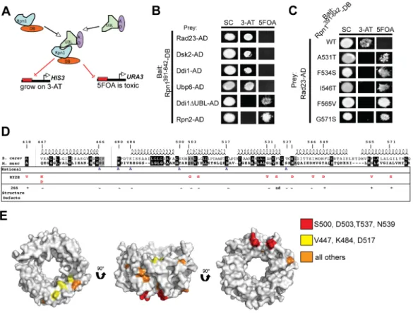
interaction between Rpn1391-642-DB was assayed with multiple baits including: Rad23- AD, Rad23∆UBL-AD, Ubp6, Ubp6∆UBL-AD, Dsk2-AD and Rpn10-AD. A full-length Rpn1-DB bait was also tested for interactions with these prey. Unexpectedly, in all tested combinations, growth was not seen on medium lacking uracil and induction of lacZ was only detected after several hours. However, growth on 50 mM 3-AT for Rad23-AD and Dsk2-AD in combination with Rpn1391-642-DB was observed (Figure 2.1). However, when the polarity of this interaction was reversed so that Rpn1391-642-AD was tested for interaction with prey, no productive interactions were detectable (data not shown). Taken together, these results are consistent with a very weak interaction occurring between Rpn1-DB, both the full-length and truncated forms, and the tested prey proteins.
Expression of baits on a 2µ plasmid enhance positive and false-positive signals
We postulated that interaction between Rpn1-DB and its prey might be improved by over expression of one of the interacting partners. It is possible that Rpn1-DB and Rad23-AD are interacting with endogenous proteins, and we might detect an increased reporter gene signal if we increase the pool of prey or bait proteins. To query this hypothesis, a high copy number 2µ Y2H pAD vector was created. In testing the strength of the interactions with this clone, several observations were made. Firstly, the importance of the polarity of the interaction was again observed, positive interactions were detected between Rpn1-DB pairs and activation-domain-carrying baits and not vice versa (data not shown).
Furthermore, there is an increase in putative negative interacting partners. Unexpectedly, pAD-2µ vectors cause growth on 5FOA, in most tested cases. For instance, pAD-2µ-
Rad23 grows on uracil whereas its CEN/ARS plasmid counterpart does not, but it also grows on 5FOA (Figure 2.1). Hence, the pAD-2µ vectors appear to create false positive and negative signals and would not be optimal for use in a RY2H screen.
Isolation of putative Rpn1 IDAs
Using the strongest forward-yeast two-hybrid interactions we could detect (those between Rpn1391-642-DB and Dsk2 and Rad23 CEN/ARS baits, we proceeded with the RY2H screen. The Rpn1391-642-DB allele library, containing over 500,000 individual clones, was co-transformed with either Rad23-AD or Dsk2-AD. Transformations were plated
directly onto medium containing 5FOA and incubated at 30°C until putative IDAs grew.
A total of 964 5FOAR colonies appeared and were screened in a Rad23/Rpn1 assay. For the Dsk2/Rpn1 assay, a total of 322 colonies were screened. Of those, 90 Rpn1-Rad23 IDAs and 22 Rpn1-Dsk2 IDAs appeared positive when screened for the proper reporter phenotypes and were subsequently sequenced. For the Rpn1-Rad23 screen, 22% of all putative positives contained a mutation, either silent (2.2%), a truncation or frameshift (3.3%), or a single or double mutation (16.7%). For the Dsk2-Rpn1 screen, again, roughly 22% of all putative positives contained a mutation of some sort, with 18.2% of these clones containing single or double mutations (Table 2.1). When these putative positive Rpn1 IDAs were retransformed with either Rad23 or Dsk2, only 10% of Rad23/Rpn1 IDAs retested as positives and 75% of Dsk2 clones. The mutations identified in this screen are shown (Table 2.2).
Rpn1 IDAs appear universal in their ability to disrupt binding to UBL proteins
The Rpn1 IDAs identified in the Rad23 and Dsk2 screens were tested for their ability to interact with other UBLs, including Rad23, Dsk2, and the deubiquitinase UBL-
containing enzyme, Ubp6. It was found that in all tested cases, every Rpn1 IDA
hampered to some degree the interaction with any tested UBL in comparison to the wild- type interaction. Additionally, similar levels of 3-AT were found necessary for inhibition of growth (Table 2.3).
Putative Rpn1 IDA alleles have wild-type phenotypes when introduced into Rpn1 null cells
To assess the physiological consequences of the putative Rpn1 IDAs I identified in the RY2H screen, mutant LEU2 plasmids were plasmid shuffled into rpn1Δ haploid cells sustained by a low copy URA3 plasmid containing RPN1. Transformants were plated onto 5FOA to evict the wild-type RPN1 plasmid, and clones sustained by the
mutagenized plasmid were sought. All alleles of rpn1 were viable with the exception of rpn1-F534S (Figure 2.2).
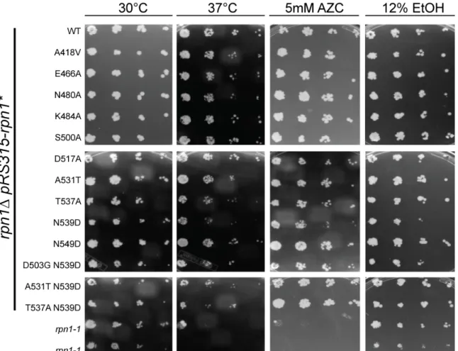
To identify mutants that exhibited broad defects in the ubiquitin–proteasome system, we tested these alleles for growth defects. Proteasome mutants often show growth defects under conditions that induce protein misfolding, such as in a variety of proteasome assembly mutants (Saeki et al., 2009). Strains lacking multiple ubiquitin receptors, such as the double and triple UBA-UBL strains, rad23∆dsk2 and
rad23∆dsk2∆ddi1∆, are also sensitive to protein misfolding, salt, and ethanol stress
(Husnjak et al., 2008; Kim et al., 2004; Lambertson et al., 1999). I postulated that if any of the identified Rpn1 IDAs diminished binding of UBA-UBL proteins in vivo, they would show sensitivity to stress conditions. Two rpn1-1 mutant isolates were also used in this assay as positive controls. The rpn1-1 strain is an unmapped mutation within RPN1 that was identified in a genetic screen for mutants that accumulate 3-hydroxy-3-
methylglutaryl-CoA reductase (Hampton et al., 1996). The rpn1 mutants were tested for their ability to grow on YPD medium at 30oC and 37oC. In addition, their ability to grow in the presence of ethanol (EtOH) and the proline analog AZC (L-azetidine-2-carboxilic acid) was also tested. Proteasome mutants exhibit stress when grown on AZC, as it promotes misfolding in proteins and presumably overloads the proteasome (Fowden and Richmond, 1963). Surprisingly, all of the rpn1 mutant strains show similar viability in comparison to wild type under all tested conditions (Figure 2.3). As expected, the rpn1-1 strains show sensitivity to elevated temperature and AZC.
The rpn1 mutants were also tested for ubiquitin conjugate accumulation by Western blotting cell extracts. All of the mutants seemed to show wild-type levels of Ub conjugate accumulation (data not shown).
Rpn1 mutant proteins assemble into fully intact and active 26S proteasomes
Cell extracts were resolved in non-denaturing gels in the presence of ATP and MgCl2 and visualized with the fluorogenic substrate SUC-LLVY-AMC. Rpn1 IDA mutants
contained wild-type ratios of double-capped 26S proteasomes, single-capped 26S proteasomes, and 20S core particles (Figure 2.4A). Thus, we conclude that there are no
assembly defects. Additionally, using a fluorogenic chymotrypsin activity assay, mutant proteasomes were found to have wild-type levels of chymotrypic activity (Figure 2.4B).
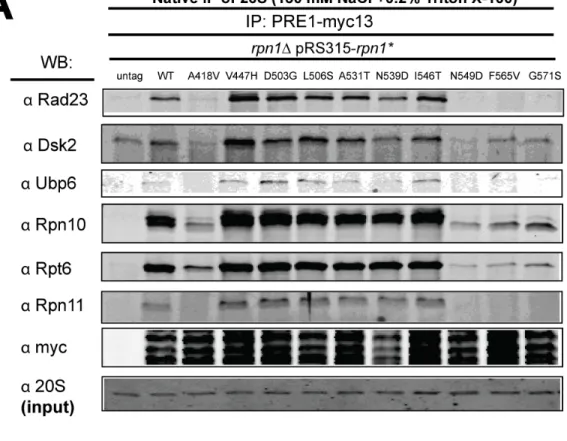
RY2H derived Rpn1 IDA alleles do not impair the interaction between the proteasome and the UBA/UBL proteins in vivo
Despite the rpn1 mutants showing no other physiological signs that they may have impaired delivery of ubiquitinated substrates to the proteasome, I performed
immunoprecipitation experiments to examine recruitment of UBA-UBL proteins to mutant proteasomes. To determine which UBA-UBL proteins were able to interact with 26S proteasome complexes from wild type and rpn1 mutants, I subjected strains
expressing Pre1-Myc to native immunoprepcipitation. In wild-type cells, Rad23, Dsk2, and Ubp6 were detected (Figure 2.5A). These UBA/UBL proteins were also detected in rpn1 mutants V447H, D503G, L506S, A531T, N539D, and I546T (Figure 2.5A and data not shown). Notably, rpn1 mutants A418V, N549D, F565V, and G571S seemed to destabilize proteasomes during the IP procedure, as they showed reduced levels of the UBA-UBL proteins Rad23, Dsk2, and Ubp6, but also of proteasome subunits Rpn10 (base), Rpt6 (base), and Rpn11 (lid), indicating that the proteasome subcomplexes were disassociated during the immunoprecipitation (Figure 2.5A). Because the presence of the proteasome inhibitor MG262 has been shown to increase proteasome stability (Kleijnen et al., 2007), the rpn1 mutants that showed proteasome instability were reassayed with exogenous 2 µM MG262 added to the extracts and under milder native buffer IP conditions (75 mM NaCl and 0.1% NP40). Under these conditions, all mutants that had showed instability in the prior assay now remained assembled, with the exception of
rpn1-A418V. In addition, none of these mutants interfered with the proteasomes’ ability to co-immunoprecipitate with Rad23 or Dsk2 (Figure 2.5B and data not shown).
Discussion
Approximately 250 of the residues that had previously been determined to be necessary for binding UBA-UBL proteins were in included in the aforementioned RY2H screen (Elsasser et al., 2002; Seeger et al., 2003). A ~1 50 residue span was identified within this ~ 250 amino acid fragment, containing 11 residues that perturbed Rad23 and/or Dsk2 binding to Rpn1 within the context of a yeast two-hybrid interaction.
However, when these mutations were used to replace endogenous Rpn1 within a yeast cell and probed for their ability to bind UBA-UBL proteins, unexpectedly, binding was not diminished.
It is hard to ascertain why such a discrepancy in binding occurred between the results of the genetic screen and the results of following full-length physiologically relevant protein interactions.
Considering the large number of IDAs identified that decreased proteasome stability (4 IDAs out of 11 total IDAs), it is plausible that the RY2H screen selected for residues that enhanced misfolding of Rpn1, which in turn eliminated binding of UBA- UBL proteins. Since a truncated version of Rpn1 was used in the screen, misfolding events are conceivable. Supporting this claim is an electron microscopy study of Rpn1 which shows that truncated forms of Rpn1 do not fold correctly (Effantin et al., 2009).
Interestingly, one residue uncovered in the RY2H screen caused lethality. Since Rpn1 is the largest subunit of the proteasome, misfolding events that lead to improper scaffolding
of adjacent subunits could very well be a lethal event. It may be interesting to do in vitro studies with mutant Rpn1-F534S protein to determine if this residue causes severe misfolding of Rpn1 that impinges on proteasome function.
Another reason true IDAs were not identified in the RY2H screen may be due to the inherent nature of the proteasome to interact with GAL1-10 promoter in vivo through binding the activation domain of (AD) of Gal4. Gal4-AD interacts with proteasome subunits, Sug1/Rpt6, Sug2/Rpt4, and Rpn1, Rpn2 (Gonzalez et al., 2002). This may have allowed Rpn1-DB to naturally interact with Gal4-AD containing proteins. Admittedly, since all forward Y2H interactions were quite weak (Figure 2.1) this may be the weakest argument. However, the possibility exists that this natural tendency for endogenous proteasome subunits to interact with Gal4-AD may have compounded other intrinsic problems with this genetic screen.
It is also possible that in the context of the RY2H screen the interactions between Rpn1 are truly impaired with UBA-UBL proteins. However, in vivo, a lot of binding redundancy e