The neurochemistry underlying higher cognitive functions in the primate fronto-striatal network
By
Seyed-Alireza Hassani
Dissertation
Submitted to the Faculty of the Graduate School of Vanderbilt University
in partial fulfillment of the requirements for the degree of
DOCTOR OF PHILOSOPHY in
Psychology
May 12, 2023 Nashville, Tennessee
Approved:
Thilo Womelsdorf, PhD Carrie K. Jones, PhD Alexander Maier, PhD
Mark Wallace, PhD
Copyright © 2023 Seyed-Alireza Hassani All rights reserved
Acknowledgements
First and foremost, I would like to sincerely thank my PhD supervisor and scientific mentor, Dr. Thilo Womelsdorf. Starting from when we met, during my undergraduate studies, he showed excitement in my ideas and fostered my curiosity about the brain and cognition. He spent countless hours supporting me and my scientific endeavors and with great patience and care has taught me much, scientific and otherwise.
I would like to thank my committee members, Dr. Carrie K. Jones, Dr. Alexander Maier and Dr. Mark Wallace for their invaluable feedback and support.
I especially would like to thank Dr. Carrie K. Jones for her wisdom and support as a collaborator in our cholinergic projects. I would also like thank Dr. Paul Tiesinga and Dr. Matthijs van der Meer for their computational support as collaborators. Last but not least, I would like to thank our collaborators Dr. Janusz Pawliszyn and the Pawliszyn lab, in particular Dr. Sofia Lendor, for their support in the neurochemical sampling.
I would like to thank all past and present lab members and co-authors for providing invaluable scientific and social support. I would like to thank Dr. Mariann Oemisch in particular for her mentorship and training, Dr. Benjamin Voloh for making difficult times in my life bearable and being a wonderful role model, Adam Neumann for his patience, experimental support and for pushing me to be better, Dr. Kianoush Banaie Boroujeni and Robert Louie Treuting for their impact on my research and friendship, and Dr. Marcus Watson and Dr. Christopher Thomas for their invaluable technical support and friendship.
Finally, I would like to thank my friends and family from the bottom of my heart. My friends for helping keep me sane and grow in unexpected ways. My parents who gave up comfortable lives and promising futures to give me and my sister the best future possible, my sister for her unconditional love, my loving partner, Elizabeth Senft who has supported me and loved me through even the hardest parts of my studies and our wonderful dog Cabbages.
Table of Contents
Page
List of Figures ... ix
Chapter 1 Introduction ... 1
1.1 NEUROMODULATORY SYSTEMS ... 1
1.1.1 Neuromodulatory projections ... 1
1.1.2 The adrenoceptors of the noradrenergic system ... 2
1.1.3 The muscarinic receptors of the cholinergic system ... 3
1.1.4 Spatial scale of neuromodulatory action ... 4
1.1.5 Receptor densities ... 4
1.2 NEUROMODULATORS AND BEHAVIOR ... 6
1.2.1 The role of neuromodulators in psychiatric disorders... 7
1.2.2 Pharmacological intervention ... 8
1.3 THE FRONTO-STRIATAL NETWORK ... 10
1.3.1 Insights from lesion studies ... 11
1.3.2 Convergent neuromodulation ... 11
1.4 THE MULTI-MODULATORY BRAIN ... 12
1.4.1 The interaction between multiple neuromodulatory systems ... 13
1.4.2 Making multi-modulator measurements ... 14
1.5 THESIS OVERVIEW ... 14
Chapter 2 Multi-Neuromodulator Measurements across Fronto-Striatal Network Areas of the Behaving Macaque using Solid-Phase Microextraction... 17
2.1 ABSTRACT ... 17
2.1.1 New and noteworthy ... 17
2.2 INTRODUCTION ... 18
2.3 MATERIALS AND METHODS... 20
2.3.1 Animals ... 20
2.3.2 SPME protocol and Fabrication of SPME Probes ... 21
2.3.3 MRI Guided Electrophysiological Mapping of Target Tissue ... 22
2.3.4 SPME Sampling and Post-Processing Procedures ... 22
2.3.5 Neuromodulator detection and quantitation ... 25
2.4 RESULTS ... 25
2.5 DISCUSSION ... 30
2.5.1 Extracellular Concentrations of Glutamate, Dopamine, Acetylcholine and Choline. ... 30
2.5.2 Reliable Measurement of Individual Differences of State Specific Neuromodulatory Tone. ... 32
2.5.3 Qualities and Advantages of SPME. ... 33
2.5.4 Future direction and improvements to the SPME neurochemical sensing. ... 36
2.5.5 Implications for Understanding and Treating Psychiatric Disease States. ... 38
2.6 CONCLUSION. ... 39
2.7 ACKNOWLEDGMENTS ... 39
2.8 CONFLICTS OF INTEREST ... 39
2.9 AUTHOR CONTRIBUTIONS ... 39
Chapter 3 Dose-Dependent Dissociation of Pro-cognitive Effects of Donepezil on Attention and Cognitive Flexibility in Rhesus Monkeys... 41
3.1 ABSTRACT ... 41
3.2 INTRODUCTION ... 42
3.3 METHODS AND MATERIALS... 45
3.3.1 Nonhuman Primate Testing Protocol ... 45
3.3.2 Drugs and Procedures ... 45
3.3.3 Behavioral Paradigms ... 46
3.3.4 Neurochemical Confirmation of Drug Effect ... 46
3.3.5 Statistical Analysis... 48
3.4 RESULTS ... 48
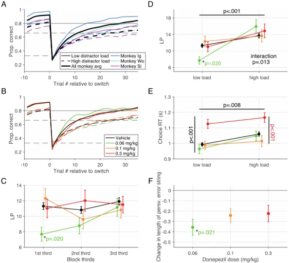
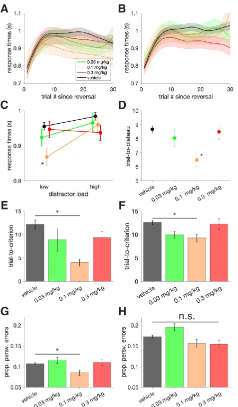
3.4.1 Dose-dependent improvement of visual search accuracy and slowing of choice reaction times ... 48
3.4.2 Dose-dependent improvement of flexible learning performance ... 51
3.4.3 Dissociation of attention and learning improvements, but slowing is correlated ... 52
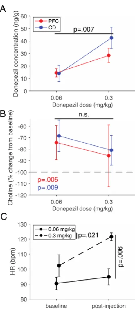
3.4.4 Determination of extracellular donepezil and choline levels in the prefrontal cortex and anterior striatum 53 3.5 DISCUSSION ... 54
3.5.1 Different donepezil dose-ranges for improving interference control and flexible learning ... 55
3.5.2 Quantifying extracellular levels of donepezil and choline in prefrontal cortex and striatum... 58
3.6 ACKNOWLEDGEMENTS ... 60
Chapter 4 M1 selective muscarinic allosteric modulation enhances cognitive flexibility and effective salience in nonhuman primates ... 61
4.1 ABSTRACT ... 61
4.1.1 Statement of significance ... 61
4.2 INTRODUCTION ... 62
4.3 RESULTS ... 64
4.3.1 M1 PAM VU0453595 enhances learning ... 66
4.3.2 Improved cognitive control with M PAM VU0453595 ... 67
4.3.3 M1 PAM VU0453595 has no consistent effect on interference control ... 68
4.3.4 Double dissociation of VU0453595 and Donepezil for cognitive flexibility and interference control .. 70
4.4 DISCUSSION ... 72
4.4.1 M1 PAM enhances learning and extra-dimensional shifts ... 72
4.4.2 M1 PAM reduces perseverative responding ... 75
4.4.3 M1 PAM has no consistent effect on interference control over distractors ... 75
4.4.4 Limitations ... 77
4.5 CONCLUSION ... 78
4.6 METHODS AND MATERIALS ... 78
4.6.1 Subjects ... 78
4.6.2 Compounds and Procedures ... 78
4.6.3 Behavioral Paradigms ... 79
4.6.4 Statistical Analysis... 80
4.7 FINANCIAL DISCLOSURES ... 80
4.8 ACKNOWLEDGEMENTS ... 80
4.9 AUTHOR CONTRIBUTIONS... 80
Chapter 5 A computational psychiatry approach identifies how alpha-2A noradrenergic agonist Guanfacine affects feature-based reinforcement learning in the macaque ... 81
5.1 ABSTRACT ... 81
5.2 INTRODUCTION ... 81
5.3 RESULTS ... 86
5.3.1 Reinforcement learning mechanisms underlying faster versus slower learning ... 91
5.4 DISCUSSION ... 96
5.4.1 Alpha 2A noradrenergic action supports multiple routes to behavioural flexibility ... 97
5.4.2 Reinforcement learning modeling of behavioural drug effects advances computational psychiatry. 100 5.5 METHODS ... 102
5.5.1 Subject and apparatus ... 102
5.5.2 Behavioural paradigm... 103
5.5.3 Experimental procedures for dose identification testing protocol... 104
5.5.4 Experimental procedures for optimal dose testing protocol... 105
5.5.5 Behavioural analysis of learning trials ... 106
5.5.6 Testing for trial-by-trial differences of the probability of rewarded choices. ... 106
5.5.7 Testing for the consistency of learning differences across blocks within sessions. ... 107
5.5.8 Reinforcement learning modeling ... 108
5.5.9 Bayesian learning modeling ... 110
5.5.10 Hybrid Bayesian-Reinforcement learning modeling ... 111
5.5.11 Model optimization, evaluation and comparison ... 113
5.6 ACKNOWLEDGMENTS ... 113
5.7 AUTHOR CONTRIBUTIONS... 113
Chapter 6 2A adrenoceptor stimulation in primates supports fronto-striatal functions by enhancing reward prediction error encoding ... 115
6.1 ABSTRACT ... 115
6.2 INTRODUCTION ... 115
6.3 MATERIALS AND METHODS ... 117
6.3.1 Subjects and apparatus ... 117
6.3.2 Behavioral task ... 119
6.3.3 Statistical measure of learning ... 120
6.3.4 Drug dosing... 120
6.3.5 Electrophysiological recordings and unit isolation ... 120
6.3.6 Putative cell type classification ... 121
6.3.7 Multi-linear regression ... 121
6.3.8 Model variables ... 123
6.4 RESULTS ... 123
6.4.1 Guanfacine enhances reversal learning and post-error adjustment ... 123
6.4.2 Pupils constrict with guanfacine ... 125
6.4.3 Guanfacine reduces pairwise firing correlations in ACC ... 125
6.4.4 Guanfacine enhances encoding of reward prediction errors during learning ... 126
6.4.5 Guanfacine modulates signaling which stimulus will be chosen after the attention cue onset ... 129
6.4.6 Guanfacine enhances outcome encoding particularly for putative interneurons ... 129
6.5 DISCUSSION ... 131
6.5.1 The 2A adrenoceptor and cognitive flexibility. ... 132
6.5.2 Enhanced outcome and RPE encoding without increased proportion of encoding neurons. ... 133
6.5.3 Spatial and cell-type specificity... 134
6.5.4 Insights from 2A stimulation: norepinephrine and behavior. ... 136
6.6 CONCLUSIONS ... 137
6.7 ACKNOWLEDGEMENTS ... 138
Chapter 7 General discussion ... 139
7.1 EVALUATING PHARMACOLOGICAL INFLUENCE ON MULTIPLE COGNITIVE DOMAINS ... 139
7.2 BROADER FRAMEWORKS FOR NEUROMODULATORY ACTIONS ... 140
7.4 PERSPECTIVE ON NEUROMODULATION ... 143
7.5 CONCLUSION ... 144
Appendix A: Supplemental information for Chapter 2 ... 145
A.1 SUPPLEMENTARY METHODS CHEMICALS,REAGENTS AND MATERIALS ... 145
A.2 FIGURES ... 148
A.3 TABLES ... 150
Appendix B: Supplemental information for Chapter 3 ... 151
B.1 SUPPLEMENTAL METHODS ... 151
B.2 SUPPLEMENTAL RESULTS... 161
B.3 SUPPLEMENTAL DISCUSSION ... 166
B.4 FIGURES ... 171
B.5 TABLES ... 174
Appendix C: Supplemental information for Chapter 4 ... 177
C.1 SUPPLEMENTAL MATERIALS AND METHODS ... 177
C.2 SUPPLEMENTAL RESULTS ... 179
C.3 SUPPLEMENTAL DISCUSSION ... 180
C.4 FIGURES ... 183
Appendix D: Supplemental information for Chapter 5 ... 185
D.1 SUPPLEMENTARY RESULTS... 185
D.2 SUPPLEMENTARY METHODS ... 189
D.3 FIGURES ... 191
Appendix E: Supplemental information for Chapter 6 ... 194
E.1 SUPPLEMENTAL METHODS ... 194
E.2 SUPPLEMENTAL RESULTS ... 195
E.3 FIGURES ... 197
References: ... 203
List of Figures
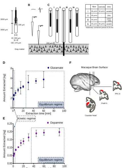
Fig 2.1 SPME sampling procedure, extraction time profiles and measurement locations. ... 24
Fig 2.2 Changes in SPME sampling over time. ... 26
Fig 2.3 SPME sampling of the macaque brain. ... 29
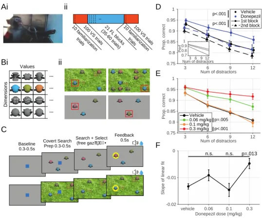
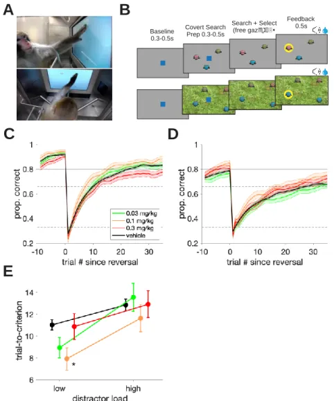
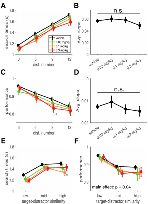
Fig 3.1 Task design, meta-structure and visual search performance as a function of distractor number. ... 44
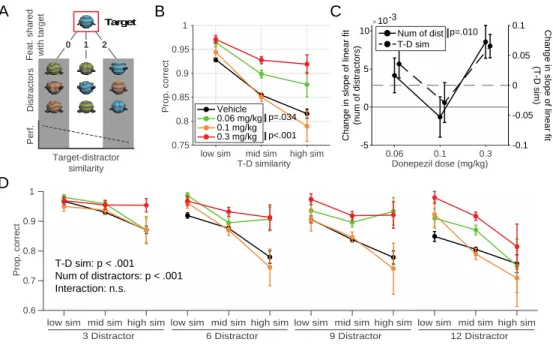
Fig 3.2 Visual search task performance and change in difficulty through increasing distractor numbers and target- distractor similarity. ... 47
Fig 3.3 Feature learning task learning curves and performance. ... 49
Fig 3.4 The relationship between the visual search task and the feature learning task... 52
Fig 3.5 In-vivo extracellular measurements of choline, donepezil as well as donepezil’s effect on heart rate. ... 55
Fig 3.6 Theoretic curves. ... 58
Fig 4.1 Task design and feature-reward learning task performance enhancement by VU0453595 ... 65
Fig 4.2 Feature-reward learning task efficiency and cognitive flexibility improvements with VU0453595 ... 69
Fig 4.3 Distractor effect and interference control are not consistently impacted by VU0453595 ... 71
Fig 5.1 Feature-based reversal learning task. ... 84
Fig 5.2 Dose-dependent improvement of reversal learning performance. ... 87
Fig 5.3 Comparison of reversal learning on Guanfacine days versus Control days. ... 92
Fig 5.4 Reinforcement learning (RL) modeling of reversal learning during drug and control sessions... 93
Fig 5.5 Performance and parameter values for the most-predictive RL model ... 94
Fig 5.6 Parameter values for the Feature-Weighting + Decay RL model applied to different sets of reversal blocks showing slow and fast learning. ... 96
Fig 5.7 Relation of model parameters underlying reversal performance. ... 98
Fig 6.1 Feature-based reversal learning task with simultaneous electrophysiological recordings. ... 118
Fig 6.2 Enhanced learning speed and improved post-error behavioral adjustment with guanfacine... 124
Fig 6.3 Guanfacine-mediated changes in neural activity correlations to stimulus variables during the feedback epoch. ... 126
Fig 6.4 Guanfacine-mediated changes in neural activity correlations to outcome variables during the feedback epoch. ... 127
Fig 6.5 Guanfacine-mediated changes in neural activity correlations to RPEs during the feedback epoch. ... 128
Fig 6.6 Guanfacine-mediated changes in neural activity correlations to outcome variables during the feedback epoch for broad and narrow spiking neurons. ... 130
Fig 6.7 Guanfacine-mediated changes in neural activity correlations to outcome variables during the attention cue onset epoch for broad and narrow spiking neurons. ... 132
Fig A1 Behavioral task that the monkeys were engaged in 148 Fig A2 Experimental procedure from SPME probe fabrication to quantitation. ... 149 Fig B1 Search reaction time in the visual search task and its relationship with distractor number. 171
Fig B2 The relationship between performance and reaction times in both the visual search task and FL task. ... 172 Fig B3 The relationship between the set-size effect of visual search performance as a function of distractor number versus target-distractor similarity. ... 172 Fig B4 Search reaction times within the visual search task as a function of target-distractor similarity and distractor number... 173 Fig B5 Theoretical curves. ... 173
Fig C1 VU0453595 enhances multiple measures of learning performance. 183
Fig C2 VU0453595 does not impact the speed of processing. ... 184 Fig D1 Examples of learning at varying speeds estimated with an ideal observer estimate of choice confidence. 191 Fig D2 Consistency of reversal learning benefit on Guanfacine days versus Control days. ... 192 Fig D3 Performance for the seven RL models with worse log likelihood and inferior sum of squared error for the most-predictive RL model... 193
Fig E1 Individual monkey performance curves and pupil diameter. 197
Fig E2 Guanfacine-mediated changes in spiking properties. ... 198 Fig E3 The change in the proportion of neurons that significantly regress to each tested variable during the feedback epoch. ... 199 Fig E4 The change in the proportion of neurons that significantly regress to each tested variable during the attention cue onset epoch. ... 200 Fig E5 The best fit regressors for explaining the activity of each respective brain region. ... 201 Fig E6 Guanfacine-mediated changes in neural activity correlations to stimulus variables during the attention cue onset epoch separated by putative cell types... 202
List of Tables
Table 4.1 Comparison of performance metrics with the best-doses of VU0453595 and Donepezil. ... 77 Table 5.1 Meta-survey of cognitive effects from systemic Guanfacine administration in non-human primates. ... 85 Table A1 A comparison of methods capable of measuring single or multiple neurochemicals in vivo. 150 Table B1 A summary of the literature testing donepezil’s cognitive effects in nonhuman primates. 174 Table B2 A summary of choice papers testing donepezil’s cognitive effects in rodents. ... 175 Table B3 A summary of observed dose-limiting side effects. ... 176
Chapter 1 Introduction
Neuromodulators add a dynamic component to the neural computations of neurons. These compounds act at metabotropic receptors and alter synaptic, neuronal and circuit behavior through intracellular second messenger systems. Their actions can have profound effects on synaptic plasticity, firing behavior of individual neurons, and even larger scale circuit activity like local field potentials observable in circuits ranging from invertebrate central pattern generators all the way to the primate neocortex (Bargmann, 2012; Marder, 2012; Marder et al., 2014; Thiele and Bellgrove, 2018). They are thought to define internal ‘states’ which adjust local neural activity to best support particular behaviors and actions.
1.1 Neuromodulatory systems
The characteristics of major neuromodulatory systems, namely norepinephrine (NE), acetylcholine (ACh), dopamine (DA) and serotonin (5-HT), well supports their ability to influence a large number of local circuits in a non-uniform manner. Throughout this document, I will focus on the noradrenergic and cholinergic systems and their involvement in the prefrontal cortex (PFC), anterior cingulate cortex (ACC) and the striatum.
1.1.1 Neuromodulatory projections
First, neuromodulatory nuclei project to multiple brain regions spanning the cortex, basal ganglia, hippocampus, cerebellum and even other neuromodulatory nuclei (Foote et al., 1983;
Mesulam et al., 1983; Aston-Jones and Cohen, 2005a; Newman et al., 2012; Nomura et al., 2014;
Avery and Krichmar, 2017). The pontine locus coeruleus (LC), the primary source of NE in the brain innervates the entirety of the cortex, including the PFC, ACC and hippocampus, as well as parts of the basal ganglia although with notably sparser projections (Foote et al., 1983; Aston- Jones and Cohen, 2005a; Nomura et al., 2014). The basal forebrain (BF), one of two sources of ACh, innervates the cortex, including the PFC, ACC hippocampus, amygdala and more while the brainstem cholinergic system innervates the basal ganglia including the ventral tegmental area
(VTA) as well as the cortex to a much lesser extent (Mesulam et al., 1983; Newman et al., 2012).
Furthermore, the LC has projections to the BF while cholinergic stimulation of the LC is known to elicit a neural response (Berridge and Foote, 1991; Avery and Krichmar, 2017).
Although the BF is known to be made up of several smaller nuclei (Avery and Krichmar, 2017) the LC has, until recently, been thought of as a homogenous nucleus. However, recent results from both ex vivo and in vivo experiments have demonstrated different LC cell populations based on waveforms and encoding, as well as distinct sub-populations of cells projecting to specific targets (Chandler et al., 2014; Totah et al., 2018; Bari et al., 2020; Breton-Provencher et al., 2022;
Su and Cohen, 2022).
1.1.2 The adrenoceptors of the noradrenergic system
Second, they have a wide range of receptor families, with different sensitivities for their respective neuromodulators, that couple with different g proteins and interact with various intracellular signaling pathways and transcription factors (Caulfield, 1993; Arnsten, 2000). For NE, there are three families of adrenoceptors: the , 1 and 2 with the lowest to highest affinity for NE in that order. The adrenoceptors are coupled to Gs and have a facilitatory interaction with cAMP while in contrast the 2 adrenoceptors are Gi/o coupled with an antagonistic relationship with cAMP signaling. The role of cAMP on neural activity flips when comparing posterior, sensory cortices to the PFC such that the 2 adrenoceptors enhance PFC function but impair sensory cortical activity while adrenoceptors enhance sensory cortical activity with some evidence of supporting a 1 mediated disruption of PFC function (Arnsten, 2000; Ramos et al., 2005). The 1 adrenoceptors are Gq/11 coupled and behave in an opposite manner to 2 adrenoceptors, enhancing sensory processing while having a negative impact on PFC function (Datta et al., 2019).
The well-defined differences in adrenoceptor sensitivity for NE combined with their frequently opposing actions lends the noradrenergic system to a Yerkes-Dodson style inverted-U function (Yerkes and Dodson, 1908) with too high or too low concentrations resulting in suboptimal behavior (Aston-Jones and Cohen, 2005a; Arnsten, 2015). The 2 adrenoceptors also
have the added function of being pre-synaptic auto-receptors for the noradrenergic system, however, they are expressed predominantly post-synaptically (U’Prichard et al., 1979; Arnsten and Pliszka, 2011). Previous work shows that auto-receptor activation reduces the slower tonic firing of the LC, associated with arousal (Rajkowski et al., 1994), while leaving glutamate-driven phasic responses, associated with stimulus driven activation (Aston-Jones and Cohen, 2005a), intact suggesting that auto-receptor activation does not simply lead to a non-discriminant reduction in LC activity (Aston-Jones et al., 1991a).
1.1.3 The muscarinic receptors of the cholinergic system
The cholinergic system contains two major families of receptors: the ionotropic nicotinic receptors and the metabotropic muscarinic receptors. I will focus on the neuromodulatory actions of ACh through the muscarinic receptors as nicotinic receptors allow for direct depolarization. The muscarinic ACh receptors (mAChRs) themselves contain 5 subtypes (M1-M5) which can be further split into M1-like mAChRs paired with Gq/11 (M1, M3 and M5), generally associated with promoting neural excitability, and M2-like mAChRs paired with Gi/o (M2 and M4), generally associated with reducing neural excitability (Caulfield, 1993; Lucas-Meunier et al., 2003; Langmead et al., 2008;
Brown, 2010; Jones et al., 2012; Thiele, 2013). Although M1 receptors bind to Gq/11, depending on its phosphorylation state, the -arrestin signaling pathway could instead be activated (DeFea, 2008; Tobin, 2018).
The M1 mAChRs are the most prevalent of the muscarinic receptors and are expressed post-synaptically throughout the brain, for example in the hippocampus, striatum, and the cortex including the PFC and ACC (Mrzljak et al., 1993; Langmead et al., 2008; Tsolias and Medalla, 2022). The M2 mAChRs are expressed in the hippocampus, striatum and cortex, including the PFC, primarily as pre-synaptic auto-receptors (Mrzljak et al., 1993; Rouse et al., 1997; Tsolias and Medalla, 2022). The M3 mAChRs are expressed in the cortex and hippocampus while the M5
mAChRs are expressed in the substantia nigra, with both being expressed to a lesser degree than the M1 mAChRs (Langmead et al., 2008; Jones et al., 2012). The M4 mAChRs are expressed most prominently in the striatum both post- and pre-synaptically (Langmead et al., 2008; Jones et al., 2012). Notably, pharmacological targeting of muscarinic receptors has led to adverse side effects
through peripheral receptor activation credited to the M2 and M3 mAChRs (Foster, 2022), although there is evidence for M1 mAChR involvement as well (Cruickshank et al., 1994; Rook et al., 2017).
The mAChR localization and activation in posterior sensory cortices differ from the PFC, however, in contrast to the noradrenergic system, they do not seem to have different post-synaptic effects, but rather seem to serve different functions (Herrero et al., 2008; Galvin et al., 2018; Dasilva et al., 2019a), although excessive mAChR stimulation has been shown to disrupt PFC functions like rule selectivity (Major et al., 2018).
1.1.4 Spatial scale of neuromodulatory action
Third, there is evidence to suggest that the release of neuromodulators may take part in synaptic spillover or utilize a volume-transmission mechanism allowing for the activation of extra- synaptic receptors and those expressed by distal neurons (Umbriaco et al., 1994; Mrzljak et al., 1995; Mather et al., 2016; Disney and Higley, 2020). This is in opposition to wire-transmission referring to communication between a pre- and post-synaptic neuron (Sarter and Lustig, 2020).
Through such mechanisms, a spatial gradient of neuromodulatory concentration is created with the site of release containing the highest concentration. This, combined with the variable sensitivities of different receptor families can create interesting patterns of receptor activation. One theory posits that this exact process, with noradrenergic spillover stimulating receptors at the point of release with 1 and 2 receptors being activated in the proximal and distal periphery respectively, creating a so-called ‘hotspot’ (Mather et al., 2016) with recent empirical evidence to support it (Ghosh and Maunsell, 2022).
1.1.5 Receptor densities
Lastly, the specific pattern of expression of both muscarinic and noradrenergic receptors varies between brain regions and plays an important role in their ability to exert control over local circuits. For the various neuromodulatory receptor sub-types, expression profiles vary across regions and laminae (Zilles et al., 2004; Palomero-Gallagher et al., 2008; Froudist-Walsh et al., 2021; Rapan et al., 2022). The laminar distribution of most neuromodulatory receptors seems to be well preserved between different areas in the macaque, with the notable exception of the M2
mAChR (Rapan et al., 2022). This seems to also be consistent in the human ACC, where the expression of M1, M3, 1, and 2 receptors is highest in layers 2-4 with some differences in the expression density of noradrenergic (primarily 1) receptors between the dorsal and ventral banks (Palomero-Gallagher et al., 2008). In the primate, amongst the cortical regions, the ACC seems to boast the highest raw concentrations of multiple neuromodulatory receptors among which are the M1, 1, 2 and other dopaminergic and serotonergic receptors (Froudist-Walsh et al., 2021). On the other hand, there was no observable difference in the expression of cholinergic or noradrenergic receptors, except for M3 mAChRs, between dorsal and ventral sub-divisions of area 46 (Rapan et al., 2022). There is, however, a significantly higher expression of 2 adrenoceptors along the entire principal sulcus relative to surrounding areas (Rapan et al., 2022). Note that not all major neuromodulatory families of interest were always accounted for in the above studies but they do provide invaluable data for the receptors that are reported.
The identity of the cell type, whether they are excitatory or inhibitory in nature for example, is also an important variable that determines neuromodulatory action on local circuits. Within the striatum, the M1 mAChR is widely expressed and the M2 mAChR is primarily pre-synaptic while the M4 mAChR is the primary subtype responsible for regulating dopaminergic signaling and is highly expressed in D1 containing direct pathway spiny neurons (Hersch et al., 1994; Moehle and Conn, 2019). Cortically, in rhesus macaques, a comparative anatomy study found M1 expression in both the lateral PFC and ACC to be extensive with almost all parvalbumin, calretinin and calbindin positive interneurons showing M1 with no area differences, while expression in excitatory neurons was higher in the lateral PFC than the ACC (Tsolias and Medalla, 2022). On the other hand, post-synaptic M2 expression was higher in the ACC than the lateral PFC for interneurons and lower for excitatory neurons, while pre-synaptic M2 expression co-localized with the spines of excitatory, but not inhibitory, neurons more in the layer 3 of the lateral PFC compared to ACC (Tsolias and Medalla, 2022). This suggests strong cholinergic influence on both circuits with the ACC likely to experience more inhibitory outcomes from M1 and M2 mAChR stimulation than the lateral PFC. Lastly, in dorsolateral PFC (dlPFC) layer 3 excitatory synapses also show strong localization and responsiveness to M1 stimulation (Galvin et al., 2020a). The dense expression of mAChRs in both the ACC and lateral PFC suggests they play a crucial role for local circuit functioning which is supported by many studies showing severe behavioral consequences
after exposing these circuits to scopolamine, a muscarinic antagonist (Bartus and Johnson, 1976;
Zhou et al., 2011).
Within the noradrenergic system, 2 adrenoceptors are documented to play a role in controlling dopamine release (Trendelenburg et al., 1994; Hara et al., 2010). Cortically, the 2 and
1 adrenoceptors are also documented at the dlPFC layer 3 excitatory spines where they have enhancing and detrimental effect on synaptic efficacy respectively (Wang et al., 2007; Datta et al., 2019). Previous reports using rodents suggest that but not adrenoceptors enhance interneuron driven inhibitory action on pyramidal cells (Kawaguchi and Shindou, 1998). While another study showed NE selectively depresses excitatory action on inhibitory interneurons in the rat PFC (Wang et al., 2013). More recently, the cell-type localization of adrenoceptors was reported for the macaque frontal eye field (FEF) which results suggest may be extended to area 46 (Lee et al., 2020). It was found that 2A and 2 adrenoceptors were the most abundant relative to 1 and 1 adrenoceptors although all were found on both excitatory and inhibitory neurons consistent with previous results in primate dlPFC and mouse medial PFC (Liu et al., 2014; Xing et al., 2016).
Furthermore, , 1, and 2 expression in calbindin positive interneurons was significantly higher than both calretinin and parvalbumin positive interneurons. Lastly, long range projecting excitatory neurons were found to have a greater expression of all adrenoceptors than other excitatory neurons (Lee et al., 2020). This suggests that NE plays an important role in the PFC’s capability to exert influence over other brain regions.
These characteristics of neuromodulatory systems allow for widespread and non-uniform action on local circuits across cortical and subcortical regions. This enables them to modulate behavior dependent on the current internal state of the individual.
1.2 Neuromodulators and behavior
What are the actual behavior and cognitive processes that these neuromodulatory systems support? We can explore this question from two complementary perspectives: disease states with loss of neuromodulatory neurons as well as exogenous compounds that activate, antagonize, modify or otherwise interact with existing neuromodulatory receptors. Both approaches allow for
the observation of changes in behavior that can provide insights into the specific contribution of each system or receptor to specific aspects of cognition.
1.2.1 The role of neuromodulators in psychiatric disorders
Extensive loss of neuromodulatory neurons is observed in the post-mortem brains of individuals with Alzheimer’s disease (AD), Parkinson’s disease and more (Whitehouse et al., 1981; Mesulam et al., 1983; Delaville et al., 2011). Furthermore, the extent of neuronal loss in neuromodulatory nuclei has been described to precede and predict the disease progression in AD (Schmitz and Nathan Spreng, 2016; Fahnestock and Shekari, 2019). Similarly, deafferentation or lesioning of neuromodulatory nuclei can lead to profound behavioral deficits that parallel symptoms in disease states.
For example, selective lesions of the nucleus basalis of Meynert or of cholinergic inputs to the PFC impaired spatial working memory (WM) and attention but not learning, decision making or episodic memory in macaques (Voytko et al., 1994; Croxson et al., 2011). In rats, cholinergic lesions of the BF disrupted visual attention and shifted latencies to maintain response accuracy (speed-accuracy tradeoff) to a stimulus of an unknown modality (visual vs auditory), while more specific lesions of the nucleus basalis magnocellularis disrupted feature binding while leaving learning and retrieval intact (Muir et al., 1992; Turchi and Sarter, 1997; Botly and de Rosa, 2009).
In contrast, lesioning of the LC in rats impaired performance in a visual detection task only with increased attentional demand through the addition of distractors or temporal unpredictability (Dalley et al., 2001). The same study found elevated ACh tone in the PFC being significantly attenuated after dissociating performance with reward while NE, which was only elevated transiently after task onset, maintained high tone throughout after the dissociation of reward from performance. Studies have also compared cholinergic and noradrenergic lesions to better dissociate their functionality in cognition. In rats, noradrenergic but not cholinergic deafferentation of the medial PFC disrupted performance in an intra-/extra-dimensional set shifting task (McGaughy et al., 2008). Although these studies suggest distinct functions for the cholinergic and noradrenergic systems, a recent study found that lesions to both the LC and BF were required to induce memory deficits in rats although WM disruption only required LC lesions (Leo et al., 2023).
These studies suggest that ACh is critical for acuity and attention-based behaviors while NE helps adjust behavior in light of changing task rules and environmental statistics.
1.2.2 Pharmacological intervention
Due to the variability in the observable symptoms of individuals based on progression, co- morbidity and other variables, accurate diagnoses have remained a major challenge for psychiatric disorders. The research domain criteria (RDoC) project attempts to resolve this by providing guidelines for the classification of mental disorders based on the behavioral and cognitive processes that brain networks have been identified to support (Cuthbert and Insel, 2013). This is partially based on the heavy overlap in the, often multiple, cognitive processes that are disrupted in different psychiatric disorders (Millan et al., 2012). Individual cognitive processes that are impacted to some degree in multiple disorders may reflect insults to one or more neuromodulatory systems. Thus, the RDoC framework may assist in the association of particular cognitive processes to neuromodulatory perturbations. Neuromodulatory receptors are common targets for pharmacological interventions aimed at addressing and reducing psychiatric symptoms. The goal of these pharmacological agents is often to supplement reduced neuromodulatory tone by (1) increasing the longevity of endogenous neuromodulators, (2) directly acting as agonist or antagonist at the orthosteric site on receptors, or (3) potentiating endogenous neuromodulatory receptors through actions at allosteric sites.
The degradation of ACh in the synaptic cleft is conducted by the enzyme acetylcholine esterase. Compounds that disrupt the activity of this enzyme are referred to as acetylcholine esterase inhibitors (AChEIs), which have achieved mild success in the treatment of AD symptoms (Li et al., 2019; Marucci et al., 2021). One such compound is donepezil, and although it is FDA approved for the treatment of dementia in AD, due to overlapping symptoms across disorders, its efficacy has also been studied in attention deficit hyperactivity disorder (ADHD), schizophrenia, and more (Sugimoto, 2001; Yoo et al., 2007). Studies utilizing donepezil in primates provide evidence for the cholinergic system’s involvement in multiple cognitive domains such as attention and vigilance (Rupniak et al., 1997; Tsukada et al., 2004a; Hassani et al., 2021), working memory (Buccafusco and Terry, 2004; Callahan et al., 2013) and even reasoning and problem solving
(Vardigan et al., 2015). While rodent studies support these findings, they also robustly test learning and memory enhancement with donepezil (Luine et al., 2002; Spowart-Manning and van der Staay, 2005; Bartko et al., 2011). For the noradrenergic system, NE is not degraded but is removed from extracellular space through the norepinephrine transporter which is targeted by drugs such as methylphenidate and atomoxetine. These drugs are FDA approved for the treatment of ADHD and have been shown in rodent studies to increase both dopaminergic and noradrenergic tone in PFC but only methylphenidate increases striatal dopamine (Bymaster et al., 2002; Swanson et al., 2006;
Koda et al., 2010; Kodama et al., 2017). Empirical evidence suggests that both methylphenidate and atomoxetine confer their pro-cognitive benefits in the PFC through 2 adrenoceptors (Andrews and Lavin, 2006; Koda et al., 2010), or alternatively through striatal dopamine (Swanson and Volkow, 2002; Kodama et al., 2017), or both (Gamo et al., 2010). Studies in primates, supported by rodent studies, implicates these drugs with enhanced attention, impulse control, WM, distractor filtering and more (Chamberlain et al., 2006; Seu et al., 2009; Gamo et al., 2010; Kodama et al., 2017; Callahan et al., 2019; Higgins et al., 2020).
Alternatively, neuromodulatory receptors can be directly targeted with agonists or antagonists. However, because receptors from the same neuromodulatory system respond to the same endogenous actor (e.g. NE or ACh), their orthosteric sites are similar and thus make the development of receptor selective compounds difficult. For example guanfacine, an 2A adrenoceptor selective agonist still has affinity for 2B and 2C sub-types although at 15-60 times lower affinity (Uhlen and Wikberg, 1991; Uhlen et al., 1994). Nevertheless, guanfacine has provided great insight into the mechanism of the 2A adrenoceptor mediated enhanced in spatial WM (Wang et al., 2007). Clinically, guanfacine is FDA approved for the treatment of ADHD but has been explored for schizophrenia, autism spectrum disorder and more (Arnsten and Jin, 2012).
Outside of WM enhancement, primate studies show guanfacine is capable of enhancing associative learning, attention and distractor filtering as well (O’Neill et al., 2000; Wang et al., 2004, 2007;
Hassani et al., 2017). Although successful in the case of guanfacine, other drugs with lower specificity, such as Xanomeline, an M1/ M4 mAChR agonist suffer from peripheral side effects despite having highly efficacious pro-cognitive effects (Thorn et al., 2019).
Unlike orthosteric binding sites, pharmacological agents with affinity towards allosteric sites can be far more selective. Allosteric compounds for various metabotropic receptors have shown promise as pharmacological agents (Foster and Conn, 2017), with mAChR allosteric compounds raising a lot of excitement for the treatment of AD, schizophrenia and other disorders (Korczyn, 2000; Conn et al., 2009; Jones et al., 2012; Tobin, 2018). In particular, a number of M1
mAChR positive allosteric modulators (PAMs) have been developed with several undergoing clinical trials. Studies in primates and rodents have shown that M1 PAMs enhance WM, learning, executive functioning, attention and more with dramatically less dose-limiting adverse effects than non-PAM alternatives (Shirey et al., 2009; Chambon et al., 2012; Uslaner et al., 2013, 2018; Lange et al., 2015a; Vardigan et al., 2015; Rook et al., 2018a).
Due to the heavy overlap in symptoms and cognitive deficits between psychiatric disorders, the same compounds are explored as therapeutic agents in multiple disorders (Yoo et al., 2007;
Arnsten and Jin, 2012; Millan et al., 2012; Melancon et al., 2013). This strongly suggests the involvement of neuromodulatory systems in multiple disorders as the mechanism of cognitive deficits but also therapeutic targets.
1.3 The fronto-striatal network
The fonto-striatal network, namely the dlPFC, ACC and caudate nucleus of the striatum (CD) are critical and heavily interconnected brain regions for the performance complex tasks.
There is strong reciprocal connectivity between the ACC and dlPFC (Arikuni et al., 1994; Barbas, 2000; Heilbronner and Hayden, 2016; Nácher et al., 2018), both of which send excitatory projections to the striatum in a topographic manner (Haber and Knutson, 2010). The fronto-striatal network supports functions such as credit assignment, updating object and action values, maintaining of abstract rules, shifting behavioral strategies and more (Buckley et al., 2009; Morris et al., 2016; Asaad et al., 2017; Hikosaka et al., 2017; Izquierdo et al., 2017; Bartolo and Averbeck, 2020; Monosov et al., 2020; Boroujeni et al., 2022). In support of their function, neurons in these areas encode object and feature values (Kim and Hikosaka, 2013; Atallah et al., 2014; Bichot et al., 2015; Asaad et al., 2017; Oemisch et al., 2019; Boroujeni et al., 2020), reward expectation
(Kennerley et al., 2009; Kaping et al., 2011; Monosov, 2017), and reward prediction errors (RPEs) (Matsumoto et al., 2007; Glimcher, 2011; Oemisch et al., 2019).
1.3.1 Insights from lesion studies
Lesion studies can help reveal the functional role of different brain regions in a variety of behaviors. In humans and monkeys, lesions of the ACC disrupt the integration of history about rewards of feedback needed for the optimization of behavior as well as impair shifting behavioral strategies and response sets, particularly after erroneous outcomes when such adjustments are most needed (Kennerley et al., 2006; Buckley et al., 2009; Gläscher et al., 2012; Sheth et al., 2012;
Kuwabara et al., 2014; Mansouri et al., 2020). Comparatively, in human and monkey lesion studies, dlPFC lesions disrupted maintenance of task rules in WM, conflict monitoring and selective attention of relevant feature dimensions (Mansouri et al., 2007, 2020; Rossi et al., 2007;
Buckley et al., 2009; Minamimoto et al., 2010; Gläscher et al., 2012). As for the striatum, studies in monkeys with selective lesions of the ventral striatum resulted in deficits in utilizing reward to learn stimulus values (Rothenhoefe et al., 2017) while selective lesioning of the medium striatum resulted in deficits for the updating of stimulus values after reversal events (Clarke et al., 2008).
These studies provide strong evidence for the dissociable contributions of the individual areas within the fronto-striatal network for robust and flexible behavior.
1.3.2 Convergent neuromodulation
The fronto-striatal network is critical in the regulation of neuromodulators. The LC and serotonergic raphe nucleus have strong reciprocal projections to the PFC and ACC (Aston-Jones and Cohen, 2005a; Avery and Krichmar, 2017), and the BF works in concert with the ACC to implement action plans (Khalighinejad et al., 2020). As described previously (see section 1.1.5), the neuromodulatory influence of the PFC and ACC involves expression of neuromodulatory receptors on the vast majority of neurons, with particularly dense expression on interneurons and excitatory projection neurons (Goldman-Rakic et al., 1990; Lee et al., 2020). While striatal dopamine, critical for RPE signaling (Schultz et al., 1997; Glimcher, 2011), is partially regulated by both cholinergic (Cachope et al., 2012; Cachope and Cheer, 2014; Moehle et al., 2017; Mohebi
et al., 2022) and noradrenergic signals (Trendelenburg et al., 1994; Hara et al., 2010). Although neither the dlPFC nor the ACC has particularly dense expression of neuromodulatory receptors relative to other areas within the PFC, perhaps with the exception of M2 mAChRs (Rapan et al., 2022), layer 3 dlPFC recurrent connections between pyramidal neurons are documented to be particularly sensitive to neuromodulatory actions (Arnsten et al., 2010; Cools and Arnsten, 2022).
These recurrent connections between pyramidal neurons support the maintenance of information in WM through persistent ‘delay’ activity. Furthermore, these excitatory NMDA synapses are morphologically unique, with elongated post-synaptic spines expressing leaky channels that lead to low fidelity conduction by default (Arnsten et al., 2010). However, through intracellular signaling cascades, these synapses can be dynamically strengthened through the actions of 2A adrenoceptors (Arnsten and Goldman-Rakic, 1985; Li and Mei, 1994; Mao et al., 1999; Wang et al., 2007), M1 mAChRs (Zhou et al., 2011; Major et al., 2015; Galvin et al., 2020a), nicotinic (7) cholinergic receptors (Yang et al., 2013), nicotinic (4/2) cholinergic receptors (Sun et al., 2017), and mGluR3 metabotropic glutamate receptor (Jin et al., 2018). Alternatively, these synapses may be weakened by the over stimulation of NE or dopamine in an inverted-U manner through the activation of 1 adrenoceptors (Datta et al., 2019), adrenoceptors (Ramos et al., 2005), and D1 dopaminergic receptors (Vijayraghavan et al., 2007, 2016; Wang et al., 2019).
These synapses demonstrate how the simultaneous action of multiple neuromodulators may converge to modulate local circuit activity and demonstrate the interconnected nature of the neuromodulatory systems.
1.4 The multi-modulatory brain
Neuromodulatory systems heavily interact with one another. They can do this directly, for example through innervation from one neuromodulatory nucleus to another (Jones and Cuello, 1989; Aston-Jones et al., 1991b; Avery and Krichmar, 2017), or through convergent actions on local circuits (for example, see 1.3.2). It is therefore difficult to probe the behavioral contributions of single receptor sub-type or even a single neuromodulatory system in behavior. A trade-off exists between identifying receptor-specific contributions to the modulation of single neurons and local circuits and confidence in the manipulation being causal and sufficient for behavioral adjustment.
Both are ultimately required to gain a better understanding of receptor-specific contributions to cognition and developing efficacious interventions for the clinical population.
1.4.1 The interaction between multiple neuromodulatory systems
As already described, neuromodulatory receptors are abundant throughout the brain and fronto-striatal network and converge on individual neurons and even synapses (see sections 1.1.5 and 1.3.2). Activation of these receptors is not only non-mutually exclusive, but rather likely due to the overlap in the activity of neuromodulatory nuclei. Although serving different functions, neurons in the LC, BF, dopaminergic ventral tegmental area and serotonergic raphe nucleus may respond to similar sensory-evoked events such as reward acquisition and presentation of surprising or salient stimuli (Aston-Jones et al., 1994; Mirenowicz and Schultz, 1994; Parikh et al., 2007;
Bromberg-Martin et al., 2010; Glimcher, 2011; Bouret and Richmond, 2015; Hangya et al., 2015;
Luo et al., 2015; Monosov et al., 2015; Breton-Provencher et al., 2022). Furthermore, even outside of their respective nuclei, neuromodulators may interact through hetero-receptors. A well- documented example of such a case is the cholinergic mediated release of dopamine in the striatum (Zhang and Sulzer, 2004; Cachope and Cheer, 2014). A recent study shows that the activity of cholinergic interneurons and dopaminergic release are linked in the striatum independent of the firing rate of dopaminergic neurons in the ventral tegmental area (Mohebi et al., 2022). This suggests that the application of even receptor-specific pharmacological agents could implicate other neuromodulatory systems.
A special relationship also exists between NE and dopamine due to dopamine being the pre-cursor molecule to the synthesis of NE. This also means that they are similar enough structurally that the norepinephrine transporter is capable of removing dopamine from extracellular space (Morón et al., 2002; Devoto et al., 2020). Furthermore, noradrenergic terminals releasing NE have been shown to contain dopamine, with LC activity being linked to extracellular dopamine in the PFC (Devoto et al., 2005, 2019, 2020).
1.4.2 Making multi-modulator measurements
The strong interactions between the neuromodulatory systems suggests that in order to understand the role of one neuromodulatory system on cognition and behavior, we should ideally be measuring as many neuromodulators as possible. This would not only lead to more confident results by ruling out confounds and contributions of other neuromodulatory systems, but also be informative about the simultaneous and convergent actions of neuromodulatory systems. Several theories and models take into account the multi-modulatory nature of the brain (e.g. Yu and Dayan, 2005; Doya, 2008).
Methodological advances in the past few decades for neuromodulatory detection have mostly focused on increasing temporal resolution, specialized for a few compounds at most (Heien et al., 2004; Dale et al., 2005; Jacobs et al., 2010). Recent optical methods using fluorescent proteins such as dLight are promising (Patriarchi et al., 2018; Salinas et al., 2022) with more generic metabotropic (g-protein coupled receptor) tracking likely to be the future, although currently not capable of tracking multiple neuromodulatory signals to my knowledge (Jing et al., 2019). Thus, micro-dialysis still remains the most widely used method capable of reporting multiple neuromodulators in vivo during active behavior (Anderzhanova and Wotjak, 2013;
Kennedy, 2013). It is by no means a perfect method however, and has certain weaknesses and barriers to entry such as requiring a long stabilization period before recording.
1.5 Thesis overview
Probing the contribution of neuromodulatory systems on the fronto-striatal network and its cognitive functions is a worthwhile endeavor that could help forward our basic science understanding of neuromodulators as well as better support clinical populations. My work outlined here attempts to support studying the multi-modulator brain and parse out the contributions of individual receptor sub-types through the comparative use of specific and non-specific pharmacological agents.
In the second chapter, we describe efforts for the development of a multi-modulator measurement technique to supplement micro-dialysis (Hassani et al., 2019). A critical focus was
placed on multi-site recordings for reasons described above such that asymmetries in neuromodulatory tone and activity could be captured. We report concentrations of glutamate, ACh, dopamine, and choline simultaneously in the dlPFC, premotor cortex and CD in a stable and reliable manner in two monkeys. Since then, continued work with our collaborators has allowed us to report serotonin and GABA as well. Expanding the space of available tools for multi- modulatory measurements is critical for more accessible and creative experiments that will undoubtedly reveal more about the individual, synergistic and antagonistic role of neuromodulatory systems.
In the third chapter, we utilize the AChEI donepezil to explore how the cholinergic system modulates multiple facets of higher order cognition (Hassani et al., 2021). By evaluating the variable contributions of such a non-specific drug on different cognitive domains, we can learn more about the role of the endogenous cholinergic system. Our results suggest that the peak of inverted-U curves of the dose – performance relationship in two tasks varies based on their cognitive demands. Thus, the ‘optimal’ donepezil dose promoting improved cognitive flexibility and improved attentional filtering differed from one another.
In the fourth chapter, we utilize a more specific M1 PAM utilizing the same behavioral setup as chapter three in order to isolate the M1 specific contributions of cholinergic neuromodulation on behavior relative to the unspecific AChEI donepezil (Hassani et al., 2023, in review). We find that the M1 potentiation by an M1 PAM is important for enhancing cognitive flexibility but not attentional mechanisms.
In the fifth chapter, we utilize the non-WM contribution of 2A adrenoceptors on cognition by identifying and utilizing an efficacious dose of guanfacine for enhancing selective attention and cognitive flexibility (Hassani et al., 2017). Using hybrid Bayesian-reinforcement learning models we propose a non-WM mechanism of 2A action involving enhanced scaling of RPE signals.
In the sixth chapter, we empirically show that RPE signals in the fronto-striatal network are indeed enhanced with guanfacine in order to improve cognitive flexibility (Hassani and Womelsdorf, 2023, in prep). Simultaneous single unit recordings in the dlPFC, ACC and CD
reveal that guanfacine does not change firing rate statistics or the proportion of neurons encoding task, outcome or latent (model-derived) learning relevant variables, but instead results in enhanced outcome and RPE encoding to allow for faster behavioral adjustments after unexpected outcomes.
Furthermore, fast spiking putative inhibitory neurons were specifically identified as contributing to the enhanced outcome encoding observed after guanfacine administration.
Chapter 2 Multi-Neuromodulator Measurements across Fronto-Striatal Network Areas of the Behaving Macaque using Solid-Phase
Microextraction
2.1 Abstract
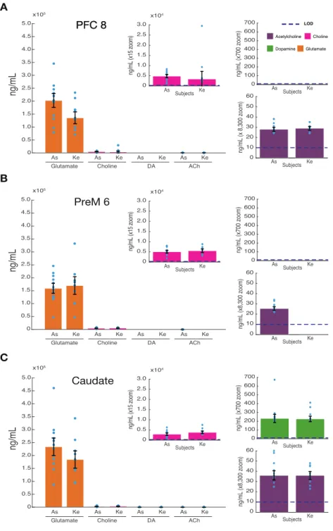
Different neuromodulators rarely act independent from each other to modify neural processes but are instead co-released, gated, or modulated. To understand this interdependence of neuromodulators and their collective influence on local circuits during different brain states, it is necessary to reliably extract local concentrations of multiple neuromodulators in vivo. Here we describe results using solid phase microextraction (SPME), a method providing sensitive, multi- neuromodulator measurements. SPME is a sampling method that is coupled with mass spectrometry to quantify collected analytes. Reliable measurements of glutamate, dopamine, acetylcholine and choline were made simultaneously within frontal cortex and striatum of two macaque monkeys (Macaca mulatta) during goal-directed behavior. We find glutamate concentrations several orders of magnitude higher than acetylcholine and dopamine in all brain regions. Dopamine was reliably detected in the striatum at tenfold higher concentrations than acetylcholine. Acetylcholine and choline concentrations were detected with high consistency across brain areas, within monkeys and between monkeys. These findings illustrate that SPME microprobes provide a versatile novel tool to characterize multiple neuromodulators across different brain areas in vivo to understand the interdependence and co-variation of neuromodulators during goal directed behavior. Such data will be important to better distinguish between different behavioral states and characterize dysfunctional brain states that may be evident in psychiatric disorders.
2.1.1 New and noteworthy
Our manuscript reports a reliable and sensitive novel method for measuring the absolute concentrations of glutamate, acetylcholine, choline, dopamine and serotonin in brain circuits in- vivo. We show that this method reliably samples multiple neurochemicals in three brain areas
simultaneously while nonhuman primates are engaged in goal directed behavior. We further describe how the methodology we describe here may be used by electrophysiologists as a low barrier to entry tool for measuring multiple neurochemicals.
2.2 Introduction
Extracellular concentrations of neuromodulators influence firing regimes, input-output relationships and neural interactions in local circuits and long-range brain networks (Marder, 2012;
Thiele and Bellgrove, 2018), and are dysregulated in virtually all psychiatric disorders (Millan et al., 2012; Avery and Krichmar, 2017). Accumulating evidence suggests that these fundamental roles of neuromodulators for circuit functioning are unlikely realized by single neuromodulators operating in isolation. Rather, neuromodulatory systems are heavily intertwined (Gobert et al., 1998; Avery and Krichmar, 2017; Moehle et al., 2017), and operate simultaneously on individual cells and circuits (Arnsten et al., 2010; Marder, 2012; Hassan et al., 2015; Santana et al., 2018). In each circuit, local mechanisms exert control over the release of neuromodulators from terminals of brainstem-originating projection neurons. This local control proceeds through activation of pre- synaptic glutamatergic receptors (Wang et al., 1992; Ghersi et al., 2003; Pittaluga et al., 2006;
Luccini et al., 2007; Grilli et al., 2009; Pittaluga, 2016). These insights suggest that an understanding of the contribution of neuromodulators to circuit functioning requires measuring, simultaneously, multiple neuromodulators in conjunction with ongoing glutamatergic neurotransmitter concentrations and action. Consistent with this conclusion single neuromodulator theories often fail to account for all observable symptoms in psychiatric diseases (Remy et al., 2005; Halliday et al., 2014; Bohnen et al., 2015).
Despite the accumulating evidence for the interdependence of neuromodulator actions, few methods exist for their simultaneous measurement in vivo and across multiple brain areas (Appendix A, Table A1; https://github.com/att-circ-contrl/SPME_paper_SI.git). Most of these existing neurochemical sensing methods allowing multi-neuromodulator sampling have a barrier to entry by requiring specialized equipment and trained experts preventing data collection by scientists who are otherwise interested in the role of endogenous and exogenous neuroactive chemicals in cognition and psychiatric disorders. Electrochemical methods such as fast scan cyclic
voltammetry (FSCV) and amperometry have sub-second temporal resolution, but are limited to the measurement of a few compounds (Dale et al., 2005; Jacobs et al., 2010) and are challenging and not robust for wide-spread in vivo application in nonhuman primates yet, although several labs have recently reported success (Quintero et al., 2007; Schluter et al., 2014; Disney et al., 2015;
Yoshimi et al., 2015; Schwerdt et al., 2017; Vartak et al., 2017; König et al., 2018). Imaging techniques such as positron emission tomography (PET) are also limited to the measurement of one or a few compounds simultaneously (Fisher et al., 1995). Microdialysis (MD) paired with mass spectrometry is the most commonly used method for measuring multiple neuromodulators in awake behaving animals. MD provides a data collection method which is then analyzed post hoc to identify and quantify collected analytes. It operates with a semi-permeable membrane which allows for the continuous collection of the available extracellular neuromodulators through passive diffusion, and can even be used to locally release pharmacological agents (Watson et al., 2006;
Buck et al., 2009; Perry et al., 2009; Anderzhanova and Wotjak, 2013; Kennedy, 2013). However, MD does have several disadvantages. MD disrupts the tissue during its initial placement of the probe or a guiding cannula resulting in damage-induced release of neuromodulators that can last several hours before stable measurements become possible. Moreover, MD has low affinity for hydrophobic compounds and comparatively broad spatial and temporal resolution that is in the range of 200-400 μm in diameter and 10-20 minutes, respectively. These values are dependent on the surface area of the permeable membrane, the exact method of MD, flow rate, resolution of detection methods for analytes of interest, tissue tortuosity and more (Watson et al., 2006;
Anderzhanova and Wotjak, 2013; Kennedy, 2013).
Here, we set out to address some of these limitations with a novel protocol for measuring multiple neuromodulators in vivo in discrete 20 minute intervals using probes optimized for solid phase microextraction (SPME) (Pawliszyn, 2000, 2012). SPME probes are thin (200 μm) wires of arbitrary length coated with an inert porous polymeric matrix using biocompatible binder on one end where molecules with appropriate size and affinity migrate via passive diffusion and are retained by weak intermolecular interactions (see Methods). SPME provides an alternate method for data collection which can then be analyzed by tools such as mass spectrometers. This method has been shown to extract in neural tissue dynamic changes in dopamine (DA) and serotonin (5- HT) levels with comparable precision to MD (Cudjoe et al., 2013; Cudjoe and Pawliszyn, 2014).
Additionally, due to the similarity of SPME probes to commonly used microelectrodes in electrophysiological recordings, relatively minor adjustments will allow for the adaptation of conventional microelectrode driving systems for SPME use. This, combined with post collection analysis through standard chemistry facilities makes SPME an attractive and easy-to-use tool for electrophysiology labs.
SPME has the potential to be a powerful new tool to compliment the mentioned methods well suited for neurochemical profiling that spans both multiple neuromodulators as well as multiple brain regions simultaneously. Such data will allow for global observation of slow neuromodulator dynamics that could better inform our hypotheses and help relate global neuromodulator levels to electrophysiology and behavior.
Thus, the ability of SPME to report major neuromodulators as well as glutamate and GABA were tested in two behaving rhesus macaques. Probes were repeatedly and simultaneously inserted into two cortical regions and the striatum to observe inter-areal differences between extracellular neuromodulator concentrations. We found that extracellular concentrations of glutamate, dopamine, acetylcholine and choline could be reliably distinguished and differed systematically between brain regions.
2.3 Materials and methods
2.3.1 Animals
Data was collected from two 8 year-old male rhesus macaques (Macaca mulatta) weighing 8-12 kg. All animal care and experimental protocols were approved by the York University Animal Care Committee and were in accordance with the Canadian Council on Animal Care guidelines.
Details regarding the experimental setup, recording procedures, and reconstruction of recording sites have been described previously (Oemisch et al., 2015). Briefly, animals were implanted with a 20 mm by 28 mm recording chamber over the frontal region of the right hemisphere guided by stereotaxic coordinates (Paxinos et al., 2000) and MR images. The animals were seated in a custom made primate chair and head stabilized with their eyes 65cm away from a 21’ LCD monitor