PART I: NEW MATERIAL OF CORYPHOMYS BUEHLERI SCHAUB, 1937, AND DESCRIPTION OF
A SECOND SPECIES OF THE GENUS
K. P. APLIN
Australian National Wildlife Collection, CSIRO Division of Sustainable Ecosystems, Canberra
and Division of Vertebrate Zoology (Mammalogy)
American Museum of Natural History ([email protected])
K. M. HELGEN
Department of Vertebrate Zoology National Museum of Natural History Smithsonian Institution, Washington and Division of Vertebrate Zoology (Mammalogy)
American Museum of Natural History ([email protected])
BULLETIN OF THE AMERICAN MUSEUM OF NATURAL HISTORY Number 341, 80 pp., 21 figures, 4 tables
Issued July 21, 2010
Copyright
E
American Museum of Natural History 2010 ISSN 0003-0090CONTENTS
Abstract . . . 3
Introduction . . . 3
The environmental context . . . 5
Materials and methods . . . 7
Systematics . . . 11
CoryphomysSchaub, 1937 . . . 11
Coryphomys buehleriSchaub, 1937 . . . 12
Extended description ofCoryphomys buehleri. . . 12
Coryphomys musseri,sp. nov. . . 25
Description . . . 26
Coryphomys,sp. indet. . . 34
Discussion . . . 40
Species diversity inCoryphomys. . . 41
Phylogenetic affinities ofCoryphomys. . . 43
Paleoecology ofCoryphomys. . . 63
Conclusions . . . 68
Acknowledgments . . . 69
References . . . 69
2
Large collections of fragmentary animal bones excavated from archaeological contexts in East Timor between 1968 and 2002 provide new material referable to the recently extinct, gigantic murine genusCoryphomys. We document the upper and lower dentition and palatal anatomy of C. buehleri Schaub, 1937, and identify and name a second species ofCoryphomys, based on differences in molar size and morphology and skeletal robusticity. Alternative interpretations of the observed morphological and metric variability (sexual dimorphism, resource-based polymorphism, sample heterochroneity) are each carefully assessed and rejected, and we conclude that the genus comprised two species of approximately similar body size. Preserved cranial elements of both species of Coryphomys feature a high degree of anatomical specialization, including an unusual elaboration of the maxillary sinus complex. Though the specialized anatomy ofCoryphomysinvites consideration of its phylogenetic relationships, this exercise is hindered by a demonstrable high level of homoplasy (i.e., multiple, independent evolutionary losses and gains) in many of the key craniodental features traditionally surveyed within Murinae, while other features are insufficiently well surveyed for broad-scale analysis.
Nevertheless, our comparisons highlight two potentially related lineages among the geographically proximate Murinae—the Philippine Phloeomyini and the Australo-Papuan Hydromyini. The remains ofCoryphomysare relatively scarce in all the archaeological samples, but distributional evidence suggests that both species ofCoryphomyswere found primarily in upland habitats. Late Pleistocene samples document their former presence at lower elevations, possibly reflecting cooler climatic conditions at that time.
INTRODUCTION
Timor is located near the eastern end of the Indonesian archipelago and is the largest and highest of the Lesser Sunda Islands (fig. 1). The surrounding region is geologi- cally young and tectonically active, the product of late Tertiary collision between three major earth units—the Asian and Australian continental plates, and the Pacific oceanic plate. Plants and animals of Asian and Australian affinities intermingle across the region and it was this melding of two such different biotas that inspired the nascent biogeographer Alfred Russel Wallace, and continues to excite the imagination of stu- dents of historical biogeography today.
Timor was one of the first of the Lesser Sunda Islands to be explored biologically, with collections made during the Baudin expeditions of 1801 and 1803, and subse- quently by Mu¨ ller, Wallace, and others (see Hellmayr, 1914, and Mayr, 1944, for the history of ornithological collecting). These expeditions encountered a distinctive avi- fauna, including a significant number of endemic species and subspecies. In contrast, the early expeditions encountered a fairly impoverished mammal fauna made up of the familiar suite of domesticates and commen- sals, and a moderate number of bat species,
the majority of which were subsequently found elsewhere within the Indonesian archi- pelago and beyond (see Goodwin, 1979, for a summary). Indeed, prior to the 1990s, the only potentially endemic Timorese mammals were two shrews (Crocidura tenuis: treated as a synonym of C. fuliginosa [Blyth, 1835] by Jenkins [1982] but maintained by Corbet and Hill, 1992: 42; and C. macklotti: included within C. fuliginosa by Jenkins [1982] but considered a synonym ofC. tenuisby Corbet and Hill, 1992) and one bat, the enigmatic Nyctophilus timorensis (see Goodwin, 1979;
Kitchener et al., 1991d, for discussion of this taxon). The impoverished nature of the Timorese mammal fauna was further high- lighted by subsequent discovery through the course of the 1900s of endemic nonvolant mammals on Seram and several other Mo- luccan islands (summarized by Helgen, 2003) and Flores (e.g., Sody, 1941; Kock, 1974).
More recent surveys of the Lesser Sunda Islands, particularly by staff of the Museum Zoologicum Bogoriense and the Western Australian Museum (Kitchener and Mar- yanto, 1993, 1995; Kitchener and Suyanto, 1996; Kitchener et al., 1991a, 1991b, 1991c, 1991d, 1994), also resulted in discovery of several additional endemic mammals includ- ing one native rodent extant on Timor, Rattus timorensis(Kitchener et al., 1991a).
3
The first hint of a much higher endemic mammalian diversity on Timor came to light during excavations by Alfred Bu¨ hler (see Sarasin, 1936) in a limestone cave near Nikiniki in southwest Timor. A damaged mandible and an incomplete femur of a very large rodent were forwarded to Schaub (1937), who described them as a new fossil murid, Coryphomys buehleri. The dental morphology ofC. buehleriwas later revisited by Stehlin and Schaub (1951: 348) who regarded the morphology of the anterior portion of m1 to be unparalleled among murines. Not long after, additional Timorese rodent material was recovered by Th. L.
Verhoeven from archaeological contexts at Lian Leluat on the Maubesi River, southern Timor (Verhoeven, 1959). Hooijer (1965) described and figured a total of three dentaries and though he noted considerable morphological variation, especially in the form of the anterior moiety of m1, he concluded that a single species was repre- sented.
Large samples of prehistoric mammal remains from Timor first came available through archaeological excavations by Ian Glover (1986) over the period 1966–1967.
Glover excavated a total of five sites using systematic methods and retrieved animal remains from contexts dated back as far as the terminal Pleistocene. The enormously abundant and surprisingly diverse rodent specimens were turned over to Jack Mahoney
for detailed study. Mahoney distinguished a total of four large murids including C.
buehleri, four smaller native murids, and two commensal murids, the latter found in association with remains of other introduced mammals including domesticates, and he figured each of the four ‘‘giant’’ rats (Maho- ney, appendix 3 in Glover, 1986). Musser (1981b) examined some of this new material and commented briefly on the possible affinities of the Timorese rodents, pending publication of Mahoney’s full account.
Sadly, Mahoney did not complete his studies of the Timor rats before his death in 1985.
Since that time the Glover material has passed through several sets of hands with the intention of completing his work, but so far this goal remains unfulfilled.
Additional impetus to document the pre- historic Timorese rodents arose after recom- mencement of archaeological research in East Timor in 2000 by archaeologists from the Australian National University, Canberra.
Excavations in five cave sites, one of them studied previously by the Portuguese archae- ologist Antonio de Almeida, again produced abundant vertebrate remains from contexts ranging from near contemporary back to more than 38,000 BP (O’Connor et al., 2002;
O’Connor and Aplin, 2007; Veth et al., 2005).
Our studies of the combined Glover and newly excavated collections inspired a new interpretation of taxic diversity within this fascinating prehistoric fauna. Like Mahoney Fig. 1. Regional map showing the location of Timor and other significant islands and continental landmasses of Wallacea. The grey shading indicates the approximate extent of land that is exposed with a sea level depression of 100–120 m below present level. Note that Timor remains isolated from other major islands and continental landmasses during episodes of lowered sea level.
4 BULLETIN AMERICAN MUSEUM OF NATURAL HISTORY NO. 341
and Musser, we recognize a total of four genera of large to gigantic murine rodents, and a total of five native smaller murines.
However, we depart from previous assess- ments in distinguishing a higher diversity among the large-bodied rats, with a mini- mum of eight species in the four genera. The total prehistoric murine fauna of Timor thus comprised at least 13 species, some with clear affinities with the murine fauna of Melanesia, others linked to the contemporary murine fauna of Southeast Asia, and others again of obscure affinities and potentially more an- cient origin.
The scale of the descriptive and compara- tive effort needed to document the prehistoric Timorese murine fauna requires that it be divided among a number of contributions.
This first paper sets the scene by describing the physical, biological, and archaeological context of the prehistoric Timorese rodents, and presents our conclusions on the taxon- omy of the genus Coryphomys, with two species recognized. We also present our preliminary observations on the phylogenetic relationships and paleoecology of this fasci- nating genus of murine rodent.
THEENVIRONMENTALCONTEXT
GEOGRAPHY: Timor is an impressive island even by global standards, with an area of 28,418 km2 and a maximum elevation of 2962 m. It is the largest and highest of the Lesser Sunda Islands, and one of the more varied in terms of climatic regimes and vegetation. It is also one of the more isolated, with a deep oceanic trench along the northern side of the island separating it from the Inner Banda Arc, a chain of volcanic islands that runs east from Flores and includes Lembata, Pantar, Alor, and Wetar. Further to the west, and again separated by very deep water, lie Savu and Sumba islands, while to the south, more than 450 km across the Timor Sea, extends the northwestern coastline of Aus- tralia. Timor has a significant satellite island at its southwestern end—Roti, with an area of 1227 km2and an elevation of 430 m. The two islands are separated by a 10 km wide strait with a sill depth of ca. 100 m.
A high central massif running the full length of Timor includes several peaks rising
to over 2000 m. The northern side of the massif rises abruptly from the coast, whereas in the south and southwest the ranges are fringed by broad alluvial and coastal plains.
Rugged relief with steep slopes, often in excess of 40%inclination, thus typifies much of the habitat on Timor, broken locally by valleys, uplifted sedimentary basins and uneven limestone plateaux.
Timor Island is divided politically in two roughly equal parts—an Indonesian portion in the west (forming part of the province of Nusa Tenggara Timur) and the independent nation of Timor Lorosa’e (East Timor) in the east.
GEOLOGY: Timor is located in the Neo- gene collision zone between the northwest continental margin of Australia and the Banda Island arc system and has a complex geology that has been subject to contrasting tectonic interpretations (Price and Audley- Charles, 1987; Charlton, 1991). A paleogeo- graphic framework for Eastern Indonesia based on tectonic and geological evidence is provided by Hall (2002).
Rocks belonging to four major tectono- stratigraphic units are exposed on Timor, namely: (1) basal, unmetamorphosed sedi- mentary rocks of the Australian continental shelf, ranging in age from Permian to Paleocene; (2) metamorphic rocks of variable age, extruded along the subduction zone; (3) volcanic fore-arc ophiolites of late Tertiary age; and (4) Neogene to Quaternary sedi- mentary formations, including postorogenic molasse sediments and uplifted coral reefs, the latter present locally to 1400 m and often highly karstic (Audley-Charles, 1968; Mid- dleton et al., 2006).
For biogeographers, the key points of interest are the age of subaerial emergence and of subsequent uplift of Timor. These are not known with any precision but are constrained by stratigraphic and radiometric data. Berry and Grady (1981) dated the main phase of deformation in the metamorphic Aileu Complex to between ca. 8 mya and 5.5 mya, and interpreted this phase as marking the main arc-continent collision.
Kaneko et al. (1987) suggested that exhuma- tion of the metamorphic belt doming com- menced in the late Miocene in West Timor but attributed major uplift to doming and
high-angle faulting of the welded sedimentary stack during the Quaternary, as a response to slab break-off of subducting Australian continental crust. Evidence of rapid uplift of Timor through the Quaternary comes from dated flights of coral terraces, with the uplift rate during the late Quaternary esti- mated at 0.5 m per 1000 yrs for northeastern Timor (Chappell and Veeh, 1978). Even higher local rates are implied by de Smet et al.’s (1989) suggestion of more than 2 km of uplift since ca. 5 mya in central West Timor.
CLIMATE: Timor enjoys a tropical oceanic climate but the pattern of rainfall is highly variable, due to the complex interaction of two monsoonal systems and local topogra- phy (Monk et al., 1997). From November to May, monsoonal winds blow from the north and bring thunderstorms and rain to most of the island but especially over the central ranges. From June to October, offshore winds from northern Australia cross the Timor Sea to bring rain to the southern side of the central ranges. Mean annual rainfall ranges from less than 600 mm on the north coast to over 3000 mm in mountainous areas along the south coast. Mean daily tempera- tures range from 23u–31uC in the lowlands to 15u–24uC at 1000 m. November is the hottest month, and July the coldest. Precipitation generally exceeds evaporation between De- cember and April, but there is a deficit between May and November (Monk et al., 1997).
VEGETATION: Less than 15% of the primary forest cover of Timor survives today and even this is highly fragmented, with the largest remnants mainly confined to the mountains and pockets along the south coast. Monk et al. (1997) reconstructed the original cover as a mosaic of evergreen and semievergreen rainforests, principally located along the southern side of the central ranges, and deciduous forests, with a largely com- plementary northern distribution but also found on the southern coastal strip. Small pockets of mesophyll vine forest occurred in the southeast of the island. All forest com- munities above 600–1000 m were ever- green, with broadleaf communities probably dominant. Drier sites probably supported sclerophyllous vegetation featuring endemic Myrtaceae including Eucalyptus urophylla
(Whitmore et al., 1989; Martin and Cossalter, 1977, 1979). Above 1500 m, all forests support a heavy mantle of mosses, lichens, and epiphytes, giving way to mosses alone above 1800 m. At the highest elevations, high wind shear creates stunted (elfin) tree com- munities and patches of low heath dominated by plants of ‘‘Himalayan’’ affinity (e.g., Rubus rosifolins,Ranunculusspp.). Mangrove and beach communities occupy suitably sheltered sites around the coastal margin.
Today, most of the land below 1000 m supports savannah woodland of casuarinas and eucalypts (mainly Eucalyptus alba), usually with a weedy understory heavily grazed by goats, cattle (mainly banteng or Balinese cattle), and horses (Metzner, 1977).
This community presumably developed over millennia through the practice of traditional shifting agriculture, which involves wide- spread burning at the end of the dry season.
Soil loss from steep slopes following initial forest destruction has also contributed to a radical change in the nature of the Timorese environment and there is little evidence of regeneration to original forest types. In the mountainous regions, the creation of open pasture by repeated burning has converted much of the adjoining forest to sclerophyll communities dominated by E. urophylla.
Commercial extraction of sandalwood (San- talum album) over many centuries has also affected forest communities (WCMC, 1995) and there has been a recent sharp increase over the last few decades in the rate of deforestation, in response to both population growth and commercial plantations and broad-acre agriculture (Bouma and Kobryn, 2004).
HUMAN PREHISTORY: The earliest evi- dence for human occupation of Timor dates to around 40,000 years ago (O’Connor and Aplin, 2007), and there is growing evidence for continuous occupation after that time, at least in the coastal regions. The earliest populations presumably lived by hunting and gathering, possibly combined with basic arboriculture, with little archaeological evi- dence for cultural or economic change prior to the appearance of various ‘‘Neolithic’’
markers (pottery, remains of various domes- ticates) in the late Holocene, dated to,3500 BP (Glover, 1986; O’Connor and Aplin,
6 BULLETIN AMERICAN MUSEUM OF NATURAL HISTORY NO. 341
2007). A notable exception is the sudden appearance during the early Holocene of a New Guinean marsupial,Phalanger oriental- is, presumably as a consequence of increas- ing human interaction between the Lesser Sunda Islands and Melanesia. The remains of now-extinct rodents occur in all the exca- vated sites, often co-occurring with Neolithic artifacts and the bones of domesticates. The same layers also contain the bones of two commensal murines (Rattus exulans and R.
rattus) that probably accompanied early agriculturalists into Eastern Indonesia some 3500 years ago (Bellwood, 1997). Whether these co-occurrences faithfully reflect the survival of the ‘‘giant’’ rats into recent millennia cannot be answered until such time as direct radiocarbon dating is undertaken on relevant bones and teeth.
The archaeological record of Flores Island to the west features a much longer record of human occupation, with strong evidence for initial colonization prior to 780,000 years ago (Morwood et al., 1998, 1999) and still controversial evidence for local evolution of a now extinct hominin species (Brown et al., 2004; Morwood et al., 2004; Jacob et al., 2006). To date, Timor has not produced any evidence for a long history of human occupation. However, it should be noted that poorly dated Quaternary sediments (Ainaro Formation of Audley-Charles, 1968) on Timor have produced fossil remains of three large vertebrates (a pygmy probosci- dean Stegodon sp., a ‘‘giant’’ turtle, and a
‘‘giant’’ varanid lizard [Verhoeven, 1964;
Hooijer, 1969a, 1969b]), none of which have
yet turned up in any of the archaeological assemblages. Exactly when and why these
‘‘megafaunal’’ species became extinct on Timor is not known. However, if the extinc- tions on Timor are linked to human activ- ities, as they so often seem to be (Burney and Flannery, 2005), we might reasonably postu- late a longer history of human occupation of Timor.
MATERIAL ANDMETHODS
THEARCHAEOLOGICALSITES: The collec- tions we studied come from excavations in nine archaeological sites, all located in East Timor (fig. 2). All the sites are shallow caverns or shelters developed in uplifted coralline limestones. The sites are clustered in five regions, as follows:
1. Northern flank of the Baucau Plateau, west of Baucau township on the north coast of East Timor. The plateau is approximately 400 km2 in area and comprises a series of Quaternary coral limestone terraces that rise from sea level to 500 m. Two sites were excavated by Ian Glover and his team.
Lie Siri (TL): a large, open cavern located just over 1 km from the coast, at an elevation of 240 m, and at approximately 8u269S, 126u229E. The cave deposit was excavated to a maximum depth of 2.05 m and produced a maximum carbon 14 (14C) determination of 7270 6 160 BP (ANU-236) from scattered charcoal fragments. Glover (1986) inferred a basal age of ca. 10,000 BP for the deposit and recognized a total of seven ‘‘horizons’’
(number from deepest to most surficial;
corresponding to sedimentological and cul- Fig. 2. Map of Timor showing the location of major towns and the various archaeological sites that have produced fossil murine remains.
tural subdivisions of the stratigraphic col- umn). Horizons Vc to VII contained pottery and the remains of domesticates. Jaws and teeth of large murids were obtained from horizons II to Vc.
Bui Ceri Uato (TB): a large, open cavern located around 2 km inland, 175 m and at approximately 8u279S, 126u229E. The deposit was excavated to a maximum depth of 1.45 m, with a total of 10 horizons identified. Pottery and bones of domesticates were found in horizons V to X and bones of large murids from horizons I to VI or VII. Glover encountered problems with radiocarbon dat- ing of this site but more recent14C determi- nations place the basal layers of Bui Ceri Uato in the terminal Pleistocene (Selimiotis, unpublished data cited by Oliveira, 2006: 95).
2. Central highlands of East Timor at about 600 m and just over 20 km from the nearest coast. Two sites located east of the township of Venilale, on the eastern side of Hatu Ariana, were excavated by Glover.
Uai Bobo 1 (TO1): a small, enclosed cave at approximately 8u389S, 126u239E. Excavation reached a maximum depth of 1.4 m, with a total of eight horizons recognized. The lowest
14C date obtained is 3470 6 90 BP (ANU- 414), but Glover (1986: table 65) inferred a basal age of around 9000 BP. Pottery and the bones of domesticates was found in horizons III to VIII, and bones of large murids from horizons I to V. Very large numbers of bones of small murids and bats were found in horizons I to IV; Glover attributed the accumulation of these remains to a nonhu- man predator. We agree and suggest an owl as the most likely agent.
Uai Bobo 2 (TO2): a small, enclosed cave located adjacent to Uai Bobo 1. Excavation reached a maximum depth of 4.9 m in a loose, dry deposit. Thirteen horizons were distin- guished, with a 14C determination for Ho- rizon I of 13,400 6 520 BP (ANU-238).
Pottery and bones of domesticates were found in horizons VII or VIII to XIIII. Large murids were recovered from horizons I to X.
A large quantity of small mammal bones was also recovered from this site.
3. Coastal scarp at the eastern end of East Timor, in the vicinity of Tutualu village.
Lene Hara (LH): a large, open cavern located about 5 km east of the village of Tutuala, 100 m and at approximately 8u249S, 127u169E. The site was excavated in 1963 by Antonio de Almeida (Almeida and Zbysz-
weski, 1967), reinvestigated by Glover in 1966 (Glover, 1986: 17) and then re-excavated by the ANU team in 2000–2002. The deposit has a complex stratigraphy and excavations in different parts of the site produced cultural and faunal assemblages that span three periods: from 30,000–35,000 BP; from 18,000–25,000 BP; and from ca.10,000 to recent (O’Connor et al., 2002; O’Connor and Aplin, 2007). Small samples of large murid remains were obtained from each strati- graphic context.
Jerimalai (JM): This site is located close to Lene Hara but is at slightly higher elevation and somewhat further inland. Excavated by O’Connor (2007) in 2005 to a maximum depth of 1.5 m. Basal14C dates in each of two excavated pits fall in the range 37,000–39,000 BP, but most of the stratigraphic profile spans the terminal Pleistocene and Holocene.
Small samples of large murid remains were obtained from each stratigraphic context.
4. Adjacent to Lake Ira Laloro, the largest freshwater lake in Timor, located at ca. 334 m in Laute´m Province, central to the eastern peninsula of East Timor. Two sites were excavated by the ANU team in 2001. Both caves are in a cliffline located a few hundreds of meters from the northern margin of the lake floodplain.
Matja Kuru 1 (MK1): a large, open cavern, at approximately 8u269S, 127u089E. Excavation reached a maximum depth of 1.55 m. The basal part of the deposit is terminal Pleisto- cene, but the bulk of the sediments accumu- lated during the period 5600–4600 BP (Veth et al., 2005). Large samples of murid remains were obtained from these levels.
Matja Kuru 2 (MK2): a large, open cavern located a few hundreds of meters to the east of Matja Kuru 1. Excavation by the ANU team reached a maximum depth of 2 m. The sequence produced a basal date around 32,000 BP but features an apparent hiatus in deposition over the period 30,000–15,000 BP. Large samples of ‘‘giant’’ rat remains were obtained from this site.
5. The Com area, on the north coast of the eastern peninsula.
Telupunu cave: a moderately large, open cavern located approximately 5 km inland from the coast, at 260 m, and at approxi- mately 8u229S, 127u029E. Excavation by Spriggs in 2002 reached a depth of 1.5 m.
The greater part of the sequence is preceramic and contains sparse cultural and faunal
8 BULLETIN AMERICAN MUSEUM OF NATURAL HISTORY NO. 341
remains, with a basal age of around 13,500 BP (Veth et al., 2005). Preservation of organic remains including bone in this deposit is superior to that of all others.
ASSOCIATION OF UPPER AND LOWER
DENTITIONS: As reported previously by both Mahoney (appendix 3 in Glover, 1986) and Musser (1981b), the Timorese ‘‘giant’’ murids fall into three major groups based on their dental morphology: (1) specimens with highly cuspidate, complex molars (Coryphomysand Mahoney’s genus A); (2) specimens with high- crowned, transversely laminate molars (Ma- honey’s genus B); and (3) specimens with low- crowned molars with cusps fused into trans- verse laminae (Mahoney’s genus C). Upper and lower dentitions of each general group are easily paired. However,Coryphomysand Mahoney’s genus A are similar in general size and morphology and greater care is needed to correctly associate upper and lower molars.
To do so, we paid attention to the following attributes of the specimens: (1) relative upper and lower molar row lengths (in murines length of the upper molar row usually exceeds that of the lower molar row, usually by 4%–
6%); (2) the morphology and dimensions of the upper and lower incisors (in murines width of the upper incisors almost always exceeds that of the lowers; one exception is Nesokia indica); and (3) the relative abun- dances of the various morphological forms recognized within the complex-toothed group. Using these criteria, it is possible to confidently associate upper and lower denti- tions with each of the individual species.
ALLOCATION OF DISSOCIATED CRANIAL
ELEMENTS: As is commonly the case with archaeological assemblages, almost all the cranial specimens of the Timorese murines consist of individual dissociated elements, the majority with some damage. In the present collection, two partial crania are available, each referable to a different genus, and these provide the key to allocation of other less complete specimens. One of the two partial crania is thoroughly burnt and fragmented but could be reconstructed to provide an almost complete cranium belonging to Ma- honey’s Genus A. This specimen is critically important as it allows for direct association of an upper molar morphology with various
other craniodental elements including the premaxilla and upper incisor, and the frontal, petrosal, and ectotympanic bones. The sec- ond specimen consists of a palate with associated interorbital portions of the neuro- cranium, referable to a species of Mahoney’s Genus B. Both specimens will be described in a later contribution of this series. Judging from the relative abundance of the tooth- bearing elements, these two genera are the most abundant taxa overall in the collection.
With these specimens as a reference point, the large number of dissociated specimens of other bones could be assessed as to their probable affinity. As a result, it is possible to generically assign the majority of isolated upper incisors and premaxillae, frontal bones, and petrosal bones, and to assign some specimens to species level.
The isolated petrosal bones from large murines showed striking morphological var- iation and particular effort was made to allocate these elements. An initial analysis produced four morphological groups. One well-defined group, represented by five ex- amples, includes the petrosal associated with the burnt cranium of Mahoney’s Genus A. A second distinctive group, including several dozens of specimens, consists of petrosals that are morphologically very similar to the petrosal ofRattus norvegicus, albeit of much larger size. This group is referred with confidence to Mahoney’s genus B, for two reasons: (1) this genus is also most abun- dantly represented among the jaws and teeth;
and (2) its dental morphology also suggests an affinity with Rattus and its close allies (members of the Rattus Division, sensu Musser and Carleton, 2005). The third and fourth groups are represented by three and two petrosals, respectively. These groups presumably represent Coryphomys and Ma- honey’s Genus C, the latter of which shows strong dental similarities with various Aus- tralo-Papuan murines including Uromys, Solomys, andMelomys. Petrosals of the third group are referred with confidence to Maho- ney’s Genus C, for three reasons: (1) they are consistently smaller than petrosals of the fourth group (judging from mandible dimen- sions, Genus C is a smaller murine than Coryphomys); (2) they do not show any significant variation in size or morphology
(consistent with the lack of significant size variation in the dental sample of this taxon);
and (3) they are consistent in general morphology with petrosals of Uromys and Melomys. In contrast, the two petrosals of the fourth group are larger, differ markedly in morphology from those of Uromys and Melomys, and differ considerably in size, consistent with our identification of two species within the Coryphomyssample.
None of the abundant postcranial remains from the various sites are associated with other skeletal elements, either postcranial or cranial. Though great morphological diver- sity is present in nearly every skeletal element, the task of allocating all of this dissociated postcranial material to particular taxa is one for the future.
TERMINOLOGY ANDMEASUREMENTS: For naming the principal cusps and crests of the molar teeth of Coryphomys spp. we have followed the descriptive terminology em- ployed by Musser in his numerous works (e.g., Musser, 1981a; Musser and Heaney, 1992), based for the upper molars on the system introduced by Miller (1912) and
modified by Misonne (1969) and for the lower molars on the terminology of Weerd (1976: 44). This terminology is illustrated here (see fig. 3) for each of Coryphomys buehleri and Lenothrix canus, the latter to identify structures that are not represented in the dentition of Coryphomys. Where appro- priate, we identify the equivalent structures in alternative terminologies as used by others.
We use the term ‘‘lamina’’ to refer to the more or less transverse rows of cusps that typify both the upper and lower molars of murine rodents. The first upper molar has three such laminae (anterior, middle, and posterior) while each of the second and third upper molars has two laminae each. In the lower molars, each tooth has two primary laminae that are also called ‘‘chevrons’’ in some contexts; this is supplemented in the case of the first lower molar by an additional structure—the anteroconid—that is attached to the front of the anterior lamina.
Osteological terminology of the mamma- lian skull remains highly multifarious, with many different terms often in use for what are undoubtedly homologous features in Fig. 3. Nomenclature of upper and lower molar crown structures in each of Lenothrix canus(after Musser, 1981) and Coryphomys buehleri. A, upper molars of Lenothrix canus; B, upper molars of Coryphomys buehleri;C,upper molars ofLenothrix canus;D,upper molars ofCoryphomys buehleri. For upper molars principal cusps are numbered according to Miller’s (1912) system; these are identified in the text with the prefix ‘‘t.’’Abbreviation:pc, posterior cingulum. Lower molar cusps are labelled according to the system of Weerd (1976).Abbreviations:a-cen, anterocentral cuspid;a-lab, anterolabial cuspid;a-lin, anterolingual cuspid; alc, anterolabial cusplet;alic, anterolingual cusplet; ed, entoconid;hd, hypoconid, md, metaconid,pc, posterior cingulid;pd, protoconid;plc, posterolabial cusplet.
10 BULLETIN AMERICAN MUSEUM OF NATURAL HISTORY NO. 341
different groups of mammals. Wahlert (e.g., 1974, 1985) has established a systematic terminology for cranial foramina across all major groups of rodents. For the most part here, we have followed his usage. However, for osteological features of the auditory region that relate to the passage of arteries and veins, we have adopted the terminology of Wible (e.g., Wible et al., 2001) as this better reflects both the embryology and phylogeny of the mammalian blood vascular systems.
There is a substantial body of work on the functional anatomy of the masticatory mus- cles of rodents, especially of Rattus norvegi- cus(e.g., Hiiemae, 1971; Hiiemae and Hous- ton, 1971; Weijs and Dantuma, 1975), and a growing body of comparative work on other groups of muroids (Bekele, 1983; Satoh, 1997, 1999; Satoh and Iwaku, 2004, 2006, 2008). In contrast, osteological features that relate to the attachment of tendons and aponeuroses are generally given light treat- ment in standard anatomical sources. To more accurately describe these features, we have adapted the terminology of Satoh and Iwaku (2006) for subdivisions and apo- neuroses of the primary masticatory muscles.
For other elements of muscular anatomy, we have used the more conventional terminology of Greene (1968).
The internal osteology of the mammalian nasal cavity has attracted little general attention in the paleontological literature and virtually none in the case of fossil murines. Fortunately, for laboratory rats and mice, there is a rapidly growing literature on the anatomy, histology and embryology of the olfactory and glandular tissues of the nasal cavity (e.g., Jacob and Chole, 2006) and this provides both a basic terminology for major spaces and a basis for reconstruction of soft tissue relations in fossil murines.
However, as this literature generally does not identify the osteological expression of soft tissues features, we have been obliged to introduce some new terminology for this region of the skull.
Craniodental measurements were taken with handheld digital calipers, to the nearest 0.01 millimeter (mm). Upper and lower molar lengths are maximal coronal dimen- sions taken along the approximate midline of
the tooth, with the body of the caliper held parallel to the occlusal plane.
INSTITUTIONAL ABBREVIATIONS: The Ti- morese fossil samples are housed in three collections, identified by the following pre- fixes: Australian Museum, Sydney (Palaeon- tology): AMF; Australian National Wildlife Collection, CSIRO, Canberra (Palaeontol- ogy): ANWCP; American Museum of Nat- ural History, New York (Modern mammals):
AMNH. Comparative specimens in the mod- ern mammal collection of the Australian National Wildlife Collection, CSIRO, Can- berra (Mammals) are identified by the prefix ANWCM.
DENTAL ENUMERATION AND ABBREVIA- TIONS: Teeth of the upper and lower molar series are designated by superscript and subscript numbers, e.g., first upper molar is M1, second lower molar is m2. Symmetry of teeth is indicated by a prefixed R for right side and L for left side; e.g., right first lower molar is R m1.
ANATOMICAL ABBREVIATIONS: The ab- breviations used for upper and lower molar cusps are shown in figure 3. Other abbrevia- tions are listed beneath each figure in which they are used.
SYSTEMATICS Family Muridae Subfamily Murinae CoryphomysSchaub, 1937
TYPE SPECIES: Coryphomys buehleri Schaub, 1937.
CONTENT: Coryphomys buehleri Schaub, 1937,Coryphomys musseri, sp. nov.
REVISED DIAGNOSIS: A genus of large- bodied, complex-toothed murines distin- guished from all others by the combination of the following morphological features:
molar crowns moderately hypsodont and weakly inclined; anterocentral cuspid promi- nent on m1; principal lingual cusps of upper molars (t1, t4) isolated from central cusps until molars attain advanced state of wear; a discrete cusp t7 united to cusp t8 on each of M1–2; posterior cingulum forming an ele- vated, transverse occlusal ridge behind cusps t8–t9 on each of M1–3, and barring contact between central row cusps on successive
molars; upper and lower third molars almost equal in area to the corresponding second molars; upper and lowers incisors large and with simple, D-shaped cross-sections; incisive foramen very short and narrow, penetrating only a short distance into palatal lamina of maxilla; zygomatic plate tall and narrow, with a straight anterior margin and shallow zygomatic notch; stem of stapedial artery extremely reduced such that promontorium lacks a conspicuous stapedial sulcus; rostrum shortened; and maxillary sinus complex enlarged and elaborate, causing a marked constriction of both the posterior nasophar- ynx and the infraorbital sulcus.
Presence of a large cusp t7 on each upper molar distinguishes Coryphomys from the majority of previously described murine genera. Among genera with large cusps t7, Coryphomys is distinguished from all except Carpomys and Spelaeomys (Hooijer, 1957;
Musser, 1981b) by the presence of a sub- stantial posterior cingulum on all three upper molars. It is distinguished fromCarpomysby its highly cuspidate molar form (cusps are largely fused into transverse laminae in Carpomys spp.) and the absence of labial cusplets on m2–3. It differs fromSpelaeomys in numerous details of molar morphology including the greater size of the posterior cinguli on all upper molars, the absence of cusp t3 on M2–3, and the absence of accessory labial cuspids and cusplets on all lower molars.
REMARKS: Under Article 32.5.2.1 of the Fourth Edition of the International Code of Zoological Nomenclature (International Commission on Zoological Nomenclature, 2000: 40) the original specific epithet bu¨hleri is adjusted tobuehleri.
Coryphomys buehleri Schaub, 1937 HOLOTYPE: Naturhistorisches Museum Basel, NMB A.P.1. A right dentary with m1–3 and root of incisor; preserving condylar process but with damage to coronoid and angular processes. Figured by Schaub (1937:
figs.1 and 2). Not examined for this study.
REFERRED MATERIAL: AMF 68831: Uai Bobo 1 (TO1/2), R Dentary with m1–3;
AMF 68789: Bui Ceri Uato (TB/2), L Dentary with m1–2; AMF 68850: Lie Siri
(TL/E 5C/C3), R Dentary with m1–2;
ANWCP1: Jerimalai (JM/B/19) L m2;
ANWCP2: Matja Kuru 1 (MK1/A/17), L M3; ANWCP3: Matja Kuru 1 (MK1/AA/
28), R M2; ANWCP4: Matja Kuru 1 (MK1/
AA/25), L M3; AMF 68838: Lie Siri (TL/E/5C/C3), L maxilla with M1; AMF 68751a: Uai Bobo 1 (TO1/3), L palatal fragment (partial maxilla and palatine) with M1–3; AMF 68751b: Uai Bobo 1 (TO1/3), R maxilla fragment with M1; AMF 68751c:
Uai Bobo 1 (TO1/3), L zygomatic plate;
ANWCP5: Matja Kuru 1 (MK1/AA/10), R M1; ANWCP6: Matja Kuru 2 (MK2/D/32), R m1; ANWCP7: Matja Kuru 2 (MK2/D/
41), L M3; AMF 68824: Uai Bobo 1 (TO1/3), R M1; ANWCP8: Matja Kuru 1 (MK1/AA/
10), L M2.
REMARKS: Given the presence on Timor of various other equally large murines, it is possible that the murine femur referred by Schaub (1937: fig. 3) toCoryphomys buehleri may not belong to this taxon. Its identity will be reassessed following allocation of the abundant postcranial remains now available from the many excavated assemblages.
Only one of the three dentaries (no. 2) attributed to C. buehleri by Hooijer (1965) actually belongs to this species. Hooijer’s specimen no. 1 is referable to Mahoney’s Genus A, while his no. 3 belongs to the new species of Coryphomys described below.
Ironically, Coryphomys buehleri, the only formally described member of the prehistoric Timorese murine fauna, is also one of the rarest of all the large-bodied taxa in the fossil samples.
Attribution of a maxillary dentition to this species is based on the recognition of a morphologically distinctive subset of Cor- yphomys specimens that are also slightly larger in all dental dimensions relative to those of the second species.
EXTENDEDDESCRIPTION OF
CORYPHOMYS BUEHLERI
UPPER MOLARS (fig. 4; table 1): The upper molars are moderately hypsodont but comprised of variably discrete and partially united cusps arranged in more or less transverse series. The occlusal plane of the molar row is tilted outward (i.e., lingual
12 BULLETIN AMERICAN MUSEUM OF NATURAL HISTORY NO. 341
cusps are significantly lower than labial cusps) but there is little indication of helical torsion along the molar row.
No maxillae without molars are confi- dently assigned to this species. The M1 has four roots. The anterior root, supporting the anterior lamina, is broad and kidney shaped with the concavity facing the rear. Two small circular roots on the lingual side of the tooth support cusps t4 and t7. A posterolabial root, intermediate in size, supports cusps t8 and t9 and the associated posterior cingulum. The root pattern for M2 cannot be determined from available specimens. Two isolated M3 show a broad anterior root supporting cusps t1 and t4–6, a somewhat narrower root supporting cusps t7–8 and a small labial rootlet above cusp t9 and partially united to the front of the posterior root.
M1: This tooth is represented by a total of five specimens (fig. 4A–E). They are broadly
ovate in occlusal outline but have a weakly concave labial margin.
The anterior lamina consists of three discrete, columnar cusps. The central cusp t2 curves forward and downward from a bulbous base, narrowing toward an occlusal surface that is rounded anteriorly but flat- tened on each of the labial, lingual, and posterior margins. The labial cusp t3 is positioned level with cusp t2. It is firmly adpressed to the labial surface of cusp t2 but is fully encircled by enamel for approximately one third of its total height. Above that level, the two cusps are united but with well- defined anterior and posterior grooves that extend to the crown base. Cusp t3 also has a bulbous base and narrower apical portion.
The occlusal surface is ovate, with the long axis oriented at approximately 45u to the coronal major axis. Cusp t1 is isolated from the cusp t2–3 complex by a deep cleft that Fig. 4. Occlusal views and one lingual view of upper molars referred toCoryphomys buehleri. Occlusal views: A,AMF 68751a: associated left M1–3;B, AMF 68751b: a right M1 (same individual as AMF 68751a);C,AMF 68838: a left M1;D,AMF 68824: a right M1;E,ANWCP5: a right M1 (missing cusps t1–2);F,ANWCP3: a right M2;G,ANWCP8: a left M2;H,ANWCP7: a left M3;I,ANWCP4: a left M3;
J,ANWCP2: a left M3. Lingual view:K,AMF 68751a: associated left M1–3. The lingual view illustrates the moderately hypsodont condition of the molars inC. buehleriand the slight posterior inclination of the cusps on M1, trending to vertical on M3. Scale bar beneath A also applies to B–J and represents 5 mm. A separate 5 mm scale bar is provided for K.
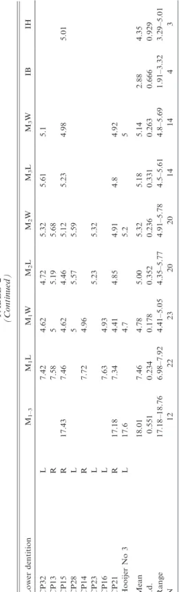
TABLE1 MeasurementsofUpperandLowerDentitioninSpecimensAttributedtoCoryphomysbuehleri Measurementsaremolarlengths(L)andwidths(W)andthebreadth(IB)anddepth(ID)ofthelowerincisor.Samplesizes,means,standarddeviations, andrangesarealsoshown. UpperdentitionSymmetryM1–3M1LM1WM2LM2WM3LM3W CP2L5.975.12 CP3R5.695.51 CP4L5.55.22 AMF68838L8.516.15 CP7L5.595.53 AMF68824R8.56 AMF68751aL19.498.565.775.865.85.45.31 AMF68751bR8.65.66 Mean19.498.545.905.785.665.625.30 s.d.0.0460.2210.1200.2050.2490.175 Range8.5–8.65.66–6.155.69–5.885.51–5.85.4–5.975.12–5.31 N1442244 Lowerdentitionm1–3m1Lm1Wm2Lm2Wm3Lm3WIBID AMF68831R18.847.755.145.365.565.505.39 AMF68789L7.625.145.45.483.154.88 AMF68850R8.125.375.716.163.40 CP19L5.426.28 CBP6R8.55 HolotypeR19.98.55.45.96.25.405.60 HooijerNo.2L20.15.25.75.80 Mean19.618.005.165.475.875.505.393.284.88 s.d.0.6770.4100.1520.2360.3560.0710.2050.177 Range18.84–20.17.62–8.125.14–5.375.36–5.715.48–6.285.4–5.55.39–5.83.15–3.40 N356562321
14 BULLETIN AMERICAN MUSEUM OF NATURAL HISTORY NO. 341
extends more than three quarters of the way to the crown base. The cusp is bulbous basally but narrows apically and folds against the anterolingual surface of cusp t5.
A weak posterolingual groove on cusp t1 produces a kidney-shaped occlusal surface which is oriented at approximately 45uto the main coronal axis. The occlusal surface of cusp t1 lies in a plane slightly above that of cusps t1–2 and t5.
The middle lamina consists of a united but lobular cusp t5–6 complex and a discrete but adpressed cusp t4. Cusps t5 and t6 are approximately equal in bulk but differ in shape. The central cusp t5 is columnar and laterally compressed, and is smaller in occlusal area than cusp t2. Cusp t5 is firmly connected to cusp t6, but the cusp boundary is marked by strong posterior groove. Cusp t6 is an elongate, transversely oriented structure with a bulbous base, a rounded posterior surface, a flattened anterior surface and a well-developed posterolingually direc- ted ridge. Cusp t4 is similar in basic shape and orientation to cusp t1, but differs in the presence of a well-developed anterolabial ridge that abuts against the corresponding ridge from cusp t4. As a result, the occlusal surface of cusp t4 is broadly arcuate (concave anteriorly) and continuous with the cusp t5–6 complex. The occlusal surface of cusp t4 lies in the same plane as cusp t1 and is thus slightly above that of cusps t5–6.
The posterior lamina consists of a trans- versely oriented t8–9 complex and a promi- nent t7 that is firmly connected to cusp t8.
The central cusp t8 is similar in size and shape to cusp t5. The boundary between cusps t8 and t9 is well marked in relatively unworn specimens by an apical cleft but this is rapidly obliterated by wear. The lateral margin of cusp t9 supports a broad an- terolabially directed ridge that ascends to the crown base. With progressive wear, this ridge produces an elongation of the occlusal sur- face of cusp t9. Cusp t7 is discrete apically from t8 but soon unites to this cusp with wear. In contrast, the fissure separating cusp t7 from cusp t4 extends almost to the crown base, thereby keeping the two cusps separate, even under advanced wear.
The posterior cingulum is an elevated transverse ridge that forms the posterior
surface of the tooth. The cingulum shows a variable pattern of connections. On both sides of AMF 68751 (fig. 4A–B) it is firmly connected labially to the point of union of cusps t8 and t9, but remains well separated from the posterior surface of cusp t7 as it rises toward the crown base, creating a lingually open cingular shelf. On AMF 68838 (fig. 4C) the labial end of the cingulum is united high to the anterior surface of cusp t9, thereby enclosing a distinctive cingular fossette.
AMF 68824 (fig. 4D), with more advanced wear, also has a fully enclosed fossette with broad cingular connection at both labial and lingual ends. Two small pimples on the elevated portion of the ridge create a weakly bicuspid occlusal outline on AMF 68751 (fig. 4A–B).
M2: Three specimens are available (fig. 4A, F–G), each at a different wear stage.
The less worn example, part of the complete molar series in AMF 68751 (fig. 4A), is described first. In both size and cuspal arrangement the M2 closely resembles M1, save for the absence of elevated cusps t2–3.
Cusp t1 is more vertically oriented than on M1 but is similarly folded against the anterolingual surface of cusp t5. Its posterior surface is rounded and the occlusal surface is correspondingly ovate rather than kidney shaped. The occlusal surface of cusp t1 lies in a plane slightly above that of cusp t5. Cusp t3 is represented by a small nubbin positioned near the crown base in the fold that marks union of cusps t5 and t6.
Cusps t5 and t6 of the middle lamina are firmly united but cuspal limits are marked by strong anterior and posterior grooves. The central cusp t5 is columnar and laterally compressed, and is slightly larger than the serial homolog on M1. Cusp t6 is larger in occlusal area than cusp t5. It is broader and more transversely oriented than the serial homolog on M1 but otherwise similar in form. Cusp t4 is similar in basic shape and orientation to cusp t1 but is larger in occlusal area. It lacks the anterolabial ridge seen on cusp t4 of M1 and this has a simpler, ovate occlusal outline. The occlusal surface of cusp t4 lies slightly above that of cusps t5–6, but the contrast is less marked than on M1.
The posterior lamina is an almost exact duplicate of this structure on M1 except that cusp t7 is slightly larger in occlusal area. The posterior cingulum is connected at its lingual end to the posterior surface of cusp t7. The labial end is deeply separated from cusp t9 by a cleft that ascends almost to the crown base.
ANWCP3 (fig. 4F) is in a more advanced state of wear. It shows comparable features save for a more elevated connection between the labial end of the posterior cingulum and the posterior surface of cusp t9. Cusps t7–t9 and the posterior cingulum are united by a common dentin pool in this specimen, while cusps t4 and t1 each remains discrete from cusp t5. ANWCP8 (fig. 4G) is even more heavily worn. Cusps on both laminae are united into common dentin pools but cusp t9 remains isolated from the labial end of the posterior cingulum.
M3: Four specimens are available (fig. 4A, H–J). The least worn examples of this tooth (AMF 68751, fig. 4A; ANWCP4, fig. 4I) are only slightly shorter and narrower than M2, but they differ in numerous morpho- logical features. Cusp t1 is more conical in form but bears a short ridge on its labial surface, directed to the front of cusp t5.
Cusps t5 and t6 are firmly united into a transversely oriented lamina with a common dentin pool. There is no delimiting anterior groove, and no cusp nubbin or fossette in the position of cusp t3. However, a posterior groove indicates the relative contribution of the two cusps, with cusp t6 being slightly the larger. Cusp t4 is smaller and more trans- versely oriented than it serial homologs on M1–2; the occlusal plane is only slightly above that of cusp t5 and the two cusps are united by a common dentin pool.
Cusps t7 and t8 are united into a broad, transversely oriented structure that is gently convex anteriorly. The boundary between the two equal-sized cusps is marked by a slight indentation of the anterior margin and a corresponding flexion of the occlusal surface.
Cusp t9 is a smaller columnar cusp that is firmly applied to the labial surface of cusp t8 but projects posterior to this cusp.
The posterior cingulum forms a robust, transversely oriented lamina. It is united high on the crown to the posterior surfaces of cusps t7 and t9 and has a rounded and
bulbous posterior surface. The occlusal sur- face consists of two facets, the lingual facet paralleling that on cusp t7.
The most heavily worn example of M3 (ANWCP2, fig. 4J) has the posterior cingu- lum joined by a common dentin pool to t9 but still discrete from cusp t7 at the lingual end.
LOWERINCISOR: The incisor is retained in AMF 68789 (fig. 9F) and lacks only the tip.
The body of the tooth is approximately D- shaped in cross-section and has a width-to- depth ratio of 0.645 (table 1). A smoothly curved strip of enamel is present along the ventral and lower lateral surfaces of the tooth, extending just over half way up the lateral surface of the tooth and terminating at the widest point of the tooth. Enamel is absent from the medial surface, save for a 0.34 mm rim corresponding to the thickness of the enamel layer. Pale orange pigment is present medially in a 1.6 mm wide band.
Otherwise the enamel is unpigmented. The enamel-free dorsal half of the incisor bears a weak dorsolateral groove. The medial surface is very slightly concave. The occlusal facet is incomplete but terminates posteriorly in a distinct step.
LOWER MOLARS (fig. 5, table 1): The lower molars are also moderately hypsodont.
The enamel is smooth on all surfaces, irrespective of intensity of wear. The occlusal plane of the molar row has a moderate degree of helical torsion, changing from transversely horizontal on m1 to lingually inclined (i.e., lingual cusps slightly lower than labial cusps) on m3.
No dentaries without molars are confi- dently assigned to this species; hence, infor- mation on molar root patterns is based on careful examination of teeth in situ. The m1 has two roots. The posterior root has similar dimensions on the labial and lingual sides and appears to bear a posterior groove; it probably occupies a symmetrical and possi- bly weakly bilobed alveolus. In contrast, the anterior root appears much broader labially than lingually, and the alveolus is presumably comma shaped with an anterior ‘‘head’’ and a posterolabial ‘‘tail.’’ The m2 appears to have two broadly united, transverse roots, one supporting each of the two principal chevrons. The posterior root, as exposed on
16 BULLETIN AMERICAN MUSEUM OF NATURAL HISTORY NO. 341
AMF 68789, is solidly united to the root apices and shows an increase in width with depth. This firm anchoring of the molars presumably accounts for the high rate of retention of molars in preserved dentaries of this taxon. The alveoli of m3 are exposed on AMF 68850. The anterior alveolus is broad and contained a fused root that terminated in short separate nubbins. The posterior alveo- lus is narrower and more circular.
m1: This tooth is represented by four new examples (fig. 5A–D). They have a rectangu- lar outline that is broadest across the posterior
lamina and narrows slightly to the front. The anteroconid is relatively large and equals the two posterior lophids in occlusal area. The principal cuspids are weakly united, even in heavily worn examples. The occlusal surfaces of the labial and lingual rows of cuspids are elevated externally and slope down to a central occlusal valley. The external surfaces of the labial cuspids are vertical, whereas those of the lingual cuspids are slightly bowed in less heavily worn examples.
The anteroconid shows three principal cuspids arranged in a somewhat variable Fig. 5. Occlusal views of lower molars referred toCoryphomys buehleri.A,AMF 68831: associated right m1–3;B,AMF 68789: associated left m1–2;C,AMF 68850: associated right m1–2;D,ANWCP6: an isolated right m1;E,ANWCP1: an isolated right m2. Scale bar represents 5 mm and applies to all images.
pattern. The anterolingual cuspid is consis- tently the largest and most discrete of the three. It has an ovate occlusal outline, with the labial end adpressed against the front of the protoconid and a long axis that swings approximately 30uforward of transverse. The anterolabial cuspid is transversely aligned with the posterolabial end of the anterolin- gual cuspid but it is considerably smaller and has a more rounded occlusal outline. In all specimens the two cuspids are deeply sepa- rated almost to their bases. The anterocentral cuspid is more variable in size, morphology, and relations. In AMF 68850 (fig. 5C) it is a simple rounded cuspid that is broadly con- nected to the anterior surface of the ante- rolabial cuspid. All other examples show a bicuspid anterocentral cuspid. In ANWCP6 (fig. 5E) it is divided into two subequal components by an anterolabial groove; the posterolabial end is adpressed against the anterolabial cuspid. AMF 68831 (fig. 5a) shows a larger labial component and a smaller, pimplelike lingual component at- tached high on the anterolingual side; the combined anterocentral cuspid in this exam- ple is equally discrete from each of the anterolabial and anterolingual cuspids.
AMF 68789 (fig. 5B) has the anterocentral cuspid divided into a larger lingual compo- nent and a smaller labial component, the combined cuspids having a specific attach- ment to the anterolabial cuspid. In all cases, the occlusal surface of the anterocentral cusp is elevated anteriorly and slopes down to the rear, thereby blocking the longitudinal oc- clusal valley of m1. The type specimen, as described and illustrated by Schaub (1937:
fig. 2), has a divided anterocentral cuspid.
Hooijer’s (1965: pl. I) specimen no. 2, herein referred to C. buehleri, possesses an undi- vided cuspid.
Two specimens show small accessory cuspules attached to the anteroconid. AMF 68831 (fig. 5A) has a small cuspule attached to the posterolingual base of the anterolin- gual cuspid. The other (ANWCP2, fig. 5C) has a basal cuspule attached to the posterior surface of the anterolabial cuspid; a weak buttresslike ridge from the base of the protoconid joins this structure to produce a partial cingulum across the base of the anterolabial flexid. These cuspules appear to
be absent from the holotype and Hooijer’s specimen no. 2, judging from the respective illustration and figure.
The anterior lamina of m1 is made up of two elongate cuspids arranged in a forward- facing chevron. The labial protoconid is elongate and has an occlusal surface that is variably rectilinear or weakly dumbbell shaped, the latter condition produced by shallow concavities on both the anterolabial and the posterolingual surfaces. The lingual metaconid is D-shaped, flattened anteriorly, curved posteriorly, and smaller in occlusal area than the protoconid. In two specimens (AMF 68831, fig. 5A; AMF 68850, fig. 5C) the metaconid abuts the anterior end of the protoconid without overlap. In the other two (AMF 68789, fig. 5B; ANWCP6, fig. 5D), the protoconid overlaps the front of the metaconid, so that the latter abuts the lingual face of the protoconid. In both conditions the two cuspids remain narrowly separate almost to the crown base.
Hooijer’s (1965: pl. I) specimen no. 2, herein referred to C. buehleri, has the two cuspids abutting without overlap. The anterolophid chevron has an anterior angle of approxi- mately 130uand a posterior angle of approxi- mately 70u.
The posterior lamina of m1 repeats the basic structure of the anterior lamina but with minor differences in cusp size and orientation. The labial hypoconid is virtually identical in size and orientation to the protoconid, even to the presence of a low anterolabial buttress that partially encloses the posterolabial flexid. The lingual entoco- nid is slightly larger in occlusal area than the metaconid. Centrally, the hypoconid and entoconid are more firmly adpressed and the two cuspids show a transversely contin- uous dentin pool in more heavily worn examples. The hypoconid and entoconid abut without overlap on one specimen (ANWCP6, fig. 5D), but in the other three the hypoconid overlaps the front of the entoconid. They abut in Hooijer’s (1965: pl. I) specimen no. 2, herein referred to C. buehleri. The posterior chevron has an anterior angle of approxi- mately 115uand a posterior angle of approxi- mately 90u. There is no contact between the anterior and posterior laminae above the level of the crown base.
18 BULLETIN AMERICAN MUSEUM OF NATURAL HISTORY NO. 341