/S
^ \Human Remains from La Florida, Quito, Ecuador
DOUGLAS H. UBELAKER
SMITHSONIAN CONTRIBUTIONS TO ANTHROPOLOGY • NUMBER 43
Emphasis upon publication as a means of "diffusing knowledge" was expressed by the first Secretary of the Smithsonian. In his formal plan for the institution, Joseph Henry outlined a program that included the following statement: "It is proposed to publish a series of reports, giving an account of the new discoveries in science, and of the changes made from year to year in all branches of knowledge." This theme of basic research has been adhered to through the years by thousands of titles issued in series publications under the Smithsonian imprint, commencing with Smithsonian Contributions to Knowledge in 1848 and continuing with the following active series:
Smithsonian Contributions to Anthropology Smithsonian Contributions to Botany Smithsonian Contributions to the Earth Sciences Smitfisonian Contributions to the Marine Sciences
Smithsonian Contributions to Paleobiology Smithsonian Contributions to Zoology
Smithsonian Folklife Studies Smithsonian Studies in Air and Space Smithsonian Studies in History and Technology
In these series, the Institution publishes small papers and full-scale monographs that report the research and collections of its various museums and bureaux or of professional colleagues in the world of science and scholarship. The publications are distributed by mailing lists to libraries, universities, and similar institutions throughout the world.
Papers or monographs submitted for series publication are received by the Smithsonian Institution Press, subject to its own review for format and style, only through departments of the various Smithsonian museums or bureaux, where the manuscripts are given substantive review.
Press requirements for manuscript and art preparation are outlined on the inside back cover.
I. Michael Heyman Secretary
Smithsonian Institution
Human Remains from La Florida, Quito, Ecuador
Douglas H. Ubelaker
Smithsonian Institution Press
Washington, D.C.
2000
Ubelaker, Douglas H. Human Remains from La Florida, Quito, Ecuador. Smithsonian Con- tributions to Anthropology, number 43, 28 pages, 14 figures, 10 tables, 2000. Excavations from
1984 to 1988 at the site of La Florida in suburban Quito, Ecuador, discovered six very deep shaft tombs dating to about AD 340 (Chaupicruz phase of the Regional Development period).
Analysis of the human remains recovered from the tombs indicates the presence of at least 76 individuals. Data are presented to support archeological interpretations of mortuary proce- dure and social status of individuals. Cultural observations include perimortem sharp-force trauma, cranial deformation, interproximal grooves, and evidence of squatting postures on foot bones. Frequencies of skeletal indicators of pathology are low compared to other prehistoric samples from Ecuador, suggesting relatively good health. Biological evidence of status differ- ences were largely confined to carbon isotopes, suggesting high-status individuals consumed more maize.
OFFICIAL PUBLICATION DATE is handstamped in a limited number of initial copies and is recorded in the Institution's annual report, Annals of the Smithsonian Institution.
Library of Congress Cataloging-in-Publication Data Ubelaker, Douglas H.
Human remains from La Florida, Quito, Ecuador / Douglas H. Ubelaker.
p. cm.—(Smithsonian contributions to anthropology ; no. 43) Includes bibliographical references.
1. Florida Site (Quito, Ecuador) 2. Indians of South America—Ecuador—Quito Region—Antiquities. 3. Indians of South America—Anthropometry—Ecuador—Quito Region. 4. Human remains (Archaeology)—Ecuador—Quito Region. 5.
Burial—Ecuador—Quito Region. 6. Quito Region (Ecuador)—Antiquities. I. Title. II.
Series.
F3721.1.Q55U24 2000
986.6'13—dc21 99-049217
© The paper used in this publication meets the minimum requirements of the American National Standard for Permanence of Paper for Printed Library Materials Z39.48—1984.
Page
Introduction 1 Methods 2 Tomb Contents 2
Tomb C-l 2 Tomb C-2 4 Tomb P-l 9 Tomb P-2 13 Tomb P-3 14 Tomb P-4 17 Test Excavations 21 Biological Analysis and Interpretation of Human Remains 21
Individual Representation 21 Cultural Alterations 23 Living Stature 24 Pathology 24 Cranial and Mandibular Measurements and Observations 25
Stable Isotope Analysis 25
Summary 27 Literature Cited 28
FIGURES
1. Location of La Florida site, Ecuador 1 2. Depression on posterior sagittal suture of 17- to 19-year-old female, C-l, E-2 . . . 3
3. Colles fracture of distal right radius of 20-year-old adult, sex undetermined, C-2,
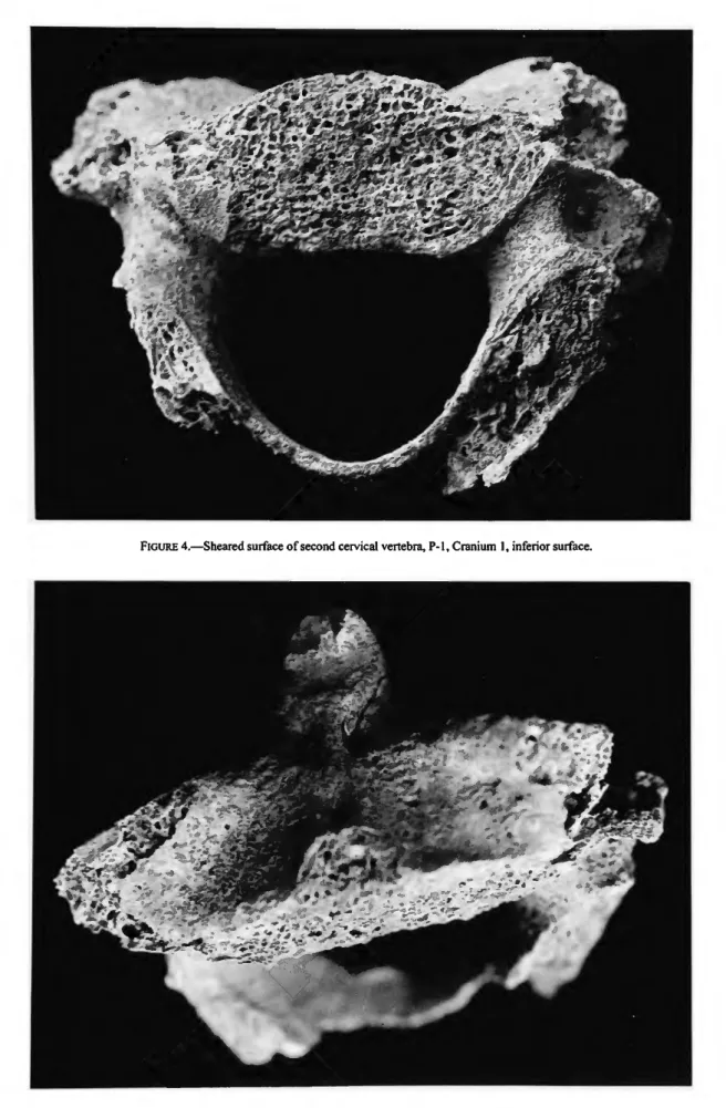
E-7 5 4. Sheared surface of second cervical vertebra, P-1, Cranium 1, inferior surface . . . 11
5. Sheared surface of second cervical vertebra, P-l, Cranium 1, ventral surface . . . 11
6. Occlusal surfaces of mandibular teeth, P-l, Cranium 2 12 7. Interproximal grooves on mandibular right first premolar (distal surface) and sec-
ond premolar (mesial surface), P-l, Level 3 13 8. Pubic bones of 45- to 50-year-old female, P-3, E-5, showing deep pitting 15
9. Exposure of pulp cavity through extreme attrition in the maxillary left first molar
of the 45- to 50-year-old female, P-3, E-5 16 10. Interproximal groove on the distal buccal surface of the maxillary first molar
illustrated in Figure 9, P-3, E-5 17 11. Healed fracture on sternal end of right rib, P-4, E-1B 18
12. Cribra orbitalia in both orbits of 18- to 19-year-old female, P-4, El-D 19
13. Polished indentation of human femur, P-4, Cranium 2 20 14. Polished indentation of human femur, P-4, Cranium 2 21
TABLES
1. Permanent teeth recovered from tomb P-l, La Florida 13 2. Deciduous teeth recovered from tomb P-l, La Florida 13 3. Individuals represented within tombs at La Florida 23 4. Distribution of ages at death for each tomb at La Florida 23
in
5. Frequencies of antemortem tooth loss, carious lesions, alveolar abscesses, and
hypoplasia in the La Florida samples 24 6. Distribution of dental calculus in La Florida samples 25
7. Cranial and mandibular measurements of high- and low-status males and females
from La Florida 25 8. Cranial and mandibular observations of high- and low-status males and females
from La Florida 26 9. Age at death, sex, status, and stable carbon (C) and nitrogen (N) isotope data in
La Florida samples 26 10. Comparison of stable carbon (C) and nitrogen (N) isotope values of high- and
low-status individuals from La Florida 27
Human Remains from La Florida, Quito, Ecuador
Douglas H. Ubelaker
Introduction
Excavations in the area of suburban Quito, highland Ecuador (2940-3030 m altitude), known as "La Florida" (0°08'45"S, 78°30'10"W) revealed six high-status shaft tombs that have been radiocarbon dated (Doyon, 1995:70) to about AD 340 (uncalibrated) ±80 (Figure 1). The excavations were sponsored by the Museum of the Banco Central del Ecuador in Quito, Ec- uador, and were concentrated within section OPQLF-1 of the one kilometer La Florida site. The shaft tombs invade a slightly earlier mound, and according to project director Leon G. Doy- on (1988), fill for the construction of the mound was taken from an earlier midden.
The tombs are of complex construction and extraordinary depth (12.5-15.1 m) and are laden with diverse and exotic of- ferings. Artifacts such as metals, marine shell, fine textiles, abundant ceramics, and two emerald beads attest to the high status of the occupants. Doyon (pers. comm., 1988) interprets the tombs as being constructed for the noble elite of a ranked social structure existing at that time. Human interments re- covered represent the elite, accompanied by sacrifices, sev- ered heads, or interments of representatives of other classes in service to the elite (Doyon, 1995). The tombs are temporally assigned to the Chaupicruz phase (ca. AD 1-500) of the Re- gional Development period.
At the invitation of Doyon, during the months of August 1988 and July to August 1989 I analyzed the human skeletal re-
Douglas H Ubelaker, Department of Anthropology, National Museum of Natural History, Smithsonian Institution, Washington, D.C. 20560- 0112.
Review Chairperson: Dennis Stanford, Department of Anthropology, National Museum of Natural History, Smithsonian Institution, Wash- ington, D.C, 20560-0112.
Reviewers: William M. Bass, Department of Anthropology, The Uni- versity of Tennessee, Knoxville, Tennessee, 37996-0720; Leon G.
Doyon, Department of Anthropology, Yale University, New Haven, Connecticut, 06520-8277; John Verano, Department of Anthropology, Tulane University, New Orleans, Louisiana, 70118-5670.
mains recovered from the excavations. The analysis presented herein is largely limited to biological information gleaned from the recovered human remains. All archeological details, includ- ing mortuary analysis, interpretation of burial position, and procedure, will be reported separately by Doyon, supplement- ing published information (Doyon, 1988, 1995). Detailed skel- etal inventories of each relevant unit are presented herein to fa- cilitate future interpretations of this complex and ritually significant site.
FIGURE 1.—Location of La Florida site, Ecuador.
METHODS
The analysis was completed in Doyon's laboratory near the site with the able assistance of Doyon and Megan Criley. All of the human remains had been cleaned prior to my arrival, great- ly facilitating the study.
The six tombs had been labeled by Doyon with "P" or "C."
The P numbers refer to those tombs that had been refilled by the La Florida population to the original surface level, appar- ently using the same soil that had been removed. The C num- bers refer to those tombs with entrances that had collapsed. Ap- parently, all the tombs included a central internal pit that, except in P-2, was left empty and was covered with wood be- fore fill material was added above. Eventually, the wood cover- ing collapsed, allowing the fill above to enter the central pit, re- sulting in considerable mixing. Burials in P-2 were placed within the central pit, which was not covered, and only the burial bundle itself collapsed.
Within each tomb, bone concentrations were labeled with
"E" or "S" numbers or with some other individual indication.
The E numbers refer to what appeared at excavation to be con- centrations of human remains. The S numbers refer to "special"
materials that appeared at excavation to be nonceramic and nonhuman. Each tomb contained two or more high-status indi- viduals as indicated by inclusion of a bundle burial in the or- ganic remains as well as by the quantity and type of associated sumptuary materials. Within most tombs, one individual dis- played a burial pattern and was associated with cultural materi- als that suggested that his/her death was the reason for the tomb's construction. This individual is termed the "principal burial." High-status individuals in addition to the principal burial were termed "companion burials." These individuals, found adjacent to the principal burial, were associated with high-status artifacts, but the associated artifacts were less abun- dant and were of inferior quality to those associated with the principal burial. Many of these high-status skeletons were in- complete, frequently missing bones of the legs, and some are represented only by cranial bones.
A third category of interment found within the tombs was termed "sacrificial burials." These remains generally lacked as- sociated artifacts and were found in a variety of positions. The nature of these positions (hands in faces, protective positions) suggested to Doyon (pers. comm., 1988) that many of them, if not all, were buried alive as sacrifices.
In addition, articulated crania, mandibles, and some bones of the upper torso were found at the entrance (surface level) of four of the tombs. These were interpreted by Doyon (pers.
comm., 1988) as representing body parts. Disarticulated groups of bones also were encountered in association with the articu- lated crania.
Identification of individual skeletons during excavation and analysis was complicated by (1) the collapse of the soil fill into the central pit (except in P-2), (2) the proximity and frequent superimposition of individuals, (3) modifications of postcranial remains and the possible placement of disarticulated body parts
near completely articulated individuals, (4) highly variable bone preservation, and (5) partial looting of two tombs (P-l and C-2). All individual groups of human bones were given E numbers, and distinct crania were so labeled when identifiable as human. In addition, some skeletal evidence that either was recovered from passing soil through a screen or that for some other reason could not be associated securely with only one hu- man skeleton during excavation is referred to in the text as
"general."
Tomb Contents TOMB C-l
INVENTORY.—This tomb was 15.1 m deep and about 52.1 m3 in volume. Nine groups of bones were identified.
C-l, E-l (sacrifice): Field notes indicate this feature was found separately within the tomb and is believed to represent a sacrifice. Bones present include fragments of both humeri, both ulnae, and both femora; both tibiae; one left temporal and two right temporals; both sides of the mandible; one second cervi- cal vertebra; four thoracic vertebrae; one lumbar vertebra; one right fourth metatarsal; and two ribs. The size of the right tem- poral and united epiphyses on the distal tibia and femoral head suggest an age at death of about 15 to 16 years. The maximum diameter of the femoral head is about 36 mm, suggesting fe- male sex. The one orbit present lacks evidence of disease. All permanent teeth of one individual are present except for the right second maxillary molar and the mandibular third molars.
The roots of the maxillary third molars are only about 50%
complete, supporting the age estimate of about 15 to 16 years.
Teeth present representing a second individual include the maxillary first and second right premolars.
C-l, E-2 (principal burial): This represents the fragmentary skeleton of a young adult or late-adolescent female. Bones present include all long bones except the tibia and fibula. Also present are a fragmentary cranium, fragments of both sides of the mandible, pelvis, all cervical vertebrae, 11 thoracic verte- brae, five lumbar vertebrae, sacrum, 13 ribs, and the following bones of the hand: both lunates, right greater multangular, both capitates, left hamate, left second metacarpal, left fifth meta- carpal, six proximal hand phalanges, and 10 middle hand pha- langes. The right greater multangular of a second individual also is present.
Various features of the pelvis, including a wide sciatic notch, elevated auricular area, and a well-developed preauricular sul- cus indicate female sex.
The extent of epiphyseal union indicates an age at death of between 17 and 19 years. Epiphyses are not united on the ster- nal ends of the clavicles and are partially united on the proxi- mal humerus and iliac crest. The roots of the maxillary third molars are 50% formed, and those of the mandibular third mo- lars are about 75% formed.
The sagittal suture of the cranium is prematurely closed en- docranially. The cranium shows marked flattening on its supe-
rior surface, with a circular depression 45 mm in diameter along the posterior sagittal suture (Figure 2). Presumably, this represents an old, healed, depressed fracture that could have initiated the early union of the sagittal suture. Other anomalies include a bony extension on the right fifth lumbar vertebra be- tween the transverse process and the sacrum.
A shallow, thin incision approximately 6 mm long is located on the superior surface of the head of the right femur. This inci- sion appears fresh and most likely was made during excava- tion.
The maximum length of the right humerus (29.5 cm) sug- gests a living stature of about 157 cm (5 ft, 2 in) using Trotter's 1970 formulae for white females (Ubelaker, 1989:61).
Maxillary teeth present include the right central incisor, left lateral incisor, right canine, left premolars, both first molars, right second molar, and both third molars. All 16 mandibular teeth are present.
C-l, E-3 (sacrifice): Only fragments of the following bones are present: right ulna, cranium, and both sides of the mandible. The bones appear to represent a young adult, proba- bly between the ages of 23 and 26 years. Sex cannot be esti- mated.
All maxillary teeth are present except the left second molar.
Mandibular teeth present include the left central incisor, left ca- nine, all four premolars, and right first molar.
C-l, E-4 (sacrifice): This individual is represented only by a fragmentary cranium, the left mandible, and the second cervi- cal vertebra. The extent of dental attrition suggests an age at
death of between 22 and 26 years. No reliable estimate of sex can be made.
Maxillary teeth present include the canines, right premolars, right first molar, and left third molar. Mandibular teeth present include the right lateral incisor, right first premolar, left second molar, and right third molar.
C-l, E-5 (companion burial): This individual is represent- ed by all major long bones except the tibiae and fibulae. Other bones present include the right scapula; cranium; mandible;
first and second cervical vertebrae; two other cervical verte- brae; 10 thoracic vertebrae; first, second, and third left meta- carpals; two proximal hand phalanges; one middle hand pha- lanx; and 13 ribs.
All permanent teeth are present. Twenty-nine teeth repre- senting at least two other individuals also are present. These consist of two maxillary right central and lateral incisors, two right canines, and two right first premolars. Additional teeth present consist of the maxillary second premolars, first molars, left second molar, third molars, and the following mandibular teeth: right central incisor, lateral incisors, canines, first premo- lars, and first molars.
All cranial sutures are ununited. The following epiphyses were not united: proximal femur, greater trochanter of femur, iliac crest, proximal humerus, proximal ulna, distal radius, proximal radius, and proximal clavicle. The ilium and ischium were not united. The distal humerus was in the process of unit- ing. The roots of the third molars were about 50% complete.
These data collectively suggest an age at death of about 15 years.
FIGURE 2.—Depression on posterior sagittal suture of 17- to 19-year-old female, C-l, E-2.
Female sex is suggested by the small size of the bones and the appearance of the cranium.
C-l, S-l (human remains found mixed with a large quantity of animal bones): Bones labeled S-l consist of cranial frag- ments and fragments of the left humerus, one left tibia, one fragmentary fibula, one left and one right temporal, and one middle hand phalanx. Teeth represent at least two adults: two of all maxillary right teeth, two left maxillary central incisors, two left maxillary lateral incisors, one left maxillary canine, two left maxillary first premolars, one maxillary left second premolar, two of each of the left maxillary molars, one right mandibular lateral incisor, two right mandibular canines, two of all mandibular premolars, two of all mandibular first and second molars, two mandibular right third molars, and one mandibular left third molar.
The bones appear to be mature, but no reliable estimate of age at death or sex can be made.
C-l, S-2: The following fragments were mixed with animal bones: both femora, one tibia, one right innominate, one lum- bar vertebra, and one left talus. The individual appears to repre- sent a young adult, but no reliable estimate of sex can be made.
C-l, S-4 (mixed with animal bones): The following bones of an immature skeleton are present: left humerus, left radius, right clavicle, both scapulae, left temporal, right mandible, left innominate, one first cervical vertebra, and two left ribs. Maxil- lary teeth present are the right central incisor, both canines (crowns complete, roots 80% formed), right first premolar, and both second molars (crowns complete, roots 50% formed).
Mandibular teeth present are the right canine, right first premo- lar (crown complete, root 80% formed), right second premolar (crown complete, root 50% formed), right first molar (crown complete, root complete), and right second molar (crown com- plete, root 50% formed).
The size of the bones and the extent of dental calcification suggest an age at death of about 10 years.
C-l, General: A small quantity of human remains was re- covered from sifted soil. These bones apparently originate from the other identified individuals within C-l. They consist of a right temporal, a manubrium, a fragment from an adult acetab- ulum, and a very eroded femoral head. Teeth include one each of all maxillary teeth except the left incisors. Also present are the mandibular right first premolar, mandibular left and right first molars, and mandibular left and right second molars.
SUMMARY.—Analysis of all human remains recovered from tomb C-l suggests that at least eight individuals are present.
Among these, the context of two young females, aged approxi- mately 17 to 19 years (E-2) and 15 years (E-5), suggested to Doyon (pers. comm., 1988) that they represent high-status indi- viduals. The remaining six individuals consist of a female aged about 15 to 16 years (E-l), a 23- to 26-year-old adult of unde- termined sex (E-3), a 22- to 26-year-old adult of undetermined sex (E-4), two adults of undetermined sex (S-l, S-2), and a child (S-4) of about 10 years of age.
TOMB C-2
INVENTORY.—This tomb was 15.1 m deep and about 56.5 m3
in volume. Thirty-two groups of bones were available for anal- ysis.
C-2, E-l (sacrifice): Bones present include fragments from all long bones; the right scapula; cranium; both sides of the mandible; right innominate; both patellae; four thoracic verte- brae; one lumbar vertebra; sacrum; both hand naviculars; right lunate; right triquetral; right greater multangular; right lesser multangular; right capitate; right hamate; all metacarpals ex- cept the left fifth metacarpal; seven proximal hand phalanges;
one middle hand phalanx; right calcaneus; both tali; both cuboids; both foot naviculars; right first cuneiform; both sec- ond cuneiforms; left third cuneiform; left and right first, sec- ond, and third metatarsals; left fourth metatarsal; one foot pha- lanx; and eight ribs.
Teeth present include all right maxillary teeth, the left maxil- lary incisors, canine, and second and third molars, and all man- dibular teeth.
Epiphyses are united on the proximal femur, distal ulna, dis- tal radius, and the head of the humerus and are not united on the distal femur and proximal fibula. The third molars show completely formed crowns, but the roots are only about 75%
formed. These data suggest an age at death of about 16 to 17 years.
A wide sciatic notch and elevated auricular area strongly suggest female sex for this individual.
A left humerus length of 27.8 cm suggests a living stature of about 151 cm (4 ft, 11.5 in) using various stature equations of Trotter (Ubelaker, 1989:61).
C-2, E-2 (sacrifice): Only fragments of the cranial vault, left maxilla, and mandible are present. All teeth are present.
Extra teeth belonging to a second adult also are present and in- clude the maxillary canines, maxillary right first premolar, maxillary right first and second molars, maxillary left third mo- lar, mandibular canines, mandibular right first premolar, and all six mandibular molars.
The extent of dental attrition suggests an age at death of be- tween 22 and 25 years. No reliable estimate of sex or stature can be made.
C-2, E-3 (sacrifice): Bones present consist of the right hu- merus, both radii, both ulnae, left femur, right tibia, one frag- mentary fibula, left clavicle, left scapula, both temporals, right maxilla, both sides of the mandible, left patella, first cervical vertebra, four other cervical vertebrae, right calcaneus, right ta- lus, both cuboids, right foot navicular, right first metatarsal, all five left metatarsals, left distal first foot phalanx, two left ribs, and one right rib.
Teeth present include all maxillary teeth and all mandibular teeth except the left second premolar and left second molar.
The small size of the bones suggests likely female sex. The extent of dental formation of the third molars (maxillary roots 75% formed, mandibular roots 50% formed) suggests an age at
death of between 16 and 18 years. No estimate of stature can be made.
The left femur shows extreme platymery on the proximal end, with an anterior-posterior diameter of 18 mm and a medial-lat- eral diameter of 29 mm.
C-2, E-4 (sacrifice): Bones present consist of the left hu- merus, left femur, right innominate, both patellae, two cervical vertebrae, five thoracic vertebrae, two lumbar vertebrae, left calcaneus, left talus, left second cuneiform, and ribs. The bones are nearly of adult size, but the ischium is not united to the ili- um, and the epiphysis for the iliac crest is separate. The epiphy- sis for the proximal end of the humerus is partially united and small. These data suggest the remains originate from a female of between probably 15 and 17 years of age. No teeth are present.
C-2, E-5 (sacrifice): Bones present consist of the distal end of a tibia, distal femoral epiphysis, fragments of a fibula, left temporal, left calcaneus, and a right talus. Lack of union of the distal femoral epiphysis and a humeral head diameter of 37 mm suggest the remains represent a female between the ages of 14 and 17 years.
C-2, E-6 (sacrifice): Bones present consist of both femora;
both tibiae; both fibulae; both scapulae; left innominate; both patellae; four cervical vertebrae; five thoracic and five lumbar vertebrae; both calcanei; both tali; both cuboids; both foot nav- iculars; three right cuneiforms; both first, second, and fifth metatarsals; two left first foot phalanges; six other foot phalan- ges; two middle foot phalanges; one distal foot phalanx; four other distal foot phalanges; and several ribs.
The right innominate shows a deep preauricular sulcus and other strong indications of female sex.
The epiphysis for the iliac crest is not united, although the bones are of adult size. The proximal right tibia shows recent epiphyseal union. This suggests an age at death of between 16 and 19 years.
The maximum length of the right tibia was estimated at 30.2 cm, which suggests a living stature of about 146 cm (4 ft, 9.5 in) using Genoves' regression equation for Mexican females (Ubelaker, 1989:62).
C-2, E-7 (sacrifice): Bones present include both humeri;
both radii; both ulnae; both femora; left tibia; one fibula; both temporals; both maxillae; both sides of the mandible; both pa- tellae; first cervical vertebra; two other cervical vertebrae; both hand naviculars; both lunates; left triquetral; one pisiform; both lesser multangulars; left capitate; right hamate; left first, sec- ond, third, and fourth metacarpals; right third and fifth meta- carpals; four proximal hand phalanges; two middle hand pha- langes; left calcaneus; left talus; both foot naviculars; right second and third cuneiforms; five left metatarsals; two proxi- mal foot phalanges; and three distal foot phalanges.
All teeth are present. The right mandibular third molar is im- pacted. The right maxillary third molar shows a root 90%
formed.
The appearance of the bones and the extent of dental forma- tion suggest an age at death of about 20 years. No reliable esti- mate of sex can be made. The estimated length of the left tibia (32.4 cm) suggests a stature of about 157 cm using the male formula or 152 cm using the female formulae of Genoves (Ubelaker 1989:62); an average estimate is about 155 cm (5 ft, lin).
The right radius shows a Colles fracture near the distal end, with considerable destruction of the distal articular surface (Figure 3). The right femur displays considerable platymery.
FIGURE 3.—Colles fracture of distal right radius of 20-year-old adult, sex undetermined, C-2, E-7.
C-2, E-8 (sacrifice): Bones present consist of both humeri, right radius, both ulnae, both femora, right tibia, one fragmen- tary fibula, right clavicle, both scapulae, both temporals, both maxillae, both sides of the mandible, both innominates, one cervical vertebra, seven thoracic vertebrae, three lumbars, one sacrum, one right hand navicular, one right lunate, one right lesser multangular, one right capitate, one right hamate, one left second metacarpal, one right third metacarpal, one right fourth and one right fifth metacarpal, four proximal hand pha- langes, three middle hand phalanges, one distal hand phalanx, one left first cuneiform, and several rib fragments. All teeth are present.
Various features of the innominates strongly indicate female sex.
The extent of dental attrition and other aging data suggest an age at death of between 25 and 30 years.
Maximum lengths of the right ulna (22.0 cm) and the right radius (20.4 cm) suggest a living stature of about 150 cm (4 ft, 11 in) using combined white female and black female formulae of Trotter (Ubelaker 1989:61).
A well-remodeled healed fracture is located on the midshaft of the left radius.
C-2, E-9 (sacrifice): Bones present include all of the long bones except the right tibia and right clavicle. Also present are the scapulae, temporals, maxillae, both sides of the mandible, innominates, patellae, first and second cervical vertebrae, two other cervical vertebrae, four thoracic vertebrae, two lumbar vertebrae, sacrum, all carpal bones except the pisiforms and right lesser multangular, all metacarpals, four proximal hand phalanges, seven middle hand phalanges, six distal hand pha- langes, both calcanei, both tali, left cuboid, both foot navicu- lars, left first cuneiform, both second cuneiforms, right third cuneiform, both second metatarsals, left third metatarsal, both fifth metatarsals, both proximal first foot phalanges, six other proximal foot phalanges, two middle foot phalanges, three dis- tal foot phalanges, and several ribs.
All teeth are present except the maxillary left central incisor.
Various features on the innominates as well as the morpholo- gy of the other bones strongly indicate female sex.
The extent of dental attrition suggests an age at death of be- tween 21 and 24 years.
Maximum length of the left tibia (31.6 cm) suggests a living stature of about 150 cm (4 ft, 11 in) using the formula of Gen- oves for Mexican females (Ubelaker, 1989:62).
C-2, E-10 (sacrifice): Bones present consist of the right hu- merus, right femur, right tibia, one fibula, right scapula, mandi- ble, and left pubis. All teeth are present. Teeth of a second adult individual also are present. These consist of all maxillary teeth except the left central incisor and all mandibular teeth ex- cept the canines, right second molar, and third molars. The third molars of the main individual show only initial root for- mation, suggesting an age at death of about 13 years; no esti- mate of age can be made for the second individual. No reliable estimate of sex can be made for either individual.
C-2, E-ll (sacrifice): Bones present consist of one left and one right humerus, one left and one right radius, one left and one right ulna, one tibia, one left and one right clavicle, one left and one right scapula, one left and one right temporal, one left and one right maxilla, both sides of the mandible, seven cervi- cal vertebrae, six thoracic vertebrae, one left and one right hand navicular, both lunates, one left and one right pisiform, one left greater multangular, one right capitate, one right hamate, 10 metacarpals, 12 proximal hand phalanges, eight middle hand phalanges, four distal hand phalanges, one left calcaneus, one left and one right talus, one left first and one left fifth metatar- sal, two foot phalanges, and 14 ribs. A second individual is probably indicated by the 12 proximal hand phalanges (normal number for one person is 10).
All teeth of one individual are present except the maxillary right canine and left first molar, missing postmortem, and the mandibular second molars, missing antemortem.
The size of the bones indicates female sex. The extent of dental attrition suggests an age at death of between 23 and 26 years.
Maximum length of the right radius (21.0 cm) suggests a liv- ing stature of about 153 cm (5 ft, 0 in) using the formulae for females of Trotter (Ubelaker, 1989:61).
C-2, E-l2 (sacrifice): Bones present consist of the left hu- merus, both femora, both tibiae, right clavicle, both scapulae, left temporal, both maxillae, both sides of the mandible, first cervical vertebra, right calcaneus, right talus, and six ribs.
All maxillary teeth are present except the left incisors. Man- dibular teeth present consist only of the right first molar, left premolars, and left molars. The mandibular left third molar is not yet erupted. The roots of the maxillary third molars are about 50% formed.
The extent of root formation on the maxillary third molars suggests an age at death of about 16 years. No estimate of sex can be made.
C-2, E-l3 (sacrifice): Bones present consist of both hu- meri, left radius, left ulna, left femur, both tibiae, both clavi- cles, left scapula, both temporals, both maxillae, both sides of the mandible, both innominates, three cervicals, four thoracics, three lumbars, and eight ribs.
There are seven deciduous and 11 permanent teeth present.
The deciduous teeth are the maxillary right canine, all four maxillary molars, and the mandibular first molars. All perma- nent teeth except the mandibular left lateral incisor are present, but only the four maxillary incisors, two maxillary first molars, mandibular central incisors, mandibular right lateral incisor, and mandibular first molars have erupted. The extent of forma- tion of the permanent teeth suggests an age at death of about nine years.
C-2, E-14 (sacrifice): These immature remains consist of the right humerus, right radius, right ulna, left femur, right clavicle, left scapula, right temporal, both maxillae, both sides of the mandible, left ilium, six ribs, six carpal and tarsal bones, and three vertebrae. Ten deciduous teeth and all permanent
teeth are present. The deciduous teeth are the maxillary right lateral incisor, right maxillary canine, all four maxillary mo- lars, the mandibular left canine, the mandibular first molars, and the mandibular right second molar.
The extent of dental formation suggests an age at death of about eight years.
C-2, E-l5 (sacrifice): Bones present consist of both hu- meri; right radius; both ulnae; both femora; left tibia; fragments of at least one right fibula; both scapulae; both temporals; both maxillae; both sides of the mandible; one gladiolus of the ster- num; both innominates; both patellae; first and second cervical vertebrae; three other cervical vertebrae; seven thoracic verte- brae; five lumbar vertebrae; right-hand navicular; both lunates;
right triquetral; one pisiform; right greater multangular; right lesser multangular; both hamates; all right metacarpals; first four left metacarpals; 11 proximal hand phalanges; three mid- dle hand phalanges; one distal hand phalanx; both calcanei;
both tali; left cuboid; both foot naviculars; left second cunei- form; right third cuneiform; all left metatarsals; first, second, fourth, and fifth right metatarsals; two first proximal foot pha- langes; two other middle foot phalanges; two first distal foot phalanges; five distal foot phalanges; and six ribs.
All permanent teeth are present except the maxillary right second molar and the maxillary left second premolar.
The morphology of the left innominate as well as the size and appearance of the other bones suggests male sex.
The extent of dental attrition as well as other features sug- gests an age at death of between 22 and 25 years.
The maximum length of the right ulna (24.0 cm) suggests a living stature of about 160 cm (5 ft, 3 in) using Trotter's formu- la for Mexican males (Ubelaker, 1989:62).
C-2, E-16 (sacrifice): Bones present include all long bones except the left clavicle. Also present are the scapulae, tempo- rals, both maxillae, both sides of the mandible, ilia, ischia, pu- bes, 22 ribs, a total of 28 carpals and tarsals, and 18 vertebrae.
Deciduous maxillary teeth present include the right lateral inci- sor, the canines, and the molars. Deciduous mandibular teeth include the right central incisor and the four molars. All perma- nent teeth are present, but only the mandibular right lateral in- cisor, mandibular first molars, and maxillary left first molar are erupted.
The extent of calcification of the permanent teeth suggests an age at death of about seven years. No estimate of sex can be made reliably.
C-2, E-l 7 (sacrifice): The bones present are immature and consist of both humeri, both radii, right ulna, both femora, left tibia, left clavicle, left scapula, both ilia, right ischium, left pu- bis, both patellae, various ribs, a total of 10 carpals and tarsals, and 27 vertebrae. All permanent teeth are present.
The extent of dental calcification suggests an age at death of between 13 and 14 years. No reliable estimate of sex or stature can be made.
C-2, E-l9 (principal and companion burials): This numera- tion refers to multiple burials that had been disturbed by looters
during the excavation. The numbers 19A, 19B, and 19C had been assigned to leg bones of what were originally believed to be three individuals that had not been disturbed by the intrud- ers. The remaining bones were mixed.
C-2, E-19A: Bones present, representing two individuals, include two left femora and one right femur, one left tibia, one fibula, two left patellae, one right patella, one proximal hand phalanx, one proximal first foot phalanx, and one other foot phalanx.
The size and morphology of the bones suggest that one indi- vidual is a young female between the ages of 22 and 26 years, and the other is a young male between the ages of 19 and 22 years.
C-2, E-19B: This individual is represented only by the right ulna, both femora, left tibia, right patella, right lunate, right greater multangular, right capitate, left second metacar- pal, one proximal hand phalanx, and one distal hand phalanx.
The individual appears to represent a large, young adult male between the ages of 22 and 30 years.
C-2, E-19C: Bones present include a left ulna, left femur, two right tibiae, one fibula, two patellae, left talus, two left naviculars, one right cuneiform, and one right first metatarsal.
Most bones appear to represent a young adult male between the ages of 20 and 25 years. One tibia present has a maximum length of 33.5 cm, suggesting a living stature of about 159.4 cm (5 ft, 3 in) using Genoves' formula for Mexican males (Ubelaker, 1989:62).
C-2, E-19ABC, General: Bones labeled in this manner con- sist of one left humerus; both radii; one left ulna; fragments of a femur, tibia, and fibula; one left innominate; one lumbar ver- tebra; one right navicular; one left capitate; one left hamate;
one left and one right second metacarpal; one left and one right third metacarpal; one right fourth metacarpal; one left fifth metacarpal; 11 proximal hand phalanges; six middle hand pha- langes; two distal hand phalanges; one right talus; and one right rib. One left pubis from this bone group shows slight parturi- tion pits on the dorsal surface and appears to represent a 25- to 30-year-old female. One femoral head diameter measures 37 mm, well within the female range.
C-2, E-l9, Bones Mixed by Looters: Bones labeled in this manner consist of three left and two right humeri, one left and two right radii, two left ulnae and one right ulnus, one right fe- mur, one right tibia, fragments of a fibula, two left and two right clavicles, three left and two right scapulae, three left and three right temporals, three mandibles, one gladiolus, two manubria, one left and one right innominate, two first cervical vertebrae, two second cervical vertebrae, nine other cervical vertebrae, 13 thoracic vertebrae, two lumbar vertebrae, one sacrum, two left naviculars, one right navicular, one right lu- nate, one left and two right greater multangulars, one left and one right capitate, one right hamate, one left and one right first metacarpal, one left and one right second metacarpal, one right third metacarpal, one right fifth metacarpal, three proximal
hand phalanges, four middle hand phalanges, two distal hand phalanges, one right second metatarsal, and various ribs.
One left pubis is from a male, aged about 28 to 30 years. One right scapula appears to be male, another female. A right radi- us, right femur, and right ulna show recent epiphyseal union.
The head of one left humerus, stained green (apparently from exposure to copper salts), measures 38 mm in diameter and thus probably represents a female. The head of a right humerus, also stained green, measures about 43 mm in diameter, proba- bly indicative of male sex. Three left temporals present appear to represent at least one male and one female.
Twenty teeth are labeled as originating from a "large male with copper." Maxillary teeth include the canines, premolars, first molars, and right second molar. Mandibular teeth include all right teeth, except the right central incisor, and the left sec- ond premolar and molars. The teeth represent a young adult be- tween the ages of 22 and 28 years.
Teeth labeled as "extra teeth" and "robbed, mixed," consist of 107 permanent teeth from at least four individuals. These in- clude one complete set of 32 teeth and one set of 31 teeth miss- ing only the mandibular left central incisor. The remaining maxillary teeth consist of two of each incisor, two right ca- nines, two of each right premolar, one right first molar, one right second molar, one left canine, one left first premolar, and one of each left molar. The remaining mandibular teeth are two left and two right lateral incisors, two right canines, one left ca- nine, two left and two right first premolars, two right second premolars, one left second premolar, two right first molars, two right second molars, one right third molar, one left first molar, two left second molars, and one left third molar.
Overall, the dental and skeletal inventory total for E-l9 shows that four individuals are represented by the teeth, left hu- meri, left ulnae, and left femora. These appear to represent two males and two females. The two females are of ages 22 to 26 and 25 to 30 years. The two males are of ages 19 to 22 years and 22 to 30 years.
C-2, E-20 (sacrifice): Bones consist of both humeri, left ul- na, both femora, both tibiae, left scapula, left mandible, left and right innominates, 10 thoracic vertebrae, five lumbars, one sacrum, one right navicular, right lunate, right greater multan- gular, left lesser multangular, right capitate, left second meta- carpal, right third metacarpal, four proximal hand phalanges, two middle hand phalanges, and four ribs.
Maxillary teeth present include the right incisors, right ca- nine, right first premolar, right first molar, and left first and third molars. Mandibular teeth present are the right incisors, right canine, right and left molars, left lateral incisor, and left premolars.
The extent of dental formation suggests an age at death of between 17 and 20 years.
C-2, Cranium 1 (severed head): An isolated cranium was found with nonhuman scapula at the tomb entrance. Bones present are a right frontal and other cranial fragments. The bones approach adult size but are very gracile. They represent
an individual between the ages of 12 and 25 years. No teeth are present.
C-2, Section 1, General: These few bone fragments were recovered from the general area of C-2, section 1, and could represent E-6, E-7, E-8, or E-l 1. Bones present are two distal hand phalanges, three (2-5) proximal foot phalanges, and one middle foot phalanx. The bones are of adult size, but no esti- mate of sex or age can be made.
C-2, Section 2, General: The following bone fragments and teeth were found within C-2, section 2, and could relate to E-l, E-3, E-4, E-5, E-6, E-7, or E-8. Bones present are a right fibula, right scapula, right maxilla, right patella, five thoracic vertebrae, five proximal and two middle hand phalanges, one right fourth metatarsal, one right fifth metatarsal, one proximal first foot phalanx, eight (2-5) proximal foot phalanges, and three ribs. Only three teeth are present: one maxillary third mo- lar with initial root formation and the first and second right mandibular premolars.
C-2, Section 3, General: The following immature fragmen- tary remains were recovered from C-2, section 3: ulna, right scapula, a total of 12 carpals and tarsals, and one vertebra.
Teeth present include one deciduous right maxillary lateral in- cisor with occlusal dentin exposure, the permanent central right maxillary incisor, and the maxillary right first molar (root 25%
formed). These bones probably relate to E-l4 and/or E-l6.
C-2, Section 4, General: Bones mixed from this sector in- clude one left capitate, one proximal hand phalanx, two middle hand phalanges, one distal hand phalanx, and one right first metatarsal. Permanent teeth present are the maxillary right inci- sors. An immature left pubis (child) also is present.
C-2, Section 5, General: The following permanent teeth were recovered from this feature: the maxillary left second mo- lar, mandibular right incisors, mandibular first premolar, and mandibular second molar. The mandibular second molar shows 80%i root formation.
C-2, Section 6, General: Adult bones from this sector in- clude the right humerus, right ulna, left temporal, right maxilla, left innominate, left patella, one (3-7) cervical vertebra, right triquetral, left fourth metacarpal, two proximal hand phalanges, one proximal first foot phalanx, and one (2-5) proximal foot phalanx. Permanent teeth present include all incisors, all ca- nines, the maxillary right premolars, mandibular right premo- lars, all four first molars, maxillary right second molar, and maxillary right third molar. Many of these remains likely repre- sent the same individual as E-5.
C-2, General: These bones consist of two left humeri and one right humerus; two left and two right radii; two left and two right ulnae; one right femur; two left tibiae; one right patel- la; one left navicular; one left lunate; one left capitate; one left hamate; two left first metacarpals and one right first metacar- pal; one left second metacarpal; one right third metacarpal; one left fifth metacarpal; four proximal hand phalanges; one middle hand phalanx; two right calcanei; one left and two right tali;
one right cuboid; two left naviculars; one right first cuneiform;
two right second cuneiforms; two left third cuneiforms; one each of the left and right first, second, third, and fourth meta- tarsals; one right fifth metatarsal; one proximal first foot pha- lanx; three distal foot phalanges; and various cranial fragments.
Teeth in this category include one each of all permanent teeth except the maxillary left central incisor, the mandibular right first premolar, the mandibular right third molar, and the mandibular left central incisor.
SUMMARY.—At least 21 individuals are represented by hu- man remains from C-2. Burials interpreted by Doyon (pers.
comm., 1988) as representing principal and companion burials consist of four individuals, an adult male likely between the ages of 19 and 22 years, a male between ages 22 and 30, a fe- male aged 22 to 26, and a female aged 25 to 30.
Interpretation of the remaining individuals is complicated by the likely commingling within the tomb of remains of different individuals. Careful inventory and sorting of the remains by age, sex, and bone morphology suggest that at least 17 addi- tional individuals are present. These individuals consist of a 16- to 17-year-old female (E-l), a 22- to 25-year-old adult of unde- termined sex (E-2), a 15- to 17-year-old female (E-4), a 14- to 17-year-old female (E-5), a 16- to 19-year-old female (E-6), a 25- to 30-year-old female (E-8), a 21- to 24-year-old female (E- 9), a 13-year-old of undetermined sex (E-10), a 23- to 26-year- old female (E-l 1), a 16-year-old of undetermined sex (E-l 2), a nine-year-old (E-l3), an eight-year-old (E-14), a 22- to 25- year-old male (E-l5), a seven-year-old (E-l6), a 13- to 14-year- old (E-17), a 17- to 20-year-old of undetermined sex (E-20), and the cranium of a 12- to 25-year-old. The remains in E-3 and E-7 may relate to other burials in tomb C-2 (see "Biological Analysis and Interpretation of Human Remains," below).
TOMB P-l
INVENTORY.—This tomb was almost completely disturbed by looters during excavation. It measured 14.2 m deep and about 51.3 m3 in volume. Thirteen groups of remains were available for analysis.
P-l, E-l (sacrifice): Originally, this was identified as a complete skeleton, but it was disturbed by looters. Bones present are a fragmentary left scapula, both temporals, right maxilla, right mandible, one cervical vertebra, and several ribs.
All permanent teeth are present except the maxillary right sec- ond molar, maxillary left canine, mandibular left central inci- sor, and mandibular left second premolar.
No estimate of sex can be made, but the extent of dental attri- tion suggests an age at death of 18 to 21 years.
P-l, E-2 (sacrifice): Although E-2 was recognized during excavation, no remains were recovered specifically from this feature.
P-l, E-3 (sacrifice): This feature was disturbed, and re- mains originating from E-l and E-2 possibly are mixed in. The following fragmentary remains are present: both humeri, left radius, left ulna, fibula, scapula, nine thoracic vertebrae, one
lumbar, one right third metacarpal, both fourth and fifth meta- carpals, three proximal hand phalanges, and six middle hand phalanges.
Adult teeth present consist of the maxillary lateral incisors and right canine and the mandibular right lateral incisor, right premolars, and right first molar.
The skeleton appears to represent a young adult of undeter- mined sex, 18 to 25 years of age.
Three middle hand phalanges of a child, six to nine years of age, also are present.
No evidence of disease was noted.
P-l, E-4 (sacrifice): This originally complete skeleton was disturbed by looters. Very fragmentary remains of a right hu- merus and an unsided femur, fibula, and patella are present.
The remains are of young adult size (perhaps between 20 and 24 years of age). Sex cannot be estimated reliably.
P-l, E-5 (sacrifice): The bones recovered from this feature generally relate to a single individual but were disturbed by looters. A large quantity of very fragmentary bones are present.
Recognizable fragments include one left and one right humer- us, one left radius, one left and one right ulna, one left femur and two right femora, two tibiae, one right fibula, one left and one right clavicle, one right scapula and one other scapula, three left and two right temporals, two mandibles, one left and one other innominate, one left and one other patella, one (3-7) cervical vertebra, one thoracic vertebra, five lumbar vertebrae, one left hand navicular, one right hamate, one left first meta- carpal, one left and one right third metacarpal, one right fourth and one right fifth metacarpal, two proximal hand phalanges, four middle hand phalanges, two distal hand phalanges, one left calcaneus, one left and one right talus, one cuboid, one left first metatarsal, one left and one right fourth metatarsal, one left and one right fifth metatarsal, two proximal first foot pha- langes, two (2-5) proximal foot phalanges, one distal first foot phalanx, and fragments of ribs.
One left pubis within these remains shows characteristics of a female between the ages of 27 and 32 years. Other bones ap- pear to originate from a large male. An ununited distal femoral epiphysis of adult size indicates that an adolescent also is rep- resented.
A large number of teeth are present, including three com- plete sets of permanent teeth. A fourth set is complete except for the mandibular left central incisor, mandibular left canine, mandibular left second premolar, mandibular right second pre- molar, and maxillary left second premolar. Additional fully formed permanent teeth include the maxillary right and left lateral incisors, left central incisor, right first premolar, right first molar, both third molars, and the mandibular right second molar.
Twenty-five additional developing permanent teeth are present. Maxillary teeth present are the right incisors, the left central incisor, both canines, all four premolars, the four sec- ond and third molars, and the left first molar. Mandibular teeth present are the left canine and all premolars and molars.
Deciduous teeth present consist of the following maxillary teeth: two right lateral incisors, one right canine, one first and one second right molar, one left lateral incisor, and two first and two second left molars. Mandibular teeth present include one right and one left canine, one right first molar, two right second molars, and two first and two second left molars. The deciduous teeth all originate from at least two children between the ages of six and seven years.
P-l, E-6 (sacrifice): This isolated cranium and mandible was identified in the field after other aspects of this feature were disturbed by looters. Analysis revealed cranial fragments, a left temporal, and both sides of the mandible. The bones were of adult size. A small mastoid process on the left temporal sug- gests female sex.
Maxillary teeth present are only the central incisors and right canine. Mandibular teeth are the right lateral incisor, both first premolars, the right second premolar, the right first and second molars, and the left second molar. The extent of dental wear suggests an age at death of about 30 years.
P-l, E-7 (severed head): An isolated cranium, mandible, and clavicle were recovered from this feature. Analysis re- vealed a right clavicle, both temporals, both maxillae, both sides of the mandible, first and second cervical vertebrae, and three other cervical vertebrae. A fragment of the acromial pro- cess of a right scapula also is present. All 32 permanent teeth are present.
The size and robusticity of the remains indicate male sex.
The extent of dental eruption, dental attrition, and epiphyseal union indicates an age at death of between 20 and 23 years.
No evidence of disease was noted, but a slight incision, 4 mm in length, is located on the dorsal surface of one of the lower cervical vertebrae. The presence of soil within the inci- sion indicates it was not of recent origin. The fine nature of the incision suggests it was made with a sharp blade.
P-l, E-8 (principal and companion burials): Originally, this feature consisted of multiple complete skeletons with asso- ciated high-status artifacts. The principal burial and companion burials were represented but were subsequently disturbed and mixed by looters. Analysis revealed the presence of two left humeri and one right humerus, two left and two right radii, two left and three right ulnae, one left and one right femur, one right tibia, one fragmentary fibula, one left and one right clavi- cle, one left and one right scapula, two left and two right tem- porals, one right maxilla, both sides of the mandible, two glad- ioli of the sternum, one manubrium, one left and one right innominate, one second cervical vertebra, three (3-7) cervical vertebrae, three thoracic vertebrae, five lumbar vertebrae, one sacrum, one right hand navicular, one left lunate, one right less- er multangular, one right capitate, one right hamate, one left first metacarpal, one right second metacarpal, two right third metacarpals, one left fourth metacarpal, one right fifth metacar- pal, four proximal hand phalanges, three middle hand phalan- ges, one distal hand phalanx, one left calcaneus, one left talus, and fragments from nine ribs. Copper stains are located on the left scapula, one right temporal, and the left mandible.
Two complete adult permanent dentitions are present, miss- ing only one left mandibular central incisor.
Bone morphology suggests at least one male and one female are present. Both are between 20 and 24 years of age.
No evidence of disease was noted.
P-l, E-9 (sacrifice): This feature consists of a distinct, par- tially disturbed skeletal assemblage. Bones present include one right humerus, one left radius, and one (3-7) cervical vertebra.
Teeth present include all maxillary teeth from two adults ex- cept for one left central incisor. Mandibular teeth present are one right canine, the 12 molars, and one right first premolar.
The four deciduous second molars from one immature individ- ual also are present.
One adult right humeral head measures 35 mm in diameter, suggesting female sex.
The extent of dental formation suggests the adults are of ages 16 to 19 and 21 to 26 years.
P-l, Shelf: This feature was originally identified as likely representing a complete, articulated skeleton, but it was dis- turbed by looters. Analysis revealed the following very frag- mentary bones: right radius, right ulna, left mandible, three (3-7) cervical vertebrae, one first metacarpal, one right second metacarpal, and one proximal hand phalanx. The following maxillary teeth were identified: both lateral incisors, right ca- nine, right first premolar, and right second molar.
No estimate of sex can be made. The extent of dental attri- tion suggests an age at death between 18 and 25 years. No dis- ease was noted. Some of these remains may have originated from E-3.
P-l, Cranium 1 (severed head): This cranium, with articu- lated mandible and cervical vertebrae, was found at the en- trance to the tomb with an associated tibia and fibula. Bones present are cranial fragments, left tibia, fibula fragments, both temporals, both maxillae, both sides of the mandible, and the first and second cervical vertebrae. All permanent teeth are present except the maxillary central incisors.
Cranial morphology suggests male sex.
The extent of dental attrition indicates an age at death of about 20 years.
No evidence of disease is present, but the inferior surface of the second cervical vertebra has been sheared. The affected surface is very planar along all of the body, with soil embedded within the exposed trabeculae (Figures 4, 5). No fine cut marks are present. The alteration apparently was produced by a large blade applied with considerable force.
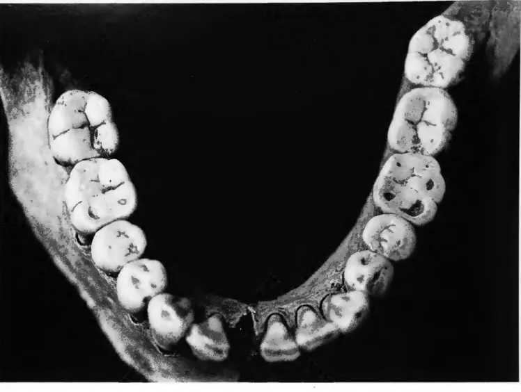
P-l, Cranium 2 (severed head): This cranium, with articu- lated mandible and cervical vertebrae, also was found at the tomb entrance. Bones present are both temporals, both maxil- lae, both sides of the mandible, and the first three cervical ver- tebrae. All 32 permanent teeth are present.
Morphology of the cranium indicates male sex. The extent of dental attrition (Figure 6) and other factors indicate an age at death of 25 to 30 years.
No cut marks were noted, however, an oval-shaped lesion is present on the right medial surface of the mandible just above
FIGURE 5.—Sheared surface of second cervical vertebra, P-l, Cranium 1, ventral surface.
FIGURE 6.—Occlusal surfaces of mandibular teeth, P-l, Cranium 2.
the lower margin. Some reactive bone is present at the superior surface. The depression measures 7 mm x 13 mm x 3 mm deep. The lesion is smooth-walled and opens internally.
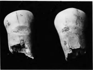
P-l, Level 3: This isolated mandible was found at the tomb entrance. In addition to both sides of the mandible, a left tibia is present. All mandibular teeth except the central and right later- al incisors are present.
No estimate of sex can be made. The extent of dental attri- tion indicates an age at death of between 25 and 30 years.
Interproximal grooves are located at the cervical junctions of the distal surface of the mandibular right first premolar and the mesial surface of the contiguous second premolar (Figure 7).
The grooves are 2 mm and 1 mm wide, respectively.
P-l-H: This feature consists of general mixed bone dis- turbed by looters. Bones present are a left humerus, left scapu- la, left and right temporal, left maxilla, left mandible, left patel- la, second cervical vertebra, and one other (3-7) cervical vertebra.
All maxillary teeth from one individual are present. Addi- tional maxillary teeth are the right incisors, left lateral incisor, right canine, right molars, and left third molar. Mandibular teeth present are the right incisors, both canines, both first pre- molars, both left molars, and the second and third right molars.
An additional mandibular third molar is present with 25% root formation.
One temporal shows a very large mastoid process, indicating male sex. This left temporal also is copper stained.
SUMMARY.—Analysis suggests that at least 20 persons are represented by the human remains originating in Tomb P-l.
The principal and companion burials (E-8) consist of a male and female, both likely between the ages of 20 and 24 years.
The remaining 18 individuals include an 18- to 21-year-old of undetermined sex (E-l), an 18- to 25-year-old of undetermined sex (E-3), a young adult of undetermined sex (E-4), a 27- to 32- year-old female (E-5), a six- to seven-year-old child (E-5), a fe- male of about 30 years (E-6), a 20- to 23-year-old male (E-7), a
FIGURE 7.—Interproximal grooves on mandibular right first premolar (distal surface) and second premolar (mesial surface), P-l, Level 3.
16- to 19-year-old of undetermined sex (E-9), a 21- to 26-year- old of undetermined sex (E-9), a 20-year-old male (severed skull number 1), a 25- to 30-year-old male (severed skull num- ber 2), and a mandible from a 25- to 30-year-old of undeter- mined sex (Level 3).
The minimum of 20 individuals was suggested by analysis of both permanent and deciduous teeth present (Tables 1, 2). The dental data suggest the presence of at least two additional chil- dren and four additional adults.
TOMB P-2
INVENTORY.—Human remains from this tomb originate from three individuals, an adolescent and two children. Bones from all three individuals were commingled. Individual E num- bers were assigned but are difficult to assess. All burials in this tomb were assigned high status by Doyon (pers. comm., 1988).
The tomb measured 12.5 m deep and about 27.2 m3 in volume.
Four groups of remains were available for analysis.
Adolescent: Bones of the adolescent consist of the left hu- merus; left radius; fragments of an ulna; right femur; right scapula; left temporal; both innominates; first and second cer- vical vertebrae; two thoracic vertebrae; two lumbar vertebrae;
sacrum; one left lunate; left triquetral; left capitate; left hamate;
left second, third, fourth, and fifth metacarpals; four proximal, two middle, and two distal hand phalanges; and several ribs.
Teeth of the adolescent consist of the permanent maxillary right lateral incisor, right canine, right premolars, right first molar, and first and second left molars. Permanent mandibular teeth are the right second premolar, first and second right mo- lars, and first and second left molars.
The extent of dental formation, bone size, and epiphyseal union suggest an age at death of between 17 and 19 years.
The appearance of the ilium and other bones suggests female sex.
The mandibular right third molar is peg-shaped and unerupt- ed. In addition, a left orbit shows active porosity (cribra orbita- lia).
Children: Bones of the two children consist of one left and one right humerus, one left and one right radius, two left and two right clavicles, two left and two right scapulae, one left temporal and two right temporals, two left maxillae and one right maxilla, two left mandibles and one right mandible, one right ilium, one sternum, three left and five right ribs, and 23 centra and six transverse processes of the vertebrae.
The younger child is represented by the following deciduous teeth: maxillary lateral incisors, canines, and molars, and man- dibular canines and molars. Permanent teeth include an unerupted first molar with about 25% root formation, a canine with initial root formation, and a second molar with 50%
crown formation. These data suggest an age at death of about five years.
Erupted teeth of the older child are the four deciduous maxil- lary and mandibular first and second left molars, the permanent maxillary right first molar, and the mandibular right first mo- lars. Other permanent teeth are present, but they are neither erupted nor fully formed. The extent of dental development suggests an age at death of about 7.5 years.
TABLE 1.—
Maxillary teeth Right 11 Right 12 Right C Right PM1 Right PM2 Right Ml Right M2 Right M3 Left 11 Left 12 LeftC LeftPMl Left PM2 Left Ml LeftM2 LeftM3
Permanent teeth recovered from tomb P-l Number present
15 18 18 16 14 15 15 16 14 17 13 14 13 14 14 16
Mandibular teeth Right 11 Right 12 Right C Right PM1 Right PM2 Right Ml Right M2 Right M3 Left 11 Left 12 LeftC LeftPMl Left PM2 Left Ml LeftM2 LeftM3
, La Florida.
Number present 11 13 13 15 13 17 17 16 7 11 13 15 10 14 16 14
TABLE 2.—
Maxillary teeth Right 11 Right 12 Right C Right Ml Right M2 Left 11 Left 12 LeftC Left Ml LeftM2
-Deciduous teeth recovered from tomb P-l Number present
0 2 1 1 2 0 1 0 2 3
Mandibular teeth Right 11 Right 12 Right C Right Ml Right M2 Left 11 Left 12 LeftC Left Ml LeftM2
, La Florida.
Number present 0 0 1 1 3 0 0 1 2 3