PHEROMONES IN FREE-LIVING AND PARASITIC NEMATODES
Thesis by Andrea Choe
In Partial Fulfillment of the Requirement for the Degree of
Doctor of Philosophy
CALIFORNIA INSTITUTE OF TECHNOLOGY Pasadena, California
2012
(Defended June 17, 2011)
© 2011 Andrea Choe All Rights Reserved
Acknowledgements
To begin with, I must dedicate this thesis to several people that have helped to define my personal beliefs and perspectives:
1) My mother, Jennie, who showed me inspiration 2) My father, Gregory, who showed me discipline 3) My brother, Daniel, who jedi-mind tricked me into becoming fearless 4) Dima Kogan, for making the journey the destination 5) Paul Sternberg, for making science an adventure.
Before meeting Paul Sternberg, my friends told me that I simply must rotate in his lab. I can see why, given that he is probably one of the very few people that understand the way that I think about science. I had not gone into this thinking about a career, but rather “how can I get Paul to pay for this thing I need to study this other cool thing?” This is a man that will not bat an eyelash in the face of curiosity-driven science. Having said that, he will be the first to tell you to redo an experiment before turning it into a real project. I am not the first, nor will I be the last, to say that I could never imagine doing graduate work in any other lab.
I must thank the members of my committee for their insightful discussions and collaborations, without which I could not enjoy the multidisciplinary perspectives that have driven this thesis forward.
I have so many members of the Sternberg lab to thank. In retrospect, my bay-mates were extraordinarily invaluable to my daily thought process. For this reason, I have to thank Steven Kuntz and Ali Mortazavi for enthusiastically talking about anything and everything from worm genomes to British TV shows about evil butlers. I remember when I was having contamination issues with my bacterial cultures, Steven would willingly sniff the flasks and we would discuss the subtleties of its odor like two wine snobs. Of course I must thank Oren Schaedel for his support throughout the years. I have never argued with anyone as much as I have argued with Oren and yet I can always count on him for pretty much anything. I must thank Adler Dillman, one of the last remaining field biologists, for shamelessly loving science. Lastly, I must thank Dmitriy Kogan for creating the automated worm detection software that most of my thesis has relied upon.
Preface
As far as I can remember, I have always been curious about behavior, but several particular influences come to mind when I think about how the interest really developed. One such influence comes from John Steinbeck’s novel, East of Eden. When it was adapted for Elia Kazan’s film in 1955, several key components of the novel seemed to be left out; most notably the servant character, Lee, whose personal philosophies laid the foundation for the central theme: control of instinct. Lee offers the doctrine that humans have choice to alter their innate fears and desires, contrary to the protagonist Adam’s deterministic belief that we are born with rigid instincts. Such is an important lesson for his son Cal, whose impulse to wreak havoc is appended by the profound realization that he can simply choose to reject the need to destroy things. I still have a vivid memory of the scene in which James Dean, who plays Cal, clutches his curled fist with conflict.
I often think about the tug-of-war between nature vs. nurture and the emotional drives that we can/cannot choose to follow or ignore. I think about the correlations between aggression/passivity and specific genes and sizes of structures in our brains, leading me to further wonder how much of our behavior is predetermined. I think about the infamous serial killer Ted Bundy, who claimed, “I just couldn’t help it” and wonder if there is some truth to that.
I’ve also spent a great deal of my life working with children with developmental disabilities, witnessing the subtle ways that autistic individuals can sabotage their relationships by prioritizing specific needs over friendship. Sometimes those needs are to rock back and forth 10 times before answering the friendly question, “Do you want to play?”, increasing the frequency of this rocking pattern when the other has finally left from insult. When I look at their expression, it seems to read: “I don’t have a choice.”
Finally I come to the worm. People often ask me why I chose to study the worm when it seems that my interests are so strongly aligned with human pathologies and disabilities. I disagree that the two are mutually exclusive, as I have felt no greater gratification than poking and prodding at one of Earth’s most successful creatures. I am not interested in the worm such that I may study humans; rather I am interested in worms because they are interesting to study life. There exists a broader forum for the questions that I seek to answer; questions that feel the weight of a more diverse perspective. How does instinct evolve? What changes behavior? What is behavior? It is no coincidence that I have migrated from research of the mouse, to the fly, to the worm. With less, we can find more, and I believe that the work that is presented here will begin to reveal that there is indeed much, much more.
Abstract
Nematodes are among the most diverse phyla of animals, occupying almost every ecological niche available[1]. Their ubiquity has led to a number of problems for civilization, including the loss of crops and the spread of neglected tropical diseases.
Because they are responsible for a broad range of agricultural and human diseases, many pheromone-mediated nematode behaviors have been described but very few pheromones have been identified.
We report, via high-performance liquid chromatography electrospray ionization mass spectrometry, the discovery that many free-living and parasitic nematodes secrete small- molecule pheromones called ascarosides. These pheromones, called ascarosides, were first found to play a role in sex attraction and induction into a stress-resistant diapausal life stage in the free-living organism, Caenorhabditis elegans. We have performed a double-blind purification of the female sex pheromone in the sour paste nematode Panagrellus redivivus and report that the female sex pheromone is composed of at least two ascarosides. We have also found that both free-living and parasitic nematodes respond to different concentrations of ascarosides through attraction or repulsion, demonstrating cross-species communication.
These results suggest that ascarosides could be a universal nematode cue, similar to the role of N-Acyl homoserine lactones in bacteria quorum sensing.
Because ascarosides are nonvolatile, they can only mediate close-range communication.
Nematodes have a well-characterized capacity for long-range chemoattraction to a range of
volatile cues. However, no studies have been done towards characterizing natural volatile cues derived from nematodes. Here I describe the discovery of volatile cues are produced by male-female species in the genus Caenorhabditis, but are lacking in the hermaphroditic species C. elegans, C. briggsae, and C. sp11. These volatile cues attract males (and sometimes females) from other Caenorhabditis species, demonstrating a cross-species gonochoristic cue.
TABLE OF CONTENTS
Acknowledgements ... iii
Abstract ... v
Table of Contents ... vii
Chapter I: Introduction ... 1
Evolution and Ecology ... 2
The Study of Behavior ... 7
Nematode Response to Chemicals ... 10
Nematode Pheromones ... 12
References ... 18
Chapter 2: Source of the mate-finding cue in Caenorhabditis elegans ... 23
Abstract ... 24
Introduction ... 25
Results ... 27
References ... 44
Chapter 3: Aqueous and volatile pheromones in the genus Caenorhabditis…22 Abstract ... 23
Introduction ... 24
Results ... 26
References ... 44
Chapter 4: Ascarosides are pheromones for both free-living and parasitic nematodes ... 76
Abstract ... 77
Introduction ... 78
The Panagrellus redivivus female sex pheromone is an ascaroside ... 80
Ascarosides are broadly present in many nematodes ... 84
Different species of nematodes respond to ascarosides ... 89
Discussion ... 91
Methods ... 92
Supplementary Figures ... 95
References ... 97
Conclusions ... 99
CHAPTER 1 Introduction
Evolution and Ecology
Nematodes (roundworms) occupy virtually every ecological niche available [1], making it difficult to classify them by lifestyle. They are found to inhabit sulfurous sediment, volcanic ash, deep-sea trenches, human lymph nodes, pig intestines, plant roots, whale placenta, arctic ice, and many other diverse ecosystems [2-7]. Despite their diversity, nematologists have cast a general scientific divide between parasitic (plant, vertebrate, invertebrate) and free-living (soil, fresh, marine, brackish, or estuarine water) nematodes* [8]. This divide has led to differences in nomenclature (e.g. larval vs. juvenile), creating the illusion that free-living and parasitic nematodes have very different life cycles, when in reality all nematodes have five main life stages, partitioned by four molts [9].
Figure 1: Nematodes have a remarkably uniform body plan. The dorsal and ventral muscles oppose each other in contraction, creating the nematode’s trademark wave-like movement. Adapted from Croll (1970)[10].
*However, many nematode species may occupy both free-living and parasitic lifestyles during different parts of their life cycle.
Another common trait among nematodes is their similar body plan (see Figure 1), despite the wide range of body size, ranging from 0.3 millimeters to 8.4 meters† in length [11]. The typical nematode has a pseudocoelomate, cylindrical body that tapers at both ends [12] and an intestinal, reproductive, endocrine, and nervous system [13] held together by hydrostatic pressure. Because nematodes lack a true coelom, traditional tree topologies position them in the ancestral position when compared to two other major model systems, the mouse and the fly (Figure 2a) [14]. Alternatively, nematodes have been clustered with arthropods for their shared ability to molt (Figure 2b) while others predict that they may actually be closer to vertebrates (Figure 2c) [14].
Figure 2: Comparison of three major model organisms: Mus musculus represents “the vertebrate”, Drosophila melanogaster represents “the arthropod”, and Caenorhabditis elegans represents“the invertebrate”. Model (a), which places C. elegans as the outgroup, is favored by the molecular data, including sequence comparison of RNA polII and III, mitochondrial rDNA, cytochrome c, and 18S rRNA[14]. Taken from Fitch, D.H.A. (2005) WormBook[14].
The appropriately named Placentonema gigantissima is an 8-meter-long parasitic nematode that dwells within the
The representative nematode for these comparative analyses is the soil-dwelling Caenorhabditis elegans; which is the first metazoan to have its full-genome sequenced [15]. Comparative analysis between C. elegans and other nematodes have proven to be important, given that nematodes are incredibly diverse within their phylum and can offer much insight into how specific changes in molecular function and development arise [14].
In fact, very small differences in their body anatomy has led to the successful invasion of many different habitats [16]. For example, nematodes from the order Enoplina have developed setae (bristle-like protrusions) on their head, which allow worms to attach to a surface and prevent backslide during peristaltic propulsion [17] (see Figure 3A and 3C).
Punctations found in the cuticle of nematodes from the order Chromadorida (see Figure 1F) are tentatively classified as ornamentations, however some speculate that these may actually be canals that run through the cuticle for a yet undefined function (Baldwin, unpublished).
Figure 3: Example of divergence of nematode anterior morphology
A) Thoracostoma sp (Enoplina). B) Acromoldavicus mojavicus (Tylenchina: Cephalobomorpha).
C) Enoploides sp. (Enoplina). D) Pontonema cf. parpapilliferum (Oncholaimina). E) Ceramonema sp. (Plectida). F) Latronema sp. (Chromadorida). G) Actinca irmae (Dorylaimida). Taken from De Ley, P. (2006) WormBook[18].
The marine nematode Oncholaimus campylocercoides is able to survive their anoxic, sulfidic environment by developing S-8 rings and polysulfur chains in their epidermis (which disappear upon reintroduction of oxygen) [19]. These are just a few of many morphological adaptations that have helped to establish the ubiquity that nematodes enjoy.
Nematodes also have a wide range of behaviors which include chemotaxis, food-seeking, host-seeking, mate-finding, mating, swarming, dispersal [8], nictation (this is otherwise best described as “flailing”), stress-resistance, response to mechanical stimuli, egg-laying‡,
response to electric currents, response to gravity, response to light, and learning [20].
Variation in these aforementioned behaviors is important for successful adaption to different hosts and/or environments. Such behaviors can even be specific to certain life stages.
Figure 4: Leaping behavior of Steinernema carpocapsae towards insect host S. carpocapsae, during its infective juvenile life stage, is often found standing on its tail and waving its head side to side. This is thought to help them sample odors from the air in order to detect passing insect hosts. When an insect host is within proximity, they bend into a loop and fling towards the host [21].
For example, many nematodes have a stress-induced alternative life stage in which their body cavity becomes sealed off to the external environment, allowing them to survive desiccation. This life stage is often associated with nictation, a behavior in which the nematodes can balance on their tail and wave their heads back and forth, presumably to attach onto a passing animal that may transport it towards more favorable conditions (more
food, less population density, change in pH, etc.). In the insect-parasitic nematode, Steinernema carpocapsae, this behavior is extended such that they may leap towards passing hosts at a distance that is nine times its body length [22] (see Figure 4). Leaping is not seen in all insect-parasitic nematodes, nor do all nematodes from the genus Steinernema have this ability, making it a particularly interesting trait for studying the evolution of behavior (Dillman, personal communication).
There are many combinations of behavioral variants that help nematodes adapt to their given environment, many of which we still do not understand. It is only with a combined look at ecology and evolution that we might begin to appreciate the complexity of nematode behavior.
The Study of Behavior
Animal behavior is traditionally described as the integration of intrinsic and extrinsic inputs, which are observed as set of actions elicited by a given organism (or set of organisms) [8]. The goal for any ethologist is to understand what this actually means, requiring the careful investigation of precise and measurable stimuli, as well as mechanisms underlying an individual’s response. Let’s take, for example, one of the landmark experiments that helped to establish the field of ethology in the 1930s; the study of territorial fighting behavior in three-spined sticklebacks (Gasterosteus aculeatus).
During the spring mating season, males develop a red coloration on their bellies and defend their territories by fighting off any other males of their species, which they recognize by their red underbelly [23]. This behavior is interesting because of its specificity to a
developmental life stage (redness only occurs in adult males) and clear motive (territorialism specific to mating season).
Figure 5: To identify specific triggers for aggression in male sticklebacks, Tinbergen created several models with different traits and presented them to males. Some were realistic replicas of the male stickleback, but lacked any red coloration, whereas several other models lacked many of the characteristics of a fish, but had red underbellies. Adapted from Tinbergen (1948)[23].
It was with these experiments that Nikolaas Tinbergen demonstrated the concept of fixed action patterns (FAP). FAPs are sequences of instinctive behaviors that are observed when an individual is presented with a “releaser”§ stimulus [24]. Because FAPs occur without prior exposure or training, they are one of a few types of behaviors that are considered instinctive. In this study, Tinbergen made several models of sticklebacks and presented them to live male sticklebacks (see Figure 5). He found that the male sticklebacks ignored the realistic model lacking a red underbelly and attacked the unrealistic red-bellied models,
§ It was thought that specific stimuli would “release” a set of behaviors that are constantly suppressed and thus require an unlocking mechanism.
proving that the sticklebacks react only to the red and neglect most other characteristics [25]. As with any discovery, this conclusion led to more questions. How did this behavior evolve? Can sticklebacks only detect a narrow range on the visible spectra, or are there special receptors for longer wavelengths that trigger a response to the aggression center of the brain? Do males still behave this way when there are a plethora of females and viable nesting sites? Because the study of this behavior involves investigation from many angles, Tinbergen defined a set of questions that should be asked of every animal behavior [26]:
1. Causation (mechanism): What are the stimuli that elicit the response?
2. Development (ontogeny): How does the behavior change with age?
3. Evolution (phylogeny): How does the behavior compare with similar behavior in related species and how might it have arisen through the process of phylogeny?
4. Function (adaptation): How does the behavior impact survival and reproduction?
I firmly believe these are the bare minimum number of tenets to consider when studying any behavior. These are the tenets that I will continue to address, towards the goal of revealing multidisciplinary perspectives throughout the investigation of pheromone- mediated nematode behavior.
Nematode Response to Chemicals
“Obviously mononchs (predatory nematodes) hunt by the aid of some sense other than sight…picture these ferocious little mononchs engaged in a ruthless chase in the midst of stygian darkness. We may imagine them taking up the scent of the various small animals upon which they feed… pursuing them with relentless zeal that knows no limit but repletion.” –Nathan A. Cobb, founder of Nematology (1917).
There are two main categories of nematode locomotive response to chemical cues: kinesis and taxis [10]. Kinesis describes movement that lacks directional orientation and is affected by the intensity of the stimuli. For example, if a photophobic nematode is exposed to light, it may simply increase its speed and return to its normal speed upon removal of light. In this situation, the nematode is not avoiding light in a directional manner but rather spending more time in optimal conditions. The other type of nematode locomotive behavior is taxis, which involves the directed orientation towards or away from the stimuli.
Nematodes have several possible methods of responding to chemical gradients via taxis (see Figure 6) [27].
Figure 6: Nematodes can respond to chemical gradients (taxis) by forward sampling, klinotaxis, or tropotaxis [27].
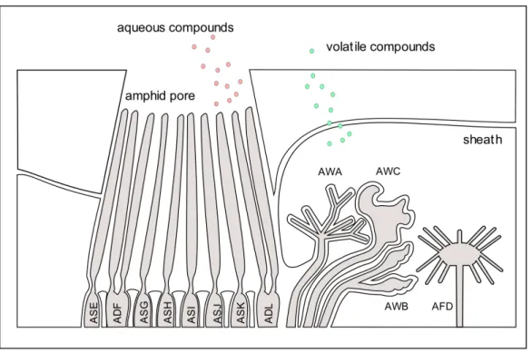
In forward sampling, nematodes compare two different samples (and presumably different concentrations of a chemical) at different time intervals separated by forward movement. In klinotaxis, nematodes compare left and right samples in their environment with side-to-side movement of their receptors (in this example, the receptors are in their head). In tropotaxis, nematodes use more than one receptor in different locations of their body to sample from two different sources at the same time. Studies have demonstrated that klinotaxis is the most likely method of decision-making in C. elegans [28, 29]. C. elegans has several types of sense organs (which are largely conserved across nematodes): the anteriorly located paired amphids, inner and outer labial papillae, cephalic papillae, and the posteriorly located phasmids (see Figure 7) [30, 31]. Studies in a number of species have shown that the amphids are the primary chemosensory organ [32, 33]. They are the largest and most complex of the anterior sense organs and come in pairs, surrounding the nematode head region.
Figure 7: C. elegans sensory organs. Taken from Bargmann, C.I. (2006) WormBook [34].
The ability of C. elegans to respond to a wide variety of olfactory and gustatory cues is mirrored in the organization and structure of the amphid cilia, which are made up of both
Figure 8: Closeup of amphid sensory openings in C. elegans. Adapted from Bargmann, C.I. (2006) [34].
C. elegans’ amphid structure has eight pairs of dendritic processes that are exposed to the outer environment (gustatory) and four pairs of wing-shaped dendritic process that are buried beneath the sheath (volatile) and thus require gases to penetrate for exposure [35].
Because both types of chemical cues are important for long and close range chemotaxis, it is important to consider both volatile and nonvolatile cues in the investigation of nematode pheromones.
Nematode Pheromones
Pheromones are the most fundamental communicative cue for most organisms, possessing many advantages over other types of signals, such as their utility in darkness and energetic
efficiency (less than a microgram of a simple compound can produce a signal that can last for days) [36]. The word “pheromone” is limited to chemical interactions within species, although its general usage has implied otherwise. The correct nomenclature for chemical communication is as follows (see Figure 9):
Figure 9: Classification of chemical communication signals [37-39].
Many studies have proven that nematode sex pheromones exist, but few have tested for the effects of these pheromones on other species [40]. Therefore, most nematode sex pheromones are limited to their intra-specific definition by default that attempts have not yet been made to prove otherwise. For example, the first pheromone discovered in C.
elegans is the ascaroside, ascr#1, for its small role in the induction of the diapausal life
semiochemicals
(inter- and intra-specific interactions)pheromones sex pheromone
(intra-specific interactions)
allelochemicals
(inter-specific interactions)
epidietic pheromone
alarm pheromone
allelomone kairomone synomone
apneumone
-
+
+ +
X
negative response in recipient
positive response in recipient
positive response in both emitting and responding
signal produced by non-living matter response between or within sexes
regulate population densities
warning or protective signals
elegans can induce dauer formation in C. briggsae[41], no studies have been performed to demonstrate the utility of ascarosides in non-C. elegans dauer formation. This is surprising, given that there exist many examples of intraspecific chemical communication between (and within) vertebrate and invertebrate species[42-45] and is perhaps attributable to the convergence of interest in C. elegans throughout the past few decades. In fact, only one other pheromone has been identified from a nematode species other than C. elegans:
vanillic acid (see Figure 10) is the female sex pheromone in the soybean cyst nematode, Heterodera glycines[46]. A significant amount of work has been done in this plant parasitic nematode, perhaps because it is responsible for a loss of ~ $500 million annually[47].
Figure 10: Female sex pheromone of Heterodera glycines: vanillic acid
Isolation of sex pheromones in C. elegans has led to the discovery that a blend of at least three ascarosides (see Figure 11) mediate mate finding[48]. It is surprising that ascarosides played a role in both mate finding and dauer formation[49], given that they are two very different survival strategies (see Figure 12). Recent work has also revealed that ascarosides
play a role in aggregation (see Figure 12) by attracting both C. elegans males and hermaphrodites to gather in groups for long periods of time (Srinivasan, unpublished).
Figure 11: General structure of ascarosides: ascarylose sugar ring + modifications in lipid tail. Adapted from Edison (2009)[50].
Figure 12: Role of ascarosides in C. elegans: different ascarosides (and some overlapping) play a role in mate finding, aggregation (Srinivasan, unpublished), and induction into the diapausal life stage known as dauer [48,
General Ascaroside Structure
3,6-dideoxy-L-mannose “ascarylose” + R1 primary fatty acid derived side chain (R2 glucose for ascr#4, H for the rest; R3 indole for ascr#9, H for the rest)
mate finding aggregation dauer formation ascr#2
ascr#3 ascr#8
icas#3 icas#5 icas#9
ascr#1 ascr#3 ascr#5 icas#9
Hermaphrodites produce these ascarosides to attract
males
These ascarosides induce aggregation of both males
and hermaphrodites
These ascarosides affect the percentage dauers formed in a population with unfa-
vorable conditions
This has led to the further investigation of C. elegans ascarosides, which are secreted in combinatorial blends that vary across different life stages[52]. Not much is known about the biosynthetic pathway for ascarosides, although it has been found that the signaling deficient C. elegans mutant, daf-22, fails to produce several ascarosides[48, 53]. daf-22 was first discovered for the abnormal dauer formation phenotype observed in mutants[54].
It encodes the C. elegans ortholog of human sterol carrier protein SCP2, which catalyzes the final step of peroxisomal fatty acid beta-oxidation[55]. daf-22 mutants are found to accumulate massive amounts of fatty acyl-coAs (up to 100-fold), causing severe developmental defects and abbreviated lifespan, suggesting that ascaroside biosynthesis is essential to C. elegans homeostasis and that the conversion of toxic long-chain fatty acids has provided a subset of readily-excreted pheromones that are then utilized for other purposes[56]. This would be interesting and would help to explain the conservation of ascarosides in other nematodes (as will be discussed later), given that the presence of these pheromones may be linked to conserved metabolic pathways that are conserved between nematodes. DAF-22 fusion protein tagged with GFP is expressed in the intestine, hypodermis, and body wall[55], lending weight to the theory that ascarosides are produced in the intestine and excreted through the mouth, anus, or excretory pore.
It is clear that ascarosides are important pheromones for several behaviors (and a symptom of good homeostatic balance) in the free-living nematode, C. elegans. However much work remains to be done on other nematodes, especially since there exist over 25,000 known nematode species[57]. Because investigation of both close and distant relatives to C.
elegans would help to reveal general nematode mechanisms, I have begun my work with other nematodes, such as the sour paste nematode, Panagrellus redivivus. This approach
has contributed towards a greater understanding of nematode pheromones and cross- species nematode communication.
References
1. Platt, H.M., "Foreword", in The phylogenetic systematics of free-‐living nematodes, S. Lorenzen, Editor 1994, The Ray Society: London.
2. Nussbaumer, A.D., Attachment mechanism in a highly specific association between ectosymbiotic bacteria and marine nematodes. Aqua. Microb. Ecol, 2004. 34: 239-‐246.
3. Andrassy, I.Z., Evolution as a basis for the systematization of nematodes, 1976, London: Pitman.
4. Gubanov, N., Giant nematode from the placenta of Cetacea; Placentonema gigantissima. Dokl Akad Nauk SSSR, 1951. 77(6): 1123-‐1125.
5. Tietjen, J.H., Ecology of deep-‐sea nematodes from the Puerto Rico Trench area and Hatteras Abyssal Plain. Deep-‐Sea Research, 1989. 36: 1579-‐1594.
6. Aben-‐Athar, J., Mechanism producting elephantiasis in filariasis (F. wucheria and F. bancrofti). Rev Bras Med, 1951. 2: 89-‐94.
7. Lutz, A., Zur Frage der Uebertragung des menschlichen Spulwurms. Weitere Mittheilungen, 1888. 3(14): 425-‐428.
8. Gaugler, R.B., Nematode Behaviour 2004, Cambridge: CABI.
9. Lee, D., "Life Cycles," in The Biology of Nematodes, D. Lee, Editor 2002, Taylor &
Francis: New York. 61-‐72.
10. Croll, N.A., The Behaviour of Nematodes 1970, New York: St. Martin's Press.
11. Yeates, G., "Ecological and Behavioural Adaptation", in Behaviour of Nematodes, R.B. Gaugler, Editor, 2004, CABI. 1-‐24.
12. Altun, Z.F.H., "Introduction", in Wormatlas 2009, doi:10.3908/wormatlas.1.1.
13. Gibbons, L.M., "General Organisaion", in The Biology of Nematodes, D. Lee, Editor 2002, Talyor & Francis: New York. p. 31-‐59.
14. Fitch, D.H.A., "Introduction to Nematode Evolution and Ecology", in Wormbook, The C. elegans Research Community, Editor, 2005,
doi/10.1895/wormbook,1.19.1, http://www.wormbook.org.
15. Blaxter, M., Caenorhabditis elegans is a nematode. Science, 1998. 282: 2041-‐
2046.
16. Coghlan, A., "Nematode Genome Evolution", in Wormbook, doi/10.1895/wormbook.1.15.1, The C. elegans Research Community, Editor, 2005.
17. Hyman, H.L., Further Notes on the Occurence of Chitin in Invertebrates.
Biological Bulletin, 1966. 130: 1-‐149.
18. De Ley, P., A quick tour of nematode diversity and the backbone of nematode phylogeny, in Wormbook, D.H.A. Fitch, Editor, 2006.
19. Thiermann, F., Sulphfide tolerance of the marine nematode Oncholaimus campylocercoides-‐ a result of internal sulphur formation. Marine Ecology-‐
Progress Series, 2000. 193: 251-‐259.
20. Lee, D., "Behaviour", in The Biology of Nematodes, D. Lee, Editor 2002, Taylor &
Francis: New York. 369-‐388.
21. Hallem, E.A., et al., A sensory code for host seeking in parasitic nematodes.
Current biology, 2011. 21(5): 377-‐83.
22. Campbell, J.F.K., et al., How and why a parasitic nematode jumps. Nature, 1999.
397(485-‐486).
23. Tinbergen, N., Social releasers and the experimental method required for their study. Wilson Bull, 1948. 60: 6-‐52.
24. Cummings, B., in Biology, N.A. Campbell, Editor, 1996.
25. Tinbergen, N., The Study of Instinct, 1971, Oxford: Clarendon Press.
26. Tinbergen, N., On Aims and Methods in Ethology. Zeitschrift für Tierpsychologie, 1937. 20: 410-‐433.
27. Ward, S., Nervous System of Nematodes, in The Organization of Nematodes, N.A.
Croll, Editor, 1976, Academic Press: London. 365-‐382.
28. Bargmann, C.I. and H.R. Horvitz, Control of larval development by chemosensory neurons in Caenorhabditis elegans. Science, 1991. 251: 1243-‐6.
29. Bargmann, C.I. and Mori, I. Chemotaxis and Thermotaxis. 1997.
30. Ward, S., et al., Electron microscopical reconstruction of the anterior sensory anatomy of the nematode Caenorhabditis elegans. The Journal of comparative neurology, 1975. 160(3) 313-‐37.
31. Jones, J.T., Nematode Sense Organs, in The Biology of Nematodes, D. Lee, Editor, 2002, Taylor & Francis: London. 353-‐368.
32. Trett, M. et al., Functional and evolutionary implications of the anterior sensory anatomy of species of root lesion nematodes (genus Pratylenchus). Revue de Nematologie, 1985. 8: 341-‐351.
33. Bargmann, C.I., J.H. Thomas, and H.R. Horvitz, Chemosensory cell function in the behavior and development of Caenorhabditis elegans. Cold Spring Harbor symposia on quantitative biology, 1990. 55: 529-‐38.
34. Bargmann, C.I., Chemosensation in C. elegans. WormBook : the online review of C. elegans biology, 2006. 1-‐29.
35. Perkins, L.A., et al., Mutant sensory cilia in the nematode Caenorhabditis elegans.
Developmental biology, 1986. 117(2): 456-‐87.
36. Wilson, E.O., Sociobiology1975, Cambridge: The Belknap Press of Harvard University
37. Riga, E., Orientation Behavior, in Nematode Behaviour, R.B. Gaugler, A.L., Editor, 2004, CABI: Cambridge. 63-‐90.
38. Huettel, R.N., Chemical communicators in nematodes. Journal of Nematology, 1986. 18(1): 3-‐8.
39. Perry, R.N., et al., Behaviour and sensory responses, in The Physiology and Biochemistry of Free-‐living and Plant-‐parasitic nematodes, R.N.W. Perry, D.J., Editor ,1998, CAB International: Wallingford. 75-‐102.
40. Chasnov, J.R., et al., The species, sex, and stage specificity of a Caenorhabditis sex pheromone. Proceedings of the National Academy of Sciences of the United States of America, 2007. 104(16): 6730-‐5.
41. Jeong, P.Y., et al., Chemical structure and biological activity of the Caenorhabditis
42. Arvedlund, M., McCormick, M.I., Fautin, D.G., Bildsoe, M, Host imprinting and possible imprinting in the anemonefish Amphiprion melanopus (Pisces :Pomacentridae). Marine Ecology-‐ Progress Series, 1999. 188: 207-‐218.
43. Fiedler, T.E., Holldobler, B., Seufert,P., Butterflies and ants-‐ the communicative domain. Experientia, 1996. 52: 14-‐24.
44. Noldus, L.P et al., Moth sex pheromones adsorption to leaf surface: bridge in time for chemical spies. Physiological Entomology, 1991. 16: 329-‐344.
45. Hardie, J., Nottingham, S.F., Powell, W. Wadmans, L.J., Synthetic aphid sex-‐
pheromone lures female parasitoids. Entomologia Experimentalis et Applicata, 1991. 61: 97-‐99.
46. Jaffee, H., Huettel, R.N., DeMilo, A.B., Hayes, D.K. Rebois, R.V., Isolation and identification of a compound from soybean cyst nematode, Heterodera glycines, with sex pheromone activity. Journal of Chemical Ecology, 1989. 15(2031-‐
2043).
47. Cooperative State Research, E., and Extension Service United States Department of Agriculture. Keeping the Profits in Soybeans. 2000; Available from: http://www.csrees.usda.gov/newsroom/impacts/00index/soybeans.htm.
48. Srinivasan, J., et al., A blend of small molecules regulates both mating and development in Caenorhabditis elegans. Nature, 2008. 454(7208): 1115-‐8.
49. Butcher, R.A., et al., Small-‐molecule pheromones that control dauer development in Caenorhabditis elegans. Nature chemical biology, 2007. 3(7): 420-‐2.
50. Edison, A.S., Caenorhabditis elegans pheromones regulate multiple complex behaviors. Current opinion in neurobiology, 2009. 19(4): 378-‐88.
51. Butcher, R.A., J.R. Ragains, and J. Clardy, An indole-‐containing dauer pheromone component with unusual dauer inhibitory activity at higher concentrations.
Organic letters, 2009. 11(14): 3100-‐3.
52. Kaplan, F., et al., Ascaroside expression in Caenorhabditis elegans is strongly dependent on diet and developmental stage. PloS one, 2011. 6(3)
53. Pungaliya, C., et al., A shortcut to identifying small molecule signals that regulate behavior and development in Caenorhabditis elegans. Proceedings of the National Academy of Sciences of the United States of America, 2009. 106(19):
7708-‐13.
54. Golden, J.W. and D.L. Riddle, A gene affecting production of the Caenorhabditis elegans dauer-‐inducing pheromone. Molecular & general genetics : MGG, 1985.
198(3): 534-‐6.
55. Butcher, R.A., et al., Biosynthesis of the Caenorhabditis elegans dauer pheromone. Proceedings of the National Academy of Sciences of the United States of America, 2009. 106(6): 1875-‐9.
56. Joo, H.J., et al., Caenorhabditis elegans utilizes dauer pheromone biosynthesis to dispose of toxic peroxisomal fatty acids for cellular homoeostasis. The Biochemical journal, 2009. 422(1): 61-‐71.
57. Hugot, J.P., Baujard, P, Morand, S., Biodiversity in helminths and nematodes as a field of study: an overview. Nematology, 2001. 3(3): 1999-‐208.
CHAPTER 2
Source of the mate-finding pheromone in Caenorhabditis elegans
Abstract
Characterization of the nature of the sex pheromone in nematodes is important for understanding and potentially regulating populations of nematodes that affect parasitism, agriculture, and important ecologic processes. Because the free-living nematode C. elegans remains one of the best studied model organisms, it serves as a good starting point for addressing this subject. Previous studies in C. elegans have shown very little about the source of the mate-finding cue and about the processes that regulate its production and release. Also, there has yet to be any study on the volatile components of the mate-finding cue, which may be largely responsible for long-range attraction.
One way of revealing sources of the mating pheromone is to utilize a bioassay comparing attraction of males to secretions collected from wild-type, mutant, and laser-ablated hermaphrodites. These experiments will help identify necessary cellular components of the mating pheromone and possible mechanisms that help to regulate its synthesis and secretion.
Introduction
Over 30 species, but relatively few genera, of free-living, plant-parasitic, and animal- parasitic nematodes have been shown to exhibit pheromone-mediated behavior[1, 2]. Being the most abundant metazoan (by individual count) on earth, nematodes play a significant role in many important processes, such as infectious diseases, agricultural sustainability, and biogeochemical regeneration. Because the identification of mechanisms controlling nematode mating and growth carry such broad implications, it has been of great interest to characterize the nature of the mate-finding cue and how it affects populations across multiple genera. However, not much is known about the site of sex pheromone production in nematodes[3]. Possibilities might include secretions through the vulva from the gonad, excretions from the digestive tract or excretory pore, or simply from the cuticle to the environment directly.
A range of studies support the likelihood that female sex pheromones are produced by the gonad and exit via the vulva. Because ascarid sex organs are large enough to dissect, they have been tested in isolation and it has been reported that both male and females of A. suum are attracted to sexual organs from the opposite sex[4]. It has been suggested that the female pheromone in the mouse pinworm, Aspicularis tetraptera, is secreted from the glandular cells of the female pulvilus (part of the reproductive organ)[5]. Studies on the source of the female sex pheromone in the free-living nematode Panagrellus silusiae also suggest that the gonads are a likely source[6]. Males from the rodent parasitic nematode species, Heligmosomoides polygyrus, demonstrate sexual behavior by flaring their copulatory bursa in the presence of females. However, when the females were treated to prevent release from the vulval area, males failed to flare their copulatory bursa, suggesting
that the sexual cue is secreted from the female vulva [7].
Several studies have suggested an alternative source of the sex pheromone. Simon and Sternberg (2002) reported evidence of a sexually dimorphic mate-finding cue in C. elegans, yet they found that vulvaless mutants were as attractive as wild-type hermaphrodites, implying that the vulva is not necessary for the release of the cue. Chasnov et al. (2007) found conflicting results, reporting that C. elegans males failed to show significant attraction for hermaphrodites. Instead, they characterized a female-derived attractant in the closely related, male-female species, Caenorhabditis remanei. They found that the female somatic gonad is required for the production and secretion of C. remanei attractant, but that the vulva is not necessary for its release. I have long speculated that male-female cues are much stronger than hermaphroditic cues, given that male-female species must find each other in nature and hermaphrodites do not require males. This would explain the discrepancy between the Simon and Chasnov study (and support Simon’s conclusion that a hermaphrodite cue exists), given that Chasnov et al. collected secretions at a concentration of 1worm per 20 microliters of buffer, which may be sufficient for stronger cues, but may be hidden by dilution.
Here I consider the chemoattraction assays that have previously been used to study mate finding in C. elegans and describe the parameters that have influenced the development of my own bioassay. I also describe a series of experiments that suggest findings contrary to both previous experiments: I report that C. elegans hermaphrodites have a mate-finding cue and that the vulva is necessary for its release.
Results
There have been three independent studies on the mate-seeking behavior of C. elegans males to hermaphrodites, each of which have influenced the development of my own behavior assay and have contributed to my perspectives on nematode chemical signaling. I will discuss the first two in this chapter and describe the third in the following chapter.
1. Simon and Sternberg (2002)[8]: This study provided the first evidence of a mate- finding cue in the free-living nematode C. elegans. They describe a sexually dimorphic mate-finding cue produced by hermaphrodites (and not males) that attracted males (and not hermaphrodites).
Figure 1: Simon and Sternberg (2002) Assays used to score for male chemoattraction to hermaphrodites[8].
(bacteria = gray) (cue = red dot)
They used several types of assays (see Figure 1), which utilized an uncoordinated mutant hermaphrodite, unc-52, to stay in one place on a bacterial lawn such that a male may be subsequently tested for response to the hermaphrodite-conditioned region. They found that males are attracted to, reverse direction of movement frequently, and remain in regions conditioned with hermaphrodites. Males were also observed to be more effective at finding their mates in a shorter amount of time on pre-conditioned lawns vs. unconditioned lawns.
Next, they used vulvaless mutants, with strains containing let-23 and lin-3 mutations, to demonstrating that vulvaless mutants are as attractive as wild-type hermaphrodites.
Although each individual was scored (via microscopic examination) for lack of a complete vulva, genetic mutants can never guarantee full removal of the affected organ or tissue. For this reason, I am skeptical from this evidence that the vulva fails to play a role in sex pheromone secretion. In an effort to reproduce these experiments, I used the same uncoordinated mutants and observed that the individuals tended to move > 1.5cm within 1 hour, thus I was unable to verify or disprove these findings by direct comparison.
2. Chasnov et al. (2007) [9]: This study reports that C. elegans does not have a cue, contrary to the Simon and Sternberg (2002) findings. In these experiments, worm secretions were collected from hermaphrodites (C. elegans and C. briggsae) and females (C. remanei and C. brenneri). Adult males were isolated and tested for attraction to the hermaphrodite or female cue collected from their own species. Chasnov et al. used several different bioassays, each utilizing a paralyzing agent (sodium azide) to hold males within a scoring region once they enter (see Figure 2). They found that hermaphrodite-derived cues fail to attract males from their own species whereas female cues attract all males,
via the simultaneous mutation of loss-of-cue and gain-of-sperm production. I find this to be a misleading conclusion, given that the assay had several design flaws (which actually lead to a very different, more interesting conclusion).
Figure 2: Chasnov et al. (2007) Assays used to score for male chemoattraction to hermaphrodites or females [9].
(bacteria = gray) (buffer = pink) (cue = red dot) (buffer + cue = pink + red dot) (sodium azide = dark blue)
The first problem is that the worm supernatant was collected at a concentration of 5 individuals per 100 µL at 25°C. That is a very low concentration to collect from, given that each individual has about 15x their body volume in buffer that they are secreting into.
Although I don’t know enough about how effective dilute pheromone might be in nature, I would say that erring on the less concentrated end of the spectrum is not sufficient when making the claim that something does not exist. I would at the very least lyophilize the buffer to test a concentrated smaller volume before ruling out the existence of an attractant.
Also most experiments with C. elegans are performed at 20°C, even though some speculate that their natural environment in compost might be closer to 25°C. Because we do not know enough about this matter, I cannot comment on whether this is better or worse for the design of this assay.
Secondly, these assays utilize a scoring method that prohibits individuals from sampling both scoring regions before deciding on a preference. Because sodium azide is combined with the control and cue region, a male may choose to enter one region or the other but cannot leave their first choice before they become paralyzed. This is a favorite method used for chemotaxis assays, as described by Bargmann and Mori (1997)[10]. However, this method only works if there is actually a chemical gradient. If a sufficient chemical gradient has not formed by the time that males are placed between the two regions, they would be forced to move in either direction without any information. An exception would exist in the presence of a volatile cue, which would provide information if sufficient amounts of volatile cue were to be released from the point source during the duration of the chemotactic event. It is my guess that this is precisely what happened, given that male-
hermaphrodites (self-fertilizing). Let us now address the first condition of this assumption: an insufficient chemical gradient is present at the time of the assay. This could only be answered by the knowledge of how quickly the attracting compound moves through 1.5% agar during the amount of time that it would take to absorb into the agar (which is the deciding event for when males are placed on the assay). Upon recreation of this assay, it took approximately 20 minutes for a drop of water to absorb onto a 2 mm layer of 1.5% agar mounted on a glass microscope slide (as opposed to the several hour overnight incubation period used in the Simon and Sternberg (2002) study, perhaps explaining the discrepancy between these findings). Although we do not know how quickly the C. elegans attractant moves through agar, I have come to understand that the C. elegans pheromone has a significant lipid component, which would probably not facilitate effective distribution in a polar environment. The distinction between volatile and nonvolatile cues would be useful in clarifying these findings. This could be easily done if the paralyzing agent was removed and response to each scoring region was observed. It is therefore surprising that Chasnov et al. instead chose to create a third volatile assay (see Figure 3), which further verified that females did indeed produce a volatile cue.
Figure 3: Chasnov et al. (2007) Assay used to score for male volatile chemoattraction to females[9].
(buffer = pink) (cue = red dot) (buffer + cue = pink + red dot) (sodium azide = dark blue)
From these findings, I conclude that C. remanei and C. brenneri females have a volatile cue. Whether or not hermaphrodites have a cue or females have a nonvolatile cue cannot be concluded from this experiment.
Lastly, Chasnov et al. addressed the source of the female volatile cue by performing a set of laser ablations on C. remanei. They ablated the somatic gonadal precursor cells, Z1 and Z4, and found that females did not produce an attractive cue, suggesting that the female volatile cue is made in (or depends on) the somatic gonad. Next they ablated the germ line gonadal precursors, Z2 and Z4, and found that females were still attractive, suggesting that the germ line is not necessary for production of a female cue**. Lastly, they ablated the anchor cell, which allowed for full formation of the gonad but prevents the development of the vulva.
They report that anchor cell-ablated females are still attractive, despite the lack of a vulva, suggesting that the release of the cue did not depend on the vulva. This would make sense, given that a volatile cue would likely diffuse through the cuticle.
Developing a new bioassay
I have reproduced both the Simon and Sternberg (2002) and Chasnov et al. (2007) experiments. I do not favor the use of uncoordinated hermaphrodites, not only because I found them to be surprisingly coordinated, but also because the control for this experiment is the manual disruption of the bacterial lawn, with the assumption that making it look as unkempt as the area left behind by the uncoordinated hermaphrodites would suffice. I have observed that nematodes are able to burrow into the agar surface, sometimes leaving the
** When Z2 and Z3 are ablated, the somatic gonad fails to extend its arms properly (which normally depends
surface looking as if it were degraded via enzymatic secretion††. This phenomenon has yet to be investigated, however I would not be surprised if the agar surface previously occupied by the hermaphrodite experienced changes different from the addition of a sex pheromone. This leads me to support the collection of a cue into a buffer, such that response in the buffered region may be compared to response in a cue-conditioned region.
This approach provides two advantages: removal of plate-to-plate variation and removal of user bias. To address the first point, each agar plate has its own variations (moisture, texture, angle of surface, etc.) that could affect the results between plates. This variation leads me to support the observation of nematodes when presented with both the cue and control on the same agar plate. Lastly, I do not support the use of a paralyzing agent, given that I have no information about the chemical distribution of unidentified compounds and hence have no idea if a sufficient gradient would be present during the experiment.
I began using several types of behavior assays and appreciated the absence of a paralyzing agent. Without a paralyzing agent, I may observe nematodes entering a region and responding (reverse, turn, re-enter). After much trial-and-error, I have developed an assay that reproducibly demonstrates the production of a C. elegans hermaphrodite sex pheromone (see Figure 4).
†† I am reminded of a popular clothing brand, Raquel Allegra, which sells extra soft t-shirts. She reveals that her secret to the soft textile is that they are recycled from old shirts worn by prisoners at the LA County Jail, whose sweat simply degrades the cotton fibers.
Figure 4: Because our behavior assay does not utilize a paralyzing agent, recording is required to observe individuals as they move in and out of scoring regions. (buffer = pink) (buffer + cue = pink + red dot)
Figure 5: Rationale for chosen parameters of the behavior assay
I describe the rationale for every parameter in Figure 5, with the goal of collecting high concentrations of hermaphrodite-derived cue (without causing stress) and eliciting a reproducible male response to their attractant. After placing a drop of buffer and hermaphrodite-incubated buffer on the bacterial lawn, I would immediately place 5 males on each dropping area equidistant from the foci of the scoring region (see Figure 4). The immediacy of the drop absorption on a bacterial lawn (5 seconds) is a great advantage over drop placement onto agar (ranges from 2-20 minutes depending on the thickness and %
agar). The first observation that I made was that the males would distribute randomly (star-shaped dispersion) and move quickly throughout the lawn until they came to the region conditioned with the hermaphrodite-derived supernatant. The males would then slow down and reverse such that they would stay within that area (see Figure 6). Once other males would enter this region, they would repeat the same behavior and often times attempt to mate with other males.
Figure 6: Observation of male attraction to a hermaphrodite-incubated buffer (right circle)
The next step would be to establish a metric for scoring attraction to the cue. I can appreciate why the sodium azide method is so popular, given that you can start the assay and come back an hour later to score by simply counting the # males paralyzed in either region. I could see by the star-shaped distribution of their dispersal that they would distribute evenly upon release (see Figure 6) therefore scoring with an azide technique would hide any holding effect of a cue. I could do one of two things: 1) adopt the azide technique and allow the cue to settle in overnight with the hope that a chemical gradient would form and the cue would not disintegrate during this time or 2) figure out how to
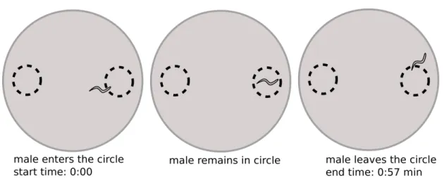
because the first method relies on hidden information, but also because I find it informative to watch males interact with the cue time and time again. I could see that they demonstrate several types of taxis: their heads move side to side (klinotaxis) and reverse when their head region, but not tail, became removed from the conditioned region (tropotaxis). I also observed kinesis, as males tended to speed up once they left the conditioned region and slowed down upon re-entering. I began to score “worm events” as the duration of time between when each worm entered a scoring region and when it left the scoring region (see Figure 7).
Figure 7: Calculating worm events: start time when the worm enters the circle, end time when the worm leaves the circle
It can become challenging to keep track of 10 worms that are entering the circle, and it is also very time consuming to manually score worm events. Although I still like the approach of comparing time spent in each scoring region, I dislike the metric of scoring worm events for several reasons (see Figure 8). Perhaps the most obvious problem is when the experimenter must manually score entry and exit times when there are ten worms simultaneously moving throughout both regions. When worms cross over, the experimenter
must make a best guess to assign worm identity. This becomes particularly challenging when multiple worms collide, making the decision arbitrary. There are also many gray zones, for example when a worm almost entirely leaves the scoring region but reverses back. An experimenter must decide whether to score that as one worm event or two. Trials can be performed one worm per plate but that would be very time consuming.
Figure 8: Problems with user error/bias with calculating worm events
Lastly, the metric of scoring each worm event as n=1 can be problematic. With a trial of 20 minutes, there may be hundreds of worm events and thus n can be > 100. If one worm enters the scoring region repeatedly, each reentry will increase the value of n, therefore increasing the significance (and decreasing the standard deviation) of that phenotype with false amplification.
I was given the suggestion to use an automated approach to scoring attraction, such that the labor and bias of scoring manually would be alleviated. Dmitriy Kogan wrote a program for this purpose, using a set of optical filters to exaggerate the contrast between the worms and the background, which would allow for the detection of worm pixels within each scoring region (see Figure 9).
Figure 9: Optical thresholding allows for the detection of worm pixels in each scoring region, which could then be used for comparison
The extraction rate is one frame per second (total 1200 frames per 20 minute trial), which allows for sufficient resolution of worm entry and exit.
Figure 10: Data output in the form of percentage worm occupancy in each scoring region, over 20 minutes (1 frame/second)
The output shows the percentage worm occupancy, per scoring region, over 1200 frames (see Figure 10). If worms are attracted to a region and accumulate, this percentage worm occupancy increases over time (see Figure 10, green line). If the worms are not attracted to a region, this percentage would rise with entry, plateau during their duration in the scoring region, and decline with exit (see Figure 10, red line). To be certain that this method finds individual worms accurately, I took a segment of video where two worms in a scoring region exit in sequence (see Figure 11).
0 0.005 0.01 0.015 0.02 0.025 0.03 0.035 0.04 0.045
0 2 4 6 8 10 12 14 16 18 20
Circle worm occupancy (%)
Time (minutes)
Left circle Left circle mean Right circle Right circle mean
Figure 11: The data reflects changes in worm occupancy over time
The data output reflects these events accurately, diminishing in equal ratios of worm occupancy upon exit.
I then take the integral under each curve and apply them to an attraction index:
% W.O. cue - % W.O. control
% W.O. cue +% W.O. control.
Time spent equally in each region would result in a score of 0, perfect attraction to the cue is +1, and perfect repulsion to the cue is -1.
In my first set of experiments, I incubate 50 C. elegans L4 hermaphrodites (the life stage preceding sexual development), 50 young adult (YA) hermaphrodites (the life stage where the vulva first becomes exposed to the external environment), 50 adult hermaphrodites (the life stage where eggs develop), and 50 adult males each in 50 µL M9 buffer. They are
incubated at 20°C for 6 hours and the supernatant was collected and stored at -20°C until the time of the experiment. I report that C. elegans males are attracted to young adult and adult hermaphrodites, but not to L4 hermaphrodites or adult males (see Figure 11).
C. elegans males to hermaphrodites at different developmental stages and males
Figure 11: C. elegans males are attracted to young adult and adult hermaphrodites, but not to L4 hermaphrodites or adult males. -1= perfect repulsion, +1= perfect attraction, 0=no difference
These results support the findings from the Simon and Sternberg (2002) study and conclude that C. elegans indeed produces a sexually dimorphic cue. It makes sense that L4 hermaphrodites are not attractive, given that they are yet unable to mate. The young adults and hermaphrodites are equally attractive, which seems more advantageous for the young adult since they have not yet produced eggs, however the adults may continue to produce more eggs and would likely still benefit from sexual reproductiom.
Next I looked to see if closely related species followed the same rules of attraction. I tested several species from the genus Caenorhabditis: the hermaphroditic species C. briggsae and
-‐1 0 1
control L4 herm YA herm Adult herm Adult male

![Figure 6: Nematodes can respond to chemical gradients (taxis) by forward sampling, klinotaxis, or tropotaxis [27].](https://thumb-ap.123doks.com/thumbv2/123dok/11715902.0/17.918.160.839.645.910/figure-nematodes-respond-chemical-gradients-sampling-klinotaxis-tropotaxis.webp)

![Figure 2: Chasnov et al. (2007) Assays used to score for male chemoattraction to hermaphrodites or females [9]](https://thumb-ap.123doks.com/thumbv2/123dok/11715902.0/35.918.253.764.288.860/figure-chasnov-2007-assays-score-chemoattraction-hermaphrodites-females.webp)
![Figure 3: Chasnov et al. (2007) Assay used to score for male volatile chemoattraction to females[9]](https://thumb-ap.123doks.com/thumbv2/123dok/11715902.0/37.918.167.801.775.1009/figure-chasnov-2007-assay-score-volatile-chemoattraction-females.webp)