Processing at Primary Chemosensory Neurons
Thesis by
Dhruv Zocchi
In Partial Fulfillment of the Requirements for the degree of
Doctor of Philosophy
CALIFORNIA INSTITUTE OF TECHNOLOGY Pasadena, California
2020
(Defended January 28. 2020)
© 2020 Dhruv Zocchi
ACKNOWLEDGEMENTS
Giovanni Zocchi, Mukta Zocchi, Angelica Zocchi
Elena Zocchi, Lina Zocchi, Mahendra Pal Singh, Uma Singh Rae Demars
I would like to thank my advisors, Yuki Oka and Elizabeth Hong, who saw me start, and end, graduate school, respectively.
I also want to thank the members of my thesis committee, Michael Dickinson, Markus Meister, and Paul Sternberg for their help and support throughout.
ABSTRACT
Chemosensory perception involves the detection of chemical compounds. In animals, there are 2 chemical senses: taste, and olfaction. The two are related in that they utilize ligand-gated receptors, expressed in primary sensory neurons, to detect chemical stimuli from the surrounding environment. However, the processing of these inputs is quite different in the two systems, leading to divergent roles for olfaction and taste in sensory perception. This dissertation highlights some of these differences, by looking at
processing of ethologically relevant stimuli at the very peripheral receptor neurons. The work is divided into 2 parts: water sensing by the mammalian taste system, and CO2
sensing by the Drosophila olfactory system.
In Chapter 1, I talk about water sensing in the mammalian taste system. Initiation of drinking behavior relies on peripheral water detection. It is likely that this detection is mediated, at least in part, by the taste system. Here, I have shown that acid-sensing taste receptor cells (TRCs) that were previously suggested as the sensors for sour taste, also respond to water. This response is mediated by a bicarbonate-dependent molecular mechanism, likely involving the Carbonic Anhydrase enzyme family. Furthermore, optogenetic stimulation of the acid-sensing TRCs in thirsty animals induces robust licking responses towards the light source, even in the absence of water. Conversely, thirsty animals lacking functional acid-sensing TRCs show compromised discrimination between water and non-aqueous fluids. Taken together, this work reveals the cellular mechanism of water detection by the mammalian taste system.
In chapter 2, I talk about CO2 sensing in the fruit fly. The Drosophila olfactory system responds to most odors with the activation of a large subset of its olfactory receptors (ORs). This broad activation is a consequence of the ORs having affinity to multiple chemical compounds. In contrast, a small number of odors, like CO2, elicit responses in only single ORs. These ORs are, in contrast to most ORs, very narrowly tuned, generally responding only to that one odor. It has been assumed up until now that the specificity of these unique ORs is inherited by the olfactory receptor neurons (ORNs) they are
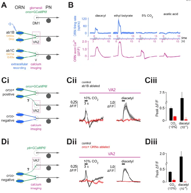
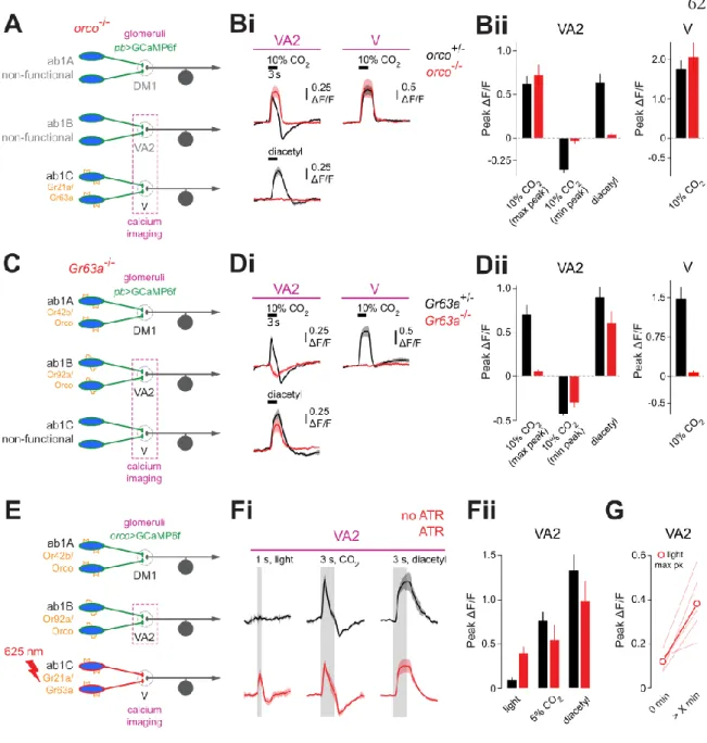
expressed in, and even in the projection neurons (PNs), that the ORNs synapse onto. I show here that CO2,though it activates only a single OR, the GR63a/GR21a hetero-dimer complex, actually activates multiple ORN axon terminals. This activation is due to lateral excitatory connections between axon terminals of the GR63a/GR21a expressing ORNs, and at least 4 other ORN types. Focusing on one of these ORNs, Ab1B, I show the lateral connections bypass the ORN cell bodies, only driving responses at the axon terminals.
Consequently, Ab1B ORN axon terminals receive 2 sources of excitatory input, a feed- forward excitation from its endogenous OR, and a lateral excitation from GR63a/GR21a.
This effectively divides the ORN into 2 compartments, distinct in their odor tuning.
Finally, I show that lateral excitation is a general feature of the ORN circuit by silencing the feed-forward input of another ORN class, Ab1A. The Ab1A cell body is completely silent, but the axon terminals retain odor responses from lateral excitatory inputs. Thus, there is a lateral flow of odor information between multiple ORNs of the Drosophila olfactory system.
PUBLISHED CONTENT AND CONTRIBUTIONS
Zocchi, D., Wennemuth, G., Oka, Y. (2017). “The Cellular Mechanism for Water Detection in the Mammalian Taste System.” In: Nature Neuroscience 20, pp. 927-933. doi:
10.1038/nn.4575
DZ participated in the conception of the project, carried out the experiments, analyzed data, and wrote the manuscript.
TABLE OF CONTENTS
Acknowledgements...iv
Abstract ...v
Published Content and Contributions...vii
Table of Contents...viii
List of Illustrations and/or Tables...ix
Author note...x
Chapter I: The detection of water by the mammalian taste system...1
Introduction...2
Water elicits responses from the taste system...2
Acid sensing TRCs are responsible for water responses...4
Carbonic anhydrases function as molecular detectors for water...5
Optogenetic activation of sour sensing TRCs drives appetitive licking responses in thirsty mice...6
Silencing acid sensing TRCs affects fluid discriminatory ability in thirsty mice...8
Silencing acid sensing TRCs does not affect aversion to sour tastants...9
Discussion...9
Methods...11
References...42
Chapter II: Lateral flow of information at primary olfactory afferents... 46
Introduction ... 47
Multiple ORN classes respond to CO2 ... 48
VA2 CO2 responses switch from a low to high responsive state...49
Mismatch in odor tuning between VA2 ORN soma and axon terminals...50
CO2 responses in VA2 originate from activity in V…..………...51
DM1 ORNS also receive lateral inputs………...53
Mixed excitation and inhibition confers special response Properties on ORN axons………...54
Discussion………...55
Methods………....58
References………....81
LIST OF FIGURES
Chapter 1 Page
1. Water responses in the mammalian taste system ... 16
2. Water activates the acid sensing taste pathway ... 18
3. Carbonic anhydrases mediate taste responses to water ... 20
4. Stimulation of acid sensing TRCs drives drinking responses ... 22
5. Genetic ablation of ChR2-positive geniculate neurons by DTx ... 24
6. Water induced taste signals provide a cue for fluid discrimination ... 26
7. Acid sensing taste pathway is not essential for sour aversion ... 28
8. Ionic effects on taste responses induces by water ... 30
9. Taste responses in TRPM5 KO and PKD2L1TeNT mice ... 32
10. CA independent taste responses and the kinetics of PKD-2L1 taste responses ... 34
11. Light-induced taste nerve responses in PKD2L1ChR2 mice...36
12. Ectopic expression of ChR2-EYFP in the geniculate ganglion...38
13. Acid-sensing TRCs are important for fluid discrimination, but not for water consumption………...40
Chapter 2 Page 1. CO2 elicits responses in multiple glomeruli of the antennal lobe ... 63
2. CO2 responses switch from a low to a high state over the course of a recording ... 65
3. Mismatch in odor tuning between Ab1B cell bodies and axon terminals ... 67
4. The inhibitory component of the CO2 response is orco dependent, the excitatory component is Gr21a/Gr21a dependent ... 69
5. Mismatch in odor tuning between cell bodies and axon terminals of a different olfactory channel ... 71
6. Interaction of inhibitory and excitatory components of CO2 responses leads to selective filtering of CO2 versus other odor stimuli………....73
7. CO2 does not drive spiking responses at Ab1B cell bodies………...75
8. Odor responses persist in Ab1A terminals when Ab1A
cell bodies are silenced………...77 9. Residual odor responses in Ab1A axon terminals when
the Ab1A receptor is silenced are correctly identified as
DM1 responses, and not due to out of plane fluorescence………...79
AUTHOR’S NOTE
The work written about in the first chapter of this thesis was done in the lab of Yuki Oka from October 2014 – March 2017. The work referenced in the second chapter was done in the lab of Elizabeth Hong from January 2017 – July 2019.
C h a p t e r 1
THE DETECTION OF WATER BY THE MAMMALIAN TASTE SYSTEM
SUMMARY
Feelings of thirst trigger water drinking behaviour, which, in turn, is essential for maintaining body fluid homeostasis.1-3 Initiation of drinking behaviour therefore relies on both internal state, as well as peripheral water detection.4-7 While the neural circuits underlying thirst have been well studied, it is still unclear how mammals recognize water in the oral cavity. Here, we identified the cellular logic of water taste in the mammalian taste system. We show that a single class of taste receptor cell (TRC) mediates taste responses to water. We identified this cell population as the acid-sensitive taste receptor cells (TRC) that was previously suggested as the sour taste sensor.8-11 Genetic silencing of these TRCs abolishes water- evoked responses. Conversely, optogenetic stimulation of acid-sensitive TRCs in thirsty animals induced appetitive drinking responses toward light even in the absence of water.
Moreover, our functional manipulation studies indicate that the activities of acid-sensitive TRCs are neither necessary nor sufficient to induce aversion, a typical behaviour when animals encounter sour substances. Taken together, these results show that mammalian acid- sensitive TRCs may be used to detect water in the oral cavity.
INTRODUCTION
Oral sensation, which includes the sense of taste, serves as a checkpoint prior to ingestion of nutrients. The mammalian taste system detects and differentiates between essential nutrients as well as toxic substances through taste receptors and channels expressed in TRCs.8-11,39 As an example, low concentrations of sodium are sensed by a single class of TRCs expressing the epithelial sodium channel, ENaC. Consequently, knocking out the ENaC gene abolishes appetitive salt intake in mice who have been salt deprived.12,13 Similarly, there exist dedicated receptors and TRCs for the tastes of L-amino acids, sugars, bitter, and acidic substances.14-18 Precise genetic access to the 5 different TRC classes is possible via the use of specific protein markers. In particular, acid-sensing TRCs express a protein called polycystic-kidney- disease-like channel19 (PKD2L1). In contrast to the 5 basic tastes, it is still controversial whether water, another vital body nutrient, is also detected by the taste system in mammals.
Several decades of study have shown that invertebrates such as Drosophila melanogaster can sense water through specialized sensory cells in the taste system.20-22 Recent studies have demonstrated that PPK28, a member of the DEG/ENaC family is required for sensory responses to external water, as well as water-seeking behavior.22 In vertebrate species such as frogs, sheep, and cats, water has been shown to elicit electrophysiological responses in facial nerves innervating the oral cavity.23-26 Moreover, water-induced responses have been reported in taste-related neurons of the nucleus of the solitary tract in rodents.26 While these studies suggested that water perception is partially encoded by the taste system, the underlying sensing mechanisms in mammals have not been elucidated to date.
WATER ELICITS RESPONSES FROM THE TASTE SYSTEM
We reasoned that if the taste system is used to detect water in mammals, at least two criteria should be met; first, water taste should be mediated by specific cellular and molecular substrates in taste buds, and second, activation of this pathway should evoke water taste
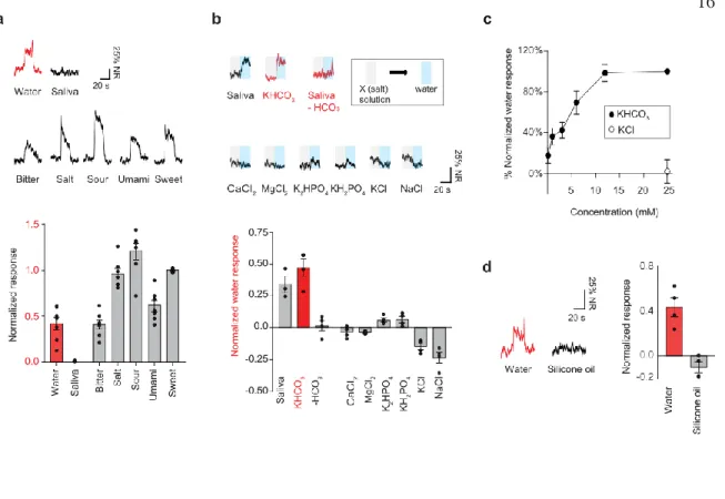
sensation to animals. To test the first of these hypotheses, we performed in vivo extracellular recordings from the chorda tympani taste nerves to explore water responses. The chorda tympani innervates all 5 TRC classes of the tongue, and so serves as a good conduit to monitor the overall activity of the taste system. Stimulating the tongue with various solutions, we observed robust spiking in response to all 5 basic tastes (Figure 1a), showing that we were effectively able to resolve spiking from each TRC population.
We then tested for water responses by probing the tongue with deionized water, eliciting robust nerve responses (Figure 1a). This demonstrated that water effectively activates the taste system, presumably driving responses in 1 or more TRC classes. How does application of deionized-water induce spiking in TRCs? The mammalian oral cavity is normally covered with a thin layer of saliva containing various ions and enzymes.27,28 Our experiments were done in deeply anesthetized animals. A consequence of the anesthetization is that the mice stop producing any saliva. Therefore, we had to constantly perfuse into the oral cavity an artificial saliva solution made up of the ionic components of normal saliva. Tastant and water stimuli were then presented sandwiched between artificial saliva perfusion.
The water responses we measured were therefore due to the wash out of the ions present in saliva with pure deionized water. These observations indicate that the ionic constituents of saliva play a key role in gating the responses. Is any one of these ions particularly important?
We sought to answer this question by making salt solutions of each of the ions present in artificial saliva, and assessing whether we could induce water responses by switching from perfusion of the single salt solution to perfusion of deionized water. Intriguingly, we found that the responses to water were only triggered when bicarbonate ion solutions were used. In other words, changing from bicarbonate solution to water triggered robust nerve responses (Figure 1b, upper middle panel), whereas bicarbonate-free saliva followed by water application failed to induce responses (Figure 1b, upper right panel). No other ions in artificial saliva induced significant taste responses (Figure 1b, lower panel), although high concentrations of phosphate exhibited minor responses (Extended Data Figure 1a). Indeed, potassium bicarbonate in baseline solution evoked dose-dependent nerve responses when
switched to water while potassium chloride had no effect (Figure 1c). Together, these results point out two important characteristics of taste responses to water; 1) the responses are induced by washing out of saliva with water, and 2) unlike invertebrate water detection,22 osmolality change by itself is not the key determinant.
When animals are thirsty, they selectively drink water over other fluids (i.e. oils). If the observed responses are the basis of water detection, one would expect the responses to be selective for aqueous solutions. In fact, application of non-aqueous silicone oil on the tongue did not evoke any significant nerve responses compared to water, indicating that the responses require aqueous solution on the oral cavity (Figure 1d).
ACID SENSING TRCS ARE RESPONSIBLE FOR WATER RESPONSES
Our results above indicate that one of the 5 TRC classes is driving responses to water. We therefore sought to identify the cellular substrate mediating these water taste responses.
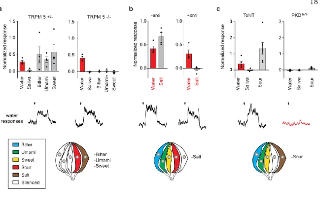
Previous studies have identified genetic markers that specifically label TRCs encoding individual taste qualities.8,14-19,29 Using these genetic handles, we systematically silenced the different TRC classes to assess which class is required for water responses. Transgenic animals lacking TRPM5, a key transduction channel for umami, sweet, and bitter,29 are unable to detect these three taste modalities, due to silencing of the corresponding TRC classes (Figure 2a). In these animals, nerve responses to water were unaffected and indistinguishable from those in control animals (Figure 2a and Extended Data Figure 2a).
This shows that the water responses are completely independent of the activity of the sweet, umami, and bitter sensing TRCs. Similarly, inhibiting the function of the sodium taste sensor, ENaC, by application of its antagonist (amiloride30) entirely suppressed sodium-evoked responses, but had no significant effect on nerve responses to water (Figure 2b). Therefore, the salt sensing TRCs can also be ruled out as the source of the water responses. Finally, we examined the involvement of acid-sensitive TRCs by genetically silencing their synaptic
machinery. To achieve this, we used transgenic animals where the tetanus toxin subunit was targeted to PKD2L1-positive cells by crossing Cre-dependent toxin (TeNT) and PKD2L1- Cre transgenic mouse lines31 (PKD2L1TeNT). Surprisingly, disrupting synaptic transmission from acid-sensitive TRCs resulted in a total loss of water responses (Figure 2c) without affecting other taste qualities, with the exception of acid responses (Extended Data Figure 2b). Altogether, these results show that the acid-sensitive taste pathway is the cellular substrate for chorda tympani responses to water.
CARBONIC ANHYDRASES FUNCTION AS MOLECULAR DETECTORS FOR WATER
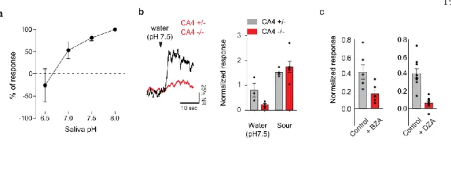
How does water drive acid-sensing TRCs to spike? Given a function of PKD2L1-expressing TRCs as acid sensors (Figure 2c), one possibility is that the water stimulus is converted to a local pH change, leading to the activation of this population. Moreover, saliva is buffered by bicarbonate, as well as phosphates, which keep the salivary pH relatively neutral.42 Given the role of bicarbonate in generating water responses, a pH change mechanism whereby the neutral saliva is replaced by a proton source seemed plausible. Consistent with this notion, water responses were sensitive to saliva pH (Figure 2d). In particular, excessive protons in saliva suppressed water responses, indicating the water responses required the saliva to be relatively pH neutral.
Carbonic anhydrase 4 (CA4) is a membrane-bound catalytic enzyme expressed by acid- sensitive TRCs,31 which reversibly catalyzes the conversion of bicarbonate and protons into CO2 and water. We hypothesized that washing out of bicarbonate from saliva reverses this reaction, which in turn increases local proton production. If this is true, pharmacological blocking or knocking out of CA4 should affect water taste responses. In fact, mice lacking CA4 exhibited significant and selective reduction in their water responses although minor residual responses remained (Figure 2e and Extended Data Figure 3a). Similarly, CA blockers like dorzolamide and benzolamide drastically suppressed water responses without
affecting other taste responses (Figure 2f and Extended Data Figure3b). These results suggest that CA4, and perhaps other CAs, function as molecular detectors that translate water stimuli into a local pH drop (Extended Data Figure 3c).
OPTOGENETIC ACTIVATION OF SOUR SENSING TRCS DRIVES APPETTITIVE LICKING RESPONSES IN THIRSTY MICE
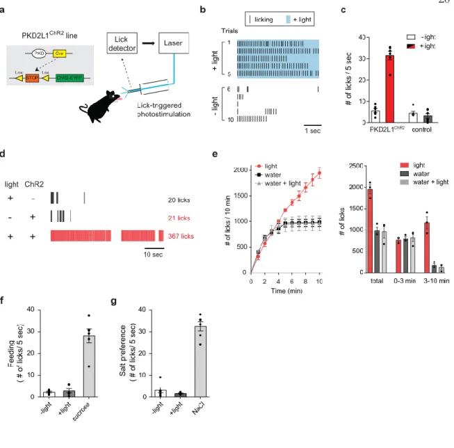
Our electrophysiological results addressed the first of the 2 criteria we framed above about water taste responses. We have established that water drives responses in a specific TRC population, and does so via the action of a dedicated molecular mechanism. The second criteria we imposed was that the activity of the dedicated TRC population should signal the presence of water to a thirsty animal. To assess this hypothesis, we utilized an optogenetic strategy by engineering animals expressing the blue light activated ion channel, channelrhodopsin32 (ChR2) in PKD2L1-expressing TRCs (PKD2L1ChR2, Figure 3a). By illuminating the tongues of these animals with blue light, we could thus effectively drive a large subset of the acid-sensing TRCs. We tested the behavioral consequences of acid- sensing TRC activation by placing the mice in a custom built gustometer with access to a water bottle. The bottle was empty except for a fiber optic cable coupled to a blue laser. The gustometer was set up such that the laser delivered a quick burst of blue light every time the animal licked at the bottle. With this closed loop stimulation setup, we were able to assess the behavioral effects of activating acid-sensing TRCs.
To our surprise, we found that thirsty PKD2L1ChR2 mice exhibited vigorous licking responses toward light in the absence of actual water (Figures 3b and c). This behavior was light- intensity dependent (Extended Data Figure 4b), and was observed both in 5-sec brief access tests, where the animals were only allowed access to the bottle for 5 second intervals (Figure 3c), as well as 1-min continuous access tests (Figure 3d, Supplementary Video 1, and 2). In contrast, neither control animals lacking ChR2 expression, or PKD2L1ChR2 mice without
photostimulation showed this behavior (Figure 3d). Therefore, artificial stimulation of acid- sensing TRCs can drive appetitive behavior in thirsty mice. If activation of acid-sensitive TRCs indeed provides a cue for water detection, then animals should be attracted to light only when they are thirsty and searching for water. Thus, we explored the effect of photostimulation on various appetites such as sugar consumption in hungry animals (Figure 3f) and salt appetite in sodium-depleted animals (Figure 3g). As predicted, animals exhibited no behavioral attraction toward light under hungry and salt-craving conditions (Figures 3f and g). Together, these results satisfy the second criteria we imposed for water responses.
We next asked whether long term activation of acid-sensitive TRCs may lead to satiation of thirst. Thirsty animals normally drink to satiety within few minutes when water is available (Figure 3e, black). Remarkably, water-deprived PKD2L1ChR2 animals showed continuous and unimpeded licking toward light during longer term behavioral sessions lasting 10 min (Figure 3e, red). However, in the presence of water, animals stopped drinking after satiation even with photostimulation (Figure 3e, gray). Together, these results clearly demonstrate that activation of acid-sensitive TRCs serves as a cue for the presence of water, but does not by itself serve as a satiation signal.
In addition to the expression in taste buds, we noticed that ChR2 was ectopically expressed in a small number of geniculate neurons, secondary taste neurons that innervate taste buds (Extended Data Figure 5). To eliminate the possibility that these neurons are involved in light-dependent drinking responses, we expressed Cre-dependent diphtheria toxin receptor in the background of PKD2L1ChR2 (PKD2L1ChR2/DTR) and ablated PKD2L1-expressing cells by injection of diphtheria toxin. Since TRCs, but not secondary neurons, regenerate over time, we were thus able to eliminate the contribution of ChR2-positive geniculate neurons. After regeneration of TRCs, we confirmed that PKD2L1ChR2/DTR mice still show drinking response toward light demonstrating that the behavior is driven by the activity of TRCs (Figure 4).
SILENCING ACID SENSING TRCS AFFECTS FLUID DISCRIMINATORY ABILITY IN THIRSTY MICE
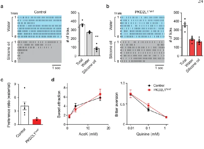
External water is likely detected through multiple orosensory systems including taste and tactile signals.33 To what extent is signaling through the acid-sensing TRCs used for identifying water? In the complete absence of taste responses to water (e.g. PKD2L1TeNT mice), animals show normal spontaneous as well as thirst-induced drinking (Extended Data Figures 6a and b). These data suggest that the taste pathway is not required for proper water identification and consumption. Instead, we wondered whether the taste pathway may play a role in discriminating water from other non-aqueous liquids. To test this idea, PKD2L1TeNT and control animals were water-deprived, and then given a choice between water and low- viscosity silicon oil in a brief taste preference test. Not surprisingly, the control group showed a strong preference to water over silicone oil (Figures 4a and c, and Extended Data Figures 6c and d). However, in sharp contrast, PKD2L1TeNT animals failed to discriminate these two fluids, consuming both silicon oil and water (Figures 4b and c). In fact, PKD2L1TeNT animals consumed significantly more silicone oil and less water compared to the control group. We observed a similar effect when using mineral oil, another taste-less fluid (Extended Data Figure 6e), although the animals eventually learned to avoid the non-aqueous stimulus, no doubt using non-taste sensory cues. Note that PKD2L1TeNT mice showed normal attraction to sweet and aversion to bitter compounds (Figure 4d), indicating that they retain taste discrimination ability. Thus, signals via the acid-sensitive taste pathway provide a cue for fluid choice and subsequent water intake.
SILENCING ACID SENSING TRCS DOES NOT AFFECT AVERSION TO SOUR TASTANTS
Previous studies have shown that acids activate PKD2L1-expressing TRCs, and eliminating these cells abolishes acid-evoked taste nerve responses.10,11,19 Based on these findings, the PKD2L1-expressing TRC population has been suggested to mediate both sour taste and
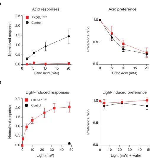
behavioral aversion. However, our results show that these TRCs can mediate both electrophysiological responses as well as behavioral attraction to water. To address this apparent conflict, we investigated if PKD2L1-expressing TRCs are required for behavioral aversion to sour tastants. Control animals with intact acid taste sensors exhibit robust aversion to citric acid (Figure 5a, left panel). In contrast, we observed similar levels of aversion in PKD2L1TeNT animals where the taste nerve responses to the acid were eliminated (Figure 5a, right panel). These data show that the taste pathway is not required for behavioral aversion to acids. On the other hand, optogenetic stimulation of the same population with ChR2 (PKD2L1ChR2 animals) triggered dose-dependent robust taste nerve firing (Figure 5b, left panel). These mice, however, showed no aversion toward water in the presence of light at any intensity (Figure 5b, right panel). Although PKD2L1-expressing TRCs are acid sensitive, our loss-of-function and gain-of-function experiments indicate that this population may not be directly involved in behavioral aversion associated with sour taste.
DISCUSSION
Peripheral water detection represents the first step in the process leading to ingestion and subsequent satiation of thirst. Since Zotterman first described water taste responses in frogs several decades ago,23 accumulating evidence supports the presence of taste detection mechanisms for water in various vertebrate species.23-26,35 Here, we used in vivo electrophysiology to elucidate mechanisms underlying water taste responses. We showed that washing out saliva with water activates the acid-sensitive taste pathway through PKD2L1-expressing TRCs. Furthermore, our data implicate that the removal of bicarbonate in saliva leads to a local pH change through the activity of CAs expressed in PKD2L1- expressing TRCs. Because over 99% of saliva is water, it seems logical that the taste system has a mechanism to detect the dilution of ions, specifically bicarbonate, as a signal of incoming water, rather than sensing water itself. Together, these studies provide key insights into the cellular and molecular basis of water detection at the periphery.
It has been previously demonstrated that PKD2L1-expressing TRCs are activated by acids, salts, and CO2.19,31,34,36,37 All 3 of these substances elicit aversive behavioral responses from mice. On the basis of these aversive behaviors, the acid-sensing TRCs were thought to encode an innate aversion, possibly linked to the sour taste perception. We now add water to the list of stimuli that activate acid-sensing TRCs. Given this large breadth of tuning, the question arises of what taste information these TRCs encode. In this study, we show that optogenetic activation of this taste pathway by light triggered appetitive licking responses in thirsty animals. Inactivation of the same population compromised animals’ fluid discriminability, demonstrating the importance of this pathway for attraction towards, and proper discrimination of water. Conversely, silencing PKD2L1-expressing TRCs had no effect on acid-induced aversive behavior. These results argue that this taste pathway by itself plays a minor role in aversion towards sour, or any other noxious substances. It may, however, be involved in sour perception/aversion under different circumstances such as in combination with other sensory signals. For example, besides the taste system, other sensory pathways including the trigeminal system also contribute to orosensation.33 Because various noxious chemicals are known to activate both taste and trigeminal nerves,36 it is conceivable that behavioral aversion to acids, salt, and CO2 is mediated by neurons in the trigeminal system in combination with signals from acid-sensing TRCs. Perceptual differences between water and sour tasting compounds may then be accounted for by the combinatorial activation of acid-sensitive TRCs and trigeminal nerves. One possible explanation is that activity in the acid-sensing TRCs alone encodes water perception, while sour perception may be created when both taste and trigeminal pathways are activated.
Notably, behavioral attraction by photostimulation of the water taste pathway was induced only when animals were dehydrated, but not food or salt depleted. Therefore, unlike sweet or bitter tastes that are linked to positive and negative values independent of internal state,40 the valence from the water taste pathway is internal-state dependent. The mechanisms underlying this valence change are unknown. While hypothalamic and reward circuits are likely involved in this process,38,41 peripheral mechanisms may also contribute to this valence
change. Future studies with neural manipulations of peripheral water pathway and central thirst circuits should help address how appetite and valence of water are encoded in the brain.
METHODS
Animals
All procedures were carried out in accordance with US National Institutes of Health (NIH) guidelines for the care and use of laboratory animals, and received approval from the Caltech Institutional Animal Care and Use Committees (IACUC). C57BL/6ByJ (B6, stock number 000664), Ai32 (stock number 012569), Rosa26iDTR (stock number 008040) mice were obtained from the Jackson Laboratory. Transgenic lines used were TRPM5 knockout29, PKD2L1-Cre 8, Rosa26-flox-TeNT41, as described previously. For optogenetic experiments, PKDCh2 mice were generated by crossing PKD2L1-Cre with Ai32 lines. Rosa26-flox-DTR was crossed with PKDCh2 line to generate PKDChR2/DTR mice for cell-ablation experiments.
CA4 knockout mice were provided by Dr. Gunther Wennemuth. Mice used for data collection were both males and females, at least 6 weeks of age. Animals were housed in a temperature-controlled environment with a 13-h light, and 11-h dark cycle, and ad libitum access to food and water unless otherwise noted.
Nerve Recordings
Mice were anaesthetized with pentobarbital (100 mg/kg) and placed in a head-fixed setup.
Body temperature was monitored and regulated using a closed-loop heating system. Chorda tympani taste nerve recordings were performed as previously described 14,35. Briefly, animals were given a tracheotomy to prevent suffocation and the right branch of the chorda tympani nerve was exposed. A high impedance tungsten electrode was hooked onto the nerve and a drop of halocarbon oil was dropped inside the surgical cavity. Stimuli were delivered using a pressurized perfusion system (AutoMate Scientific) to keep a constant flow rate. Stimuli used were: 60 mM NaCl (salt), 10 mM Citric Acid (sour), 8 mM acesulfameK (sweet), 0.1 mM cycloheximide (bitter), 50 mM monopotassium glutamate (MPG) plus 1 mM inosine monophosphate (IMP) (umami) and 5 cSt silicone oil (Aldrich), mineral oil (Aldrich).
Deionized water was either filtered (Elaga, PURELAB flex) or purchased (Ultra-purified water, Invitrogen 10977-015). All solutions were used at room temperature. Nerve responses during the 20-s tastant stimulation were integrated and analysed. Responses in each recording session were normalized to 8 mM AceK (Figure 1, 2b, c, e, f, and 5), 10 mM Citric Acid (Figure 2a), and to 25 mM KHCO3 (Figure 1c). For Figure 1b, individual responses were normalized to the average 8 mM AceK responses across entire sessions. The artificial saliva composition was as follows: 4 mM NaCl, 10mM KCl, 6 mM KHCO3, 6mM NaHCO3, 0.5 mM CaCl2, 0.5 mM MgCl2, 0.24 mM K2HPO4, 0.24 mM KH2PO4. pH was held between 7.4 and 7.6. For Figure 2d, the pH of artificial saliva was adjusted between 6 and 8. To stabilize pH of water in CA4 experiments (Figure 2e), we added 0.2 mM KH2PO4 and adjusted pH to 7.5. we Each tastant stimulation was followed by intervals at least for 40 s. Pharmacological experiments were conducted as follows: 50 mM amiloride was dissolved into tastant stimuli solutions and delivered via the pressurized perfusion system. Tastant solutions containing amiloride were presented for 20 s, preceded and followed by 5 s incubation periods with saliva containing the same concentration of amiloride. The oral cavity was incubated with a membrane-impermeable CA blocker, benzolamide (650 µM) or a membrane-permeable blocker, dorzolamide (0.5%) in water for 7 min before washing out with saliva as described previously.31
Analysis of activation kinetics of taste nerves
Activation kinetics of nerve responses was analysed in the following manner. The time points of 25% and 75% of maximum amplitude in each response were determined by MATLAB, and the rise time was calculated as the difference between these two points (Extended Data Figure 3d). The ratio of water to sour rising phase slopes was calculated as the slope of the line connecting the points at 25% to 75% of the peak amplitude of the response.
Taste preference assays
Animals were tested in a custom gustometer to measure taste preference as previously described.14 Solutions were presented to animals for 60 s per trial. Each behavioral session comprised of 10 to 30 trials depending on the number of tastants tested. Each stimulus was presented at least 5 times in one session. Presentations automatically terminated 5 s after the first lick. The number of licks in each of these 5-s windows were counted, and then averaged
across the session. Each animal was tested up to 3 sessions with the same taste repertoire.
We freshly prepared solutions for each behavioral experiment to minimize contamination of other sensory cues such as odors. Prior to all behavioural experiments, mice were water restricted for 22-48 h. For experiments that extended for 48 h, animals were provided with 0.5 mL of water after 23 h. For sucrose appetite assays, animals had no access to food for the 23 h prior to the experiments (Figure 3e). For salt appetite assays (Figure 3f), mice were injected with furosemide as previously described35 and were kept for 23 h with low-sodium diet (Envigo 90228) with free access to water. For sucrose and salt appetite experiments, we used 300 mM sucrose and NaCl. Before testing with photostimulation, animals were pre- trained to drink these solutions.
For drinking assay for silicone oil and water (Extended Figure 6c and d), animals were subjected to water-deprivation for 24 hrs, and either water or silicone oil was presented during the 5-min assay. Individual fluids were tested on separate dates; water tests were normally followed by silicone oil tests because some animals were euthanized after the silicone oil assays due to serious dehydration. We note that both control and PKD2L1TeNT animals preferred water in a long-term ad-lib drinking assay.
For quantifying spontaneous and thirst-driven drinking, animals were individually placed in their home cage and water intake was monitored for 24 hrs (ad-lib), or for 15 min (after water-deprivation for 24 hrs).
Cell ablation by injection of diphtheria toxin
PKDChR2/DTR mice were given an intra-muscular injection of diphtheria toxin fragment A (20 mg/kg BW per day, Sigma D0564) for 2 consecutive days. Expression of ChR2 on the tongue was monitored before, during, and after injections to assess amount of ablation. After the 2-day injection regime, mice were housed 3-4 weeks before being used for behavioural experiments to allow regeneration of TRCs.
Optogenetic stimulation
Photostimulation of the tongue was performed using a gustometer as described above.
Animals were subjected to a brief access taste preference assay as follows; 1) two empty bottles with and without an optic fiber, 2) solutions with and without an optic fiber. Blue laser pulses (430-490 nm, Shanghai Lasers and Optics Century Co.) were delivered through an optic fiber (1mm diameter, ThorLabs) using a pulse generator (World Precision Instruments). The stimulation paradigm was set so that every lick triggered a laser pulse of 1-sec duration in a close-loop manner. The laser power was kept at 48 mW (measured at the tip) unless otherwise noted.
Photostimulation of taste nerves
The same surgery for nerve recording was performed as outlined above. For optogenetic stimulation, the tip of an optic fiber was placed a few millimetres from the tongue while nerve responses from PKD2L1ChR were recorded. Trains of light pulses of 20 s duration were flashed onto the tongue. Each 20 s stimulation window was followed by 40 s intervals. Light trains were delivered at 8 Hz, with each pulse of a 40 ms duration. The frequency and duration was determined based on the licking behavior of mice in our behavioural assays, as well as published literature for the B6 strain (4).
Histology
Animals were euthanized with CO2 followed by cervical dislocation, and perfused with PBS followed by 4% PFA. Tongues were removed and kept in 4% PFA for 12 h followed by 30%
sucrose in PBS for overnight. Frozen sections of 20-µm thickness were prepared on slides and were post-fixed with 4% PFA for 15 min. Then the samples were incubated with blocking buffer (10% donkey serum, 0.2% Triton-X) for 2 h before incubation with primary antibodies. Primary antibodies used were goat αCA-4 (R&D Systems, 1:500, AF2414), αGFP (Abcam, 1:1000, ab6673), and rabbit αPLC-β2 (Santa Cruz Biotechnologies, 1:500, sc-206). Rabbit PKD2L1 antibody9 (1:500) was a generous gift from Dr. Matsunami at Duke University. Secondary antibodies were donkey anti-goat Cy3 (Jackson ImmunoResearch, 1:500, 705-165-147), donkey anti-rabbit Alexa-488 (Jackson ImmunoResearch, 1:500, 711- 545-152), andchicken Alexa-488 (Jackson ImmunoResearch, 1:500, 703-545-155).
Acknowledgments
We thank Brittany Ho for help with mouse husbandry. We also thank Drs. Kristin Scott,
Markus Meister, and David J. Anderson for helpful suggestions. We thank Drs. Charles Zuker and Nick Ryba for generously sharing PKD2L1-Cre and TRPM5 KO transgenic animals, Dr. Matsunami for PKD2L1 antibody, Sangjun Lee for technical support, and members of the Oka lab for helpful comments. This work was supported by Startup funds from the President and Provost of California Institute of Technology and the Biology and Biological Engineering Division of California Institute of Technology. Y.O. is also supported by the Searle Scholars Program, the Mallinckrodt Foundation, the Okawa Foundation, McKnight Foundation, and Klingenstein-Simons Foundation. Y.O. have disclosed these methods and findings to the Caltech Office of Technology Transfer.
Figure 1: Water responses in the mammalian taste system. a, Water elicits robust responses in chorda tympani taste nerves. Shown are representative integrated chorda tympani nerve responses to water and five basic tastants (upper) and their quantified data (bottom). Application of water evoked significant taste responses (n = 7 mice; p=0.0006, water vs saliva). NR, normalized response. Tastants used were: bitter (0.1 mM cycloheximide), salt (60 mM NaCl), sour (10 mM citric acid), umami (50 mM MPG + 1 mM IMP), sweet (8 mM AceK). Artificially reconstituted saliva solution (see Methods) was used as a base solution for all stimuli. b, Effects of individual ion components on water responses. Representative traces are shown (upper) where grey and blue shades denote each salt solution and water, respectively; the trace for saliva minus HCO3 was from a different animal. Average water responses elicited by different salt solutions were quantified (lower).
Removal of bicarbonate ions elicits water responses while saliva lacking bicarbonate ions or other ion species had no effect (n=3 for saliva, n=4 for other solutions; p=0.0286,
KHCO3 vs Saliva - HCO3). c, Dose dependence of water responses to potassium bicarbonate. Higher concentrations of potassium bicarbonate induced larger water responses while potassium chloride elicited no response (n=4). d, Washing-out of saliva with non-aqueous silicone oil does not induce response. Shown are representative traces to water and silicone oil (left), and quantification of responses were shown (right, n=4;
p=0.0286). Statistical significance was analyzed with two-tailed Mann-Whitney U-test.
Values are means ± s.e.m.
Figure 2: Water activates the acid-sensing taste pathway. a, Trpm 5 -/- mice show no responses to bitter, umami, or sweet tastants. However, they retain intact water responses comparable to control animals (n=4 for Trpm 5 -/- and Trpm 5 +/-). b, Application of amiloride (50 µM) completely blocked sodium responses, while it exerted a minor effect on water responses (n=4; p=0.2 for salt). c, Silencing acid-sensing TRCs (PKD2L1TeNT mice) eliminated water responses in addition to acid responses, while control animals (TeNT) show normal responses to both (n=5 for saliva in control, n=6 for the rest; p=0.0022 for water).
The data demonstrate that the acid-sensing taste pathway is required for taste responses to water.
Figure 3: Carbonic anhydrases mediate taste responses to water. a, Water responses are sensitive to saliva pH. All responses are normalized to the responses at pH 8 (n=4; p=0.0392 for pH 6.5 vs pH 7.5, two-tailed paired t-test). Note that saliva pH in healthy animals is normally neutral to basic.42 b, Representative traces from CA4 +/- and -/- mice showing responses to neutral water (adjusted to pH 7.5, left). CA4 -/- have severely reduced responses to water (n=5 for -/-, n=3 for +/-; p=0.0357, two-tailed Mann-Whitney U-test). c, Water responses before and after incubation with CA inhibitors, benzolamide (BZA) and dorzolamide (DZA). Both drugs reduced water responses (n=6; p=0.0159 for benzolamide, n=9; p=0.0007 for dorzolamide, two-tailed paired t-test). Values are means ± s.e.m.
Figure 4: Stimulation of acid-sensing TRCs drives drinking responses. a, A diagram of light-dependent drinking response. Transgenic mice expressing ChR2 in PKD2L1- expressing TRCs (PKD2L1ChR2) were subjected to a close-loop self-stimulation paradigm where each lick event induces 1-sec laser pulse through an optic fiber placed in an empty water spout. b, Photostimulation of PKD2L1-expressing TRCs induced robust drinking responses without water (trials 1-5: blue shading). In the absence of light, the same water- deprived animal did not show consistent licking (trials 6-10). Individual black bar indicates
each lick event. c, Quantification of light-dependent lick events. Number of licks was counted during the 5-sec window. Each condition was presented more than five trials and the number of licks was averaged across trials. The panel shows PKD2L1ChR2 mice (n=6, red bar) and PKD2L1-Cre control mice (n=6, black bar) with photostimulation; white bars indicate the number of licks without light (P=0.0022; two-tailed Mann-Whitney U-test). d, Photostimulation induced continuous drinking responses. Shown are representative plots illustrating the drinking responses toward light in a thirsty control animal (top), and PKD2L1ChR2 animal without (middle) or with light (bottom) during 1-min session. Total lick numbers are shown (right). e, Photostimulation does not satiate animals. Shown are cumulative number of licks from PKD2L1ChR2 animals during the 10-min behavioral sessions (left). Either water, light, or water + light was given during a session. Light stimulation induced significantly more total number of licks (n=3, P=0.0145; two-tailed paired t-test), and licks during 3-10 min (P=0.0213; two-tailed paired t-test). f,g, Food-deprived (e, n=6) or salt-depleted (f, n=6) PKD2L1ChR2 mice did not exhibit appetitive behavior toward light.
300 mM NaCl or 300 mM sucrose solutions were used as control stimuli. Values are means
± s.e.m
Figure 5: Genetic ablation of ChR2-positive geniculate neurons by diphtheria toxin.
Since geniculate neurons innervate their dendrites to TRCs, ChR2-expressing geniculate population may be activated by photostimulation of the tongue. We excluded this possibility by toxin-mediated cell ablation of this population. a, Representative immunostaining of taste buds (left panel) and geniculate ganglia (right panel) from PKD2L1ChR2 and diphtheria toxin- treated animals expressing diphtheria toxin receptor in the background of PKD2L1ChR2 (PKD2L1ChR2/DTR). After 3-4 weeks of recovery period, ChR2-EYFP expressing cells regenerated in taste buds, but not in geniculate ganglia. Scale bars, 100 µm. b, (left) Light- induced licking responses in a toxin-treated PKD2L1ChR2/DTR animal using the same behavioral paradigm described in Figure 3b. Photostimulation of the tongue induced robust drinking (trial 1-5: blue shading) after ablation of ChR2-positive geniculate neurons. These results exclude the possibility that direct photoactivation of geniculate neurons are responsible for light-driven licking behavior. Individual black bar indicates each lick event.
(right) quantification of drinking responses during 5 sec (n=3, p=0.026, two-tailed paired-t test).
Figure 6: Water-induced taste signals provide a cue for fluid discrimination. a, In a brief taste preference assay, water deprived control mice (TeNT) showed strong preference toward water over silicone oil. Shown are representative licking plots toward water and silicone oil;
fluids were presented for 8 times each (left). Individual black bar indicates each lick event.
The number of total licks as well as licks to each fluid was quantified (right) (n=6, p<0.0001 for water vs silicone oil; two-tailed paired t-test). b, In contrast, PKD2L1TeNT mice did not show preference to water and consumed the similar amount of silicone oil (n=7). c, Preference between water and oil was quantified as a ratio (n=7 for PKD2L1TeNT and n=6 for control; P=0.0012, two-tailed Mann-Whitney U-test). d, both control and PKD2L1TeNT mice exhibited dose-dependent attraction to sweet (AceK), and aversion to bitter (Quinine), indicating that PKD2L1TeNT mice retain normal taste discrimination ability (n=4 for
PKD2L1TeNT and n=6 for control). Data are show as a preference ratio to water. Values are means ± s.e.m.
Figure 7: Acid-sensing taste pathway is not essential for sour aversion. a, Normalized taste nerve responses to citric acid in PKD2L1TeNT and TeNT control animals (left). Control animals (n=6) show dose-dependent nerve responses to citric acid while PKD2L1TeNT (n=4) mice showed no response (p=0.0095, PKD2L1TeNT vs control at 20 mM citric acid).
However, both PKD2L1TeNT and control animals showed similar levels of aversion toward citric acid (right) (n=6 for control, and n=4 for PKD2L1TeNT mice), indicating that PKD2L1- expressing TRCs are not necessary for aversive behavior to sour. b, Photostimulation of PKD2L1-expressing TRCs fails to induce aversion. (left) Shown are chorda tympani nerve responses to different intensities of light in PKD2L1ChR2 and Ai32 control animals (n=3 for
0.04-32.6mW for PKD2L1ChR2 mice, n=4 for the rest; p=0.0286, PKD2L1ChR2 vs control at 48 mW). (right) Photostimulation of PKD2L1-expressing TRCs did not change preference toward water. Mice were given a bottle containing water with an optic fiber for stimulation.
Control and PKD2L1ChR2 mice showed undisturbed drinking behavior regardless of light intensity (n=7 for control, and n=6 for PKD2L1ChR2 mice). Statistical significance was analyzed with two-tailed Mann-Whitney U-test. Values are means ± s.e.m.
Extended Data Figure 1: Ionic effects on taste responses induced by water. a, Representative water responses induced by the removal of phosphate ions. In addition to bicarbonate ions, washing out of high concentrations of phosphate (KH2PO4) induced minor responses.
Extended Data Figure 2: Taste responses in TRPM5 KO and PKD2L1TeNT mice. a, Knocking out of TRPM5 has no effect on salt and sour responses. Nerve responses to salt (60 mM NaCl) in TRPM5 -/- mice were comparable to those in TRPM5 +/- control mice (n=4 for TRPM5 -/- and n=4 for TRPM5 +/-). Responses were normalized to 10 mM Citric Acid. b, Sour and water responses were specifically disrupted in PKDTeNT mice. However, response amplitudes to bitter (0.1 mM cycloheximide), salt (60 mM NaCl), umami (50 mM MPG + 1 mM IMP), and sweet (8 mM AceK) were similar between PKDTeNT (n=6) and TeNT control mice (n=5). Responses were normalized to 8 mM AceK. Data were analyzed with two-tailed Mann-Whitney U-test. Values are means ± s.e.m
Extended Data Figure 3: CA independent taste responses and the kinetics of PKD-2L1 taste responses. a, CA4 knockout mice exhibit significantly reduced responses to non- buffered water (n=9 for CA4 -/-, n=8 for CA4 -/+; p=0.0464). All other tastants evoked similar response magnitudes in both genotypes (n=6 for CA4 -/-, n=4 for CA4 +/-). b, Treatments with dorzolamide (DZA) or benzolamide (BZA) had no effect on basic taste responses (n=3). c, A proposed model for activation of acid-sensing TRCs by water and sour.
Acids (protons) directly activate PKD2L1-expressing TRCs through putative proton/potassium channels.45 On the other hand, washing out of bicarbonate with water drives catalytic reaction of CA in PKD2L1-expressing TRCs, leading to increase in local protons. d, Representative taste nerve responses to water and citric acid from the same animal (left). Response rise time (n=7, p=0.0157, middle), and ratio of the rising slopes (right) show slower kinetics of water responses compared to citric acid. Data were analyzed with two-tailed paired t-test. Values are means ± s.e.m
Extended Data Figure 4: Light-induced taste nerve responses in PKD2L1ChR2 mice. a, The tongue was stimulated with laser pulses (8 Hz, 40 ms duration) at 48 mW for 2 s. Shown is a representative trace of three sets of pulse trains. Inset shows a magnified view of a 2-s stimulation window. Each blue triangle corresponds to a laser pulse. An increase in population activity in the nerve is precisely time-locked to laser pulses. b, Total number of
licks induced by different levels of laser power. The number of licks was summed during a 1-min session (n=5). Each data point was obtained and averaged from three PKD2L1ChR2 animals. Values are means ± s.e.m.
Extended Data Figure 5: Ectopic expression of ChR2-EYFP in the geniculate ganglion.
Tissue staining of taste buds in the circumvallate papillae (top), and geniculate ganglion (secondary taste station, bottom) from a PKD2L1ChR2 animal. Shown are representative staining of ChR2-EYFP (labeled with anti-GFP antibody, left), co-labeled with anti-PKD2L1 antibody (middle); the right panels show merged images. ChR2-EYFP signals overlap with PKD2L1 expression in taste buds (top panel). However, ectopic expressions of ChR2-EYFP in geniculate neurons (arrow heads, bottom left) do not show PKD2L1 expression (bottom middle). Scale bars, 100 µm.
Extended Data Figure 6. Acid-sensing TRCs are important for fluid discrimination, but not for water consumption. a, Spontaneous (ad-lib) and thirst-induced water intake in PKD2L1TeNT and control (TeNT) mice. Both genotypes consumed similar amounts of water
(n=4 for each genotype). b, Water-deprived mice (24 h) exhibit no preference toward water and bicarbonate water (25 mM) which does not elicit water taste responses (n=4). These results suggest that non-taste signals are sufficient to drive animals for drinking even without water taste signals.c, Plots of drinking behavior of PKD2L1TeNT and control mice during 5- min consumption tests. Either water or silicone oil was presented to each animal for 5 min after 23 h water-deprivation regime. Individual black bars indicate each lick event. Average number of licks are quantified for each 10-sec period (bottom). d, Cumulative number of licks is shown during the 5-min sessions (n=4, p=0.0188 at 5 min). e, A role of taste pathway for discriminating water and mineral oil. To test if animals can discriminate water and mineral oil, water was first presented to water-deprived animals for 5 s (water), followed by 5 consecutive presentations of mineral oils (5 s each, 1-5 trials). Consistent with the results of silicone oil, PKD2L1TeNT initially consumed comparable amount of mineral oil to water (1-3 trials), but animals learned to discriminate in later trials (4-5 trials, n=3, p=0.0467, water vs 4-5 trials) possibly using other sensory cues such as olfaction and tactile. By contrast, control (TeNT) mice preferred water over mineral oil throughout the trials (n=4, p=0.0012, water vs 1-3 trials, p=0.0007, water vs all). Data were analyzed with two-tailed paired t-test.
Values are means ± s.e.m
REFERENCES
1 McKinley, M. J. & Johnson, A. K. The physiological regulation of thirst and fluid intake. News in Physiological Sciences: an International Journal of Physiology produced jointly by the International Union of Physiological Sciences and the American Physiological Society 19, 1-6 (2004).
2 Sternson, S. M. Hypothalamic survival circuits: blueprints for purposive behaviors.
Neuron 77, 810-824 (2013).
3 Abbott, S. B., Machado, N. L., Geerling, J. C. & Saper, C. B. Reciprocal Control of Drinking Behavior by Median Preoptic Neurons in Mice. The Journal of Neuroscience : the official journal of the Society for Neuroscience 36, 8228-8237, doi:10.1523/JNEUROSCI.1244-16.2016 (2016).
4 McKinley, M. J. et al. The sensory circumventricular organs of the mammalian brain. Advances in Anatomy, Embryology, and Cell Biology 172, 1-122 (2003).
5 Oka, Y., Ye, M. & Zuker, C. S. Thirst driving and suppressing signals encoded by distinct neural populations in the brain. Nature, doi:10.1038/nature14108 (2015).
6 Stricker, E. M. & Sved, A. F. Thirst. Nutrition 16, 821-826 (2000).
7 Young, J. K. Hunger, Thirst, Sex, and Sleep: How the Brain Controls Our Passions.
Rowman & Littlefield Publishers, Inc. (2012).
8 Chaudhari, N. & Roper, S. D. The cell biology of taste. Journal of Cell Biology 190, 285-296, doi:10.1083/jcb.201003144 (2010).
9 Finger, T. E. Evolution of taste and solitary chemoreceptor cell systems. Brain, Behavior and Evolution 50, 234-243 (1997).
10 Liman, E. R., Zhang, Y. V. & Montell, C. Peripheral coding of taste. Neuron 81, 984-1000, doi:10.1016/j.neuron.2014.02.022 (2014).
11 Yarmolinsky, D. A., Zuker, C. S. & Ryba, N. J. Common sense about taste: from mammals to insects. Cell 139, 234-244, doi:10.1016/j.cell.2009.10.001 (2009).
12 Chandrashekar, J. et al. The cells and peripheral representation of sodium taste in mice. Nature 464, 297-301 (2010).
13 Shigemura, N. et al. Amiloride-sensitive NaCl taste responses are associated with genetic variation of ENaC alpha-subunit in mice. American Journal of Physiology.
Regulatory, Integrative and Comparative Physiology 294, R66-75, doi:10.1152/ajpregu.00420.2007 (2008).
14 Matsunami, H., Montmayeur, J. P. & Buck, L. B. A family of candidate taste receptors in human and mouse. Nature 404, 601-604, doi:10.1038/35007072 (2000).
15 Mueller, K. L. et al. The receptors and coding logic for bitter taste. Nature 434, 225-229, doi:10.1038/nature03352 (2005).
16 Nelson, G. et al. An amino-acid taste receptor. Nature 416, 199-202, doi:10.1038/nature726 (2002).
17 Nelson, G. et al. Mammalian sweet taste receptors. Cell 106, 381-390 (2001).
18 Huang, A. L. et al. The cells and logic for mammalian sour taste detection. Nature 442, 934-938, doi:10.1038/nature05084 (2006).
19 Ishimaru, Y. et al. Transient receptor potential family members PKD1L3 and PKD2L1 form a candidate sour taste receptor. Proceedings of the National Academy of Sciences of the United States of America 103, 12569-12574, doi:10.1073/pnas.0602702103 (2006).
20 Evans, D. R. & Mellon, D., Jr. Electrophysiological studies of a water receptor associated with the taste sensilla of the blow-fly. The Journal of General Physiology 45, 487-500 (1962).
21 Wolbarsht, M. L. Water Taste in Phormia. Science 125, 1248, doi:10.1126/science.125.3260.1248 (1957).
22 Cameron, P., Hiroi, M., Ngai, J. & Scott, K. The molecular basis for water taste in Drosophila. Nature 465, 91-95, doi:10.1038/nature09011 (2010).
23 Liljestrand, G. & Zotterman, Y. The water taste in mammals. Acta Physiologica Scandinavica 32, 291-303, doi:10.1111/j.1748-1716.1954.tb01177.x (1954).
24 Shingai, T. Ionic mechanism of water receptors in the laryngeal mucosa of the rabbit. The Japanese Journal of Physiology 27, 27-42 (1977).
25 Shingai, T. & Beidler, L. M. Response characteristics of three taste nerves in mice. Brain Research 335, 245-249 (1985).
26 Rosen, A. M., Roussin, A. T. & Di Lorenzo, P. M. Water as an independent taste modality. Frontiers in Neuroscience 4, 175, doi:10.3389/fnins.2010.00175 (2010).
27 Edgar, W. M. & O'Mullane, D. M. Saliva and oral health. (British Dental Association, 1996).
28 Schneyer, L. H., Young, J. A. & Schneyer, C. A. Salivary secretion of electrolytes.
Physiological Reviews 52, 720-777 (1972).
29 Zhang, Y. et al. Coding of sweet, bitter, and umami tastes: different receptor cells sharing similar signaling pathways. Cell 112, 293-301 (2003).
30 Halpern, B. P. Amiloride and vertebrate gustatory responses to NaCl. Neuroscience and Biobehavioral Reviews 23, 5-47 (1998).
31 Chandrashekar, J. et al. The taste of carbonation. Science 326, 443-445, doi:10.1126/science.1174601 (2009).
32 Boyden, E. S., Zhang, F., Bamberg, E., Nagel, G. & Deisseroth, K. Millisecond- timescale, genetically targeted optical control of neural activity. Nature Neuroscience 8, 1263-1268 (2005).
33 Zald, D. H. & Pardo, J. V. Cortical activation induced by intraoral stimulation with water in humans. Chemical Senses 25, 267-275 (2000).
34 Bushman, J. D., Ye, W. & Liman, E. R. A proton current associated with sour taste:
distribution and functional properties. FASEB Journal : official publication of the Federation of American Societies for Experimental Biology 29, 3014-3026, doi:10.1096/fj.14-265694 (2015).
35 Hanamori, T. Effects of various ion transport inhibitors on the water response in the superior laryngeal nerve in rats. Chemical Senses 26, 897-903 (2001).
36 Oka, Y., Butnaru, M., von Buchholtz, L., Ryba, N. J. & Zuker, C. S. High salt recruits aversive taste pathways. Nature 494, 472-475 (2013).
37 Chang, R. B., Waters, H. & Liman, E. R. A proton current drives action potentials in genetically identified sour taste cells. Proceedings of the National Academy of
Sciences of the United States of America 107, 22320-22325, doi:10.1073/pnas.1013664107 (2010).
38 Dessirier, J. M., O'Mahony, M., Iodi-Carstens, M. & Carstens, E. Sensory properties of citric acid: psychophysical evidence for sensitization, self- desensitization, cross-desensitization and cross-stimulus-induced recovery following capsaicin. Chemical Senses 25, 769-780 (2000).
39 Finger, T. E. et al. ATP signaling is crucial for communication from taste buds to gustatory nerves. Science 310, 1495-1499, doi:10.1126/science.1118435 (2005).
40 Peng, Y. et al. Sweet and bitter taste in the brain of awake behaving animals. Nature 527, 512-515, doi:10.1038/nature15763 (2015).
41 Betley, J. N. et al. Neurons for hunger and thirst transmit a negative-valence teaching signal. Nature 521, 180-185, doi:10.1038/nature14416 (2015).
42 Baliga, S., Muglikar, S. & Kale, R. Salivary pH: A diagnostic biomarker. Journal of Indian Society of Periodontology 17, 461-465, doi:10.4103/0972-124X.118317 (2013).
C h a p t e r 2
LATERAL FLOW OF INFORMATION AT PRIMARY OLFACTORY AFFERENTS
SUMMARY
The Drosophila olfactory system responds to most odors with the activation of a large subset of its olfactory receptors (ORs). In contrast, a select few odors elicit responses in single ORs. In turn, these ORs are usually tuned to very few odors, a property that has been thought to be inherited by the olfactory receptor neurons (ORNs) they are expressed in. We show here that CO2,though it activates only a single OR, actually drives activity in multiple ORN axon terminals. This activation is mediated by excitatory connections between axon terminals of the GR63a/GR21a expressing Ab1C ORN, and at least 3 other ORN types. Focusing on one of these ORNs, Ab1B, we show that the lateral inputs bypass the ORN cell bodies, only driving responses at the axon terminals. Further, we find that odor responses originating from these lateral inputs are modulated between low and high amplitude states. Finally, we show that lateral excitation is a general feature of ORN axon terminals by silencing the OR driven excitation in another ORN class, Ab1A.
We find the Ab1A cell body is completely silent, while the axon terminals retain odor responses. We thus demonstrate that ORN axon terminals receive 2 sources of excitatory input, a feed-forward excitation from their OR, and a lateral excitation from other ORNs.
INTRODUCTION
The first layer of the drosophila olfactory pathway consists of 50 distinct classes of olfactory receptor neurons (ORNs), defined by their expression of 1 of 50 different olfactory receptors (ORs).1,2,3 The ORN cell bodies reside in hair-like structures called sensilla that cover the 2 antenna.4 Each sensillum houses 2-4 individual ORNs of different classes. For example, the Ab1 sensillum houses 4 ORNs: Ab1A, Ab1B, Ab1C, and Ab1D.
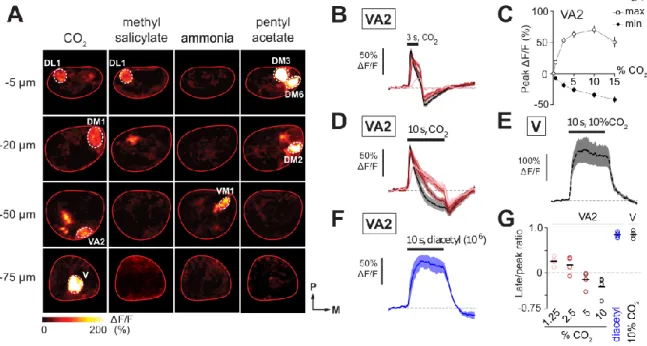
From the antenna, the ORNs project their axons via the Antennal Nerve, to the Antennal Lobes (AL) of the brain. Here, ORN axon terminals synapse onto second order neurons called projection neurons (PNs) in small, compact structures called glomeruli. There are thus 50 glomeruli for the 50 ORN classes, defining 50 olfactory coding channels.5,6 A typical odor binds with some affinity to a subset of the ORs, and so drives activity in multiple glomeruli. Most odors are thus represented with a combinatorial code in the AL.7 Certain specialized odors only drive activity in single ORs, and are instead encoded in a simpler labeled line manner. For instance, cis-Vanyl Acetate (cVA) activates Or67d specifically, and is represented by a single glomerulus in the AL.1,2,8,9,10 Similarly, CO2 has also been shown to exclusively activate the Gr21a/Gr63a heterodimer receptor complex expressed in Ab1C ORNs.11,12 Its representation in the AL is likewise believed to consist solely of activation of a single glomerulus, named the V glomerulus.13
Excitatory and inhibitory cross-talk between olfactory coding channels have been described at the PN level.14,15,16,17,18 The excitatory interactions are mediated by excitatory local neurons14,15,16 that connect PNs of different glomeruli via gap junctions. In contrast, only inhibitory interactions have been described between ORNs. These take the form of feed-forward inhibition of the ORN terminals19,20,21 as well as local ephaptic effects occurring at the cell bodies.22 Therefore, at the first layer of the olfactory pathway, excitatory responses in the 50 coding channels are thought to be completely independent of each other. In other words, tuning of any give ORN is assumed to be completely inherited from the tuning of its OR.