Cellular mRNA Export Factor UAP56 Recognizes Nucleic Acid Binding Site of Influenza Virus NP Protein
By
Andrew Kennedy Morris Dissertation
Submitted to the Faculty of the Graduate School of Vanderbilt University
in partial fulfillment of the requirements for the degree of DOCTOR OF PHILOSOPHY
in Chemical and Physical Biology May 31, 2020
Nashville, TN Approved:
Dana Borden Lacy Charles Sanders
Erkan Karakas Lauren Parker Jackson
Manuel Ascano
ii
TABLE OF CONTENTS
Page
LIST OF FIGURES ………. vii
Chapter 1 – INTRODUCTION ... 1
1.1 – Introduction ... 1
1.2 – Host mRNA Processing and Export ... 2
1.2.1 – Overview ... 2
1.2.2 – Capping ... 94
1.2.3 – Spliceosome Recruitment ... 4
1.2.4 – 3’ End Processing ... 6
1.3 – The TREX Complex ... 7
1.3.1 – Overview ... 7
iii
1.3.2 – THO Binding ... 8
1.3.3 – UAP56 Recruitment ... 9
1.3.4 – ALY/REF Recruitment ... 10
1.3.5 – NXF1/NXT1 Recruitment ... 12
1.3.6 – Export Through the Nuclear Pore Complex ... 13
1.4 – UAP56 in Detail ... 14
1.4.1 – Overview ... 14
1.4.2 – UAP56 Structure Comparison ... 14
1.4.3 – Other UAP56 Functions ... 18
1.5 – The Influenza Virus ... 19
1.5.1 – Influenza Overview ... 19
1.5.2 – Influenza Virion Structure ... 20
1.5.3 – The Viral Lifecycle ... 23
1.5.4 – NS1 ... 30
1.6 – NP structure, Function, and Interactions... 33
1.6.1 – Introduction ... 33
1.6.2 – General vRNP structure ... 33
1.6.3 – NP Structure ... 34
iv
1.6.4 – NP Phosphorylation ... 36
1.6.5 – NP-Polymerase Interaction ... 38
1.6.6 – vRNP Co-Packing ... 39
1.6.7 – Other NP-Host Interactions ... 40
1.7 – The UAP56-NP Interaction ... 42
2 – IDENTIFICATION AND CHARACTERIZATION OF UAP56 BINDING ELEMENTS TOWARDS NP ... 47
2.1 – Introduction ... 47
2.2 – Methods... 48
2.2.1 – Molecular Cloning of NP ... 48
2.2.2 – Molecular Cloning of UAP56 ... 48
2.2.3 – Protein Expression ... 49
2.2.4 – NP Purification... 49
2.2.5 – UAP56 purification ... 50
2.2.6 – GST UAP56 co-precipitation of NP ... 51
2.2.7 – Native PAGE Electrophoretic Mobility Shift Assay ... 51
v
2.2.8 – Microscale Thermophoresis ... 52
2.2.9 – Fluorescence Anisotropy ... 52
2.3 – Results ... 53
2.3.2 – Characterization of A/WSN/33 NP ... 61
2.3.3 – UAP56-NTE is the primary binding site for NP ... 63
2.4 – Discussion ... 79
3 – THE UAP56-NTE RECOGNIZES THE NUCLEIC ACID BINDING SITE OF NP IN COMPETITION WITH RNA ... 81
3.1 – Introduction ... 81
3.2 – Methods... 81
3.2.1 – Crystallization of NP... 81
3.2.2 – FITC-UAP56-NTE1-19 Crosslinking to NP ... 82
3.2.3 – Trypsin Digestion and Liquid Chromatography Tandem Mass Spectrometry (LC- MS/MS)... 83
3.2.2 – Native EMSA Titrations Examining FITC-UAP56-NTE1-19 or RNA Binding to NP* Mutants ... 85
3.3 – Results ... 86
vi
3.3.1 – Crystallization of Several NP Variants could not Resolve Binding Interface ... 86
3.3.2 – Crosslink Mass Spectrometry Reveals UAP56-NTE and NP* Residues in Close Contact ... 91
3.3.3 – Mutation of Crosslink-Identified NP Residues has Minimal Impact on NTE Binding ... 94
3.3.4 – NP* Loop Deletion and R174/R175D Mutations Abrogate RNA Binding ... 99
3.3.5 – RNA and UAP56-NTE Compete for Binding to NP ... 101
3.4 – Discussion ... 103
4 – CONCLUSIONS AND FUTURE DIRECTIONS... 108
4.1 – Conclusions ... 108
4.2 – Open Questions ... 113
4.2.1 – In Vivo Significance ... 113
4.2.2 – Further Mutational Studies ... 114
4.2.3 – The Role of the TREX Complex ... 116
REFERENCES………... 119
vii
LIST OF FIGURES
Figure Page
1.1 – mRNA Processing ... 3
1.2 – Assembly of the TREX Complex ... 8
1.3 – Structure of Yeast THO bound to UAP56 Homolog Sub2 ... 10
1.4 – Structure of Sub2 with Yra1 ... 11
1.5 – The Structure of UAP56 ... 16
1.6 – Summary of the Ten Essential Virally Encoded Proteins ... 21
1.7 – The Simplified Influenza Lifecycle ... 22
1.8 – Viral Transcription ... 25
1.9 – Crystal Structure of Viral Polymerase in Pre-initiation State ... 28
1.10 – Structure of NS1 with Host Export Receptor ... 32
1.11 – The Structure of NP ... 36
1.12 – Influenza Viral Replication ... 45
2.1 – Purification of PR8 NP ... 54
2.2 – Purification of GST-UAP56 ... 56
2.3 – PR8 NP Co-precipitates with GST-UAP56 ... 57
2.4 – Purification of NP* ... 59
viii
2.5 – NP* Co-precipitates with GST-UAP56 ... 61
2.6 – Purification of WSN NP ... 62
2.7 – WSN NP Co-precipitates with GST-UAP56 ... 63
2.8 – Constructs of UAP56 Utilized ... 64
2.9 – Native EMSAs Shows NP interaction with UAP56-NTE and RecA Domains ... 66
2.10 – Coprecipitation Demonstrates UAP56-NTE is Sufficient for Binding ... 68
2.11 – Co-precipitation of NP is Salt-Labile ... 71
2.12 – Microscale Thermophoresis of FITC-UAP56-NTE1-19 with NP* ... 74
2.13 – FA of FITC-UAP56-NTE1-19 with NP* ... 77
2.14 – EMSA Titration of GFP UAP56 Estimates Relative Affinities ... 79
3.1 – X-ray crystallography of NP with UAP56 NTE peptide ... 88
3.2 – XL-MS Reveals UAP56-NTE and NP* Residues in Close Contact ... 92
3.3 – Mutation of Crosslink-Identified NP Residues has Minimal Impact on NTE Binding ... 95
3.4 – Mutation of R174 and R175 Severely Abrogates NTE Binding ... 98
3.5 – NP* Loop Deletion and R174/R175D Mutations Abrogate RNA Binding ... 100
3.6 – RNA and UAP56-NTE Compete for Binding to NP ... 103
4.1 – A Possible Model of UAP56 NP Interaction ... 110
4.2 – Possible NP Residues to Examine for UAP56 Interaction by Mutagenesis ... 116
1 CHAPTER 1
INTRODUCTION
1.1 – Introduction
One of the defining features of eukaryotes is the separation of the transcription and processing of genetic material from its translation by the physical separation of the former into the nucleus and the latter in the cytoplasm. This provides for a much greater degree of regulation and control over the transcription process than that seen in prokaryotes. This also means that mRNAs must mature and be exported, and this involves an intricate set of cellular machinery to coordinate fine control over transcription initiation, 5’ end capping, splicing, cleavage and polyadenylation, and translocation through the nuclear pore complex. The Ren lab is principally focused on
understanding how the pieces of this machinery fit together at an atomic and structural level, and the functional implications of these interactions on mRNA processing and export. The lab particularly has an interest in the mis-regulation of the processing and export machinery and its role in disease. Often the more detailed functions of a biological process are discovered when observed as a phenotype resulting from the disruption of it. In the field of mRNA export, this has meant that several notable discoveries, including that of the primary bulk mRNA export factor NXF1/NXT1, were made based on observed interaction with viruses1,2.
Most viruses replicate in the cytoplasm, however the influenza virus is among those that replicate in the nucleus. One advantage this presents is the evasion of most of the host innate immune factors during transcription and replication, as these factors are generally confined to the
2
cytoplasm. This however also means that they have had to evolve ways to traffic components into and out the nucleus, including their mRNA transcripts. Because of this, study of influenza- host interactions can provide insight into the more fundamental aspects of mRNA processing and export.
This project looks at the interaction of a critical mRNA export factor UAP56, and an influenza viral protein called nucleoprotein. In examining the mechanism by which a virus exploits the host transcriptional process, we add to both the fields of influenza biology and mRNA processing and export. The relevant backgrounds of these fields, and their intersection is presented in chapter 1, while chapter 2 discusses our efforts to examine the minimal binding regions and the conditions which affect the UAP56 NP interaction. Chapter 3 discusses the methods to obtain an atomic resolution view of the interface between this binding pair and the role of RNA in this interaction. The final chapter concludes with a summary of findings, as well as the open questions which remain. The majority of the work described here was originally published by us in Morris et al, 20203, and figures from this paper are noted in their figure legends.
1.2 – Host mRNA Processing and Export 1.2.1 – Overview
This project is at the interface of two different fields, the basic level biochemistry of influenza and the field of mRNA processing. These fields are highly intertwined, since influenza, or indeed any virus, is an obligate parasite, and a complete understanding of influenza replication is
contingent on a knowledge of host mRNA processing and export. The aims of the lab are
3
fundamentally to understand processes in mRNA export, with the study of viruses being secondary, and so it is this area which this dissertation will begin with by reviewing the basic processes involved, beginning at transcription, through to the export of mature mRNA through the nuclear pore.
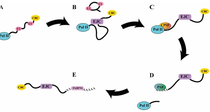
Figure 1.1 – mRNA Processing
A) In early transcription, the 7-methyl guanosine cap is bound by the Cap Binding Complex and U1 and U2 snRNPs bind to exonic junctions. B) Splicing occurs and its associated factors disengage, leaving the EJC. C) When transcription reaches the polyadenylation sequence AAUAAA, Cleavage and Polyadenylation Specificity Factor (CPSF) cleaves the transcript from Pol II elongation D) The free transcript dissociates from Pol II, which eventually terminates transcription. Poly-A polymerase (PAP) adds a variable number of adenines to the 3’ end of the nascent transcript. E) Poly-A Binding Protein Nuclear 1 (PABPN1) binds to the new 3’ poly-A tail.
4 1.2.2 – Capping
mRNA processing begins after transcription initiation. As soon as mRNA emerges from pol II, it is almost immediately subject to 5’ capping. A defining feature of eukaryotic mRNAs is the addition of the unusual nucleotide 7-methyl guanosine (7mG) triphosphate in a 5’ to 5’ linkage to the first base of a transcript. This capping at the 5’ end is essential to processing and allows the transcript to avoid rapid exonuclease degradation4. In metazoans, this is carried out by the enzyme RNGTT5. The methylation of the terminal guanine is catalyzed by RNA (guanine-7- )methyltransferase and RNMT-activating mini-protein in vertebrates6. 7mG is then bound by a large multiprotein complex called the Cap Binding Complex (CBC) which plays a role in recruiting other processing factors (see Fig. 1.1 A). It also interacts with a complex network of transcription factors which form a feedback loop to pol II which can enhance or sometimes repress premature transcription termination depending on the transcript5.
As the transcript elongates, heterologous nuclear ribonucleoproteins (hnRNP) bind along it to stabilize it. hnRNPs are a large class of small RNA binding proteins, each with varying binding sequences and specificities. These coat much of the sequence of an mRNP and the specific set of hnRNPs bound to a transcript is known to play a large part in regulating splicing, export, and later in the cytoplasm, translation7.
1.2.3 – Spliceosome Recruitment
The vast majority of transcripts also undergo splicing, that is, the removal of introns and variable incorporation of exons. Incorporation of different exons through alternative splicing is one method of controlling gene expression and function through the production of different isoforms,
5
while introns produced are the source of snoRNAs8 and often have regulatory roles as sources of lncRNAs and miRNAs9. Splicing proceeds through a very large and complex structure called the spliceosome which contains a core of large RNA-protein complexes called snRNPs with many different auxiliary factors which are associated with different steps in catalysis10. An
oversimplified mechanism of splicing is as follows. A 5’ exonic boundary consisting of a
guanine followed by a uridine is bound by the U1 snRNP, while an internal branch point adenine is bound by the U2 snRNP. The U4, U5, and U6 snRNPs form a bridge between these, and an ATP-dependent rearrangement occurs that brings the U1 and U6 sites, now holding the 5’ exonic boundary, together. A transesterification reaction occurs whereby the 2’ hydroxyl of the guanine at the 5’ exonic boundary attacks the internal branch point adenine, leading to a loop structure with the adenine sugar engaged in three different phosphodiester bonds. This creates a new free 3’ end at the 5’ exonic boundary, which engages in a second transesterification with another guanine at the 3’ exonic boundary downstream. This joins the exons and leaves a freed looped intron structure to leave, along with the U2, U5, and U6 snRNPs11, as seen in (Fig. 1.1 B).
The end result of splicing is the deposition of a set of proteins along the joined boundaries of the 5’ and 3’ splice sites. These proteins are called the Exon Junction Complex (EJC) and are located usually 24 nt upstream of the 5’-3’ boundary12. The minimal components of the EJC consist of the DEAD-box protein eIF4AIII, to which a series of other proteins (Y14, Magoh, and MLN51) bind to clamp it in place on the transcript13. The presence of the EJC, and by extension, the act of splicing is essential for mRNP export. In metazoans, the EJC is where export machinery is recruited, and so unspliced transcripts are very poorly exported14,15. The deposition of the EJC temporally separates the early stages of transcription initiation from the later stages of processing and export by signaling the completion of splicing.
6
The EJC also plays a role in quality control. It forms the center of nonsense-mediated decay, in which surveillance machinery in the form of the SURF complex can recognize premature stop codons based on their aberrant presence upstream of an EJC. This allows the cell to degrade faulty transcripts before they are translated16.
1.2.4 – 3’ End Processing
Before the transcript can be exported however, termination and polyadenylation must occur. In brief, this occurs through Cleavage and Polyadenylation Specificity Factor (CPSF), Poly A Polymerase (PAP), Cleavage Stimulation Factor (CstF), and cleavage factors I and II (CFI and CFII). As pol II escapes the promoter, its CTD becomes phosphorylated at serine 2.
Subsequently, this allows cleavage factor 1A to bind the CTD, where it is in contact with
transcripts exiting. As shown in (Fig. 1.1 C), when it recognizes a polyadenylation signal twenty nucleotides upstream of the cut site it enables cleavage17,18. The endonuclease responsible for cutting the transcript free is the CPSF73 subunit of CPSF19. After cleavage, other factors associated with phospho-serine 2 CTD eventually lead to transcription termination20. From the new 3’ end poly-A polymerase then adds on average 250 adenine residues11 (Fig. 1.1 D).
Polyadenylation of transcripts has been shown to be a prerequisite for export, and improper 3’
end processing causes transcripts to be retained and degraded. Concomitant with polyadenylation is the binding of PABPN1, which has sequence specificity to poly-A sequences and is a marker for complete 3’ processing and subsequent export from the nucleus21 (Fig. 1.1 E).
7 1.3 – The TREX Complex
1.3.1 – Overview
While the pathways above describe mRNA maturation, the export of the mRNP into the cytoplasm occurs through the Transcription Export (TREX) complex. TREX is assembled sequentially. The complete TREX complex consists of ALY, UAP56, and the THO complex, of which the THO complex is the first to be recruited22–25. TREX is also known to interact with some of the previously described mRNA maturation factors. At the 3’ end, Pcf11 is a critical transcription termination factor in yeast26, and has been shown to directly interact with Yra1, the ALY orthologue27,28. While at the 5’ end, depletion of PABPN1 significantly reduced ALY localization to the 3’ ends of mRNPs, further indicative of association, although not direct interaction, between export factors and 3’ maturation29. THO has also been shown to directly interact with Z3CH14, a poly-A binding protein and orthologue of yeast Nab2. In coordination, depletion of Z3CH14 or individual THO subunits led to an increase in transcript
polyadenylation30. Overall, TREX association with both 5’ and 3’ factors shows a physical association between steps of mRNP processing and implies a possible coordination that ensures that all maturation steps have occurred before export2. What is important to note though, is that the exact coordination between mRNA processing at the 3’ and 5’ ends and mRNA export by TREX is still unknown. Aside from splicing, it is unclear if the timing of different steps of TREX assembly and disassembly, shown in Fig. 1.2, is dictated by direct interaction with 3’ and 5’ machinery, and a complete and holistic view of coordinated processing and export is still lacking.
8 1.3.2 – THO Binding
THO is the first TREX component which is recruited2. THO is a large multi-subunit complex consisting of hTho2, hHpr1, fSAP79, fSAP35, and fSAP242,31,32. Although no structure exists of the entire complex from metazoans, the structure has been determined for yeast THO in complex with Sub2, the yeast homologue of UAP56, which shows an overall elongated boomerang- shaped structure with Sub2 sitting in the interior of the bend32.
Figure 1.2 – Assembly of the TREX Complex
A) THO associated with the Pol II CTD recognizes and binds to a deposited EJC after splicing is complete. B) UAP56 with ATP bound associates with the EJC through interaction with THO. ALY is recruited by UAP56 through interaction with its C- terminus. The full TREX complex is assembled. C) The export receptor NXF1/NXT1 binds to ALY. This activates UAP56 ATPase activity and NXF1/NXT1 is deposited on ALY, while RNA is thought to be handed off from UAP56 to ALY. D) NXF1/NXT1 displaces the ADP-bound UAP56 which is the first component to leave TREX. E) At an uncertain point before binding to the NPC, THO and ALY also leave TREX. The export receptor is left bound to the RNA and the EJC. The mRNP is now mature and docks with the basket of the nuclear pore complex through NXF1/NXT1 interaction with Nup proteins as it undergoes biased diffusion out into the cytoplasm.
9
In yeast, THO binds directly to the poll II phosphorylated CTD and is one of the first complexes present during transcription33,34. In metazoans however, THO recruitment, as well as assembly of the rest of the TREX complex is recruited to the EJC15,35,36 (Fig. 1.2 A). This difference reflects the fact that the vast majority of metazoan genes undergo splicing, while the vast majority of yeast genes do not, and so acts as a safeguard against premature export in metazoans. The structural difference that accounts for the different recruitment of THO between yeast and metazoans is not well understood.
While THO is essential for mRNA processing, a lot of the dynamics of its association with the other TREX components are still not well understood either. It is not exported to the cytoplasm, yet its association with ALY is required for binding to the export receptor NXF135,37. So, it is unclear when THO leaves the mRNP2.
1.3.3 – UAP56 Recruitment
THO, once recruited to the mRNP, then recruits UAP56 through its hHpr1 subunit. THO stimulates the ATPase activity of UAP56 by stabilizing a half open conformation which promotes ATP and RNA binding between the two RecA domains32 (see Fig. 1.2 B).
10
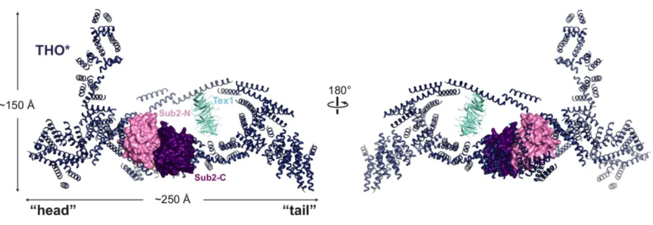
Figure 1.3 – Structure of Yeast THO bound to UAP56 Homolog Sub2
Crystal structure of the THO complex from S. cerevisiae with Tex1 protein (a part of TREX in yeast) from S. bayanus and the yeast UAP56 homologue Sub2 (PDB: 5SUQ). Resolution could not allow for the identification of individual THO subunits but did show an elongated and bent structure with two lobes. Notably, THO binding induces a primed half-open conformation of Sub2 to increase its ATPase activity. Figure taken from Ren et al. 201732.
As a central player in this project, the intimate details of UAP56 will be discussed further in a later section. Its principal role in mRNA export is the recruitment of ALY. It is likely that UAP56 binds to THO first, then is primed to bind to the mRNA by this THO-stabilized
conformation, then RNA binding allows the hydrolysis of ATP thought to be necessary for later steps2 (Fig. 1.3).
1.3.4 – ALY/REF Recruitment
The last component of TREX to be recruited is ALY, sometimes called REF. ALY recruits NXF1/NXT1, which in turn, acts as the adaptor with the nuclear pore. Unlike the other members of the TREX complex, ALY is reported to be dispensable for transcript export, at least in
Drosophila and C. elegans. However even in these studies, ALY knockdown produced some
11
level of mRNP export defect. This suggests that another factor may be functionally redundant with it38,39. This would be consistent with reports that Thoc5, part of the THO complex, is capable of binding to NXF1 directly40,41.
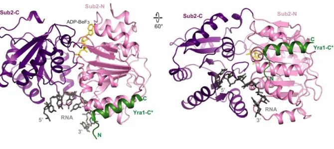
Figure 1.4 – Structure of Sub2 with Yra1
The crystal structure of the UAP56 yeast homologue Sub2 with the C-terminus of the ALY homologue Yra1 (PDB: 5SUP), demonstrating Yra1 manner of recruitment. Sub2 seen here is in the closed, pre-catalytic conformation with the non-hydrolysable ATP analog ADP-BeF3, demonstrating the common DEAD-box mechanism of inducing a kink in the bound RNA between the Rec-A domains which does not allow duplex formation. Yra1 binding stimulates the ATPase activity of Sub2 to allow release for the bound strand. Figure taken from Ren et al. 201732.
ALY directly interacts with transcripts as part of its function, yet its recruitment is mediated through protein-protein interaction with UAP56 as well as THO (Fig. 1.2 C). The C-box domain, the C-terminal-most element of the yeast ALY homologue Yra1, was crystallized with Sub2, the yeast UAP56 homologue (Fig. 1.4). It was seen to be packed against the Sub2 N-terminal RecA domain, with Sub2 in a closed RNA-bound conformation. This suggests that loading of ALY
12
onto RNA requires the ATPase activity of UAP56. In support of this, ALY stimulates ATP hydrolysis by UAP56, a mechanism conserved in yeast32,42,43.
Although it forms a complex with the receptor that delivers the mRNP to the nuclear pore, ALY itself does not leave the nucleus. In yeast, the ALY homolog Yra1 is removed as a result of ubiquitination by the protein Tom12,44. It is unclear if this same mechanism holds in metazoans.
1.3.5 – NXF1/NXT1 Recruitment
Once integrated into the TREX complex, ALY will bind to the export receptor NXF1/NXT1 at its N-terminal RNA binding domains through an N-terminal arginine rich motif on ALY37,45–49 (Fig. 1.2 D).This recruitment of NXF1/NXT1 is concomitant with the dissociation of UAP56 from the TREX complex, as demonstrated in yeast24. UAP56 and NXF1 have overlapping binding regions on the N terminus of ALY and are observed to be mutually exclusive46. NXF1/NXT1 makes protein-protein contacts with the TREX complex, but also makes direct contact with RNA through its leucine-rich repeat, RNA recognition motif, and NTF2L-like domains. It was found that NXF1 also competes with RNA for access to ALY, but that ALY significantly enhanced the NXF1/NXT1 complex affinity for RNA, leading to a model whereby ALY hands off the RNA to the NXF1/NXT1 complex48 in a sequence independent manner50,51. In line with the aforementioned dispensability of ALY, two other proteins, 9G8 and SRp20, were shown to interact with NXF1 in a structurally similar manner52.
13 1.3.6 – Export Through the Nuclear Pore Complex
To leave the nucleus, the NXF1/NXT1-bound mRNPs must travel through the NPC. For this, the export receptor interacts with Nup proteins53. NXF1 serves as the principle adaptor between the mRNP and the nuclear pore through a low-affinity, high avidity interaction with the disordered hydrophobic tails of Nup proteins in the nucleoplasmic basket of the NPC53 (Fig. 1.2 E).
Notably, unlike the typical karyopherin pathways used to import and export proteins from the nucleus, bulk mRNA export does not rely on a Ran GTP gradient31,54,55. The center of the NPC channel is filled with more unstructured hydrophobic Nup tails which are enriched in repeats of phenylalanine and glycine, or glycine-leucine-phenylalanine-glycine. This forms a hydrophobic barrier that effectively makes a gel-like phase through the center of the pore56. It is the favorable interaction of the export receptor with this milieu of hydrophobic residues that allows the mRNP to pass through the pore57. Studies of a very large mRNP called the Balbiani ring indicate that initially this contact occurs with the 5’ end of the mRNP entering the pore first58.
The directionality of movement through the pore is not any form of active pushing, but rather biased Brownian motion that presents an energetic barrier towards the mRNP sliding
backwards31,59. This bias to diffusion is provided by the removal of NXF1/NXT1 by DBP5 and its activator Gle1. This occurs as soon as the mRNP emerges into the cytoplasm, as DBP5/Gle1 are bound to Nups on the cytoplasmic face of the NPC. The absence of the NXF1/NXT1 to interact with the Nups means the protruding portion of the mRNP will not move back into the pore easily31,60–63. After this point nuclear export is complete and the various processes involved in translation can begin.
14 1.4 – UAP56 in Detail
1.4.1 – Overview
UAP56 belongs to a family of proteins called DEAD box helicases, so named after the amino acids in their catalytic signature motif. The prototypical member of this family, and the most studied is eIF4A64. The core function of these helicases is the unwinding of double stranded RNA through ATP hydrolysis. They will bind double stranded RNA through the phosphate backbone without sequence specificity, and separate the two strands, releasing them
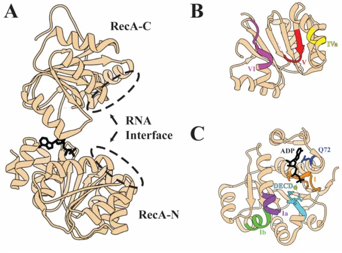
independently. In UAP56, the N-terminal RecA domain (RecA-N) consists of residues 44-251 and the C-terminal domain (RecA-C) of residues 261-428. In between is a short flexible linker with poor sequence conservation between DEAD box helicases (Fig. 1.5 A).
1.4.2 – UAP56 Structure Comparison
Several full length structures of DEAD box helicases have been crystallized32,65–67. In all
observed cases, the RecA domains have almost identical folds, however the relative orientations of RecA-N and RecA-C varies wildly without bound substrate, and they do not interact. In structures with RNA and nucleotide, both RecA domains contribute to binding the substrates through conserved motifs. The RecA domains consists of a central parallel beta sheet (six strands for RecA-C, seven strands for RecA-N) with alpha helices on both sides of the strand that run from the tail of one strand to the head of the next. RecA-N has eight helices and RecA-C has five.
The DEAD box helicase Vasa, crystallized in a closed conformation, first revealed that
unwinding is accomplished by inducing a sharp kink in the RNA that forces the strands apart65.
15
Double stranded RNA is bound in a sequence-independent manner through its phosphate backbone by motifs Ia and Ib (UAP56 a.a. 121-126 and 172-178 respectively) from one side by RecA-N while motifs IVa and V (UAP56 a.a. 324-327 and 341-347 respectively) engage the phosphate backbone from the other (Fig. 1.5 B and C). Contact occurs only on one strand of RNA, with the wedge helix in RecA-N (UAP56 a.a. 173-182) pressing against the phosphate backbone and bending the bound RNA strand in such a way that it cannot base-pair with the other strand without steric clash with the protein. The bound strand is retained, while the unbound strand floats off into solution, or otherwise disengages in the case of hairpins, and this prevents reannealing.
Both RecA domains also engage ATP together. In RecA-N, a universally conserved glutamine (Q72 in UAP56) engages the adenine base. Motif I (a.a. 89-96) positions the triphosphate moiety. Motif II, the namesake DEAD box, is actually DECD in UAP56, and coordinates the catalytic Magnesium ion between beta and gamma phosphates, as well as coordinating these phosphates along with Motif I. RecA-C contributes motif Va (a.a..348-352) and VI (a.a. 373-380) which coordinate the triphosphate from the other side and allow catalysis when the two domains come together (Fig. 1.5 B and C). RecA-C engages in a conserved aromatic stacking with the adenosine base. In the UAP56/ADP structure this stacking with F381 is lost as the RecA-C rotates outward, as are all the RecA-C contacts with the ADP phosphate groups32,66. While ATP binding is required for strand separation, its hydrolysis is not. Rather, hydrolysis is required for the release of the bound strand and for the enzyme to reset64. Unlike most helicases, DEAD box helicases have no real processivity as they do not move along a duplex strand, and typically unwind a few bases before dissociating68.
16
Figure 1.5 – The Structure of UAP56
A) A ribbon diagram of UAP56 bound to ADP from PDB: 1XTJ66. Note that the N-terminal element is not present in this structure. B) A view of the same model looking up at the C-terminal RecA domain showing conserved DEAD-box motifs. C) A 180° view looking down at the N-terminal RecA domain from above, also highlighting conserved motifs. Figure generated with UCSF Chimera69.
Some DEAD-box helicases may also have accessory domains which complement the function of the core RecA domains, often targeting them to specific sequences or structural features.
The bacterial proteins Hera and DbpA each additional C-terminal RNA binding domains involved in recognition of specific ribosomal RNA64,70,71. The yeast protein Mss116 as well as the protozoan CYT-19 also have C-terminal factors involved in enhancing the RecA core
17
helicase activity, as well as being implicated in binding double stranded RNA separately72,73. While these proteins have functions for specific RNAs, UAP56 has no additional bona fide domains and does not exhibit any sequence specificity on its own.
However, for UAP56 as well as others such as Dbp5, DDX19, and DDX25 there is an additional N-terminal sequence before RecA-N. This has some predicted helical propensity for UAP56 but is largely unstructured and unresolved in the crystal structure before residue 4566. Interestingly though, an N-terminal extension is critical for auto-inhibitory functions in DDX19, and is shown wedged between the RecA domains in a crystal structure, but is displaced by RNA and
nucleotide74, and it is believed to operate this way for the yeast DBP5 ortholog as well75. A structurally homologous N-terminus to DDX19 was shown to autoregulate the DEAD box protein DDX25, suggesting N-terminal autoregulation may be a common feature76. While UAP56 N-terminal extension (NTE) is shorter than that of DDX19 and has low homology, it too has demonstrated autoregulatory function. However, deletion of the N-terminal 43 a.a. of
UAP56, in contrast, approximately halved the ATPase activity of UAP56p,o, suggesting an auto enhancing rather than auto inhibitory role.
Dbp5 was crystalized with Gle1 and inositol hexaphosphate which showed Gle1 interaction with the N-terminus Dbp5 that prevents helical insertion between the RecA domains and structurally explains how Gle1 and inositol hexaphosphate can act as cofactors that increase Dbp5 ATPase activity by over ten-fold in vitro. Given that protein regulators can alter the activity of related DEAD box helicases, a similar regulation could possibly exist in UAP56 or Sub2 though the NTE. Indeed, even though only residues 62 onward are resolved in the THO Sub2 complex, they are firmly packed against THO, likely indicating interaction with the entire Sub2 NTE.
Correspondingly, THO increases Sub2’s ATPase activity over 3-fold32.
18 1.4.3 – Other UAP56 Functions
While not discussed in this work, it is important to note that UAP56 has other cellular functions that just its role in the TREX complex. One of the primary and critical function of UAP56
outside of the TREX complex is as an essential splicing factor. UAP56 binds to the early splicing factor U2AF65 which loads the branch point binding snRNP U2, forming the A complex. It was shows that the ATPase activity of UAP56 is what allows U2 to be deposited on the transcript RNA. Indeed, UAP56 gets its name from this function, as it was originally described as 56 kDa U2AF65 Associated Protein. This was originally discovered by a yeast two-hybrid screen for U2AF binding factors, and it was shown that immunodepletion of UAP56 from cell extract prevented splicing in in vitro assays77,78. Later studies showed its ATPase function was required at later steps in splicing, and that its helicase activity unwinds duplexed RNA from the U4 and U6 snRNPs as well79.
Another moonlighting role of UAP56 is in the generation of piRNA. These small transcripts have several functions, most notable among them is the suppression of transposable elements by a mechanism similar to RNA interference seen for siRNA and miRNA. Despite similar
functions, the biogenesis of piRNA is incompletely understood80. UAP56 together with THO has been shown to play a critical role in their transcription and export into the cytoplasm, however81–
83. This multifunctionality of UAP56 should not be surprising since it has no apparent sequence specificity and will bind any free RNA. Such a broad substrate range could mean that there are a myriad of RNA-directed processes across different pathways to which UAP56 is essential or acts on redundantly with other helicases, which have not been ascribed to it yet.
19 1.5 – The Influenza Virus
1.5.1 – Influenza Overview
Having described the host, we now turn our attention to the second aspect of this project, that of the influenza virus. Below is reviewed the basic biology of influenza to frame the context and scope of the interaction we are studying and its overlap with the host processes mRNA export processes reviewed above.
As a pathogen, influenza represents a major threat to worldwide public health. Seasonal
epidemics cause millions of cases and about 500,000 deaths annually84. In addition, pandemics of more virulent strains also occur on a sporadic basis. The most infamous of these was the 1918
“Spanish” influenza, which killed an estimated 30 million people worldwide85. There is also the ever-present threat of the very highly pathogenic zoonotic strains of the virus, or “bird flu” as it is commonly known. Certain stains are endemic to populations of animals, notably waterfowl and swine, and can cause a very high mortality rate when human transmission occurs. Should certain adaptive mutations occur that allow for easier human-to-human transmissibility in these strains, the death toll could be massive. To understand where this project and the influenza nucleoprotein fits in the larger picture of the biology of the virus, an overview of the viral lifecycle is presented here.
There are four principal types of influenza virus, A-D. While influenza C and D are rarely seen in humans, A and B are more common. The A virus mutates considerably quicker and represents the majority of all cases in humans. Additionally, it is the only antigenic type to have caused pandemics86. Influenza A strains are classified according to two of their proteins, hemagglutinin and neuraminidase, with a number denoting the order of their discovery. The combination of the
20
specific variants of these two, with H indicating hemagglutinin and N denoting neuraminidase (e.g. H5N1) defines the classification group87. Influenza is a single stranded negative sense RNA virus whose genome is divided into eight segments. Each segment encodes an open reading frame for a viral protein, although some segments encode for multiple, for a total of ten essential viral proteins, although other minor proteins also exist in many strains88. These are
hemagglutinin (HA), neuraminidase (NA), matrix protein (M1), the matrix ion channel (M2), the non-structural proteins NS1 and NS2, the two basic subunits of the viral polymerase (PB1 and PB2), the acidic subunit of the viral polymerase (PA), and the nucleoprotein (NP), which are summarized in Fig. 1.6.
1.5.2 – Influenza Virion Structure
The overall physical architecture of the individual virion was first characterized by negative stain EM studies done in the late 1960s89,90, where it was seen that individual virions were round membrane-enveloped particles typically about 100 nm in diameter with proteinaceous spikes consisting of HA and NA along the exterior embedded in the membrane with a protein coat of M1 on the interior forming a sphere. The matrix protein M1 provides the virus with structural support. It forms a coat underneath the envelope membrane which encloses the viral contents in a sphere. Tomography revealed that the interior of the capsid contains helical-looking protein
21
Figure 1.6 – Summary of the Ten Essential Virally Encoded Proteins
filaments which contain the genetic material of the virus called viral ribonucleoproteins
(vRNPs)91. M1 also interacts with the vRNPs, anchoring them to the capsid90–92. The vRNP and its functions are the area of influenza biology which are the focus of this project. In brief, they are the packaged genome segments, of which there are eight per virion. Each consists of a viral polymerase which binds the 5’ and 3’ ends of each of the single stranded RNA genome segments called viral RNA (vRNA). The remainder of the vRNA is coated by NP, which binds
independently of sequence like beads on a string to form a double helical hairpin structure capped on one end by the polymerase. The vRNP is described in detail in a later section. The structure of the virion and the viral lifecycle are described below and illustrated in (Fig. 1.7).
Protein Function
Matrix 1
Abv.
Matrix 2
Polymerase Basic Subunit 1 Polymerase Basic
Subunit 2 Polymerase Acidic
Subunit
Non-Structural Protein 1 Nuclear Export Protein
Neuraminidase Hemagglutinin
Nucleoprotein
M1 M2 PB1 PB2 PA NS1 NEP
NA HA NP
Forms interior capsid of virion. Binds to vRNPs to export them. Anchors vRNPs to capsid.
Transmembrane protein in viral envelope. Acidifies virion durring entry. Promotes membrane bending durring budding.
Catalytic subunit of the viral RNA-dependent RNA polymerase. Carries out both transcription and replication.
Integrally bound with PB1. Binds 5’ UTRs of host mRNA and host Pol II CTD for cap-snatching. Regulates PB1.
Performs endonuclease function which cleaves host mRNA 5’ UTR during cap-snatching. Binds vRNA 5’ panhandle.
Blocks host mRN A export and polyadenylation. Antagonizes innate imune factors in the cytoplasm.
Allows nuclear export of progeny vRNP through CRM1 pathway. May regulate polymerase.
Transmembrane protein at viral envelope. Cleaves sialic acid at host extra-cellular matrix to allow virion release.
Transmembrane protein at viral envelope. Binds sialic acid at host extra-cellular matrix to allow virion endocytosis.
Binds viral RNA without sequence specificity. gives structure to vRNPs. Interacts with M1, polymerase, and host factors.
22
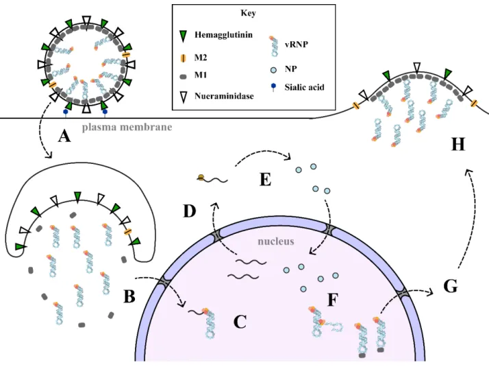
Figure 1.7 – The Simplified Influenza Lifecycle
A) A new virion adheres to the cell surface by binding of hemagglutinin to sialic acid, which triggers endocytosis. B) The early endosome acidifies and an influx of protons through the M2 channel into the virus causes disassembly of the M1 coat.
Hemagglutinin causes membrane fusion and release of contents. vRNPs are imported into the nucleus by Importin alpha. C) Transcription of viral genes begins as the viral polymerase binds and cleaves 7-methyl guanosine caps from host genes to produce its own transcripts. D) Host transcripts are retained in the nucleus by NS1 sequestration of NXF1/NXT1, whereas viral mRNAs are selectively exported E) Translation of viral genes occurs. PA, PB1, PB2, NP, NS1, and NEP are imported back into the nucleus. F) Viral replication occurs as a switch between primer-dependent and primer-independent viral polymerase activities. It is thought that the switch occurs based on the availability of free NP within the nucleus. Replication occurs first to a positive sense cRNA then replication of cRNA produces another negative sense vRNA, which are bound by NP as they emerge from the polymerase. G) Influenza M1 protein bound to vRNPs acts as an adaptor for NEP and the Crm1 export pathway. H) vRNPs migrate to the plasma membrane where hemagglutinin and neuraminidase are incorporated. M1 forms a coat along the interior and together with M2 induce membrane curvature to bud off once all eight vRNPs are incorporated.
23 1.5.3 – The Viral Lifecycle
1.5.3.1 – Viral Entry
Infection begins when a virion makes contact with the extra-cellular surface of the host cell. HA on the virus surface binds sialic acid on the extracellular matrix of host lung epithelial cell, anchoring the virion for cellular entry. Cell surface binding triggers endocytosis by the host cell via either clathrin-mediated or macropinocytosis mechanisms (Fig. 1.7 A). The endocytosed vesicle matures into a late endosome as it migrates inward from the plasma membrane, which results in its acidification93. The interior of the capsid enclosed within the M1 protein coat is also acidified by way of M2 which acts as a transmembrane proton channel. Acidification of the viral interior results in the dissociation of the vRNPs from the M1 protein and the breakup of the coat, owing to a pH dependence of its self-association94. The dissociation of the vRNP from M1 is required for its later nuclear import95. This change in pH also causes a conformational change in HA that results in the fusion of the viral membrane to the endosome, which releases the viral contents into the cytoplasm96 (Fig. 1.7 B). Of note, blocking the acidification of the viral capsid prevents any further progression of infection. M2 channel blockers like amantadine were once used as effective clinical treatments for influenza, before selective pressure led to M2 mutations which prevented drug binding97. The released vRNPs are imported into the nucleus through the nuclear pore complex in a Ran-GTP dependent manner98,99. It was shown that a non-
conventional NLS of NP at the very N-terminus recruits importin alpha /beta to utilize the import pathway typical of most nucleo-cytoplasmic protein shuttling100.
24 1.5.3.2 – Viral Transcription
Once imported into the nucleus, vRNPs begin producing mRNA transcripts of the protein encoded on that genome segment through the polymerase on the vRNP (Fig. 1.7 C). PA, PB1 and PB2 collectively form the viral polymerase, which is an RNA-dependent RNA polymerase.
A large gamut of proteins, both viral and cellular, interact with the polymerase. PB1 forms the central subunit of the complex onto which PB2 and PA anchor as well as the active site of the polymerase.101,102.
The transcriptase activity of viral polymerase depends on co-opting the normal host mRNA transcription activity of RNA polymerase II in order to snatch host-derived 5’ transcript ends with the proper 7-methyl guanosine triphosphate cap103–106 (Fig. 1.8 A). The binding of the host mRNA cap is performed through the PB2 subunit by a discrete domain in the middle of the protein107,108. To steal the cap, viral polymerase must bind to the host RNA polymerase II C- terminal domain as soon as it is activated by phosphorylation at serine 5, which it does at two sites on the surface of its PA subunit C-terminal domain109. The host mRNA is then cleaved between position 10-14106. This endonuclease function was previously thought to be performed by PB1110, but it now known that PA performs this via its N-terminal domain111. The viral polymerase then completes a positive sense mRNA by moving along the vRNA, adding viral sequence to the 3’ end of the stolen cap.
25
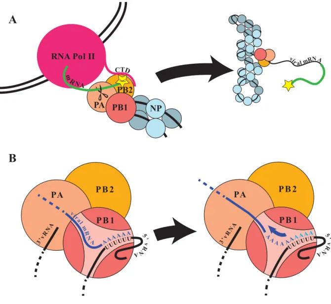
Figure 1.8 – Viral Transcription
A) schematic illustrating the viral mRNA transcription process through binding Pol II CTD and binding the 7-methyl-Guanine cap via PB2, and cleavage by PA, for subsequent elongation by PB1. B) when the polymerase reaches the 5’ of the vRNA template, a polyuridine stretch causes the polymerase to stutter and re-transcribe additional adenosine stretches to polyadenylate viral mRNA independently of the host 3’ maturation factors.
Like the host’s mRNA, viral mRNA is also polyadenylated to avoid degradation and be properly exported. Unlike the comparatively sophisticated mechanism employed for polyadenylation of host transcripts, however viral mRNAs are polyadenylated when the viral polymerase encounters
7mG
PA
PB2 PB1 RNA Pol II
NP
PA PB 2
PB 1
PA PB 2
PB 1
A
B
26
a poly-uridine tract at the 5’ end of the template. This causes it to stutter and slip, repeatedly copying this region to produce a poly-A tail on the transcript (Fig. 1.8 B)112. Notably, two of the influenza genome segments can undergo post-transcriptional splicing at nuclear speckles.
Segment 7 is exported predominantly unspliced to encode the M1 protein, but a portion is spliced to produce mRNA for M2. Similarly, segment 8 produces NS1, but if spliced instead produces NEP113. Although it is incompletely understood how the interaction occurs, NXF1/NXT1 which is the exporter for cellular mRNA, is also used for viral mRNA export112. At the same time, export of cellular mRNA is blocked by NS1114, abrogating the host’s ability to respond with production of interferon-stimulated genes (Fig. 1.7 D).
1.5.3.3 – Viral Replication
When the influenza proteins are translated in the host cytoplasm, hemagglutinin, neuraminidase, and M2 are directed to the Golgi where they undergo the typical processing of host membrane proteins and make their way to the plasma membrane where they localize to lipid rafts115. M1, NS1, PA, PB1, PB2, and NP all have nuclear localization sequences and are imported into the nucleus once produced, while NEP is small enough to enter passively (Fig. 1.7 E). When enough structural proteins have been produced for virion assembly, vRNPs switch from production of mRNA by cap snatching to self-primed replication of the vRNA to a positive sense intermediate called cRNA. What factors trigger this is not completely understood, but the availability and nuclear accumulation of free NP is thought to play a large role, as it is required for binding progeny vRNPs, but not has not been found associated with viral mRNA116. It is also known to interact with the polymerase PB2 subunit, and this may shift it into an alternate polymerase
27
activity117. NEP has also been implicated in this role. In a cell-based study, plasmids for the minimal viral proteins for replication (PA, PB1, PB2 and NP) were transfected into cells and each remaining viral protein transfected in to test them individually for their ability to regulate levels of viral mRNA, cRNA, and vRNA. NEP co-expression altered polymerase activity by suppressing mRNA production and promoting replication to cRNA118.
Unlike viral transcription, which is highly efficient because it is primed by cap-snatching, viral replication is considerably less efficient as it involves de novo synthesis without a true primer beginning at the 3’ end of the vRNA to produce an exact positive sense copy of the genome (cRNA) without any caps or polyadenylation seen for viral mRNAs. This then repeats to make another negative sense vRNA from the cRNA complement93. A crystal structure of the
polymerase complex in the pre-replication state (PDB:4WSA) led to a putative model for how de novo replication of either cRNA from vRNA or vRNA from cRNA is carried out (Fig. 1.9)119. It is thought to depend on a beta hairpin priming loop in PB1 (residues 641-657) based on
functional knowledge and structural comparison to other viral RNA polymerases from
bacteriophage120 and hepatitis C 119,121. For cRNA synthesis, this priming loop acts to stabilize the first incoming nucleotide, GTP, in a base pair with the second to last C at the 3’ terminus. An ATP then comes in on the 5’ side of this GTP in a rate limiting step to form the dinucleotide pppAgG. From this point elongation occurs, which presumably involves the repositioning of the priming helix as duplex RNA is extruded. The polymerase then replicates using the 3’ terminus being fed in as a template. It has been proposed that vRNA synthesis occurs by the same manner but involving a repositioning of the pppApG dinucleotide to the last 3’ template base after its initial formation at position 4 and 5122–124. It has also been proposed that in contrast to viral
28
transcription, replication must proceed by the transfer of the 3’ terminus from one vRNP to a non-resident polymerase. This model is not universally accepted, however125.
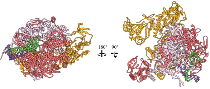
Figure 1.9 – Crystal Structure of Viral Polymerase in Pre-initiation State
PA is shown in salmon, PB1 in pink, PB2 in orange (PDB:.4WSA)119. The 5’ end of the viral RNA in green forms a hairpin bound by PA and PB1 and forms a duplex with the 3’ viral RNA terminus in purple. The endonuclease domain of PA and the cap binding domain of PB2 have rotated away from the template entrance and disengaged, promoting de novo replicative activity.
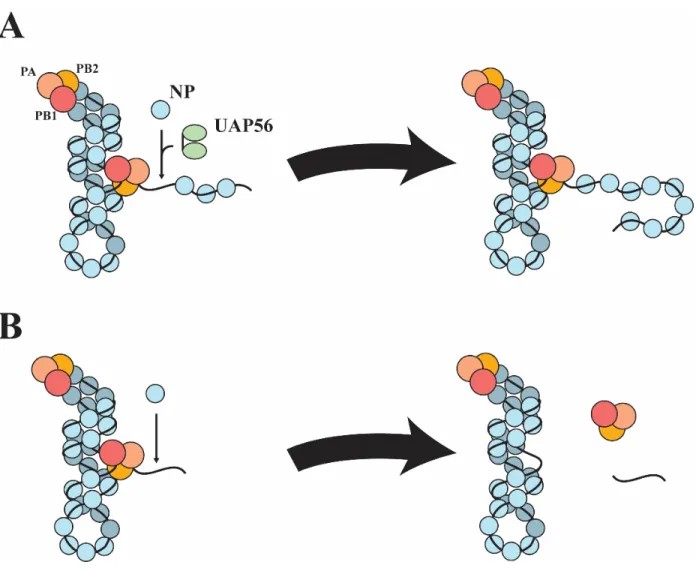
Newly synthesized cRNA and vRNA does not remain naked though. Upon emerging from the polymerase, NP covers the nascent RNA (Fig. 1.7 F). It is important to note that this process leads to abortive replication and premature termination in the absence of host factors, one of which is UAP56, to properly load NP onto new RNA. This aspect of viral replication forms the background context of this project within the scope of influenza biology and will be discussed extensively later.
180° 90°
29 1.5.3.4 – Export of vRNPs
Once produced, new vRNPs need to be exported from the nucleus to make their way to the plasma membrane. Whereas viral mRNA is exported by the NXF1/NXT1 pathway used by most cellular mRNA, vRNPs are exported by the CRM1 pathway. The M1 protein associates with assembled vRNPs through an interaction with NP mediated by RNA. As previously mentioned, M1 in complex with the vRNP prevents its nuclear import, so it is likely that M1 binding during its export serves as means to prevent re-importing vRNPs95,126 (Fig. 1.7 G). M1 also serves as the intermediary to NEP. Through its ordered C-terminal domain NEP binds to M1 associated with a new vRNP while the disordered N-terminal domain contains two non-canonical nuclear export signals that are recognized by CRM1127,128. Crm1 then allows vRNP passage through the Nuclear Pore Complex (NPC) by binding to Ran-GTP and subsequent stimulation of hydrolysis to Ran-GDP by GTPase-activating proteins that causes CRM1 to release its cargo. Because this CRM1 pathway is essential for the virus but not for host bulk mRNA export, it has been a target for anti-viral drug development129.
1.5.3.5 – Nascent Virion Assembly and Budding
Exported vRNPs then make their way to the apical plasma membrane by utilizing host cytoplasmic trafficking. The vRNP is able to bind to Rab11 and migrate with recycling
endosomes as they move outward to the plasma membrane on microtubule tracks116,130,131. M1 also assembles at the apical membrane to begin forming what will become the capsid of a new virion. There is evidence to suggest that the termini of both HA and NA, which extend into the cytoplasm, are responsible for recruiting M1 to the interior of the membrane at the budding site
30
in high enough concentrations that it can multimerize to form the interior capsid132. Through their association with M1, vRNPs are thus recruited to the budding site as well. When all 8 vRNPs have assembled at the apical membrane outward budding takes place (Fig. 1.7 H). The budding process that produces virions from the apical membrane is thought to be a product of membrane curvature induced by HA and a curved net-like lattice formed under the membrane by M1. Notably, mutations in M1 can cause virions to become elongated and filamentous133. M2 has a role in viral budding in addition to viral entry. It contains a cytoplasmic helix with a CRAC motif which recognizes cholesterol and has been demonstrated to induce the membrane curvature necessary for viral budding by its binding, both in model membranes and in vivo134,135. Once budding is complete, the last step necessary to produce an infectious virion is cleavage by neuraminidase. Like HA, neuraminidase is a transmembrane protein which forms part of the viral coat. It is a glycosidic enzyme which cleaves the terminal sugar of the sialic acid chain to allow for virion release. Notably, the most common antiviral drugs used to treat influenza such as oseltamivir and related compounds are neuraminidase inhibitors, which act as sialic acid mimetics136.
1.5.4 – NS1
As previously stated, the interest of the Ren lab in influenza is related to the way in which it hijacks the host mRNA processing and export pathways for its own use, and how this can be used to study those host pathways. Any discussion of virus-host interaction must address NS1, as its primary function is disruption of host transcription, and the study of NS1 has revealed great insights into mRNA export. The protein consists of an N-terminal RNA binding domain and a C-
31
terminal effector domain with a flexible linker in between. The NS1 protein performs many functions for the virus. Although, as the name implies, it is not a structural component of the virus, it is still considered essential. Without it, the virus is only capable of even attenuated growth in cell lines with knockouts of innate immune pathways.
One of the most critical functions of NS1 is the selective export of viral mRNA and simultaneous nuclear sequestration of host mRNA. To accomplish this, it blocks host access to the essential mRNA export factor NXF1/NXT1137. Recently, a crystal structure was published by the Ren lab showing that this occurs by direct binding to the leucine rich repeat and nucleoporin-binding domains of NXF1/NXT1 through its effector domain through two conserved residues, F103 and F138 (PDB: 6E5U)114. Binding in this manner, it acts as a mimetic and competitor to Nup98, a component of the nuclear pore complex to which NXF1/NXT1 interacts to export mRNA, and so blocks access of mature host mRNPs to the nuclear pore (see Fig. 1.10). Notably, retention of host transcripts was non-uniform and transcripts of interferon stimulated genes were enriched in the nuclear-retained RNA. This was demonstrated in vivo by showing that nuclear retention of cellular mRNA resulting from viral infection could be reversed by overexpression of either the full length NXF1 or NXF1201-616, containing the leucine rich repeat and nucleoporin-binding domains. Curiously though, overexpression of the converse NXF1 construct, NXF11-200,had a dominant negative effect in influenza infected cells, exacerbating the nuclear retention further, while lacking this effect in mock-infected cells. In agreement with the crystal structure, infection using influenza with mutations in the residues critical for NXF1 binding, NS1 F103A/F138A did not show significant nuclear retention of host transcripts114.
In line with its role in disruption of host mRNA export, NS1 also binds to CPSF30 through the same interface at F103, as revealed in an earlier crystal structure. CPSF30 is required for the
32
proper polyadenylation of host mRNAs so its disruption leads to their degradation as another mechanism to suppress host gene expression138. In a similar manner, host mRNA processing is disrupted by NS1 interaction with another polyadenylation factor PABII. Notably, this
interaction has no effect on viral mRNA as polyadenylation is performed directly by the viral polymerase instead139. NS1 also plays a role in regulating the splicing of its own transcript into the NEP coding sequence as well as the transcript of M1113.
Figure 1.10 – Structure of NS1 with Host Export Receptor
Crystal structure of influenza NS1 in complex with the export receptor NXF1/NXT1 (PDB: 6E5U)114. Note the 2-to-2
stoichiometry with two NXF1/NXT1 heterodimers to two NS1 dimerized through their RNA binding domains. Opposite sides of the effector domains of NS1 through F103 and F138 interact with hydrophobic pockets in both the LRR and NTF2L domains of NXF1, shown in insets I and II. Figure taken from Zhang et al. 2019114.
33
While not the focus of this lab, it also must be noted that NS1 has many functions in the
cytoplasm to do with the evasion of the host innate immune system. Cells contain a surveillance system consisting of proteins called pattern recognition receptors. These monitor the cell for nucleic acids, proteins, or glycans not seen in a normal cellular environment which are indicative of a pathogen140. In the case of influenza, it is usually double stranded RNA. NS1 antagonizes several different factors involved in monitoring for viral RNA, including 2’-5’ oligo-adenylate synthetase141,142, RIG-I143,144, TRIM-25143, and protein kinase R145.
1.6 – NP structure, Function, and Interactions 1.6.1 – Introduction
Having reviewed the general biology of influenza in its infection of host cells, we shift our attention now to the vRNP, and specifically NP. The replication of vRNP is the aspect of influenza biology at the center of our studies, and the context of the UAP56 NP interaction which we aim to characterize. To provide the background to understand this interaction, a general description of the vRNP, and an intimate description of NP are provided.
1.6.2 – General vRNP structure
The earliest structural information of vRNPs came from electron microscopy of sub-viral components separated by gradient ultracentrifugation, which revealed them to be flexible elongated structures of lengths between 50 and 130 nm consisting of a double helix with a loop
34
connecting the two helical strands at one end and globular mass later identified to be the polymerase at the other89,90,146. Treatment of both reconstituted and native vRNPs with RNase V1 revealed bound RNA is partially resistant to cleavage and indicated an arrangement which still contained dsRNA elements. This suggests that RNA bound to vRNPs can still retain secondary structural elements in at least some places147.
At the same time, when vRNPs are treated with nuclease specific for single stranded RNA, vRNA was highly digested, indicating that the arrangement of NP within the vRNP is such that RNA is surface accessible, likely with the RNA wrapping around the outer edge of the
helix148,149. The 3’ and 5’ ends of each viral genome segment are complementary to each other and highly conserved150,151. Mutational studies152 as well as later structures of the polymerase complex revealed that the 5’-most end forms a hairpin and further down, the 3’ end base pairs with it to form a double stranded stretch of about 14 nt to effectively link the two termini. It is this structure which the polymerase specifically recognizes and resides on at one end of the vRNP119.
1.6.3 – NP Structure
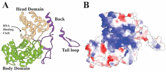
Finally, there is the nucleoprotein (NP), the central focus of this dissertation. NP is a 56 KDa protein encoded on genome segment 5. The first crystal structure available for NP from the A/WSN/1933 strain (PDB: 2IQH)153 revealed that the protein consists of a head and body domain, as well as a tail loop formed by residues 402 through 428. The head and tail domains form a contiguous crescent shaped body, with helices five through eleven as well as nineteen forming the smaller head domain and helices one to four as well as helices twelve through
35
seventeen forming the larger body (Fig. 1.3 A). Between the head and body is a large groove on one side opposite the tail loop. Although the head and body are connected through a series of loops, they are not mobile relative to each other153. At the N-terminus is a previously identified nonconventional nuclear localization sequence which the structure revealed to be sufficiently exposed to be functional100,153. NP sequence across strains of IAV shows fairly high
conservation, with the most conserved parts being helices 6 and 7 as well as the head domain in general. The region surrounding the tail loop and helices eighteen and nineteen which follow it are the least conserved parts of the structure146. This is likely the result of their unimportance to structural stability, as most of the tail loop is unresolved in the first published crystal structure. A mutant made in the tail loop at R416 was crystallized with the tail loop in a different orientation relative to the body154. This indicates a high degree of flexibility within the loops leading to the tail loop. This flexibility very likely plays a function in the tail loop-dependent multimerization, elaborated on below, and allows for the flexibility in the angle with which each NP interfaces with its neighbor which accommodates any number of subunits to form either rings or a helical arrangement.
The principle function of NP is to provide protection and structure for the influenza genome. NP binds RNA in a sequence independent manner through contacts made between the positively charged side chains which line the cleft surface and the phosphate backbone of RNA (Fig 1.11 B). Because binding occurs through the phosphate backbone, NP is also capable of binding DNA155. In vivo however, it is almost exclusively bound to the negative sense vRNA or the positive sense cRNA replication intermediate156. As part of the formation of vRNPs, NP readily oligomerizes155. The primary interaction driving the oligomerization occurs between the tail loop and a crypt formed between several of the interior beta sheets, with critical salt bridges between
36
R416A and E339 and between R422 and E449153. Each NP inserts the tail loop into the crypt of its neighbor and polymerization occurs to form circular structures of varying sizes in vitro, even in the absence of RNA. In vivo however, the RNA binding and self-associative properties of NP lead to its accumulation onto cRNA and vRNA as they are being replicated by the influenza polymerase116.
Figure 1.11 – The Structure of NP
A) A ribbon diagram of NP from A/WSN/1933 with domains highlighted (PDB: 2IQH)153. B) The same structure with surface electrostatics. Blue is negative and red is positive charge. Figure generated with UCSF Chimera69.
1.6.4 – NP Phosphorylation
While not investigated in this study, it is important to note that NP can be phosphorylated116,157. Mass spectrometry-based phosphoproteomics showed NP to be the most heavily phosphorylated protein in influenza. Residues 3, 9, 78, 165, 188, 296, 377, 402, 457, and 472 have been
37
identified across various different strains as subject to phosphorylation to various extents158–162. Notably, the exact pattern of phosphorylated residues depends on strain, and changes over the course of infection163.
Control of phosphorylation and dephosphorylation of serine 165 was shown to regulate the ability of NP to form oligomers. Phosphomimetic S165E critically inhibits oligomerization of NP by disrupting the interface between subunits, producing NP which exists as monomers in vitro, and cannot produce virions in a reconstitution assay. The phospho-deletion mutant S165A, however also exhibited reduced titer, suggesting that phosphorylation at this residue may be used to regulate NP oligomeric state, which changes from early to late infection161.
Threonine 188 was recently identified as an important phosphorylation site. It is part of one of NP’s nuclear export signals and mutation to a phosphomimetic glutamate led to the permanent nuclear retention of NP, as well as reduced replication and transcription by the polymerase which could not create infectious progeny using a plasmid-based viral reconstitution assay. The
phospho-deletion mutant T188A had comparatively mild perturbation. While purely structural perturbation of NP itself can’t be totally ruled out, this seems to indicate that phosphorylation is at least one mechanism by which the nucleocytoplasmic trafficking of NP is regulated as part of coordinating its export during the viral life cycle160. In a similar manner, the phosphorylation status of residue 78 as well as residue 9 within the nuclear localization sequence have been shown to affect nucleocytoplasmic shuttling of NP159,162.