Proteus mirabilis and Urinary Tract Infections
Jessica N. Schaffer and Melanie M. Pearson*
Department of Microbiology, New York University Langone Medical Center, New York, NY 10016
Abstract
Proteus mirabilis is a Gram-negative bacterium which is well-known for its ability to robustly swarm across surfaces in a striking bulls’-eye pattern. Clinically, this organism is most frequently a pathogen of the urinary tract, particularly in patients undergoing long-term catheterization. This review covers P. mirabilis with a focus on urinary tract infections (UTI), including disease models, vaccine development efforts, and clinical perspectives. Flagella-mediated motility, both swimming and swarming, is a central facet of this organism. The regulation of this complex process and its contribution to virulence is discussed, along with the type VI-secretion system- dependent intra-strain competition which occurs during swarming. P. mirabilis uses a diverse set of virulence factors to access and colonize the host urinary tract, including urease and stone formation, fimbriae and other adhesins, iron and zinc acquisition, proteases and toxins, biofilm formation, and regulation of pathogenesis. While significant advances in this field have been made, challenges remain to combatting complicated UTI and deciphering P. mirabilis pathogenesis.
Proteus mirabilis is well-known in clinical laboratories and microbiology survey courses as the species that swarms across agar surfaces, overtaking any other species present in the process. Urease production and robust swarming motility are the two hallmarks of this organism. This species can be identified as a Gram-negative rod that is motile, urease- positive, lactose-negative, indole-negative, and produces hydrogen sulfide (1). It is a member of the same bacterial family (Enterobacteriaceae) as E. coli.
Disease
P. mirabilis is capable of causing symptomatic infections of the urinary tract including cystitis and pyelonephritis and is present in cases of asymptomatic bacteriuria, particularly in the elderly and patients with type 2 diabetes (2, 3). These infections can also cause bacteremia and progress to potentially life-threatening urosepsis. Additionally, P. mirabilis infections can cause the formation of urinary stones (urolithiasis).
P. mirabilis is often isolated from the gastrointestinal tract, although whether it is a
commensal, a pathogen, or a transient organism, is somewhat controversial (4). It is thought that the majority of P. mirabilis urinary tract infections (UTI) result from ascension of bacteria from the gastrointestinal tract while others are due to person-to-person transmission,
HHS Public Access
Author manuscript
Microbiol Spectr. Author manuscript; available in PMC 2016 April 01.
Published in final edited form as:
Microbiol Spectr. 2015 October ; 3(5): . doi:10.1128/microbiolspec.UTI-0017-2013.
Author Manuscript Author Manuscript Author Manuscript Author Manuscript
particularly in healthcare settings (1). This is supported by evidence that some patients with P. mirabilis UTI have the same strain of P. mirabilis in their stool, while others have no P.
mirabilis in their stools (5). In addition to urinary tract infection, this species can also cause infection in the respiratory tract, eye, ear, nose, skin, throat, burns, and wounds and has been implicated in neonatal meningoencephalitis, empyema, and osteomyelitis (1, 6). Several studies have linked P. mirabilis to rheumatoid arthritis, although others have failed to find an association (reviewed in (7) and (8)). It is thought that antibodies against hemolysin and urease enzymes are subsequently able to recognize self antigens targeted in rheumatoid arthritis patients (8).
Incidence
P. mirabilis causes between 1-10% of all urinary tract infections, varying with the geographic location of the study, the types of samples collected, and the characteristics of the patients examined. In the most recent large North American study, this species caused 4% of almost 3,000 UTI cases (9). In 2006, UTIs in the United States were the cause of 11 million physician visits and cost $3.5 billion dollars (10). This organism is more common in complicated urinary tract infections (such as patients with spinal cord injury or anatomical abnormality) and especially contributes to catheter-associated UTI (CAUTI), causing 10-44% of long-term CAUTIs at a cost of $43-256 million in the US annually (6, 11, 12).
The wide range of P. mirabilis CAUTI likely reflects differences in the population surveyed and the types of samples collected. The highest incidence of P. mirabilis CAUTI occurs in elderly patients during long-term catheterization. P. mirabilis is also a common agent of Gram-negative bacteremia, particularly in patients with concurrent UTI; in recent studies, this species was found in 5-20% of these cases and as high as a 50% mortality rate in geriatric patients (13-16).
VIRULENCE FACTORS
P. mirabilis virulence has primarily been tested using mouse or rat models of infection. Two models of ascending UTI are employed. Independent challenge and co-challenge
experiments insert bacteria directly into the bladder using a urethral catheter. In an independent challenge, each strain is tested for the ability to cause infection in the absence of other bacteria, while during a co-challenge experiment two different strains of bacteria are mixed prior to catheterization and must compete to colonize the urinary tract. A third model investigates a hematogenous route of infection, in which bacteria are injected intravenously and the ability of the bacteria to colonize the kidneys is examined.
Throughout this section, genes will be referenced by their PMI gene designations in the sequenced and annotated P. mirabilis genome, strain HI4320 (17), although other wild-type isolates have been studied.
Urease
The cytoplasmic nickel metalloenzyme urease acts by hydrolyzing urea into ammonia and carbon dioxide. The resulting ammonia is the preferred nitrogen source for many species of bacteria, and may be assimilated into biomolecules via glutamine synthetase (GlnA) or
Author Manuscript Author Manuscript Author Manuscript Author Manuscript
glutamate dehydrogenase (GdhA). Further general information on urease is reviewed in (18) and P. mirabilis urease is reviewed in (19). A direct result of urease activity and ammonia generation is an increase in local pH. In the urinary tract alkaline pH leads to precipitation of calcium and magnesium ions and the formation of urinary stones composed of magnesium ammonium phosphate (struvite) and calcium phosphate (apatite) (20). In P. mirabilis, urease is encoded by the ureDABCEFG operon (PMI3682-88). A regulator, UreR (PMI3681), is encoded on the reverse strand adjacent to this operon (21). UreR is an AraC-type DNA binding protein which positively induces urease expression in the presence of urea; a ureR mutant lacks measurable urease activity (22). UreR also positively regulates its own expression when bound to the intergenic region between ureR and ureD, and ureR
transcription is repressed by the global regulator H-NS (23). Urease expression is induced in the urinary tract of experimentally-infected mice (24, 25).
VIRULENCE
Urease mediates virulence via the production of urinary stones. These stones can block urinary flow and cause tissue damage; they can also become quite large (> 1 cm2) (26, 27).
The precipitated minerals may mix with bacteria adherent to a urinary catheter, forming a crystalline biofilm and eventually blocking urine flow through the catheter (28, 29). Similar intracellular crystals have been visualized in cultured urinary epithelial cells that have been experimentally infected with invasive P. mirabilis (30). Although other urease-positive bacterial species are associated with catheter-associated UTI, only P. mirabilis has a positive association with catheter obstruction (31). Bacteria can become embedded in these stones, which may protect pathogens from antibiotics or the immune system (32) (Fig. 1).
Furthermore, urinary stones can act as a focal point for other species of bacteria to establish UTI (19). A ureC urease mutant is incapable of forming stones, and this has a direct impact on the ability of P. mirabilis to cause UTI. When this urease mutant was tested in
independent challenge using a mouse model of ascending UTI, there was a highly- significant decrease in bacterial numbers compared to the wild-type parent in the bladder, kidneys, and urine (33). The effect was especially pronounced in the kidneys, where no mutant bacteria were detectable in most mice 48 hours post-infection. The murine 50%
infective dose for the urease mutant (2.7 × 10⁹ CFU) is 1000-fold higher than the wild type (2.2 × 10⁶ CFU) (34). From two days to two weeks post-inoculation, kidneys from mice infected with the urease mutant displayed less pathology (acute inflammation, epithelial necrosis) compared to wild-type infection. Likewise, a ureR mutant tested in cochallenge with the parent strain was unrecoverable in most mice (22). Urease activity during UTI may be influenced by polymicrobial infection; experimental co-infection of mice with P.
mirabilis and urease-positive Providencia stuartii resulted in increased urolithiasis and bacteremia despite similar bacterial loads compared to monospecies infection (35). Indeed, in vitro co-culture of P. mirabilis and P. stuartii in human urine resulted in enhanced total urease activity compared to either species alone (35).
Due to the prominent role of urease in P. mirabilis virulence, this enzyme is an active target of investigation to identify clinically useful inhibitors (36). Since the ability of P. mirabilis to generate urinary stones and crystalline biofilms is dependent upon alkaline pH, another approach to prevent catheter blockage is to acidify the urine. Similarly, mineral nucleation
Author Manuscript Author Manuscript Author Manuscript Author Manuscript
can be inhibited by reducing mineral concentration in the urine, i.e., by increasing fluid intake (37). These efforts are aimed at increasing the urinary nucleation pH (the pH at which minerals will precipitate from the urine); a lower nucleation pH is associated with increased stone formation. Preliminary results with patients consuming lemon juice are promising, with the result of increased nucleation pH (38). However, the effect of such treatments on catheter blockage has not yet been reported.
Flagella
Like many bacteria, P. mirabilis uses flagella to swim through liquids and toward chemical gradients (reviewed in (39)). In liquid culture, P. mirabilis has a short rod shape and typically possesses a few peritrichous flagella. However, on rich solid media, P. mirabilis differentiates into very long (typically 20-80 μm, although cells longer than 100 μm occur), nonseptate polyploid cells with hundreds to thousands of flagella. These swarmer cells move as a population across surfaces, and will be discussed later in this chapter. Although the flagella produced by this organism are generally similar to flagella produced by other bacteria, there are two unusual characteristics of P. mirabilis flagella. First, all genes encoding flagellar components, including the class I flagellar master regulatory genes flhDC (PMI1671-72), are found within a single 54 kb locus in the chromosome (PMI1617-72).
This is in contrast to most other flagella-producing bacteria, which have flagellar operons in disparate loci. Second, P. mirabilis encodes two flagellins, FlaA (PMI1620) and FlaB (PMI1619) (also known as FliC1 and FliC2, respectively) (40), which comprise the whip structure of the flagellum.
ANTIGENIC VARIATION
FlaA is the major flagellin. Despite the proximity of flaA and flaB, flaA is transcribed as a monocistronic message and flaB message is generally undetectable (41). Recombination between flaA and flaB can occur. This phenomenon was discovered when flaA mutants were often found to revert to a motile phenotype; these revertants produced antigenically distinct flagella that were the product of recombination resulting in a hybrid flaAB gene (41, 42).
Later studies revealed that wild-type populations of P. mirabilis are heterogeneous, with a portion of the cells possessing hybrid flaAB genes (42, 43). RNA experiments suggested that 1.0-1.5% of the total flagellin message in wild-type populations is flaAB (43).
Recombination occurs between homologous regions of flaA and flaB, and leads to deletion of the intervening sequence. Hybrid flagellins have been detected in bacteria excreted in the urine of mice experimentally infected with P. mirabilis. There may be a selective advantage to particular recombination events, as the types of rearrangements typically found depend on the bacterial environment (broth, swarm agar, or murine urinary tract) (42). The recombinant flagella are functional and may serve as a method of immune evasion during UTI or to provide motility under adverse conditions. Indeed, bacteria possessing specific hybrid FlaAB flagella are more motile under conditions of high salinity (255-425 mM NaCl) or extreme pH (5.2 or 8.2) compared to bacteria with wild-type FlaA flagella; conversely, the wild-type flagellum confers a motility advantage in low salinity (85 mM NaCl) (44).
Additionally, immune serum from mice experimentally infected with P. mirabilis reacts with both FlaA and FlaAB flagellins (45). A possible third flagellin, designated FlaC or
Author Manuscript Author Manuscript Author Manuscript Author Manuscript
FliC3, was identified by DNA-DNA hybridization (40); however, fliC3 was not annotated in the P. mirabilis HI4320 genome sequence and is not readily identifiable by nucleotide BLAST using queries that correspond to the probes used in the initial discovery of fliC3.
REGULATION OF FLAGELLA
As has been characterized for other bacteria, P. mirabilis flagellar genes are transcribed in a three-tier hierarchy (reviewed in (46)). Regulation of flagella is mediated through the class 1 flagellar master regulator genes flhDC. The regulator functions as the heteromer FlhD2C2 or FlhD4C2 (47, 48). There are multiple inputs to regulation of flhDC as well as post-
translational modification of FlhD4C2 and downstream regulation of the class II and class III flagellar genes. Perturbations in flagellar expression may lead to different outcomes with regard to swimming versus swarming, and will be discussed in detail below.
Expression of flagellar genes is regulated during experimental UTI. Within 24 h post- infection, flagella are repressed compared to an in vitro mid-logarithmic phase broth culture (25). However, by seven days post-infection, this repression is relieved (25) (Fig. 2). It is very likely that flagella are produced at some point during P. mirabilis-mediated UTI, as experimentally-infected mice produce antibodies that recognize flagella (45, 49). Flagella are also repressed during uropathogenic E. coli (UPEC)-mediated UTI (50), but are transiently expressed around 4-8 hours post-infection (51). This time coincides with bacterial ascension from the bladder to the kidneys. It is possible that P. mirabilis flagella undergo similar transient expression during early stages of ascending UTI.
ROLE OF FLAGELLA IN VIRULENCE
There are differing conclusions in the literature about the role of motility in P. mirabilis virulence. In one study, the flagellum was found to contribute to ascending UTI (52). In that report, an hpmA (hemolysin, PMI2057) mutant was compared in independent challenge of CBA/J mice to the wild-type parent and an isogenic hpmA flaD (PMI1621) double mutant.
FlaD is the capping protein of the flagellum; this mutant produces unassembled flagellin and is nonmotile. At one week post-infection, the hpmA flaD mutant was recovered from the urine, bladder, or kidneys in numbers 100-fold lower than either the wild type or the hpmA single mutant. Functional flagella contribute to bacterial spread during UTI; immobilizing antibodies prevented the ability of P. mirabilis to traverse from one kidney to the other via the ureters using a rat model (53). However, another group has reported that nonmotile P.
mirabilis strains also cause UTI (54, 55). In the first of these studies, both ascending UTI and hematogenous routes of infection were used to assess the ability of motile and
nonmotile P. mirabilis isolates to colonize the bladders or kidneys of outbred mice at seven days post-infection (54). The nonmotile strain was as infective as the motile strains. In the second study, an isogenic mutant missing the 3’ portion of flaA and the 5’ portion of flaB was compared to the wild-type parent in the ascending UTI outbred mouse model (55).
Again, at seven days post-infection, there was not a significant difference between wild type and mutant in colonization of bladder or kidneys. Taken together, it is likely that flagella contribute to P. mirabilis UTI, but the effect may be modest and dependent on the strains of
Author Manuscript Author Manuscript Author Manuscript Author Manuscript
bacteria and animal models used. The specific contribution of swarming to virulence will be considered later in this chapter.
Signature-tagged mutagenesis (STM) studies have also identified mutants with diminished or absent production of flagella that were less competitive in the mouse UTI model (56, 57).
In addition, STM identified a chemotaxis mutant (cheW), suggesting that not just motility but also the ability to move toward one or more unidentified signals contributes to bacterial fitness during UTI (56). Although eight likely methyl-accepting chemotaxis proteins (MCP) were identified in the HI4320 genome (two in the flagellar locus and six encoded elsewhere (17)), it is worth noting that the molecules sensed by these MCP have not been elucidated for P. mirabilis.
Swarming
When P. mirabilis is added to an agar surface, the bacteria grow in place for a time (which varies by medium, humidity, and temperature), differentiate into swarmer cells, and move forward as a population. The ability of P. mirabilis to swarm as an organized group across solid surfaces was first noted by Hauser in 1885 (58). DNA replication without septation occurs during swarm cell formation, which results in very long, polyploid cells. At defined intervals, the bacteria stop moving and revert to a shorter morphotype (consolidate). After a period, the bacteria redifferentiate into swarmer cells. These bacteria move across many media surfaces in a repeated process of swarming and consolidation, resulting in a
characteristic bull's-eye pattern (Fig. 3). In fact, these bacteria are named for the Greek god Proteus, who was able to “change shape at will to avoid questioning” (58). Swarmer cells are phenotypically distinct from swimmer or vegetative cells, and are characterized by great length (typically 20-80 μm) and hyperflagellation (Fig. 4). The shift to the swarmer form is accompanied by changes in lipopolysaccharide (LPS), peptidoglycan, and membrane fatty acid composition (59, 60). Swarm cells move together as a group, forming rafts of parallel cells (61). A capsular polysaccharide termed colony migration factor and an uncharacterized slime are associated with swarming cells and may be used to aid motility across surfaces (62-64). Numerous genes are differentially regulated during swarming, including genes that are not required for swarming (65, 66). Swarming motility has also been observed for other bacterial species, both Gram-negative and Gram-positive (reviewed in (67)). Specifically, P.
mirabilis shares common features with swarming by E. coli (68) and Salmonella (69, 70);
however, P. mirabilis swarming is famously robust compared to these species and will occur on most laboratory media unless inhibitors are used. Swarming does not typically occur on chemically-defined minimal media (71), and may also be controlled in the laboratory by reducing the concentration of salt to ≤0.5 g/L or by adding inhibitors (e.g., glycerol, p- nitrophenyl glycerin, or 4% agar) (72-76), although the success of these techniques may depend on incubation time, temperature, humidity, and strain of P. mirabilis. Mathematical models have been used to represent P. mirabilis swarming (77-80); these models
recapitulate the terracing that occurs during swarm-consolidation cycles and are beginning to address issues such as water channeling.
Author Manuscript Author Manuscript Author Manuscript Author Manuscript
CONTRIBUTION OF FLAGELLA TO SWARMING
Transcription of flagella is greatly increased during the initial transition from swimming to swarming, and remains high (though cyclical) throughout the swarm cycle. When
transcription of swarm and consolidate cells was measured by microarray, flaA (flagellin) was the third and sixth most highly-expressed transcript, respectively (65). Flagella are not only required for swarming motility, but they are also linked to swarmer cell differentiation.
For example, a flaD mutant, which produces flagellin but does not assemble flagella, does not swarm and fails to elongate or undergo polyploidy (40). Interaction with a surface is important to swarmer cell development. Elongation of cells can be triggered by culture in viscous fluids, such as by addition of polyvinylpyrollidone (71) or by impeding flagellar rotation through addition of anti-flagellin antibodies (81). It is believed that solid or viscous surfaces are sensed by restricted rotation of flagella, which transmit the signal to transcribe genes associated with swarming (71, 82, 83).
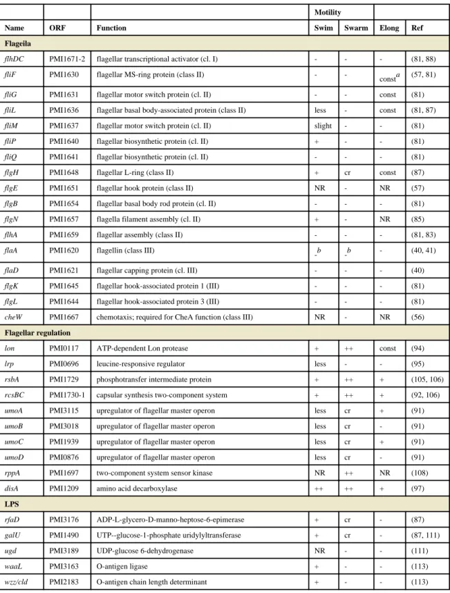
Transposon mutagenesis has been used to identify genes that contribute to swarming, with many variable phenotypes noted (swarming null or crippled swarming, failure to elongate or constitutive elongation, positive or negative for swimming motility or chemotaxis) (73, 84-86) (Table 1). As a caveat, not all of the transposon mutants described in this chapter were complemented, and there could be polar effects. Most mutations of flagellar genes lead to a motility, swarm, and elongation null phenotype (81, 83), which is in keeping with the hypothesis that swarming differentiation requires surface sensing by flagella. However, mutants that were defective in flagellar production or assembly were not universally non- motile, and had distinct phenotypes (81, 87). A flaA mutant was motility and swarming null and failed to elongate under swarming conditions; however, this mutant occasionally reverted to being motility and swarming positive (87). This was later shown to be due to recombination with the flaA and flaB flagellin genes (42). Another swarming-null, motility- positive transposon insertion in flgN (flagella filament assembly, PMI1657) was identified by Gygi et al (85). This mutation resulted in the secretion of unassembled flagellin. The few mature flagella assembled by the flgN mutant were apparently sufficient to mediate
swimming motility but did not allow the hyperflagellation required for swarming motility to occur (85). Negative feedback occurs on flhDC when flagellar assembly is blocked in an flhA mutant; restoration of elongation but not flagellation occurs when flhDC is supplied in trans (88).
Constitutive elongation flagellar mutants
In contrast to the previously described flagellar mutants, fliL (PMI1636, hook basal body) and fliG (PMI1631, flagellar motor switch) mutants exhibited a constitutively elongated (pseudoswarmer) phenotype (81, 87). The fliL phenotype is especially interesting because mutations in other genes in its operon (fliLMNOPQR) lead to a failure to elongate (81). A nonpolar fliL mutant has relatively few flagella and low levels of flaA transcription despite its elongated phenotype (89). However, transcription of the class I master regulator flhDC and the class II flagellar cascade sigma factor fliA genes is increased in this strain compared to its wild-type parent. Complementation with fliL rescues the elongation phenotype but does not restore flagellin expression. Although FliM levels are not disrupted in the fliL
Author Manuscript Author Manuscript Author Manuscript Author Manuscript
mutant, complementation with both fliL and fliM is necessary to restore swarming motility, suggesting that there is an element of fliM DNA that contributes to fliL function (89). The fliL mutant also has increased expression by vegetative cells of virulence factors (zapA and hpmA) which are induced during wild-type swarming (81). RNA-seq analysis of fliL mutant pseudoswarmer cells found that the umoA regulator of swarm cell differentiation was induced (90), similar to the induction that occurs in wild-type cells during swarming (91).
Although the mechanism of FliL-mediated swarming differentiation has not been elucidated, this protein has been proposed to sense the torque generated on the basal body when flagella encounter viscosity, or to count the rate of proton flow through the motor (81).
REGULATION OF flhDC
Cyclic regulation of flagellin contributes to the swarm-consolidation pattern of P. mirabilis, and perturbation of this regulation results in aberrant swarming. Artificial overexpression of the flagellar master regulator genes flhDC (PMI1671-2) results in earlier and faster
swarming (88). A transposon screen by Clemmer and Rather identified two similar mutants with insertions in the flhDC promoter, which had constitutively high levels of expression of these genes during swarming (92). These mutants initiate swarming sooner and swarm at a higher velocity, but lack the consolidation phase and thus do not form a bull's-eye pattern when swarming. Likewise, factors that regulate flhDC expression, transcript stability, or posttranslational modification also influence swarming behavior. As might be expected, the FlhD and FlhC proteins have high turnover and are rapidly degraded during swarming, with a half-life of approximately two minutes (93). Three regulators that are known to repress flhDC in E. coli have little or no effect on swarming when mutated in P. mirabilis: ompR, lrhA and hdfR (65, 92). Known regulators of FlhD4C2 in P. mirabilis are summarized in Table 1 and discussed in detail below.
Lon protease
A mutation in the gene encoding Lon protease (PMI0117) results in cells that initiate swarming normally but migrate faster than the wild type; complementation with lon restored the wild-type phenotype (94). The lon mutant bacteria also had a tendency to differentiate into swarm cells (elongation, increased flagellation) under non-permissive conditions such as during broth culture. This regulation is likely due to Lon-dependent degradation of FlhD (94). Expression of lon is regulated during swarming (65, 94).
Lrp
The global regulator Lrp (PMI0696) is required to initiate cell elongation and swarming.
The lrp mutant swarming phenotype can be complemented by artificial overexpression of flhDC, although this strain is not complemented for expression of hemolysin during
swarming (95). Notably, Lrp accumulates at a higher rate in P. mirabilis compared to E. coli or Vibrio cholerae during culture in rich media (a condition that is permissive for swarming) and is subject to only weak auto-repression, unlike E. coli Lrp (96).
Author Manuscript Author Manuscript Author Manuscript Author Manuscript
DisA
The DisA decarboxylase (PMI1209, decarboxylase inhibitor of swarming) is induced during swarming (97). Mutation of disA leads to increased flhDC transcript and correspondingly enhanced swarming and swimming motility. Overexpression of disA blocks flagellar class 2 (fliA) and class 3 (flaA) gene expression, but does not significantly alter flhDC transcription.
DisA is predicted to be an amino acid decarboxylase. Addition of the decarboxylated amino acid phenethylamine reduced both swarming and disA expression (97, 98). Phenethylamine also inhibited transcription of class 2 and 3 flagellar genes yet had a minimal effect on flhDC. FlhC levels are not affected by disA overexpression, suggesting that DisA activity does not destabilize this protein. It has been proposed that the DisA decarboxylation product interferes with FlhD4C2 assembly or DNA binding (97).
WosA
The wosA gene (PMI0608, wild-type onset with superswarming) encodes a predicted membrane protein that induces flhDC expression and hyperswarming when overexpressed (99). It also causes constitutive swarm cell differentiation in liquid media. Inhibition of flagellar rotation, either in a fliL mutant or in viscous broth, increases wosA expression; this regulation is only partially dependent on the presence of flagella. Transcription is also increased over time during both broth and agar culture. However, a wosA mutant only has a modest decrease in swarming compared to wild type, suggesting that wosA is one of several inputs to flhDC regulation (99).
RsmA/CsrA
Overexpression of another gene, rsmA (PMI0377, repressor of secondary metabolites, also called csrA) results in repression of swarming motility and differentiation (100). The predicted protein is 96% identical to CsrA, a positive regulator of flhDC in E. coli (101).
RsmA and CsrA appear to have opposite effects on flhDC regulation, and this warrants further investigation. However, attempts to mutate rsmA in P. mirabilis were unsuccessful.
Expression of P. mirabilis rsmA complemented an E. coli csrA mutant with regard to glycogen storage, although the effect of rsmA on flhDC expression in E. coli was not reported (100). Overexpression of rsmA decreased the half-life of hemolysin mRNA, indicating that RsmA, like CsrA, regulates by affecting mRNA stability.
Umo proteins
A screen to identify genetic regions that restore swarming motility to the motile but non- swarming flgN mutant (described above) revealed four additional genes that positively regulate flhDC (91). These four genes were designated umoA, umoB, umoC, and umoD for upregulator of the master operon (PMI3115, PMI3018, PMI1939, and PMI0876,
respectively). Expression of umoA and umoD is induced during swarming and these genes are subject to negative feedback when flagellar assembly is blocked and positive feedback by flhDC (91). Despite the plethora of sequenced genomes available since the discovery of the umo genes, umoA remains unique to species within Proteus and Morganella. The UmoB homolog IgaA has been recognized as a regulator of the Rcs phophorelay (102, 103), and UmoB homologs are widespread in the Enterobacteriaceae. The Rcs phophorelay (reviewed
Author Manuscript Author Manuscript Author Manuscript Author Manuscript
in (104)) has been implicated in swarming by P. mirabilis and is discussed below. In P.
mirabilis, UmoB is a negative regulator of swarming inhibitor disA (discussed above) (98).
TWO-COMPONENT SYSTEMS THAT REGULATE flhDC
RsbA-RcsBC
Generation of hyperswarming or precocious (early) swarming mutants has been used to identify repressors that contribute to swarming. RsbA (regulator of swarming behavior, PMI1729; also called rcsD) has twice been identified as a repressor of swarming (105, 106).
The rsbA gene encodes a phosphotransfer intermediate that is part of the RcsBCD phophorelay system. An rsbA mutant has a similar hyperswarming phenotype to that seen during flagellin overexpression (105, 106). Interestingly, overexpression of rsbA also results in precocious swarming (106). Liaw and colleagues examined the ability of transposon mutants to swarm in the presence of the swarming inhibitor p-nitrophenylglycerol (PNPG) (105) and identified a mutant with an insertion in rsbA. During swarming, this mutant expresses higher levels of flagellin, as well as other swarming co-regulated virulence factors including hemolysin, protease, and urease (105). An RsbA-mediated pathway may involve sensing of saturated fatty acids to determine a tendency toward swarming or biofilm formation (107). That is, in the presence of specific fatty acids (myristic acid, lauric acid, palmitic acid), swarming behavior is inhibited while biofilm formation and extracellular polysaccharide production is enhanced. The rsbA mutant is unresponsive to these fatty acids (i.e., hyperswarms) and is deficient in biofilm formation under permissive conditions. To further investigate the contribution of the RcsBCD phosphorelay to swarming, Clemmer and Rather constructed an rcsB (PMI1730, response regulator) mutant; this strain also had a hyperswarming phenotype (92). Thus, the RcsBCD system is likely a repressor of swarming behavior.
RppAB
The rppAB genes (PMI1696-7) form a two-component system that regulates multiple cellular functions in P. mirabilis including LPS biosynthesis and repression of flagella. An rppA mutant was identified in a transposon screen for hyperswarming mutants (108). The rppA mutant also had increased expression of virulence factors associated with swarming (HpmA) and an altered LPS profile which conferred increased susceptibility to polymyxin B. RppA regulates pmrI (PMI1045, also called arnA), which is predicted to be a bacterial UDP-glucuronic acid decarboxylase and contributes to LPS modification (109). Expression of rppA is induced in the presence of polymyxin B and repressed when 10 mM Mg2+ is added (108). This profile is similar to what is observed for the PhoPQ two-component system of Salmonella enterica (reviewed in (110)). Although RppAB has some homology to PhoPQ, another two-component system encoded by P. mirabilis has greater similarity (phoPQ, PMI0884-5) (17). The function of this second locus has not yet been reported.
Author Manuscript Author Manuscript Author Manuscript Author Manuscript
NON-FLAGELLAR LOCI THAT CONTRIBUTE TO SWARMING
LPS
Several elongation-negative mutants with defects in LPS synthesis have been identified through transposon mutagenesis screens (PMI3176 rfaD, PMI1490 galU, PMI3189 ugd, PMI3163 waaL/rfaL, an O-acetyltransferase, and a probable O-antigen chain-length determinant mutant) (87, 111-113). Swarming is connected to LPS O-antigen but not to the related enterobacterial common antigen (ECA), as a wzyE (PMI3326) mutant swarms (113).
Notably, mutation of rcsB or rcsC, and to a lesser extent rcsF, suppresses the waaL swarming deficient mutant (103, 113), showing that although O-antigen itself is not necessary for swarming, there is a regulatory link between LPS biosynthesis and swarming differentiation. UmoB and UmoD have been proposed to activate the Rcs system in an O- antigen dependent manner that is distinct from the canonical Rcs surface-sensed activation by RcsF (91, 103). Mutations in LPS also confer sensitivity to the cationic antimicrobial peptides (CAP) such as polymyxin B; P. mirabilis mutants defective in CAP resistance are either swarming negative or swarm poorly (111, 112). Mutations in at least two of these genes, ugd and galU, activate the alternative sigma factor RpoE, which leads to flhDC repression (111).
Capsule and cell morphology
A mutation in cmfA (colony migration factor, PMI3190, also called cpsF) results in a capsular polysaccharide defect. This mutant is able to elongate and become hyperflagellated, but exhibits reduced swarm velocity (63). The structure of this polysaccharide has been determined to be a tetrasaccharide repeat for one strain of P. mirabilis (64), and this polysaccharide contributes to virulence (114). Another transposon mutant with a motility and elongation-positive but swarming-deficient phenotype was localized to ccmA (curved cell morphology, PMI1961) (115). Although ccmA mutant cells become hyperflagellated and elongate under permissive conditions, the cells are curved and uneven in width. A second ccmA mutant in which 80% of the gene was deleted resulted in a less severe but still distinctive curved cell phenotype. Overexpression of ccmA results in cells with an ellipsoidal or spherical shape. CcmA is predicted to be an integral membrane protein, and its expression increases during swarming differentiation (65, 115). CcmA has been proposed to help maintain linearity during swarm cell elongation, perhaps by organizing peptidoglycan assembly (115). Immunoblotting with CcmA antibodies indicated that there are two forms of CcmA produced in wild-type cells; the larger size matched the predicted full-length protein (CcmA1), and the shorter protein corresponded to an alternative methionine at position 59 of the full-length protein (CcmA2) (115).
Zinc and iron acquisition
A study by Lai et al described an aberrant swarm mutant with a disruption in a gene encoding a zinc-transporting membrane P-type ATPase (PMI3600 ppaA) (86, 116, 117).
This mutant swarms at a lower velocity and does not fully elongate during swarming differentiation, yet swarms for longer intervals and has aberrant consolidation terracing.
Despite producing lower levels of flagellin transcript and protein as well as repressing the motility regulator lrp, it has normal swimming motility. In wild-type P. mirabilis, ppaA
Author Manuscript Author Manuscript Author Manuscript Author Manuscript
expression is induced during swarming (116). Another zinc uptake mutant, znuC (PMI1151) was subsequently found to display aberrant swarming; furthermore, wild-type P. mirabilis has a similarly altered swarming pattern in the presence of the zinc chelator TPEN (118).
Mutation of a nonribosomal peptide system involved in iron acquisition also leads to aberrant swarming (56, 119).
Multiple genes that contribute to swarming were identified in two STM screens (56, 57);
however, the roles of these genes in swarming have not been elucidated. Two other
elongation-negative transposon mutants had defects in cellular division (PMI3055 gidA) and a proline peptidase (PMI3551 pepQ) (87), although their roles in swarming differentiation have not been described further. These genes are listed in Table 1.
EXTRACELLULAR CONTRIBUTORS TO SWARMING
Glutamine
Glutamine is required for swarmer cell differentiation (71, 120). When Allison et al added a variety of components to a defined minimal growth medium that is normally nonpermissive for swarming, only glutamine triggered swarming behavior(71). Addition of the other 19 amino acids mixed together did not allow swarming within 24 hours. However, swarming occurred more rapidly and the characteristic bull's-eye pattern only developed when all 20 amino acids were present. Swarmer cell differentiation was not blocked in a glutamine transport mutant, but was inhibited by the addition of glutamine analog γ-glutamyl hydroxamate (71). Furthermore, glutamine acts as a chemoattractant for swarmer cells but not swimmer cells. In contrast, glycine, histidine, glutamate, alanine, aspartate, aspargine, tyrosine, and valine were chemoattractants solely for swimmer cells; only methionine and cysteine were chemoattractants for both cell types (71). A glutamine synthetase mutant (glnA PMI2882) is completely unable to swarm, even on rich media (120). Swarming is restored when exogenous L-glutamine, but not D-glutamine, is supplied.
Putrescine
Putrescine has been implicated as a trigger for differentiation in P. mirabilis (121). This molecule is continually produced and accumulates in the media during growth and is a component of the outer membrane in some Gram-negative bacteria, including P. mirabilis (122). In an effort to investigate cell-cell signaling by P. mirabilis, a lacZ fusion transposon screen was used to identify genes responsive to signals in spent culture supernatants (123).
One mutant, with an insertion in speA (PMI2094, arginine decarboxylase), was repressed in the presence of spent wild-type supernatant (121). However, spent mutant supernatant did not repress speA. SpeA converts L-arginine to agmatine; the gene is next to speB
(PMI2093), which converts agmatine to putrescine. Sturgill and Rather found that addition of putrescine (down to 25 μM), but not agmatine, repressed speA::lacZ expression. The speA mutant also displayed an aberrant swarm pattern comprised of very small, irregular swarm rings. An independent speB mutation had the same phenotype; taken together with the putrescine complementation of speA::lacZ repression, this suggested that the speA::lacZ phenotype was due to a polar effect on speB. Normal swarming was restored when
extracellular putrescine was added (121), but not when added to a plaP (putrescine importer
Author Manuscript Author Manuscript Author Manuscript Author Manuscript
PMI0843) speA double mutant (124). Likewise, the speB mutants swarmed when inoculated onto agar in the vicinity of an undefined nonswarming P. mirabilis mutant that still
produced extracellular signal (121). Swarming by a speB mutant can also be restored by the addition of ornithine, which can be converted to putrescine by the alternate SpeF pathway (120). Thus putrescine, which accumulates with increasing cell density, may be a signal that initiates swarming. Indeed, uptake of extracellular putrescine appears to be necessary for normal swarming, as the plaP putrescine importer mutant does not swarm as robustly as its wild-type parent (124). The effect could be mediated by putrescine forming a complex with LPS or capsular polysaccharide (125).
Arginine, histidine, malate, and ornithine
Previous approaches to defining the signals to initiate swarming involved adding substances to minimal chemically-defined media, or screening mutated bacteria for the loss of
swarming ability. A new study was able to expand the list of factors that contribute to swarming by using a rich medium (LB) with low (10mM) NaCl (120). On this medium, P.
mirabilis HI4320 does not swarm when incubated at 37 °C. Under these conditions, addition of 20 mM L-glutamine, L-arginine, DL-histidine, malate, or DL-ornithine promoted
swarming. Fumarate and agmatine promoted swarming to a lesser extent. None of these substances enhanced swimming motility, nor did they cause aberrant cell elongation in broth culture. A panel of clinical isolates responded to these swarming cues, although there was some variation in the response to each specific substance. Two of the stronger cues
(ornithine and arginine) and the weaker cue agmatine are part of the putrescine biosynthetic pathway. However, as putrescine itself was not sufficient as a swarming cue on low salt LB, and ornithine and arginine stimulated swarming in different ways and induced unique responses when pH or media were changed, these substances most likely stimulate swarming through distinct pathways (120).
Specific mutants were used to further assess the roles of the five swarming cues (L- glutamine, L-arginine, DL-histidine, malate, or DL-ornithine) (120). All five of these cues are present in human urine. Wild-type P. mirabilis cannot swarm well on urine solidified with agar; this is likely due to increased pH and crystal formation, both due to urease activity. However, a ureC urease mutant swarms on urine agar, and swarming is further promoted by all five swarming cues. Mutation of glutamine (glnA) or histidine (hisG) biosynthetic pathways led to abolished or reduced swarming, respectively; swarming was restored with exogenous supply of the corresponding amino acid. Conversely, mutation of gdhA (glutamate dehydrogenase, PMI3008) had no effect on swarming behavior (120). This finding is intriguing when compared with transcription during UTI, where gdhA is induced and glnA repressed (this is discussed later) (25).
Quorum sensing
There is no strong evidence that P. mirabilis engages in quorum sensing, despite the
coordinated behavior during swarming and the regularity with which P. mirabilis is found as part of multi-species communities. This species produces cyclic dipeptides (cyclo(ΔAla-L- Val) and cyclo(L-Pro-L-Tyr)) that were initially thought to be agonists of the Vibrio fischeri acylhomoserine lactone (AHL)-dependent LuxR quorum-sensing system (126). However,
Author Manuscript Author Manuscript Author Manuscript Author Manuscript
more recent work indicates that these cyclic dipeptides do not affect quorum sensing (127).
AHL autoinducers regulate swarming via a LuxI/LuxR-type system in Serratia marcescens (128). Although the P. mirabilis genome sequence did not reveal any putative LuxI or LuxM-type synthases (17), various exogenously-supplied AHLs affect swarming and proteolytic activity (129). P. mirabilis does encode luxS and produces the LuxS-dependent quorum sensing molecule AI-2 during swarming; however, a luxS mutant has neither a swarming nor a virulence defect (130). Despite these observations, the highly ordered P.
mirabilis swarm cycle suggests a mechanism for multicellular coordination exists (131).
TRANSCRIPTION AND METABOLISM DURING SWARMING
Despite the vigorous motility displayed during swarming, differentiated P. mirabilis bacteria are less metabolically active than consolidating cells (132). When 27 random DNA probes were hybridized with RNA isolated from different stages of the swarm process, most of the sequences were less transcribed during swarming (66). This finding was later confirmed by microarray analysis of the transcriptome of swarming P. mirabilis, in which there was a general repression of transcription of swarm cells compared to consolidate (541 genes repressed during swarming; 9 induced relative to consolidated cells) (65). Flagellin (flaA) is among the most highly-expressed genes in both swarm and consolidate despite the lack of motion observed in consolidating cells. Consolidating cells are distinct from broth-cultured cells used to inoculate a swarm plate; expression by swarm and consolidate share more in common with each other than with broth-cultured bacteria. During consolidation, bacteria are metabolically active with increased expression of genes involved with central
metabolism, respiration, nutrient uptake, and cell wall remodeling. The alternative sigma factor rpoS, associated with stationary phase and stress response, is among the genes induced during consolidation (65). Once initiated, protein synthesis is not required to maintain the swarm; swarming continues even in the presence of chloramphenicol (65, 133).
Genes co-regulated with swarming are not necessarily required or involved with the swarming cycle, including virulence genes hpmA and zapA (these virulence genes will be discussed in another section). Investigation of genes regulated during swarming has led to the identification of virulence processes. Mutation of genes involved in peptide uptake (oppB PMI1474 and dppA PMI2847) and amino acid synthesis (cysJ PMI2250) that were regulated during swarming led to minor attenuation of swarming. However, the dppA and cysJ mutants were less fit in animal co-challenge. A similar result was found for the transcriptional regulator hexA (PMI1764) (65).
Swarming occurs under both aerobic and anaerobic conditions (133). STM studies for genes involved in virulence also led to the identification of central metabolism genes (aceE and sdhC) that cause aberrant swarming when mutated, suggesting that a complete (aerobic) TCA cycle contributes to swarming (57). To further investigate how central metabolism affects swarming, selected metabolic genes were mutated and were found to affect swarming in four distinct patterns (134). Two of these classes were characterized by altered distances between rings of swarming and consolidation; TCA cycle mutations (fumC PMI1296 and sdhB PMI0568) had decreased distances, and pentose phosphate pathway mutants (gnd PMI0655 and talB PMI0006) had increased distances. Mutations in glycolysis (pfkA PMI3203 and tpiA PMI3205) resulted in reduced swarming diameter. These mutations were
Author Manuscript Author Manuscript Author Manuscript Author Manuscript
rescued by complementation with the corresponding wild-type alleles or by addition of the missing biochemical intermediate to the growth medium. Specifically, the fumC mutant was rescued by addition of succinate or malate but not fumarate; this distinction indicates fumC is acting as part of the oxidative TCA cycle during swarming and not the reduced branched TCA cycle. Mutations in gluconeogenesis (pckA PMI3015) or the Entner-Doudoroff pathway (edd PMI2760) had no effect on swarming patterns, nor did a mutation in the fumarate reductase subunit gene frdA (PMI3588) which is involved in anaerobic respiration using the branched TCA pathway (134).
Swarming also occurs in the presence of the aerobic respiration poison sodium azide supplied at growth inhibitory concentrations (133, 134). The frdA (branched TCA) mutant also swarms on azide, but the fumC mutant, used in both aerobic and anaerobic TCA cycles, is unable to swarm (134). Thus, an alternative anaerobic electron acceptor has been
proposed to provide energy during swarming. A transposon screen for mutants that are able to swarm on LB swarm agar but not in the presence of azide yielded 18 mutants. Not all of the mutated genes were expected to be involved in swarming per se, as mutations leading to increased permeability or susceptibility to azide would also be found. Two genes of particular interest that were identified are hybB (PMI0033 hydrogenase-2), which encodes an anaerobic cytochrome, and PMI2646, which encodes a putative quinine hydroxylase. To further address whether fermentation occurs during swarming, bacteria were inoculated onto swarm agar with the pH indicator phenol red. During aerobic conditions, P. mirabilis produces alkaline conditions; fermentation would have resulted in secretion of acidic byproducts. Swarming under anaerobic conditions, however, results in acidity. Taken together, the authors concluded that anaerobic respiration with a complete oxidative TCA cycle generates the proton motive force required for flagellar rotation (134).
CELLULAR INVASION
P. mirabilis uses its flagella to invade cultured cells derived from the urinary tract, including Vero (green monkey kidney parenchyma) (135, 136), EJ/28 and 5637 (transformed human transitional cell carcinoma of the urinary bladder) (66, 114, 135), NTUB1 (human
urothelium) (105), and primary human renal proximal tubular epithelial cells (HRPTEC) (52, 137). In this experimental system, P. mirabilis is highly invasive, in numbers comparable to Salmonella Typhimurium (52) or Salmonella typhi (138). Allison and colleagues found the greatest invasive capability for P. mirabilis coincided with the swarmer cell phase (135). Likewise, a hyperswarming rsbA mutant is more invasive than its wild-type parent (105). Mobley et al found that invasion was greatly impaired when flagella of swarmer cells were immobilized by the addition of antiserum (52). Furthermore, an isogenic flagella mutant (hpmA flaD) was less than 1% as invasive as the parent (hpmA).
Centrifugation of the nonmotile bacteria onto the cultured cell monolayer partially restored invasiveness to about 10% of the parental level. Invasion is aided by non-flagellar
components including the HpmA hemolysin, autotransporter protein AipA (139) and LPS modification protein PmrI (109). Using a variety of intestinal and urinary epithelial cell lines, Oelschlaeger and Tall were able to inhibit P. mirabilis invasion by blocking bacterial protein, RNA, or DNA synthesis, but not by blocking host cell (eukaryotic) protein synthesis (138). P. mirabilis colocalizes with mucins MUC2 and MUC5AC, and mucin expression has
Author Manuscript Author Manuscript Author Manuscript Author Manuscript
been found to correlate with invasion (140). Cellular invasion could be involved in the transit of P. mirabilis from the kidneys to the bloodstream. Alternately, P. mirabilis could have an intracellular population during bladder or kidney infections. Of course, cultured cells differ in fundamental ways from intact tissues (e.g. types and polarization of proteins, tight junctions). At this time, invasion of urinary cells by P. mirabilis during ascending UTI has not been investigated in much detail.
ROLE OF SWARMING IN VIRULENCE
Virulence factors, including urease, ZapA protease, and hemolysin are induced during swarming compared to broth culture or older bacteria in the interior of a swarm colony (65, 66, 141). Production of both ZapA and HpmA is increased during overexpression of the flagellar regulator umoB, and hpmA transcription is responsive to Lrp (141). The swarming ability of P. mirabilis is especially relevant in catheterized patients, as this organism is able to swarm across catheters made of silicone or latex (61, 142) (Fig. 5). Since expression of several virulence genes is increased during swarming, it is possible that P. mirabilis swarming up catheters is primed to infect the urinary tract. However, the role of swarming during UTI is debated. In one study, outbred mice intravenously infected with P. mirabilis resulted in extensive kidney infection, and long-form, swarmer bacteria were observed in the kidney parenchymal tissue 15 days post-infection (114). Transposon mutants that were motile but nonswarming caused lower rates of lethality and kidney abscesses in this model.
Several of these mutants were also less virulent compared to wild type in an ascending UTI model using suckling mice (114). In contrast, swarmer cells were very rarely found in the urinary tracts of CBA/J mice infected via bladder catheterization with GFP-expressing P.
mirabilis when examined two or four days post-infection (143). In that study, 7 of 5087 (0.14%) bacteria counted in the bladders and kidneys had an elongated swarm form (> 10 μm); no swarmer cells were observed in the ureters. Combined with the previously described reports in which nonmotile P. mirabilis was fully virulent, this suggests that swarming may not be an important contributor to UTI virulence. However, swarming might only be apparent when a catheter is in place, or during possible invasion of renal cells at later stages of disease progression.
It is noteworthy that the putrescine biosynthetic pathway also results in the formation of urea. P. mirabilis encodes genes that may catalyze the ultimate generation of putrescine and urea from ammonia and ATP (17). Because putrescine biosynthesis is required to initiate swarming, and excess urea could potentially drive the putrescine biosynthetic pathway in the opposite direction, it is possible that the abundant urea in the urinary tract represses
swarming behavior.
Dienes lines and T6SS
P. mirabilis strains are self-recognizing during swarming; that is, any given pair of isolates, when inoculated on opposite ends of a swarm agar plate, will likely swarm up to but not into each other. When this occurs, a thin clear line of demarcation remains between the two strains, called a Dienes line (144). In contrast, swarms formed by identical strains will merge. Dienes line formation has been used as a method for typing clinical strains of P.
mirabilis (145-147). When opposing swarms from two different strains meet, within one to
Author Manuscript Author Manuscript Author Manuscript Author Manuscript
two hours one of the strains will form large, rounded cells (148, 149). Over a period of hours, the rounded cells die and lyse, while the other strain dedifferentiates into vegetative cells. This rounding is not observed when a swarming strain meets nonswarming cells.
Dienes line formation is dependent upon cell-cell contact or at least very close cell
proximity (<60 μm) between opposing strains (148). When a membrane permeable to most macromolecules but not bacteria is placed between swarming strains, no Dienes line or cell rounding occurs.
The Dienes phenomenon is likely a mechanism of territoriality. When two strains (one marked with red fluorescent protein and one marked with green fluorescent protein) are mixed and placed on swarm agar, one strain dominates the resulting swarm colony, while the other strain forms rounded cells when detected at all. However, in broth culture, neither strain is dominant, and when other strains were tested for biofilm formation, dominance of a given strain did not correlate with Dienes dominance (148).
Recent studies suggest that type VI-mediated secretion of toxic effector proteins is the main mechanism of Dienes line formation (149, 150). Type VI secretion systems (T6SS)
comprise a method of protein export that generally requires cell-cell contact, whether between two bacteria or a bacterium and a eukaryotic host cell. They are widespread in Gram-negative bacteria and may be involved in virulence, commensalism, or bacterial competition (reviewed in (151-153)). A core set of structural genes are essential for function and are thought to be involved in the production or function of the secretion apparatus. This includes the hcp/vgrG genes, which respectively encode proteins that form the tube through which export occurs and a needle-like structure used to penetrate the outer membrane of the target cell.
Type VI secretion has been studied in two P. mirabilis isolates, BB2000 and HI4320. Both strains encode the core structural genes essential for T6SS function, as well as multiple putative effector operons associated with the hcp/vgrG genes (17, 154-156). In T6SS characterized in other bacterial species, the multiple hcp/vgrG homologs are thought to act as adapters between the structural components and the effectors, allowing different effector proteins to be secreted (153). In both BB2000 and HI4320, the T6SS is essential for Dienes line formation and identification of self. However, the number of effector operons present and the significance of individual operons in self-recognition varies between the two strains (155, 156).
In BB2000, a transposon screen for mutants that form Dienes lines when in competition with the parent strain (that is, the strains no longer recognize the parent strain as “self”) led to the ids operon (identification of self, idsABCDEF PMI2990-95) (149). The first two genes of this locus, idsA and idsB, have homology to hcp and vgrG, respectively. The idsD and idsE genes display the most variation between P. mirabilis strains, and are believed to be the determinants of self-recognition (149). Expression of the ids operon increases during late logarithmic and early stationary phase. During swarming, expression is highest in the center of the swarm colony where the highest cell density is observed, but expression also occurs within a subset of cells at the leading edge of approaching swarm fronts (150).
Author Manuscript Author Manuscript Author Manuscript Author Manuscript
A second transposon mutagenesis screen identified two “no-merge” mutants which form Dienes lines with both the wild-type and the idsA-F mutant (155). These mutants mapped to a previously uncharacterized five gene operon, named idr for identity recognition. As in the ids locus, the first two genes in the idr locus (idrA and idrB) have homology to hcp and vgrG, while the remaining three genes encode proteins of unknown function. IdsA, IdsB, IdsD, IdrA, and IdrB were detected using mass spectrometry in culture supernatants of wild- type BB2000 but not in a T6SS mutant, providing further evidence that these proteins are exported from the cell by the T6SS. It is apparent that competition and killing via T6SS effectors is a complex phenomenon in P. mirabilis. For example, wild-type BB2000 can dominate HI4320 in a swarm competition. However, BB2000 Δids mostly dominates HI4320 while HI4320 can dominate BB2000 idrB::tn5, suggesting that the idr operon is more important than the ids operon in BB2000 killing of HI4320 (155).
Unlike in BB2000, mutations in idsB and idsD in HI4320 do not result in a loss of self- recognition (156). However a transposon screen for HI4320 mutants which no longer recognize the wild type as self, and thus form a Dienes line with HI4320, identified hypothetical protein PMI0756 as a putative component of the T6SS. A secondary screen to identify additional mutants that no longer form a Dienes line with the PMI0756 mutant identified two groups of mutants. The first group of mutants clustered in the major T6SS structural operon (PMI0733-0749), while the second group of mutants clustered in the same operon as PMI0756 (PMI0750-0758). The first two genes in this operon are hcp and vgrG homologs, respectively; the other genes in the operon were named pef for primary effector operon. Additional targeted mutagenesis revealed that a mutation in any gene in the pef operon results in loss of self-recognition. Complementation of the pefE mutant with pefE is sufficient to restore immunity from wild-type killing, but is not sufficient to restore killing of non-immune strains. To restore both immunity to wild type and killing of the pefE mutant, complementation with pefEFG is needed (156). It is notable that although the pef and idr operons are both adjacent to the structural T6SS apparatus operon in HI4320 and BB2000, respectively, the organization and sequences of these operons are completely different (aside from the hcp and vgrG homologs at the front of both operons).
Analysis of the HI4320 genome revealed three additional putative hcp-vgrG effector operons in addition to the ids and pef operons. The proteins encoded by the hcp genes are highly similar (with the exception of PMI1332 which appears to be truncated), while the genes predicted to encode VgrG homologs are similar at the N-terminus with decreasing similarity at the C-terminus. All five of the promoters for the hcp-vgrG operons are capable of driving luciferase expression during swarming (156), though they result in different patterns of expression.
Although significant gains have been made in the understanding of Dienes line formation and T6SS competition in P. mirabilis, a number of questions remain. The function of most of the genes in the effector operons remains unclear, and it is not understood how cell death and immunity are mediated. Nor is it clear why P. mirabilis possesses several effector operons, or how they are regulated. Perhaps most importantly, it is unknown when Type VI secretion-mediated killing occurs when P. mirabilis is in its natural environments or if this phenomenon has clinical relevance.
Author Manuscript Author Manuscript Author Manuscript Author Manuscript
Fimbriae
Although the fimbriae of P. mirabilis are essential virulence factors in urinary tract
infections (Table 2), much remains unknown about their expression, physical characteristics, and biological functions. The first P. mirabilis fimbriae were identified by their ability to agglutinate red blood cells and bind uroepithelial cells. Using these methods, the mannose- resistant Proteus-like (MR/P), mannose-resistant Klebsilla-like (MR/K), and urothelial cell adhesin (UCA) fimbriae were described (157, 158). However, as P. mirabilis can
simultaneously express multiple types of fimbriae, identification of genes encoding specific fimbriae is often complicated (157). For example, the attempts to identify genes encoding UCA fimbriae led to the discovery of the P. mirabilis fimbriae (PMF) (159), and the genes associated with MR/K hemagglutination remain unidentified.
Prior to sequencing the P. mirabilis genome in 2008, five P. mirabilis fimbriae had been discovered. Sequencing revealed an additional 12 chaperone-usher fimbriae encoded in the P. mirabilis genome (17). In comparison, UPEC typically encode nine to 12 fimbrial operons (160). As the name suggests, chaperone-usher fimbriae are defined by the presence of a chaperone and an usher in the fimbrial operon. The chaperone protects the fimbrial subunits during transport from the cytoplasm to the cell surface which allows the fimbrial subunits to fold properly in the periplasm, and the usher, an integral membrane protein, releases the fimbrial subunit from the chaperone and guides it through assembly (161). Ten of the P. mirabilis chaperone-usher operons encode a homolog of MrpJ, a regulatory protein at the beginning or end of the operon, which allows the coordination of adhesion and motility, and may allow fimbrial regulation of other fimbriae (162). The following sections describe the fimbriae of P. mirabilis in order of their discovery, as well as the role of MrpJ homologs in coordinating regulation of fimbriae and flagella.
MANNOSE-RESISTANT PROTEUS-LIKE (MR/P)
Genetic organization
MR/P is the best characterized fimbria in P. mirabilis. The mrp operon (mrpABCDEFGHJ) encodes the major structural subunit (mrpA), four minor subunits (mrpBEFG), a chaperone (mrpD), an usher (mrpC), a tip adhesin (mrpH), and a transcriptional regulator (mrpJ) (163, 164).
The operon is preceded by a σ70 promoter inside an invertible element whose orientation is controlled by MrpI, a recombinase transcribed divergently from the structural operon (164, 165). When the σ70 promoter faces the structural operon, MR/P is “ON”, while when the σ70 promoter faces MrpI, MR/P is “OFF” (165). Transcription of mrpI is unaffected by the orientation of the invertable element, and mrpI is likely transcribed by a promoter outside of the invertable element (164). The MrpI C-terminal domain is homologous to the catalytic domains of the E. coli fimbrial recombinases FimB and FimE, while the N-terminal domain binds DNA (165). Like FimB, MrpI switches the invertible element both from ON to OFF and OFF to ON (165). However, when mrpI transcription decreases, mrpA transcription increases, suggesting MrpI favors switching from ON to OFF (166). In the absence of MrpI, the promoter orientation is fixed, suggesting that MrpI is the sole recombinase for the mrp
Author Manuscript Author Manuscript Author Manuscript Author Manuscript
promoter (165). To facilitate analysis of the role of MR/P fimbriae, two MR/P mutants were engineered. Both of these mutants have an insertion that inactivates mrpI; in the “MR/P L- ON” mutant, the mrp promoter is in the on position, while in the “MR/P L-OFF” mutant, the mrp promoter is in the off position.
Expression
Like many fimbriae, in vitro MR/P expression is encouraged through static culture. The percentage of bacteria with ON or OFF mrp promoters can be determined by a PCR assay, where the percentage of ON mrp promoters correlates to the percentage of cells expressing MR/P (Fig. 6) (165, 166). When a lower oxygen level is maintained in LB under shaking conditions, for example by culturing P. mirabilis in 5% O2, the mrp promoter is up to 70%
ON compared to 2% ON in aerated culture. The increased expression of MR/P under static conditions is due to decreased availability of oxygen gas, not increased levels of CO2 (166).
Oxygen-limiting conditions enrich for expression of MR/P through two mechanisms. First, the expression of MR/P lends a competitive advantage during growth in low oxygen
conditions. Although their growth during independent culture is similar, a P. mirabilis L-ON mutant will out-compete a P. mirabilis L-OFF mutant in liquid culture (166, 167). It is hypothesized that this is due to fimbrial expression and flagellar repression driving electron transport and maintaining the proton gradient (166). Secondly, oxygen limitation increases the expression of MR/P. Under limited oxygen conditions, almost twice as much mrpA is transcribed as under atmospheric oxygen, possibly because less MrpI is expressed, leading to less switching of the mrpA promoter from ON to OFF (166).
Assembly
The role of each subunit in the assembly and structure of the MR/P fimbria is partially understood. MrpA has been identified as the major structural subunit by its strong expression and location in the mrp operon (164, 168).
MrpB is homologous to E. coli PapH, which terminates fimbrial growth while anchoring fimbriae to the cell wall and modulating fimbria length (169, 170). P. mirabilis ΔmrpB displays fewer but longer fimbriae on the surface of the cell. In P. mirabilis ΔmrpB, 5% of cells were fimbriated after three 48 hour passages in static culture, while 48% of wild-type cells were fimbriated. MR/P fimbriae from an mrpB mutant are 62-fold longer than wild- type fimbriae (18 μm compared to 0.29 μm) (171). These data suggest MrpB is needed to either initiate and/or terminate assembly of MR/P (171). The mrpEFG genes are predicted to encode minor structural subunits. When MrpG is found in P. mirabilis fimbriae, it is located on short fimbriae, possibly newly synthesized, and where fimbriae meet between
aggregating bacteria (172).
MrpH is a two-domain adhesin, where the predicted MrpH C-terminus is similar to other mrp structural subunits and the larger N-terminal domain likely mediates receptor binding (Fig. 6). This two-domain structure and the presence of a proline at the last residue identify MrpH as an adhesin (163, 173). Less than 1% of mrpH mutant cells express MR/P fimbriae compared to 50% of the wild-type, suggesting that MrpH is essential for stable production of
Author Manuscript Author Manuscript Author Manuscript Author Manuscript
MR/P. Oddly, the mrpH mutant still mediates weak hemagglutination, possibly due to the expression of a different fimbria in the absence of mrpH (163). The N-terminus of MrpH has four conserved cysteine residues, which in other tip adhesins form disulfide bonds that are essential for the proper folding of the adhesin (174, 175). When the MrpH cysteine residues are mutated to serine, the MR/P fimbrial structure is produced and MrpA is found in fimbrial preparations, but there is less pellicle formation and hemagglutination, suggesting that the tip adhesin is inactivated (163). In P. mirabilis and other organisms, periplasmic disulfide bond formation is mediated by DsbA (PMI2828) (176). A dsbA mutant is deficient in MR/P production and in establishing infection in a mouse model of UTI, which is consistent with disulfide bond formation being an essential component of the MR/P structure (56, 166).
Biofilm formation
MR/P contributes to biofilm formation, presumably by mediating attachment to surfaces and autoaggregation (177, 178). P. mirabilis L-ON forms significantly more biofilm during culture in urine than a P. mirabilis L-OFF mutant or the wild type, which form similar levels of biofilms (177). Scanning electron microscopy showed that after two days, wild-type bacteria had adhered as small colonies to a cover slip, while P. mirabilis L-ON formed a full three-dimensional biofilm. By seven days however, the wild-type biofilm had become much thicker than the L-ON or the L-OFF biofilm, suggesting that the ability to turn MR/P on and off may be essential for robust biofilm maintenance (Fig. 7) (177).
Role in infection
MR/P fimbriae are expressed in the urinary tract and contribute to virulence. After transurethral infection with wild-type P. mirabilis, mice with high IgG antibody respons