ELECTROMYOGRAPHY OF MASTICATION MUSCLES IN COLLEGE ATHLETES
by
John William Ratliff
A thesis submitted to the faculty of The University of Mississippi in partial fulfillment of the requirements of the Sally McDonnell Barksdale Honors College
Oxford December 2014
r. Mark Loftin
2014 John William Ratliff ALL RIGHTS RESERVED
ABSTRACT
The purpose of this experiment was to determine if co-activation in enhanced neck muscles increased surface electromyography (sEMG) activity in the masseter while chewing with the 1st, 2nd, and 3rd molars. Sixteen football players from The University of Mississippi volunteered for this study. Football players were examined because they strengthen their neck 4 days a week to help prevent neck injuries and concussions.
Participants’ average body fat was 15.68% and average body mass was 100.69 kilograms showing that the participants were larger, muscular individuals compared to non student- athletes and represent a unique, study group. Participants performed maximum weight on a neck weight lifting machine, and then chewed Riesen candy during sEMG recording of the masseter, sternocleidomastoid (SCM), and upper trapezius of the participant’s dominant chew side. During the neck weight lifting machine trials, individuals
performed half of the repetitions with their mouth open and relaxed while the other half of the repetitions were conducted with their mouth closed and clenched. Repetitions with the mouth open generated more force from the SCM by an average of 6 Newtons. Co- activation was documented between the three muscles while performing the neck weight lifting machine and during maximum voluntary chewing on the Reisen candy. The weight of the participant had a significant positive relationship with the body fat (%) (r = 0.887), neck circumference (r = 0.604), and max weight (kg) used in the machine (r = 0.520). More weight on the neck machine and a larger neck circumference had a significant, positive relationship (r = 0.704). These data indicate a possible association between neck circumference and an increase in muscle mass in the SCM and trapezius.
Co-activation through neck exercising encourages an increase in muscle mass in the
masseter. The multiple correlations showed a significant, negative relationship between the SCM and masseter for each chew. This shows that the SCM and masseter need to be coordinated and contracted simultaneously to perform the biting task (van der Bilt et al., 2006). Long term dental effects and indirect effects of enhanced neck musculature should be taken into consideration for follow-up study.
TABLE OF CONTENTS
LIST OF TABLES ... vi
LIST OF FIGURES ... vii
DEDICATION ...viii
ACKNOWLEDGEMENTS ... ix
INTRODUCTION ...1
MATERIALS AND METHODS ...9
RESULTS ... 17
DISCUSSION ... 21
TABLES ... 28
FIGURES ... 38
REFERENCES ... 41
LIST OF TABLES
TABLE 1. Surface electromyography activity in mV.s (expressed as mean integral for raw activity ± standard deviation and the minimum and maximum) for the
masseter, sternocleidomastoid, and upper trapezius for each repetition on the neck weight lifting machine: (a) dorsal flexion and reduction with mouth open, (b) left lateral flexion and reduction with mouth open, (c) right lateral flexion and
reduction with mouth open, (d) dorsal flexion and reduction with mouth closed, (e) left lateral flexion and reduction with mouth closed, and (f) right lateral flexion and reduction with mouth closed.
TABLE 2. Bivariate correlation between the masseter, sternocleidomastoid, and trapezius for the left lateral flexion (a) with the mouth open and (b) with the mouth closed (r = correlation coefficient).
TABLE 3. Descriptive statistics for the integral of the raw (a) masseter activity (mV.s), (b) sternocleidomastoid activity (mV.s), and trapezius activity (mV.s) for the first 5 chews in each trial.
TABLE 4. Correlation matrix including mass (kg), height (cm), body fat (%), Body Mass Index (BMI), neck circumference (cm), max weight used (kg), throwing hand, writing hand, kicking foot, chewing side, average chews per trial, integral of raw masseter activity (mV.s), integral of sternocleidomastoid activity (mV.s), integral of raw trapezius activity (mV.s), force (N) with mouth open, and force (N) with mouth closed (r = correlation coefficient).
LIST OF FIGURES
FIGURE 1. Location of surface electrodes on a sample participant: (a) trapezius and (b) masseter (upper electrodes) and sternocleidomastoid (lower electrodes). The electrode on the forehead was a ground.
FIGURE 2. Significant correlation between the integral of raw masseter activity (mV.s) versus integral of raw upper trapezius activity (mV.s) during right lateral flexion with the mouth open (r = 0.441).
FIGURE 3. Significant correlations between the integral of raw sternocleidomastoid activity (mV.s) and the integral of raw masseter activity (mV.s) during chewing events 1 (r = -0.476), 2 (r = -0.622), 3 (r = -0.643), 4 (r = -0.576), and 5 (r = - 0.606).
DEDICATION
To my loving mother for always believing in me. To my dad for always watching.
“You Gotta Believe” – RWR
ACKNOWLEDGEMENTS
To Dr. Carol Britson, I thank you for the best possible advice I could receive in regard to my thesis and growing as a college student in and out of the classroom. I knew when you taught me as a freshman for Biology recitation that there was an immediate connection because of your thirst for knowledge and an intensity to find the answers. I will never forget our many meetings that I could barely stand to attend because I would be sweating by the time I got to your office. My physical sweat was nothing compared to your mental sweat from thinking about ways to make my thesis better or to keep me motivated to see the end product. I am sure I could not have picked a better advisor.
I thank my second reader, Dr. John Garner, for his input. I thank my third reader, Dr. Mark Loftin, for his suggestions and dedication of time to ensure that my thesis is as good as possible. I thank my Honors College advisor, Dr. John Samonds, for helping me plan my specific dates for my thesis. I also thank the Sally McDonnell Barksdale Honors College for making me push through the hard classes and providing me with friends when I came to Mississippi from out of state. I would also like to thank the Biology Department and Dr. Paul Lago for providing a high quality education while attending Ole Miss and for the support of my thesis.
I thank Tom Lombardo and The University of Mississippi Institutional Review Board for working with me so promptly in approving my thesis proposal so I could get started. I thank Dr. Shannon Singletary for giving consent to allow the Ole Miss football team to be the participants in my study. I also want to thank my brothers on the Ole Miss football team for being so easy to work with and volunteering to participate. I thank The University of Mississippi Interlibrary Loan Services for sending me every document I requested as soon as I asked for them. I thank Jeremy Smith for being my assistant through each experiment and making sure everything ran smoothly.
I would not be where I am today without the Ole Miss Football Strength and Conditioning Staff, specifically Coach Paul Jackson and Coach Dominic Studzinski. To Coach Jackson, thank you for instilling in me to be a lion about my business and attack everything with the utmost effort and a positive attitude. To Coach Dom, thank you for instructing my participants in the study but more importantly, for teaching me to expect nothing but great things from myself.
To my mom, Patti Ratliff, I thank you for being the only one that told me I had to finish what I started in the Honors College. I doubted myself but you knew I could do it.
You are the best mom in the world and I am so blessed to still have you in my life. To my brother, Robert Ratliff, I thank you for your constant love and support you have shown me in my schoolwork even when you wanted someone to throw the football to. You always have understood the work I must put in to reach my dreams. Mom and Robert, I love you both dearly!
To my unbelievable friends, I know most of you will never have any interest in reading this but I thank you for always being ready to hang out even though I have bailed so many times to work on this thesis. To Charlie Scott, thank you for always having my back and making me have fun when I could have been antisocial while working on this thesis. To Jordan Watson, thank you for constantly reminding that hard work pays off. I thank my girlfriend Clara Tucker for understanding when I needed to do thesis work instead of doing something fun with you. I thank Nathan Noble and his girlfriend
Courtney Deaton for the countless dinners you provided for me when I did not have time to cook. I have been so blessed to come across so many genuine and wonderful people over the years at The University of Mississippi.
INTRODUCTION
Mastication is “a sensory-motor activity aimed at the preparation of food for swallowing” (van der Bilt et al., 2006). It is an intricate process that includes many muscles in the face and muscles that insert as far down as the top of the clavicle. It
involves voluntary initiation of reflex, contraction, and relaxation of the different muscles from the brain and receptors to work efficiently to control the result of each chew
(Nelson, 2009). The teeth are important in the masticatory system for how they hold the food in place to tear apart in order to allow swallowing. The other important part of mastication is the bite force which depends on musculature and coordination of these muscles to contract simultaneously to initiate a violent, strong chew (van der Bilt et al., 2006).
The masseter (primarily hovers over the 1st, and 2nd molars) and medial
pterygoid (posterior towards the ear from the masseter) muscles are used in mandibular closing. Dang et al. (2012) state the masseter’s main role is elevating the mandible at the temporomandibular joint during a bite. Located near this joint are the 1st, 2nd, and 3rd molars which deliver the maximum amount of force when biting (Nelson, 2009).
Forrester et al. (2010) discovered that with anterior contacts between teeth (biting down on the incisors and canines) muscle activity was significantly reduced. Giannakopoulos et al. (2013) were interested if “the levator scapulae, trapezius, sternocleidomastoid (SCM), and splenius capitis co-contract (co-activate) at the different submaximum bite forces
usually generated during jaw clenching”. Co-activation is the simultaneous activation of 2 different muscles around a joint (Baratta et al., 1988).
Other muscles involved with mastication include the supra/infrahyoidal muscles (located above and below the hyoid bone at the top of the neck near the mandible), the semispinalis capitis (extends from vertebrae in the neck to the occipital bone), and the semispinalis cervicis (extends from the spinous processes to the transverse processes) which are deep neck muscles but not easily measurable with sEMG. When Hellmann et al. (2012) tested for co-activation while chewing, they found that “masticatory muscles and the neck muscles co-contract during specific motor tasks…with regard to chewing and maximum biting tasks”. Hellman et al. (2012) subjects were healthy young adults.
My study is unique in that I am investigating if enlarged neck musculature increases the surface electromyography (sEMG) activity in the masseter while biting on the 1st, 2nd, and 3rd molars of the dominant chew side.
Co-activation occurs in muscles throughout the body. For example, when someone tries to squeeze a fist as hard as they can, muscles activate from the tip of the fingertips all the way to the bicep. This co-activation is simultaneous but not of all the same intensity. The same applies for muscles during mastication when the head’s muscles and joints hold the maxilla stable during mandibular functions (Clark et al., 1993). This led Clark et al. (1993) to claim that “there would be a coupling between the mandibular motor system and cervical motor system.” A motor system is a part of the central nervous system that deals with movement throughout a specific region of the
body. The mandibular motor system deals with movement of the jaw for chewing, talking, and simply opening and closing. The cervical motor system relays information from the spinal cord to the brain (Sherwood, 2012).
From a muscle physiology perspective, Hellman et al. (2012) claimed that they found force exertion in neck muscles while examining masticatory muscle activity.
Researchers have also documented co-activation in the sternocleidomastoid. Clark et al.
(1993) showed “a clear co-activation of the SCM was present in 93% of the four repeated maximum voluntary clenching (MVC) trials” and suggests that the co-activation is necessary for the cervical muscles to provide a stable position of the maxilla for
clenching. Different muscles are involved depending on the type of masticatory activity (e.g., cutting, shearing, or grinding). In straight up-and-down movement to mash a piece of candy on the back molars, the head remains erect. The molars tend to have a flatter surface for crushing or grinding hard food. This straight up-and-down jaw movement allows researchers to focus on the erect stable head position in experiments.
Many studies, like Hellman et al. (2012), have examined mastication and examined the masseter in relation to the sternocleidomastoid. The SCM is an easily measured muscle close to the mouth but not directly attached to the face. Its main job is to contract on one side in order to turn the face in the opposite direction. Its origin is the superior margins of the sternum and clavicle while its insertion point is the mastoid region of the skull (Martini and Bartholomew, 2011). The masseter’s main job is to elevate the mandilble while chewing and extends from the zygomatic arch to the lateral surface of the mandible (Martini and Bartholomew, 2011). Clark et al. (1993) stated,
“The second finding was a progressive development of the SCM co-activation which
paralleled the masseter activation. It seems clear that the SCM level of activation was a function of the masseter level”. When the masseter elevates the mandible to chew, the SCM is activated adding muscle activity to aid in the chewing.
Co-activation is simultaneous but not necessarily equal activity between muscle groups during mastication (Giannokopoulos et al., 2013). Giannakopoulos et al. (2013) research of co-activation of neck and masticatory muscles during jaw clenching showed,
“Maximum activation of the masticatory muscles caused the most significant (P < 0.001) co-activation of the neck muscles (range 15% to 25% of their maximum voluntary contraction [MVC]).” Giannokopoulos et al. (2013) also measured the maximum contraction for the neck with different load directions in a machine designed to measure individual neck muscle activity. Lodetti et al. (2011) documented co-activation in the upper trapezius muscle but it was not as evident as the activity of the temporalis when examining bite force (Ciuffolo et al., 2005). Other studies have investigated the upper trapezius searching for its role in mastication since it is close to the masseter but also attaches to the back of the head. The trapezius extends from the occipital bone and spinous processes of thoracic vertebrae down to the clavicle and scapula (Martini and Bartholomew, 2011). It is a secondary cervical muscle that helps hold the head erect.
There are historical injuries, specifically related to the trapezius, that have occurred prior to neck strength training. At The University of Mississippi, the historical neck injury is from the famous collision Chucky Mullins put on Vanderbilt’s fullback Brad Gaines. The hit broke 4 of Chucky’s cervical vertebrae instantly paralyzing him (Cleveland, 2009). From speaking with athletes (R. Nowicki, pers. comm.) who play college football, they all strengthen neck muscles as part of daily workouts in an effort to
prevent their neck from breaking when tackling or being hit by another player. With an increase in strength found in the upper trapezius from rigorous training, I am interested to discover if co-activation and conditioning causes the force of mastication to also be strengthened. To date, no published paper has studied co-activation in subjects with enhanced neck musculature (e.g., college football players).
However, there is also a lack of information regarding co-activation in individuals who do not undergo strength training. Mansell et al. (2005) studied resistance training and head-neck stabilization in male and female collegiate soccer players where subjects did an 8 week cervical resistance training program that consisted of 3 sets of 10
repetitions of neck flexion and reduction; they showed an increase in neck strength by 15% and an increase in overall neck girth. By working out the neck muscles in addition to pectorals, lower trapezius, hamstrings, quadriceps, and gluteus maximus, all athletes potentially add more muscle mass to protect their body (P. Jackson, pers. comm.).
Football players at The University of Mississippi strengthen their neck muscles at the end of every workout 4 times a week by using a weight lifting machine. They perform 10 repetitions flexing and extending the neck in 4 different directions: flexion and
reduction about the cervical region with pressure on the back of the head, flexion and reduction about right lateral cervical region, flexion and reduction about left lateral cervical region, and flexion and reduction about the cervical region with pressure on the face. This is the one exercise they perform every workout to help build a strong neck for competition and to prevent injury.
Dr. Bob Cantu, the neurosurgeon present for all concussions related to the National Football League (NFL) court hearings, congressional meetings, and safety
panels in order to provide player safety in competition, has made it clear that “children are the most vulnerable to injury because they have weak necks and immature
musculature…Anyone playing a contact of collision sport – at any level should be strengthening their neck muscles for many reasons, not just concussions” (Hosea, 2013).
The athletes in my study play Division I National College Athletic Association football in the Southeastern Conference where competitors tend to be fast, physical, violent, and muscular. Concussions are a result of rapid movement of the brain that the skull cannot provide a cushion for resulting in temporary loss of brain function (J. Garner, pers.
comm.) These football players are at a high risk for a concussion due to the amount of force that their necks must withstand to prevent damage to the brain when two players collide.
A common physics problem in college uses examples of football players whose masses are roughly 109 kilograms colliding with each other from 18.29 meters (20 yards) apart (V. Eschenburg, pers. comm.). If they both run at around 8 meters per second and come to a complete stop in 0.2 seconds, this would be equivalent to being hit by 444.6 kilograms in the opposite direction or 4360 Newtons of force. My interpretation is that in order to be prepared for these forces, it is imperative for them to strengthen all muscles of their body, especially the neck. The football players strengthen the sternocleidomastoid, upper trapezius, scalenes, levator scapulae, splenius capitis, longissimus capitis, and semispinalis capitis involved with flexion and reduction around the cervical and lateral regions. Of these, the upper trapezius and sternocleidomastoid are the major muscles strengthened and easy to measure through sEMG.
The essential question is ‘What is the relationship between bite force and strength of neck’? Dang et al. (2012) investigated the question “is there a strong relationship between the oral maxillofacial muscle, temporomandibular joint, and neck and arm muscles”. They concluded that there is not a direct relationship between bite force and arm strength but not to count out neck musculature in un-trained individuals (Dang et al., 2012).
The goal of this experiment is to understand if the masseter activity is enhanced because of the participants’ increased musculature. The masseter is easily measurable with surface electromyography because of its origin and location on the face. Its mass is located over the 1st and 2nd molars, where the greatest bite force is measured when subjects bite on their back teeth (1st and 2nd molars) (Nelson, 2009). With knowledge of the location of the upper trapezius and SCM in relation to the masseter, I will examine sEMG in these three muscles.
Null hypothesis: There is no relationship between the circumference of the neck and activity of the masseter, sternocleidomastoid, and upper trapezius during chewing.
Alternate hypotheses: There is a positive relationship between muscle activity in the masseter and enlarged sternocleidomastoid and upper trapezius muscles. Co-
activation between all three muscles will occur during the neck weight lifting machine and during each chew. If the masseter has an increased force, this can be linked to the increased weight used on the neck machine. If the neck circumference is larger, then more weight should be used on the neck machine leading to increased bite force. I expect bite force to be lower for individuals who do not chew on the same side of their body as their dominant hand (Arima et al., 2013).
The results of this study will be used to interpret possible long-term effects on dental health from increased masticatory force as influence by enhanced neck
musculature. The association of a large neck size is a normal identifier for sleep apnea in adults 50 years of age and older (Pedrosa et al., 2011). Sleep apnea is a sleeping disorder in which breathing is interrupted during sleep causing individuals to not get enough oxygen to the brain and the of body. A common characteristic of sleep apnea is mouth breathing which has been associated with malocclusion and depressed facial features.
Paul et al. (1973) noticed that mouth breathers’ upper teeth move distally (towards the front of the mouth) causing the mandible to be less prominent. This teeth movement causes the appearance of an overbite.
METHODS
Sixteen football players from The University of Mississippi between the ages of 19 and 22 were tested in the Olivia and Archie Manning Athletics Performance Center, specifically the weight training facility, in June 2014. None of the participants had any history of facial injuries of dental issues. They were tested on their off day from training so they were hydrated and well-rested. Auditory stimuli were the normal music they listen to during workouts and volume used for external motivation (Tartagilia et al., 2008).
The participants represented every position on the football field, including offensive line, wide receiver, running back, quarterback, kicker, defensive line, linebacker, safety, and cornerback. These athletes all undergo the same neck training regime as previously described because they have heavy impact from collisions on their neck during practice and games. Each player trains using weight catered to their
maximum ability determined by the strength and conditioning staff, who all have advanced academic training (e.g., a Master’s level degree) and are certified by the National Strength and Conditioning Association (P. Jackson, pers. comm.).
Two, PowerLab 20T (LTS) electronic data acquisitions systems from
ADInstruments, Inc., were used to record data with LabChart software version 8.0.2 which allows simultaneous recording from multiple PowerLab units. Each PowerLab was equipped with a dual Bio Amp, an isolated stimulator, trigger input, 4 analog inputs, 8
digital inputs, and 8 digital outputs. One PowerLab was used to record sEMG from the masseter and sternocleidomastoid. The other PowerLab recorded sEMG from the upper trapezius. In LabChart, each muscle’s integral had a sampling rate of data taken from the muscle with a time constant decay of 0.2 seconds. The high pass frequency was 10 Hz and the low pass frequency was 200 Hz. The band pass frequency filter was set at 60 Hz to ignore the incoming signals from the computer and PowerLab systems. These were the frequency boundaries for incoming data into the PowerLabs at the sampling range of 2 mV.
The experiment was approved by the University of Mississippi Institutional Review Board (protocol #14-047). All procedures were non-invasive and did not cause pain in the participants who could stop the experiment at any time.
Each participant signed their consent for the experiment and answered a series of intake questions stating which hand, foot, and mouth side was their dominant side.
Arima et al.’s (2013) study showed that the force of mastication can be stronger on the dominant side of the body, so participants were examined on the side of the mouth in which they normally chew their food. Arima et al. also stated that “masseter muscle has a superior side of the activity corresponding to the ipsilateral side to the dominant
hand…[suggesting] that there can be a general dominant side effect in human jaw muscles possibly reflecting differences in motor unit recruitment strategies” (2013).
Participants then were analyzed for body fat percentage and Body Mass Index with an Omron Fat Loss Monitor model HBF-306C. The machine sends an extremely low-level electrical current of 50 kHz and 500 µA through the participant’s body that is picked up by muscles, blood vessels, bones, and body tissues that conduct electricity
easily, the opposite of fat. The body fat percentage was calculated with electric resistance, height, weight, age, gender, and a specification claiming each participant was an athlete.
Neck circumference and the length of the sternocleidomastoid (from origin to insertion) were measured.
Subjects’ skin was then cleaned with 70% ethanol and surface electrodes were attached to 8 different locations (Fig. 1). For the upper trapezius, the negative electrode was placed 2 cm along the upper trapezium toward the dominant chewing side from the 7th vertebrate, and the negative pole electrode was placed below the hairline parallel with the muscle fibers and directly above the other electrode (Lodetti et al., 2011). For the sternocleidomastoid, a positive pole electrode was placed at one third of the length of the sternocleidomastoid from the ear and the negative pole electrode was placed at two thirds the length of the sternocleidomastoid from the ear (Gabriel et al., 2004). For the masseter, the positive pole electrode was placed towards the ear and the negative pole was placed toward the mouth on each side of the muscle felt when the subjects clench their jaw (Lodetti et al., 2011). For grounding, one electrode was placed on the forehead and the other was placed on the wrist of the dominant chewing side.
In order to prevent injuries and to maximize results, subjects warmed up the muscles being tested with 5 neck rotations to the right and 5 neck rotations to the left, opened their mouths as wide as possible and clenching down 3 times, and performed repetitions with light weight compared to their maximum weight on the neck machine.
The neck weight lifting machine, the Core Spinal Fitness Equipment Four-Way Neck machine, is manufactured by Med X. It is used to train the flexor and extensor muscles of the neck with 4 motions: flexion and reduction about the cervical region with pressure on
the back of the head, flexion and reduction about right lateral cervical region, flexion and reduction about left lateral cervical region, and flexion and reduction about the cervical region with pressure on the face. The machine works as a pulley system with weight to counter the head movements and handles as much as 135.17 kilograms.
A strength and conditioning coach is one who must take scientific principles from such disciplines as the exercise sciences and physiology and apply them to the practical settings of program design and exercise technique to improve aspects of athletic
performance (D. Studzinski, pers. comm.). During the experiment, a strength and conditioning coach positioned each athlete into perfect posture to carry out repetitions.
Perfect posture for execution is core muscles (rectus, internal and external obliques, erector spinae, and the muscles that form the floor of the pelvic cavity) contracted (Martini and Bartholomew, 2011). The participant inhaled at the beginning of each repetition and exhales during each repetition, and gripped the handles to ensure stability.
Once the SCM, upper trapezius, and masseter were warmed up to perform
maximum contractions, the strength coach selected the maximum weight on the machine where the athlete still had perfect form and execution. At this moment, the data
recordings commenced using LabChart. Participants performed 2 repetitions with their mouth open (relaxed) (a) dorsally, (b) left lateral, and (c) right lateral flexing and
extending about the cervical region for a total of 60 degrees. Participants then performed 2 repetitions with their mouth closed (clenched) (a) dorsally, (b) left lateral, and (c) right lateral flexing and extending about the cervical region for a total of 60 degrees. Between each of these trials, the participant rested for 45 seconds before beginning the next set of
2 repetitions because during their normal routine they rest for 45 seconds betweens sets of 10 repetitions.
Two repetitions were used because it was a smooth transition between repetitions allowing the participant to avoid starting from the stationary position each time. The participant found a rhythm for the second repetition. This allowed for the
spinocerebellum to affect the second repetition by making corrective adjustments (e.g., reminding about perfect posture or full 60° rotation) from feedback from the first repetition (Sherwood, 2012). After observing athletes during earlier workouts using the neck machines, I noticed that some clench their jaws while performing flexion and reduction exercises while other athletes had their jaws opened and relaxed. With
knowledge of co-activation in the masseter with the sternocleidomastoid, I wanted to see if clenching the jaw (e.g., using the masseter in addition to the SCM) increased the muscle activity that participants used.
After measuring muscle activity on athletes using the training machine,
participants were asked to chew on a Riesen candy to measure activity in each muscle during mastication. Riesen candies are individually wrapped, hard caramels
approximately 1.25 cm wide, 1.25 cm thick, and 2.54 cm long. Van der bilt’s study shows the necessity of food in the mouth for good sEMG results, “A relatively low level of muscle activity is observed in the surface EMG of the closing muscles of subjects making pseudochewing movements without food. More muscle activity is generated if the closing movement is counteracted by food resistance” (2006). Each participant put a piece of Riesen candy in his mouth and chewed on his dominant side as violently and hard as possible for 10 seconds. If the candy was not swallowed in the 10 seconds, then
the participants were instructed to spit the candy out. The participant then rested for 45 seconds. This process was repeated for 2 more trials. After the third trial ended, subjects completed their participation in the experiment.
For the neck machine trials, I found the peak activity of the sternocleidomastoid when contracting and recorded the numbers in Hz from the integral of each of the SCM, masseter, and upper trapezius during each flexion and reduction. For the chewing trials, the main muscle contracting was the masseter so I found the peak for the masseter and recorded this value in Hz for the integral of the masseter, the integral of the SCM, and the integral of the upper trapezius. At the specific time I selected the peak for the masseter, it was not always the peak for the SCM and upper trapezius.
I wanted to calculate force generated by each participant. After entering data into a Microsoft Excel spreadsheet, I identified which contraction from the SCM was greatest between repetition 1 and 2 on the dominant chew side during flexion and reduction. I also measured the distance the weight traveled in the Med X weight lifting machine from the start of the lateral flexion to the full contraction which was the same for all participants.
After identifying which repetition number (1 or 2) showed the most EMG activity for the SCM, I measured the time at the onset of contraction until the peak muscle activity was reached. The initial velocity was zero and the final velocity was distance traveled divided by time. To find acceleration of the movement, I used the equation:
Acceleration = (VelocityFinal – VelocityInitial) / time
To find the maximum force the SCM was delivering to the weight machine, I used the equation:
Force = mass X acceleration
Mass was the kilograms the participant used for the maximum weight.
Using Microsoft Excel, I calculated descriptive statistics (e.g., mean, standard error, median, mode, standard deviation, sample variance, Kurtosis, skewness, range, minimum, maximum, sum, and count) for (a) maximum training weight, (b) body weight, (c) neck circumference, (d) Body Mass Index (BMI), (e) percent body fat, (f) integral of sEMG activity from each muscle and each repetition on the neck weight lifting machine, (g) integral of sEMG activity during the first 5 chews in each trial for each muscle, (h) maximum force exerted by the SCM with regard to mouth open and mouth closed, and (i) the average chews per trial. I also calculated descriptive statistics for all chews for the integral of the raw (a) masseter activity (mV.s), (b) upper trapezius activity (mV.s), and (c) SCM activity (mV.s).
I ran a simple correlation of max force with the mouth open versus mouth closed.
I ran simple regressions of (a) neck circumference versus max force with the mouth open and (b) neck circumference versus max force with the mouth closed. I ran multiple regressions between the SCM, upper trapezius, and masseter activity (integral) from the maximum activity from the SCM during each of the 2 repetitions: dorsal flexion with mouth open, dorsal flexion with mouth closed, left lateral flexion with mouth open, left lateral flexion with mouth closed, right lateral flexion with mouth open, and right lateral flexion with mouth closed.
I calculated the average of each of the first 5 chews in each trial for the maximum SCM, upper trapezius, and masseter activity (integral). I then performed a multiple correlation of the max SCM, upper trapezius, and masseter activity (integral) averages for each of the first 5 chewing events. Lastly, I performed a multiple correlation for (a) the
dorsal flexion mouth open, dorsal flexion mouth closed, left lateral flexion open, left lateral flexion closed, right lateral flexion open, and right lateral flexion closed; (b) each of the first 5 chewing trials with the masseter, SCM, and upper trapezius (integral) activity; and (c) with weight (kg), height (cm), body fat (%), BMI, neck circumference (cm), max weight (kg), throwing hand, writing hand, kicking foot, chewing side, average chews per trial, integral of raw masseter activity (mV.s), integral of SCM activity (mV.s), integral of raw trapezius activity (mV.s), force (N) with mouth open, and force (N) with mouth closed.
Due to co-activity I used a one-tailed test for the simple and multiple correlations.
If the integral of raw masseter activity increased, then the SCM and upper trapezius activity will also increase during co-activation in the same direction of response. There were 16 participants so there are 14 degrees of freedom represented by v (Zar, 1984). The r is the correlation coefficient and r0.05(1),14 = 0.426 shows the minimal significant
relationship (Zar, 1984).
RESULTS
Of the 16 football players that participated, 13 threw a football with their right hand, kicked a football with their right foot, wrote notes for class with their right hand, and preferred to chew food with the teeth on the right side of the mouth. One participant threw, kicked, and wrote with the right side but chewed with the left. One participant threw and kicked a football with the left side of the body but wrote notes and chewed with the right side. Only one participant threw, kicked, wrote, and chewed with the left side dominantly.
The mean body fat percentage was 15.68% ± 4.28 Standard Deviation (SD) for these participants less than individuals who do not normally work out who on average are about 8-19% (Thompson et al., 2010). The minimum body fat percentage was 6.3% and the maximum was 21.3%. The mean body mass was 100.70 kg which is above the normal male weight 88.96 kg (Thompson et al., 2010). The minimum body mass was 83.01 kg and the maximum was 128.37 kg. The mean BMI was 28.87 ± 2.84 kg/m2 with a minimum of 23.5 kg/m2 and maximum of 34kg/m2. The mean max weight used on the neck machine was 94.12 ± 15.22 kg with a minimum of 68.04 kg and maximum of 113.40 kg. The mean neck circumference was 43.56 ± 2.59 cm with the minimum at 39.50 cm and the maximum at 49.00 cm.
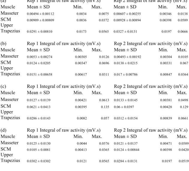
With regard to activity of each muscle during each repetition on the neck weight
integral of raw masseter activity during the second repetition of right lateral flexion and reduction with the mouth open were 0.013 ± 0.015 mV.s and 0.004 ± 0.002 mV.s respectively. The highest and lowest mean ± SD integral of raw SCM activity during the first repetition of right lateral flexion and reduction with the mouth open were 0.062 ± 0.041 mV.s and 0.009 ± 0.008 mV.s respectively. The highest and lowest mean ± SD integral of raw upper trapezius activity during the second repetition of right lateral flexion and reduction with the mouth closed were 0.042 ± 0.039 mV.s and 0.015 ± 0.007 mV.s respectively (Table 1).
There was a significant, positive relationship between the masseter and SCM in the multiple regression analysis of all three muscles during the dorsal flexion with the mouth open (r = 0.559). The masseter and trapezius did not have a significant relationship.
The SCM and trapezius were positively significant (r= 0.429). There were no significant relationships in the multiple correlation between the masseter, SCM, and trapezius for the dorsal flexion with the mouth closed.
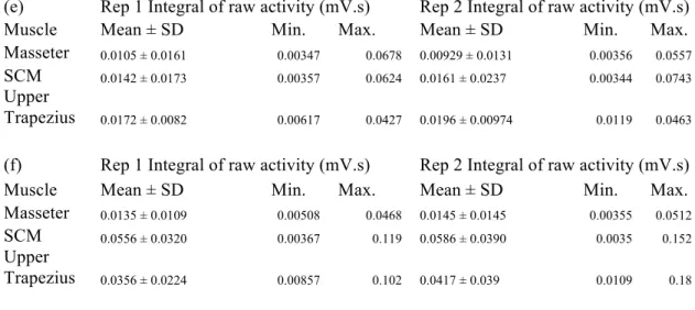
All relationships were positive and significant in the multiple correlation between the muscles for the left lateral flexion with the mouth open and mouth closed (Table 2).
The only positive significant relationship was between the masseter and the trapezius (r = 0.441) in the multiple correlation between the masseter, SCM, and trapezius for the right lateral flexion with the mouth open (Fig. 2). The SCM and masseter did not show a significant relationship nor did the SCM and trapezius when the mouth was open. There were no significant relationships in the multiple correlation between the masseter, SCM, and trapezius for the left lateral flexion with the mouth closed.
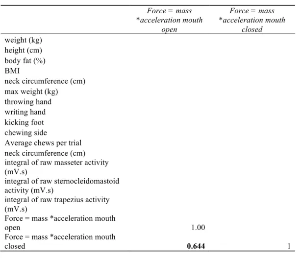
The maximum force the sternocleidomastoid exerted during the weight lifting trials occurred only when the mouth was open. The mean force for the mouth open was 17.08 ± 18.73 Newtons while the force was 11.98 ± 10.25 N when the mouth was closed.
The maximum force with the mouth open was 64.50 N while only 37.04 N with the mouth closed, and the minimum with the mouth open was 3.44 N and with the mouth closed 0.72 N. There was a significant correlation for a greater force exerted by the SCM with the mouth open versus the force with the mouth closed (r = 0.644).
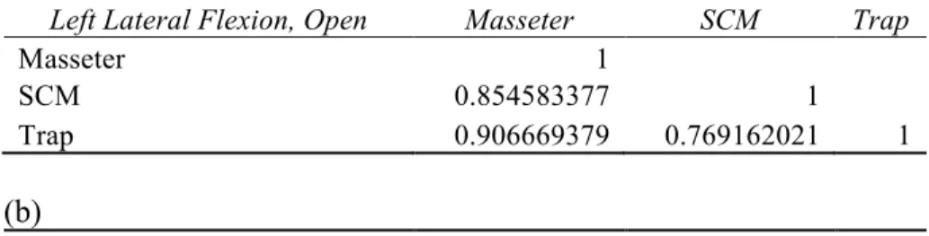
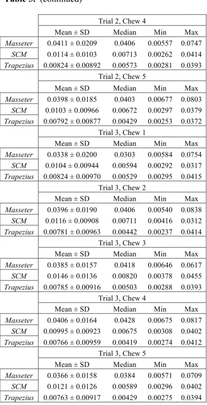
For the first 5 chews in each trial, the descriptive statistics for each muscle show co-activation (Table 3). Specifically, the integral of raw masseter activity the largest mean ± SD was in trial 2 chew 4 at 0.0411 ± 0.0209 mV.s with a minimum in trial 1 chew 1 at 0.00530 mV.s and a maximum in trial 1 chew 2 at 0.0984 mV.s. For the integral of raw SCM activity the largest mean ± SD was in trial 3 chew 3 at 0.0146 ± 0.0136 mV.s with a minimum in trial 2 chew 2 at 0.00229 mV.s and a maximum in trial 3 chew 3 at 0.0455 mV.s. For the integral of raw upper trapezius activity the largest mean ± SD was in trial 2 chew 4 at 0.00824 ± 0.00892 mV.s and trial 3 chew 1 at 0.00824 ± 0.00970 mV.s with a minimum in trial 2 chew 2 at 0.00209 mV.s and a maximum in trial 2 chew 2 at 0.0879 mV.s.
A simple regression of neck circumference (cm) versus max force with mouth open (F(1,14) = 0.29; p = 0.598; r2 = .02) or mouth closed (F(1,14) = 0.18; p = 0.676; r2 = 0.01) did not identify significant results.
For the average chews per trial, some participants averaged as many as 18.67 chews per trial while others averaged as few as 6.33 chews. The mean number of
chewing events for all of the participants was 13.40 times during each trial. The averages
for each chew of the integral of raw trapezius, SCM, and masseter activity proved to be consistent with the averages found from the descriptive statistics I ran on each of the first 5 chews from each trial.
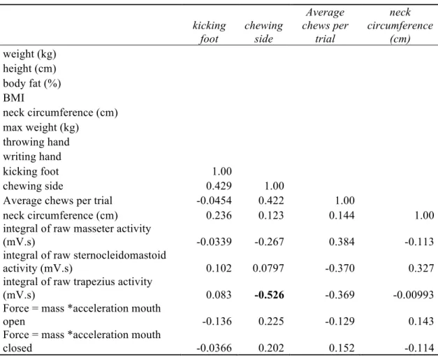
For the first five chewing events (across all trials), a multiple correlation analysis between the masseter, SCM, and trapezius showed a significantly inverse relationship between the masseter and SCM: (1) r = - 0.476, (2) r = - 0.622, (3) r = - 0.6432, (4) r = - 0.576, and (5) r = - 0.606 (Fig. 3). There was not a significant association between the SCM and the trapezius: (1) r = 0.0709, (2) r = - 0.0211, (3) r = 0.152, (4) r = 0.145, and (5) r= 0.164. The masseter and the trapezius relationships were not significant: (1) r= 0.0843, (2) r = - 0.0781, (3) r = 0.0252, (4) r = 0.0763, and (5) r = 0.220.
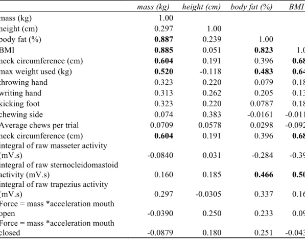
A multiple correlation analysis of mass (kg), height (cm), body fat (%), Body Mass Index (BMI), neck circumference (cm), max weight used (kg), throwing hand, writing hand, kicking foot, chewing side, average chews per trial, integral of raw masseter activity (mV.s), integral of SCM activity (mV.s), integral of raw trapezius activity (mV.s), force (N) with mouth open, and force (N) with mouth closed, several significant relationships (Table 4). Of interest is that co-activation is seen in the
significant relationship between the integral of raw masseter activity with integral of raw SCM activity (r = - 0.591).
DISCUSSION
The data recorded support my categorization of a unique set of participants for my study. Results for overall mass was higher, and body fat (%) was lower, compared to the normal American male showing that the rigorous training the participants undergo for football has a noticeable effect on their overall physique. The participants are also unique because they play football at the highest level in college athletics. This was expected because they play football at an extreme level where they must be able to endure hard collisions to prevent injury. They had thick necks with enhanced musculature and used heavy weight overall on the neck weight machine compared to the men’s basketball team (pers. obs.).
There were minimal differences in muscle activity between mouth open or mouth closed positions during the dorsal, left lateral, and right lateral flexion and reduction while using the neck weight machine. This shows that there was not a significant difference in muscle activity based on positioning of the mouth but it does support the presence of co-activation in muscles (Giannakopoulos et al., 2013). The integral of raw masseter activity showed co-activation while performing the neck machine where the main training focus is on the SCM and upper trapezius. The masseter had greater sEMG activity when doing the right lateral flexion and reduction on the machine which is most likely due to the placement of electrodes on the right side of the body because almost all of the participants chewed on the right side of their mouth.
In the multiple regression between the masseter, SCM, and trapezius for (a) dorsal open mouth, (b) left lateral flexion mouth open, (c) left lateral flexion mouth
closed, and (d) right lateral flexion mouth open. Positive significant relationships showed that co-activation occurs in the athletes while using the neck machine. The dorsal open mouth flexion and reduction was unique because only the masseter and SCM had a significant relationship which goes along with Hellman et al. (2012) who noticed co- activation in these two muscles and little activity from the trapezius. The left lateral flexion and reduction with the mouth open and the mouth closed showed all three muscles to have close positive relationships. This raises the question of why only on the left side did this occur when most of the participants were right side dominant. For future study, I hypothesize that this phenomena could be due to the body trying to balance activity on each side of the body. When performing a left lateral flexion on the machine, the right side of the neck is still activated. On the right lateral flexion the only significant relationship was between the masseter and the trapezius with the mouth open. This is unique to the data and not seen anywhere else because the upper trapezius has not been significant when the SCM was not significant. In the literature, the SCM and masseter have shown the significant relationships but because this is a unique group of participants, I hypothesize that the trapezius has a greater affect on the masseter through co-activation.
The descriptive statistics for the maximum force produced by the SCM while performing the neck machine with the mouth open and then with the mouth closed showed differences in force produced. Force produced with the mouth open was the higher force on average and with a higher maximum and a lower minimum than when the mouth was closed. However, the weakness of these forces calculated is that force
production was not the goal of the participants. I told them to focus on perfect form and execution throughout each repetition. There was a wide range in the time value used for force calculation because some participants moved slowly to maintain good form while others performed the motion more swiftly. If I had given the participants specific
instructions to do the neck weight machine as quickly as possible, these force calculations would be more useful.
The significant relationship between force produced by the SCM on the weight machine with mouth open versus mouth closed is interesting because I would expect the mouth closed to produce more force since the masseter is also maximally contracting in addition to the SCM and trapezius. As previously stated, however, the forces calculated are somewhat faulty because the participants were not told to produce as much force as possible.
I hypothesized that an increase in weight on the neck machine would lead to an increase amount of force the masseter could produce. Clark et al. (1993) states, “The second finding was a progressive development of the SCM co-activation which paralleled the masseter activation. It seems clear that the SCM level of activation was a function of the masseter level”. Indeed, the simple regressions performed on neck circumference (cm) versus max force with (a) mouth open and (b) mouth closed did not produce any significant results, and neck circumference did not necessarily mean more force was produced but this could be due to the error while giving instructions for the exercises. But through co-activation, I suspect the masseter is larger and can produce more force when biting. Even though my experiment could not directly measure the force produced by the masseter, these results indicate that neck training can increase the force of mastication.
Muscle activity on the sEMG recordings during the first 5 chews of each trial show a consistent increase-peak-decrease pattern for the participants, both individually and collectively. The strength of this data is the consistent means, minimums, and maximums for each of the three muscles because it shows a natural rhythm that all participants followed. Further research could find a way to measure exactly how muscle thickness changes during contraction (e.g., CT or MRI) but these tests are expensive.
The average chews per varied widely between each participant. With participants chewing as many as 21 times in a single trial while only chewed 5 times. The weakness of this data is that I did not tell participants to chew a specific amount of times but rather to chew as hard and violently as they could for 10 seconds.
Hellman et al. (2012) found a strong connection between the masseter and SCM during maximum voluntary clenching. These muscles need to coordinate and contract simultaneously to perform the biting task (van der Bilt et al., 2006). In the multiple correlations performed on the 5 different chews examining the relationship between the masseter, SCM, and trapezius, there was a significant negative relationship between the masseter and SCM for each chew. The correlations would have been even stronger except for two of the participants had lesser sEMG activity for the SCM and masseter. These weaker recordings were due to human error or a weak connection through the electrode because of sweat and a small amount of facial hair. A strength of this data is support for the hypothesis that co-activation would occur between the masseter and SCM.
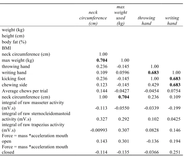
The multiple correlation with participant characteristics, average chews per trial, muscle activity, force (N) with mouth open, and force (N) with mouth closed had significant relationships. The mass (kg) of the participant had a close relationship with
the body fat (%), BMI, neck circumference (cm) and max weight used (kg) used. In essence, the heavier the participant was, the more weight he used on the neck machine.
Also, the larger neck circumference had a strong, positive relationship with max weight used (kg) used. Larger neck circumference is a result of more muscle mass in the SCM and trapezius. When participants had more body fat (%), there was a positive relationship with BMI, max weight used, and integral of raw SCM activity. The SCM and body fat (%) had a significant positive relationship which could possibly be related to long-term effects associated with sleep apnea. As stated earlier, a common identifier for sleep apnea is a large neck (Pedrosa et al., 2011).
Long-term dental effects and indirect effects of enhanced neck musculature were not addressed in this study but should be taken into consideration for follow-up study (Paul et al., 1973). Enhanced neck musculature is necessary to prevent injuries while playing a violent and physical sport, but Hosea (2013) raises the question of what effects are seen when athletes are no longer playing sports? Is sleep apnea in these football players’ futures because of protection from concussions and broken necks today (Pedrosa et al., 2011)? Hubal et al. (2005) researched the variability in muscle size and strength gained after resistance training and found there is a wide range of strength and responses to the specific training. Athletes need to take precaution from neck injuries while playing their support but need to know the potential side effects that can occur later in life as a result.
The writing hand was strongly correlated with the kicking foot and chewing side but this did not show any differences in other areas of the data. If the subject’s dominant chewing side was different than his dominant hand side, then I expected lower muscle
activity from the masseter (Arima et al., 2013). Due to the sample size of only 16
participants, only 2 of those had different dominant chew sides than the rest of their body.
This was not enough information to be able to support my hypothesis.
The integral of raw masseter muscle activity had a significant positive
relationship with the force produced by the SCM with the mouth open which is opposite of what I expected. Force with the mouth closed was expected to be greater through co- activation because there would be more muscle contraction in the SCM and the
(clenched) masseter. However, the force was much weaker when the mouth was closed.
While observing this, I think the participants were simply too focused on clenching the jaw which distracted them from performing a perfect repetition on the neck machine. The integral of raw SCM also had a positive significant relationship with force when mouth was open.
This experiment was performed during off-season training where physical contact between participants was minimal. An area for future research would be to run the same experiment during the football season when the football players are practicing with a 3.5 kilograms helmet on their heads for 2 hours a day. Their necks would be hit hard in many different ways. I hypothesize I would find a weaker, more tired neck from the constant contact (e.g., collisions and contact with the head, neck and shoulders experienced everyday during the season). If this proves to be true, I suggest football players treat their necks with ice, heat, and massages like they do for the rest of their bodies.
If I could go back and time and alter this experiment and do certain things differently, I would measure a maximum voluntary contraction through biting and the neck weight lifting machine. This would give me percentages of the muscle activity being
used allowing me to compare the percentages across all of the participants. With these comparisons, my data would be stronger if it showed similar percentages of contraction. I would also tell participants to move the weight on the machine as fast as possible and measure power of the participants’ necks instead of the force calculation I presented.
Lastly, I would place the electrodes on the masseter directly perpendicular to the ground to match the orientation of the muscle. I had the electrodes closer matched to the
mandible which missed some of the masseter activity.
Table 1. Surface electromyography activity in mV.s (expressed as mean integral for raw activity ± standard deviation and the minimum and maximum) for the masseter,
sternocleidomastoid, and upper trapezius for each repetition on the neck weight lifting machine: (a) dorsal flexion and reduction with mouth open, (b) left lateral flexion and reduction with mouth open, (c) right lateral flexion and reduction with mouth open, (d) dorsal flexion and reduction with mouth closed, (e) left lateral flexion and reduction with mouth closed, and (f) right lateral flexion and reduction with mouth closed.
(a) Rep 1 Integral of raw activity (mV.s) Rep 2 Integral of raw activity (mV.s)
Muscle Mean ± SD Min. Max. Mean ± SD Min. Max.
Masseter 0.00494 ± 0.00112 0.0034 0.0075 0.00607 ± 0.00255 0.00346 0.0138
SCM 0.00909 ± 0.00809 0.0036 0.0372 0.00928 ± 0.00894 0.00398 0.0589
Upper
Trapezius 0.0291 ± 0.00810 0.0173 0.0565 0.0327 ± 0.0131 0.0197 0.0666
(b) Rep 1 Integral of raw activity (mV.s) Rep 2 Integral of raw activity (mV.s)
Muscle Mean ± SD Min. Max. Mean ± SD Min. Max.
Masseter 0.0053 ± 0.00274 0.00305 0.0126 0.00493 ± 0.00192 0.00304 0.0105
SCM 0.0124 ± 0.0205 0.00347 0.0696 0.0138 ± 0.0215 0.00331 0.067
Upper
Trapezius 0.0151 ± 0.00658 0.00617 0.0311 0.017 ± 0.00786 0.00847 0.0364
(c) Rep 1 Integral of raw activity (mV.s) Rep 2 Integral of raw activity (mV.s)
Muscle Mean ± SD Min. Max. Mean ± SD Min. Max.
Masseter 0.0127 ± 0.0139 0.00421 0.0613 0.0133 ± 0.0145 0.00381 0.0498
SCM 0.0621 ± 0.0413 0.00395 0.135 0.06 ± 0.0397 0.00428 0.129
Upper
Trapezius 0.0286 ± 0.0143 0.0082 0.057 0.0312 ± 0.0154 0.00839 0.0661
(d) Rep 1 Integral of raw activity (mV.s) Rep 2 Integral of raw activity (mV.s)
Muscle Mean ± SD Min. Max. Mean ± SD Min. Max.
Masseter 0.0125 ± 0.0130 0.0044 0.0576 0.0121 ± 0.0137 0.00471 0.0589
SCM 0.0105 ± 0.0081 0.00413 0.0365 0.0124 ± 0.00844 0.00398 0.0420
Upper
Trapezius 0.0302 ± 0.0302 0.0121 0.0565 0.0284 ± 0.0131 0.0197 0.0519
Table 1. (continued)
(e) Rep 1 Integral of raw activity (mV.s) Rep 2 Integral of raw activity (mV.s)
Muscle Mean ± SD Min. Max. Mean ± SD Min. Max.
Masseter 0.0105 ± 0.0161 0.00347 0.0678 0.00929 ± 0.0131 0.00356 0.0557
SCM 0.0142 ± 0.0173 0.00357 0.0624 0.0161 ± 0.0237 0.00344 0.0743
Upper
Trapezius 0.0172 ± 0.0082 0.00617 0.0427 0.0196 ± 0.00974 0.0119 0.0463
(f) Rep 1 Integral of raw activity (mV.s) Rep 2 Integral of raw activity (mV.s)
Muscle Mean ± SD Min. Max. Mean ± SD Min. Max.
Masseter 0.0135 ± 0.0109 0.00508 0.0468 0.0145 ± 0.0145 0.00355 0.0512
SCM 0.0556 ± 0.0320 0.00367 0.119 0.0586 ± 0.0390 0.0035 0.152
Upper
Trapezius 0.0356 ± 0.0224 0.00857 0.102 0.0417 ± 0.039 0.0109 0.18
Table 2. Bivariate correlation between the masseter, sternocleidomastoid, and trapezius for the left lateral flexion (a) with the mouth open and (b) with the mouth closed (r = correlation coefficient).
(a)
Left Lateral Flexion, Open Masseter SCM Trap
Masseter 1
SCM 0.854583377 1
Trap 0.906669379 0.769162021 1
(b)
Left Lateral Flexion, Closed Masseter SCM Trap
Masseter 1
SCM 0.747064427 1
Trap 0.858564949 0.82869596 1
Table 3. Descriptive statistics for the integral of the raw (a) masseter activity (mV.s), (b) sternocleidomastoid activity (mV.s), and trapezius activity (mV.s) for the first 5 chews in each trial.
Trial 1, Chew 1
Mean ± SD Median Min Max Masseter 0.0338 ± 0.0220 0.0281 0.00530 0.0834
SCM 0.00807 ± 0.00767 0.00494 0.00293 0.0309 Trapezius 0.00675 ± 0.00413 0.00474 0.00309 0.0142
Trial 1, Chew 2
Mean ± SD Median Min Max Masseter 0.0377 ± 0.0226 0.0357 0.00560 0.0984
SCM 0.0103 ± 0.0104 0.00591 0.00356 0.0385 Trapezius 0.00654 ± 0.00406 0.00409 0.00284 0.0156
Trial 1, Chew 3
Mean ± SD Median Min Max Masseter 0.0356 ± 0.0211 0.0321 0.00573 0.0892
SCM 0.00913 ± 0.00723 0.00676 0.00254 0.0323 Trapezius 0.00635 ± 0.00413 0.00502 0.00271 0.0165
Trial 1, Chew 4
Mean ± SD Median Min Max Masseter 0.0377 ± 0.0209 0.0336 0.00613 0.0779
SCM 0.00965 ± 0.00919 0.00673 0.00343 0.0411 Trapezius 0.00690 ± 0.00395 0.00606 0.00248 0.0161
Trial 1, Chew 5
Mean ± SD Median Min Max Masseter 0.0364 ± 0.0171 0.0328 0.00712 0.0706
SCM 0.00982 ± 0.00690 0.00721 0.00341 0.0265 Trapezius 0.00654 ± 0.00401 0.00547 0.00263 0.0167
Trial 2, Chew 1
Mean ± SD Median Min Max Masseter 0.0345 ± 0.0195 0.0346 0.00562 0.0787
SCM 0.0109 ± 0.0115 0.00601 0.00320 0.0391 Trapezius 0.00792 ± 0.00872 0.00499 0.00284 0.0381
Trial 2, Chew 2
Mean ± SD Median Min Max Masseter 0.0399 ± 0.0238 0.0414 0.00601 0.0915
SCM 0.00978 ± 0.00938 0.00744 0.00229 0.0398 Trapezius 0.0125 ± 0.0218 0.00439 0.00209 0.0879
Trial 2, Chew 3
Mean ± SD Median Min Max Masseter 0.0391 ± 0.0165 0.0405 0.00601 0.0620
SCM 0.0106 ± 0.00964 0.00718 0.00291 0.0419
Table 3. (continued)
Trial 2, Chew 4
Mean ± SD Median Min Max Masseter 0.0411 ± 0.0209 0.0406 0.00557 0.0747
SCM 0.0114 ± 0.0103 0.00713 0.00262 0.0414 Trapezius 0.00824 ± 0.00892 0.00573 0.00281 0.0393
Trial 2, Chew 5
Mean ± SD Median Min Max Masseter 0.0398 ± 0.0185 0.0403 0.00677 0.0803
SCM 0.0103 ± 0.00966 0.00672 0.00297 0.0379 Trapezius 0.00792 ± 0.00877 0.00429 0.00253 0.0372
Trial 3, Chew 1
Mean ± SD Median Min Max Masseter 0.0338 ± 0.0200 0.0303 0.00584 0.0754
SCM 0.0104 ± 0.00944 0.00594 0.00292 0.0317 Trapezius 0.00824 ± 0.00970 0.00529 0.00295 0.0415
Trial 3, Chew 2
Mean ± SD Median Min Max Masseter 0.0396 ± 0.0190 0.0406 0.00540 0.0838
SCM 0.0116 ± 0.00908 0.00711 0.00416 0.0312 Trapezius 0.00781 ± 0.00963 0.00442 0.00237 0.0414
Trial 3, Chew 3
Mean ± SD Median Min Max Masseter 0.0385 ± 0.0157 0.0418 0.00646 0.0617
SCM 0.0146 ± 0.0136 0.00820 0.00378 0.0455 Trapezius 0.00785 ± 0.00916 0.00503 0.00288 0.0393
Trial 3, Chew 4
Mean ± SD Median Min Max Masseter 0.0406 ± 0.0164 0.0428 0.00675 0.0817
SCM 0.00995 ± 0.00923 0.00675 0.00308 0.0402 Trapezius 0.00766 ± 0.00959 0.00419 0.00274 0.0412
Trial 3, Chew 5
Mean ± SD Median Min Max Masseter 0.0366 ± 0.0158 0.0384 0.00571 0.0709
SCM 0.0121 ± 0.0126 0.00589 0.00296 0.0402 Trapezius 0.00763 ± 0.00917 0.00429 0.00275 0.0394
Table 4. Correlation matrix including mass (kg), height (cm), body fat (%), Body Mass Index (BMI), neck circumference (cm), max weight used (kg), throwing hand, writing hand, kicking foot, chewing side, average chews per trial, integral of raw masseter activity (mV.s), integral of sternocleidomastoid activity (mV.s), integral of raw trapezius activity (mV.s), force (N) with mouth open, and force (N) with mouth closed (r =
correlation coefficient). Significant relationships are identified by bold typeface.
mass (kg) height (cm) body fat (%) BMI
mass (kg) 1.00
height (cm) 0.297 1.00
body fat (%) 0.887 0.239 1.00
BMI 0.885 0.051 0.823 1.00
neck circumference (cm) 0.604 0.191 0.396 0.684
max weight used (kg) 0.520 -0.118 0.483 0.641
throwing hand 0.323 0.220 0.079 0.189
writing hand 0.313 0.262 0.205 0.134
kicking foot 0.323 0.220 0.0787 0.189
chewing side 0.074 0.383 -0.0161 -0.0110
Average chews per trial 0.0709 0.0578 0.0298 -0.0929
neck circumference (cm) 0.604 0.191 0.396 0.684
integral of raw masseter activity
(mV.s) -0.0840 0.031 -0.284 -0.391
integral of raw sternocleidomastoid
activity (mV.s) 0.160 0.185 0.466 0.502
integral of raw trapezius activity
(mV.s) 0.297 -0.0305 0.337 0.168
Force = mass *acceleration mouth
open -0.0390 0.250 0.233 0.094
Force = mass *acceleration mouth
closed -0.0879 0.180 0.251 -0.0435