Revision of the Stonefly
Family Nemouridae (Plecoptera):
A Study of the World Fauna at the Generic Level
RICHARD W. BAUMANN
m
SMITHSONIAN CONTRIBUTIONS TO ZOOLOGY • NUMBER 211
The emphasis upon publications as a means of diffusing knowledge was expressed by the first Secretary of the Smithsonian Institution. In his formal plan for the Insti- tution, Joseph Henry articulated a program that included the following statement:
"It is proposed to publish a series of reports, giving an account of the new discoveries in science, and of the changes made from year to year in all branches of knowledge."
This keynote of basic research has been adhered to over the years in the issuance of thousands of titles in serial publications under the Smithsonian imprint, com- mencing with Smithsonian Contributions to Knowledge in 1848 and continuing with the following active series:
Smithsonian Annals of Flight Smithsonian Contributions to Anthropology
Smithsonian Contributions to Astrophysics Smithsonian Contributions to Botany Smithsonian Contributions to the Earth Sciences
Smithsonian Contributions to Paleobiology Smithsonian Contributions to Zoology Smithsonian Studies in History and Technology
In these series, the Institution publishes original articles and monographs dealing with the research and collections of its several museums and offices and of professional colleagues at other institutions of learning. These papers report newly acquired facts, synoptic interpretations of data, or original theory in specialized fields. These pub- lications are distributed by mailing lists to libraries, laboratories, and other interested institutions and specialists throughout the world. Individual copies may be obtained from the Smithsonian Institution Press as long as stocks are available.
S. DILLON RIPLEY
Secretary
Smithsonian Institution
S M I T H S O N I A N C O N T R I B U T I O N S T O Z O O L O G Y • N U M B E R 2 1 1
Revision of the Stonefly
Family Nemouridae (Plecoptera):
A Study of the World Fauna at the Generic Level
Richard W. Baumann
SMITHSONIAN INSTITUTION PRESS City of Washington
1975
Baumann, Richard W. Revision of the Stonefly Family Nemouridae (Plecop- tera): A Study of the World Fauna at the Generic Level. Smithsonian Contribu- tions to Zoology, number 211, 74 pages, 186 figures, 1 table, 1975.—The Northern Hemisphere family Nemouridae is revised and divided into two subfamilies:
Amphinemurinae, new subfamily, and Nemourinae. Of the 17 recognized genera in the Nemouridae, 14 are described and figured and 3 are described as new:
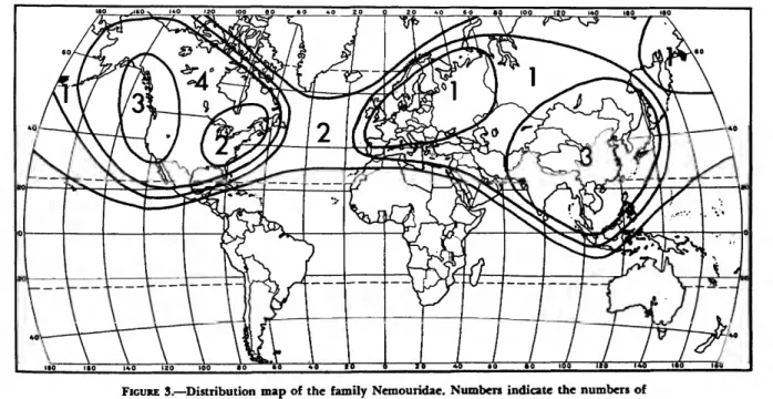
Illiesonemoura, Indonemoura, and Mesonemoura; 373 species are assigned to genera or listed as incertae sedis. Keys to subfamilies and genera are given for males, females, and nymphs. Structures of male and female terminalia are described and the terminology defined. A phylogeny is constructed showing the relationships of the genera. Distributions are given for all species, the zoogeog- raphy of the family is discussed, and a composite map of the distribution of all genera in Nemouridae is provided.
OFFICIAL PUBLICATION DATE is handstampcd in a limited number of initial copies and is recorded in the Institution's annual report, Smithsonian Year. SI PRESS NUMBER 6007. SERIES COVER DESIGN:
The coral Montastrea cavernosa (Linnaeus).
Library of Congress Cataloging in Publication Data Baumann, Richard W.
A revision of the stonefly family Nemouridae (Plecoptera) (Smithsonian contributions to zoology, no. 211)
Bibliography: p.
1. Nemouridae. 2. Insects—Classification. I. Title. II. Series: Smithsonian Institution. Smith- sonian contributions to zoology; no. 211.
QL1.S54 no. 211 [QL 505.3.N4] 591'.08s [595.7'35] 75-619077
Contents
Page
Introduction 1 Materials and Methods 2 Terminology 2 Acknowledgments 3 Morphology 3 Terminalia of Males 5 Terminalia of Females 6 Key to the Subfamilies and Genera of Nemouridae 7 Amphinemurinae, new subfamily 10 Amphinemura Ris 11 Indonemoura, new genus 12 Malenka Ricker 13 Mesonemoura, new genus 14 Protonemura Kempny 16 Nemourinae Newman, 1853 17 Illiesonemoura, new genus 18 Lednia Ricker 19 Nemoura Latreille 20 Nemurella Kempny 22 Ostrocerca Ricker 23 Paranemoura Needham and Claassen 24 Podmosta Ricker 25 Prostoia Ricker 26 Shipsa Ricker 27 Soyedina Ricker 28 Visoka Ricker 29 Zapada Ricker 30 Species Incertae Sedis 31 Phylogeny 32 Apomorphic Character States 32 Zoogeography 36 Epilogue . . . . 38 Literature Cited 38 Figures 4-186 44
i n
Revision of the Stonefly
Family Nemouridae (Plecoptera) A Study of the World Fauna
at the Generic Level
Richard W. Baumann
Introduction
The family Nemouridae is the largest in the order Plecoptera, comprising 373 species in 17 genera. The family was erected by Newman in 1853, and he included most of the present Euho- lognatha in a large diverse grouping. Klapalek (1905) divided the family into four families: C a p niidae, Leuctridae, Nemouridae, and Taeniop- terygidae. These four families and the Southern Hemisphere family Notonemouridae were recently defined as the superfamily Nemouroidea by Zwick
(1973a).
This study deals with the Nemouridae as pro- posed by Kimmins (1961) and adopted by the International Commission on Zoological Nomen- clature (1963). Kempny (1898) divided the Euro- pean species into subgenera; Kimmins (1940), lilies (1955), Winkler (1957), Hynes (1958), and Aubert (1959b) recognized four full genera: Am- phinemura, Nemoura, Nemurella, and Protone- mura. The North American subgenus Parane- moura was described by Needham and Claassen (1925), and Ricker (1952) revised the North American Nemouridae fauna and divided the species into 12 subgenera, including Paranemoura and also Nemoura and Amphinemura from Eu- rope. These subgenera were then raised to generic
Richard W. Baumann, Department of Entomology, National Museum of Natural History, Smithsonian Institution 20560.
status by lilies (1966). I describe three new genera:
Indonemoura, Mesonemoura, and Illiesonemoura and one new subfamily, Amphinemurinae, in this study.
The Asian Nemouridae have always been placed in one of the European genera even though they often did not belong: Aubert (1959a, 1967), Jewett (1958), Kimmins (1947, 1950 a and b), Kawai (1967). My study shows that, although many Asian species do belong in European genera, some belong in North American genera and others represent genera and species groups that are en- demic to the Asian portion of the Palearctic region.
The nymphs of the genera occurring in Europe are quite well known and have been treated by Hynes (1941), Brinck (1949), Despax (1951), and Rauser (1956, 1963a). The Asian and North American genera have, however, been studied very little. I have studied all available nymphal mate- rial, written generic descriptions, constructed nymphal keys, and used the characters found to help understand the phylogeny.
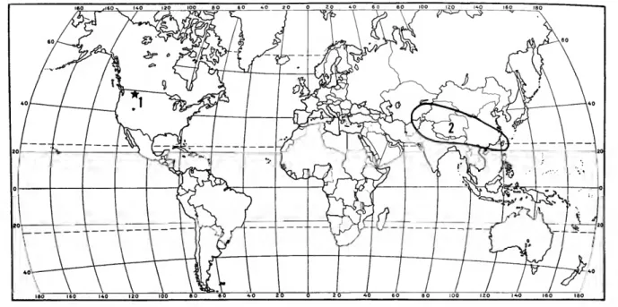
The Nemouridae are usually the dominant Plecoptera in mountain-river ecosystems, both in terms of total biomass and in numbers of species present. They are primary consumers and feed mostly on detritus. They are distributed through- out the Northern Hemisphere and a few species do just cross the equator in the Sunda Archipelago.
1
MATERIALS AND METHODS.—During this study I examined all of the North American species and most of the European and Asian species. China is the only large area from which I was unable to study many specimens. Those species not exam- ined are marked with an asterisk (*) where they appear in the "Distribution and Species Lists" at the end of each genus. This means that they were
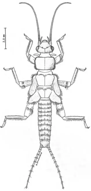
FIGURE 1.—Habitus of Zapada haysi (Ricker), mature female nymph.
placed in a genus based on figures or descriptions in the literature.
Studies were carried out with a Wild M-5 stereo- scopic microscope, which for most of the study was illuminated with a dark field base illumination system. This made it possible to study the especially fine hairs and spines on the small structures of the epiproct and paraprocts.
For detailed examination all small parts were removed from the specimens using tiny dental files or micro dissecting knives. These parts were ex- amined in small watch glasses containing a thin layer of beeswax and then stored in micro vials with a cotton plug at both ends. The white-cotton background made it much easier to see the tiny parts.
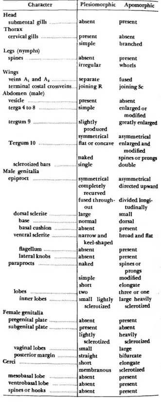
TERMINOLOGY.—Since there are many terms in existence for both male and female terminalia as listed in Brinck (1955), I am defining those most useful for the Nemouridae. I followed the work by Crampton (1918) because it contained most of the terms now in use.
Although some authors had recognized the dif- ferent lobes of the paraprocts, the parts of the epiproct had been essentially unstudied except for Zwick (1973a) where the letters "a" and "b are used for the dorsal and basal sclerites respectively.
The following list of definitions and abbrevia- tions includes those from Crampton (1918) and those coined in this study.
Basal cushion (be): rounded membranous area of the epiproct, above the base of the dorsal sclerite, between the lateral arms; found in Nemoura and closely related Nemourinae (Figures 45, 61, 118, 133).
Basal sclerite (bs): angular sclerites located on both sides at the base of the epiproct, between the tenth tergum and paraprocts, small and triangular or rectangular (Figures 9, 17, 25, 49, 57, 105, 137).
Dorsal sclerite (ds): sclerotized dorsal portion of the epiproct, with broad base and paired anterolateral extensions, cov- ering all or part of the dorsal and lateral surfaces.
Epiproct (ep): unpaired process in the tergal region of the eleventh abdominal segment, composed of dorsal, ventral, and basal sclerites; also called supra-anal process.
Epiproctal flagellum (ef): long thin sclerotized process ex- tending forward from ventral sclerite, at tip of epiproct;
as in Mesonemoura (Figures 28-30).
Hypoproct (hp): produced portion of the male ninth sternum, containing the gonopore; also called hypandrium or subgenital plate.
Inner lobe (il): paraproctal lobes located nearest midline, usually small and inconspicuous, often hidden by hypo- proct, lacking spines.
NUMBER 211
Lateral arms (la): thin darkly sclerotized anterolateral ex- tensions of dorsal sclerite, usually connected to dorsal scler- ite throughout length.
Lateral cervical sclerite (lcs): long, thin sclerotized bars lo- cated near lateral margins of cervical region (Figures 140—
147).
Lateral knobs (Ik): rounded hingelike structures located at basal corners of epiproct, formed from base of ventral scler- ite; found in Nemoura and closely related Nemourinae (Figures 45, 61, 117, 133).
Outer lobe (ol): outer lobe of paraprocts, located nearest cerci, long thin and darkly sclerotized in Amphinemurinae, broad, round and with a membranous portion in Nemour- inae; also called cereal lobe.
Median lobe (ml): middle lobe of paraprocts in Amphine- murinae, large and usually both sclerotized and mem- branous, often bearing spines.
Paraproct (pp): platelike structures situated behind the tenth tergum, located in the regions homologous with the lateral portions of the eleventh abdominal segment, may have one, two, or three lobes; also called subanal lobe.
Pregenital plate (pgp): produced portion of seventh abdomi- nal sternum of female (Figures 51, 67, 75, 123, 131, 139).
Subgenital plate (sgp): modified area of eighth abdominal sternum of female, located near gonopore; often as broadly rounded lobe or as one or more sclerotized plates.
Ventral sclerite (vs): ventral sclerotized portion of epiproct, large, flat or keel-shaped and usually bearing spines, often extending upward dorsally and modified into single or paired structures.
Vesicle (v): lobed structure arising from anterior margin of ninth sternum or hypoproct.
ACKNOWLEDGMENTS.—I heartily thank the fol- lowing people for helping me with this study by providing specimens or sharing their time and ideas: Dr. J. Aubert, Musee Zoologique, Lausanne, Switzerland; Dr. P. Brinck and Dr. C. H. Lindroth, Lund University, Lund, Sweden; Dr. P. Berthel- emy, Universite de Toulouse, Toulouse, France;
Dr. A. R. Gaufin, University of Utah, Salt Lake City, Utah, USA; Dr. D. C. Geijskes, Rijksmuseum van Natuurlijke Historic Leiden, Holland; Dr. J.
lilies, Max-Planck-Institut fur Limnologie, Schlitz, Germany; Mr. S. G. Jewett, Jr., West Linn, Ore- gon, USA; Dr. T. Kawai, Nara Women's Univer- sity, Nara, Japan; Dr. D. E. Kimmins and Mr. P.
Ward, British Museum (Natural History), Lon- don, England; Dr. M. Kohno, Aizu-Wakamatsu, Japan; Dr. A. Lillehammar, Zoological Museum, University of Oslo, Oslo, Norway; Dr. M. Meinan- der, Zoological Museum, University of Helsinki, Helsinki, Finland; Dr. W. E. Ricker, Fisheries Re- search Board of Canada, Biological Station, Nana- imo, British Columbia, Canada; Dr. B. P. Stark,
Irving, Texas, USA; Dr. K. W. Stewart, North Texas State University, Denton, Texas, USA; Dr.
L. A. Zhiltzova, Zoological Institute, Academy of Sciences, Leningrad, USSR; Dr. P. Zwick, Max- Planck-Institut fur Limnologie, Schlitz, Germany.
I am very grateful to Dr. Joachim lilies, who made it possible for me to spend a year of post- doctoral study at his institute in Schlitz, Germany, and I am grateful especially to Dr. P. Zwick for his friendship, help, and continued encouragement.
The drawings were made by Ms. Elizabeth K.
Meyers, Mr. Michael L. Druckenbrod, Ms. Debra R. Homer, and Ms. Rebecca F. Surdick. Ms. Hollis B. Williams provided valuable support in organ- izing the plates and in the preparation of the phylogenetic diagram. Special thanks are given to Mrs. Joan B. Miles for typing the manuscript.
This study was supported in part by a Sigma Xi Grant-in-Aid of Research and a research fellowship from the Max-Planck Society.
Morphology
The characters used in this study are best con- sidered by examining body sections and systems as separate entities. My study was supplemented by the works of Crampton (1918), Wu (1923), Ricker
(1952), Moulins (1968), and Zwick (1973a).
HEAD.—The head region is very similar in all nemourid genera. The modification of the last segment of the labial palpi into a large, flat, rounded structure is very distinctive, especially in the adults. Since the adults feed on algae, lichens, and other plant material, it is possible that the palpi aid in locating food. It is also possible that the enlarged palpal segments are sensory in func- tion, because adult nemourids are very active and drop immediately if the substrate upon which they are resting is disturbed.
The mandibles and maxillae are broad and modified for chewing plant material and do not vary enough from one genus to another to be use- ful for taxonomic analysis.
Branched gills that originate between the la- bium and submentum are present in Visoka. These gills are considered apomorphic because of their placement and because they are well developed with five definite branches. They are called sub- mental gills in this study but should not be con-
fused with the submental gills of the Perlodidae, which originate at the base of the submentum.
NECK OR CERVIX.—The cervical region is mostly
membranous but has long, thin, cervical or jugular sclerites along the lateral margins. These lateral cervical sclerites are lightly sclerotized and fairly distinct. They are excellent points of orientation because cervical gills, when present, are located just inside or outside of the sclerites.
Cervical gills are simple extensions of the ventral tracheae leading to the head. They do not have any muscles attached but do have osmoregulatory structures at the base, "Sensilla discoidea" (Zwick, 1973a, and Wichard and Komnick, 1974). Gills are present in all of the primitive Plecoptera families and are considered plesiomorphic in the simple unbranched condition. Modifications such as branching are considered apomorphic as is the partial or complete loss of gills. Thus a single, simple gill on each side of the lateral cervical sclerites, as exhibited by most Zapada, is consid- ered to be the most primitive condition in Nemouridae.
Cervical gills are technically called cervical gill remnants in the adult stage, but because the gills in Nemouridae are still well preserved in the adult stage, I am simply calling them gills in all cases.
THORAX (Nymphs).—The pronotum is always square or rectangular in shape and very stout. The lateral margins and much of the dorsal surface are covered with stiff spines. The mesonotum and metanotum are similar in shape but are slightly more rounded and contain large wing pads in mature specimens. The wing pads diverge outward in a wide angle from the longitudinal body axis.
Spines are usually present along the lateral mar- gins of the mesonotum and metanotum and also on the dorsal surface, especially along the anterior margins.
Presence of spines is considered an apomorphic character state, and they are found on most of the Nemouridae. Modification into specialized rows or patterns is a further apomorphic condition.
WINGS.—The wings of Nemouridae are highly uniform throughout the family. They are very simple when compared to those of most Plecoptera families and have very few crossveins. The most distinctive character is the so-called nemourid "X"
that is formed in the forewings at the cord (Figures 148-157). The "X" is formed primarily by the
terminal portion of vein Sc, which slopes abruptly to the cord, and by the presence of a costal cross- vein running between C and R at or slightly be- yond the cord. In Paranemoura the crossvein joins Sc instead of R, and I consider this a derived char- acter. Zwick (1973a) noted that the "X" in the forewings was also found in some Capniidae and Taeniopterygidae. Although it is plesiomorphic, the "X" is found in all genera of Nemouridae and is an excellent character for general field and cura- torial separation. The "X" can be skewed as noted by Lillehammer (1974) but is always present. Veins Ax and A2 are fused near the outer margin of the forewings in Soyedina. This is an apomorphic state which is sometimes variable in a single population.
Wing color is not a valid criterion for separation at the generic level because of the extreme varia- tion possible. It is, however, useful for separating species complexes as noted by Baumann and Gaufin (1972).
LEGS.—The legs are generally very uniform throughout the family. The femur and tibia are of approximately equal length and tarsal segment two is much smaller than segment one.
The nymphs can often be distinguished at the generic level by the kind and pattern of spines on the legs. All genera have some spines, but they be- come useful as taxonomic characters when they are arranged in definite patterns. Zapada, for ex- ample, has very specialized whorls of large spines on all femora.
ABDOMEN (nymphs).—All genera have fringes of spines along the posterior margins of the abdomi- nal terga. Some species have specialized stout or long spines arranged in narrow longitudinal rows, but these arrangements of spines do not help in separating genera.
ABDOMEN (adults).—The anterior abdominal segments are seldom modified. Soyedina does, how- ever, contain two species with modified tergites in the male sex: S. vallicularia Wu has tergites six to eight elevated, and S. producta Claassen has knobs on numbers two to four. The males of many spe- cies in the Nemoura species complexes Cercispi- nosa and Ovocercia exhibit modifications of ter- gites eight and nine. Some Amphinemura and Mesoncmoura species have greatly modified ninth terga, such as A. mirabilis Martynov and A. zim- mermani Joost, which are also asymmetrical.
Protonemura contains many species that have
NUMBER 211
produced areas covered with spines on terga seven, eight, and nine.
In most genera tergum ten is sclerotized and formed into a flat or concave base under the epi- proct. It also bears large spines or prongs in some species of Ostrocerca and the Cercispinosa and Ovocercia complexes. Thin paired sclerotized bars present in Nemurella, Ostrocerca, Podmosta, and Shipsa run laterally and attach to the bases of the cerci instead of the usual single, wide, median bar.
In Podmosta, Paranemoura, and Lednia the lateral margins are enlarged and modified into specialized structures that are covered with spines or hairs.
Shipsa has even larger lateral projections which extend backward beyond the tips of the cerci.
TERMINALIA OF MALES
HYPOPROCT.—The hypoproct is usually formed into a broad, flat plate which is wide at the base and tapers to a narrow tip. It is greatly enlarged and very elongate in Soyedina, Nemurella, and some species of Ostrocerca. The apex is enlarged and terminates in a truncate tip in other species of Ostrocerca. The tip bears the genital opening, and the length of the apex is dependent on the structure of the paraprocts and epiproct because they usually function together to transport sperm during mating (Zwick, 1973a).
A vesicle is present at the anterior margin of the hypoproct or ninth sternum in all genera except Paranemoura and Lednia. The vesicle is a sen- sory organ as noted by Rupprecht and Gnatzy (1974). I consider the absence of a vesicle to be apomorphic.
EPIPROCT.—The epiproct is actually composed of four sclerotized structures: dorsal sclerite, ventral sclerite, and two basal sclerites.
The basal sclerites are paired angular sclero- tized plates located at the basolateral corners.
Zwick (1973a) calls these the "b" sclerites. They are present in all genera and vary in shape from small and triangular to large and rectangular or ovoid.
The dorsal sclerite is composed of a large darkly sclerotized horseshoe-shaped base, lateral sclero- tized arms, and a large lightly sclerotized dorso- lateral area made up of many tiny scales or plates.
Zwick refers to the horseshoe-shaped base and the lateral arms as part of sclerite "a." Nemoura and
closely allied species of Nemourinae have a large membranous dorsal area located directly ahead of the horseshoe-shaped base of the dorsal sclerite. I call this area the basal cushion.
The ventral sclerite is the flat, convex or keel- shaped ventral portion of the epiproct. It is con- nected to the tenth tergum by a single or paired sclerotized bars. The ventral sclerite usually bears spines or prongs somewhere along the ventral mar- gin. It is often highly modified dorsally and ex- tends upward inside the lateral folds of the dorsal sclerite. The dorsal portion can be single but is usually paired. It is often visible on the dorsal sur- face of the epiproct and extends forward as a fla- gellum in Mesonemoura and rarely in other genera of Amphinemurinae. In Nemoura, Illiesonemoura, Zapada, and Soyedina the lateral basal corners are formed into distinct lateral knobs resembling ball and socket hinges. This derived character often obscures the lateral arms of the dorsal sclerite as they extend forward from the horseshoe-shaped base.
I consider the plesiomorphic state of the epi- proct as being similar to some species of Amphi- nemura where the dorsal and ventral sclerites are similar in size but are little modified. They bear spines somewhere ventrally and are connected to the tenth tergum by a single wide sclerotized bar.
PARAPROCTS.—These structures are very stable and provide excellent taxonomic characters. They are located ventrally in the region of the eleventh tergum. They are paired and separated by the mid- line into bilateral structures. They can be one-, two-, or three-lobed.
The subfamily Amphinemurinae has paraprocts with three lobes, and the subfamily Nemourinae has paraprocts with one or two lobes. Those with three lobes have small sclerotized inner lobes which are naked of spines. The inner lobe has a muscle connected along the inner margin. The median lobes are large, well developed, and have a sclero- tized base and membranous apex which often bears a few spines or prongs. There is a muscle that con- nects to the lobes near the apex and extends throughout the length. The outer or cereal lobes are usually small, thin and are located near the base of the cerci. They are darkly sclerotized and sometimes bear spines or prongs. These lobes have a muscle attached at the base.
In the Nemourinae, where only two lobes are
present, the inner lobes are narrow and often dif- ficult to see because they are located on the inner margins under the apex of the hypoproct. Some genera like Nemurella, Ostrocerca, Visoka, and Lednia have well-developed inner lobes which ex- tend to the base of the epiproct and possibly aid in sperm transport as noted by Zwick (1973a). The outer lobes are usually broad and membranous but sometimes have a sclerotized base. Where a single lobe is present, the inner lobe has been lost and the outer lobes cover the complete ventral region.
The primitive Plecoptera families like Austro- perlidae, Pteronarcyidae, and Scopuridae have single large paraproctal lobes. The apomorphic condition is a divided paraproct, but in the Ne- mouridae the condition of a large outer lobe and tiny inner lobe like in Nemoura is plesiomorphic and the three-lobed condition is derived.
TERMINALIA OF FEMALES
PREGENITAL PLATE.—The seventh sternum is often enlarged and modified into a pregenital plate. The median posterior margin is produced and lightly sclerotized and extends onto sternum eight. Many species in the Nemourinae have a highly developed pregenital plate which covers the genital opening and much of the eighth ster- num. Modification of sternum eight is the usual condition in Plecoptera, so formation of a pre- genital plate is taken to be apomorphic.
SUBGENITAL PLATE.—The eighth sternum is usu- ally modified into some sort of subgenital plate.
The Amphinemurinae are characterized by having paired vaginal lobes on the posterior margin.
Protonemura, Indonemoura, and Mesonemoura also have a large median plate, and Malenka and Amphinemura usually have a distinct median notch.
The Nemourinae show less development of sternum eight. It is usually narrow and covered by sternum seven. It is developed laterally in Nemu- rella and also in some Ostrocerca species. Lednia and Shipsa have small platelike areas medially and Podmosta exhibits distinctive sclerotized patterns.
In Prostoia, segments seven and eight are essen- tially unmodified.
CERCI.—Nymphs: The cerci of the nymphs are quite long and covered with spines. They are ap- proximately the length of the complete abdomen.
Whorls of spines are found at the distal margins of all segments. The length of the spines varies even between species and sometimes the ventral spines or bristles are slightly longer as shown by Harper and Hynes (1971). Intermediate spines are often present on the segments between the joints. No good generic characters have been found using the cerci, but they are useful at the specific level in small genera and when considering species in limited geographical areas.
Adults: The adult cerci are reduced to single segments. They are usually simple, membranous, and covered by short hairs. The only departures from this pleisomorphic state in the Amphine- murinae are the lengthening of the segments as in Indonemoura and the development of a mesobasal sclerotized, lobe by the males of Malenka. The mesobasal lobe is located at the base of the cerci on the dorsolateral margin.
Most of the Nemourinae also have simple cerci, but Nemoura males have a distinctive lateral sclerotized portion which usually ends in a multi- forked tip at the apex. In the Cercispinosa com- plex large spines may occur anywhere throughout the length of the cerci. The Ovocercia complex has enlarged cerci that are lightly sclerotized and modified in shape but do not bear spines. Visoka also has sclerotized cerci that end in single projec- tions.
Nemurella and Ostrocerca males have greatly enlarged sclerotized cerci. They are elongate and globose in Nemurella and naked of projections but they have a greatly enlarged base. In Ostro- cerca they are thin, elongate, and terminate in sharp points. They are bent in an S-shape in lat- eral aspect in some Ostrocerca species.
The females of most genera have simple, un- sclerotized cerci, but those of Nemoura, Nemu- rella, and Visoka are lightly sclerotized laterally and slightly modified in shape.
I submit that the occurrence of sclerotized, mod- ified cerci in scattered genera of Nemourinae is the result of convergence.
NUMBER 211
Key to the Subfamilies and Genera of Nemouridae
MALES
1. Paraprocts divided into three.lobes; with spines or prongs on middle or outer lobes (Figures 7, 15, 23, 31, 39) (Amphinemurinae, new subfamily) 2 Paraprocts single or divided into two lobes; lacking spines on outer lobes (Figures 63, 71, 95, 119, 127) (Nemourinae) 6 2. (1) Cervical gills as two highly branched structures on each side of midline, one inside and one outside of lateral cervical sclerites (Figures 140, 141) 3 Cervical gills simple, reduced or partially absent, those outside of lateral cervical sclerites may be forked (Figures 142, 143) 4 3. (2) Mesobasal lobe present at base of cerci in dorsal aspect (Figure 24); gill branches not all extending to gill base (Figure 141) (Nearctic) Malenka Mesobasal lobe absent (Figure 8); gill branches all extending to gill base (Figure 140) (Holarctic, Oriental) Amphinemura 4. (3) Cervical gills simple inside of lateral cervical sclerites and forked outside (Figure 143) (Palearctic) ProUmemura Cervical gills absent inside of lateral cervical sclerites and reduced to short stubby knobs outside (Figure 142) 5 5. (4) Paraprocts with three well-defined lobes, median lobes greatly enlarged with large apical prongs (Figure 15); epiproct without epiproctal flagellum or only very small process present (Figure 13) (Oriental) Indonemoura, new genus Paraprocts with lobes generally fused near base, median lobes relatively small, with hairs or spines apically (Figure 31); epiproct with terminal flagellum present (Figure 29) (Oriental, Palearctic) Mesonemoura, new genus 6. (1) Lateral knobs present at basal corners of epiproct; basal cushion present at dorsal base of epiproct (Figures 45, 61, 117, 133) 7 Lateral knobs absent from basal corners of epiproct; basal cushion absent from dorsal base of epiproct (Figures 69, 77, 93, 101, 125) 10 7. (6) Cervical gills present 8 Cervical gills absent 9 8. (7) Simple cervical gills present both inside and outside of lateral cervical sclerites (Figure 147); paraprocts large but without inward directed projection near apex (Figure 135) (Nearctic) Zapada Simple cervical gills only present outside of lateral cervical sclerites (Figure 144);
paraprocts large and with an inward directed process near apex (Figure 47) (Palearc- tic, Oriental) IUiesonemoura, new genus 9.(7) Paraprocts elongate and greatly enlarged (Figure 119); epiproct bilaterally asymmetrical (Figures 116-118); cerci small, unsderotized and unmodified (Figure 120); veins A, and A, joined near margin of forewings (Figure 152) (Nearctic) Soyedhta Paraprocts large and rectangular (Figure 63); epiproct bilaterally symmetrical (Figures 60-62); cerci large, mostly sclerotized, with large hooks or spines or bulbous at base (Figure 64); veins A, and A, not joined in forewings (Figure 154) (Holarctic, Oriental) Nemoura 10. (6) Dorsal sclerite of epiproct large and lateral arms well developed (Figures 68, 78, 92, 108) H Dorsal sclerite of epiproct reduced in size and lateral arms poorly developed (Figures 52, 84, 100, 124) H 11.(10) Cerci greatly enlarged and modified (Figures 72, 80); paraprocts bilobed, inner lobe large and heavily sclerotized and outer lobe mostly membranous (Figures 71, 79) ...12 Cerci small and unmodified; paraprocts with single large lightly sclerotized lobe (Fig- ures 95, 111) 13 12.(11) Cerci long, narrow throughout length and pointed at apex (Figures 80-82) (Nearctic)
Ostrocerca Cerci stout, enlarged at base and with broadly rounded apex (Figures 72-74) (Palearc- tic) Nemurella
13.(11) Tenth tergum enlarged at lateral proximal corners into long sclerotized projections (Figure 113) (Nearctic) Shipsa Tenth tergum with only stout lateral produced areas (Figure 97) (Holarctic) Podmosta 14. (10) Epiproct composed almost completely of ventral sclerite; dorsal sclerite reduced to small projections, connected at base (Figures 100-102) (Nearctic) Prostoia Epiproct composed of both dorsal and ventral sclerites, with dorsal sclerite slightly reduced in size (Figures 53, 85, 125) 15 15.(14) Paraprocts with long thin inner and mostly membranous outer lobe (Figures 55, 127);
terminal costal crossvein of forewing joining R (Figure 154) 16 Paraprocts with single large lightly sclerotized lobe (Figure 87); terminal costal cross- vein of forewing joining Sc (Figure 153) (Nearctic) Paranemoura 16.(15) Submental gills present and highly branched (Figure 146); vesicle present on ninth sternum (Figure 130) (Nearctic) Visoka Submental gills absent; vesicle absent from ninth sternum (Figure 58) (Nearctic)
Lednia
FEMALES
1. Cervical gills highly branched (Figures 140, 141), outer gills forked (Figure 143) or outer gills short and stubby with inner gills absent (Figure 142); submental gills absent; seventh sternum usually poorly developed; eighth sternum well developed, sclerotized and often notched (Figures 11, 19, 27, 35, 43) (Amphinemurinae, new subfamily) 2 Cervical gills usually simple or absent (Figures 144, 145, 147) (if branched, seventh sternum well developed and completely covering poorly developed eighth sternum) or submental gills present (Figure 146); seventh sternum usually more well developed than eighth sternum (Figures 51, 67, 75, 83, 91, 123, 131, 139) or neither segment well developed (Figures 59, 99, 107, 115) (Nemourinae) 6 2. (1) Cervical gills highly branched both outside and inside of lateral cervical sclerites; eighth sternum without large median lobe, with sclerotized vaginal lobes or sclerotization restricted to margins of narrow median notch (Figures 11, 27) 3 Cervical gills simple inside and forked outside of lateral cervical sclerites or simple out- side and absent inside of sclerites; eighth sternum well developed into large median lobe or subgenital plate, sclerotized vaginal lobes present (Figures 19, 35, 43) 4 3.(2) Gill branches not all extending to gill base (Figure 141); eighth sternum sclerotized only along median notch (Figure 27) (Nearctic) Malenka Gill branches all extending to gill base (Figure 140); eighth sternum with sclerotized vaginal lobes (Figure 11) (Holarctic, Oriental) Atnphinemura 4.(2) Cervical gills simple inside of lateral cervical sclerites and forked outside (Figure 143) (Palearctic) Protonemura Cervical gills absent inside of lateral cervical sclerites and reduced to short stubby pro- jections outside (Figure 142) 5 5.(4) Subgenital plate large and extending onto ninth sternum, usually darkly sclerotized (Figure 19) (Oriental) Indoncmoura, new genus Subgenital plate small and not extending onto ninth sternum, usually only lightly sclerotized (Figure 35) (Oriental, Palearctic) Mesonemoura, new genus 6. (1) Seventh sternum enlarged into pregenital plate which covers poorly developed eighth sternum (Figure 51); with cervical gills or veins Ax and A2 joined in forewings near margin (Figure 152) or cerci enlarged, lightly sclerotized and truncate at apex (Figure 67); submental gills absent 7 Seventh sternum enlarged or reduced; without cervical gills, without veins At and A2
joined in forewings, without cerci elongate and sclerotized (except in Nemurella which has large sclerotized patches on eighth sternum) or with submental gills present 10 7. (6) Cervical gills present 8 Cervical gills absent 9 8.(7) Cervical gills present both inside and outside of lateral cervical sclerites (Figure 147) (Nearctic) Zapada
NUMBER 211
Cervical gills present only on outside of lateral cervical sclerites (Figure 144) (Pale- arctic, Oriental) llliesonemoura, new genus 9. (7) Veins A, and A2 joined near margin of forewings (Figure 152); cerci small, membranous and round at apex (Figure 120) (Nearctic) Soyedina Veins Aj and A2 not joined in forewings (Figure 154); cerci enlarged, lightly sclerotized and truncate at apex (Figure 67) (Holarctic, Oriental) Nemoura 10.(6) Submental gills present (Figure 146) (Nearctic) Visoka Submental gills absent 11 11.(10) Terminal costal crossvein of forewings joining Sc (Figure 153) or with seventh sternum not modified but eighth sternum developed into a distinctive median platelike patch bordered by lateral sclerotized patches (Figure 59) 12 Terminal costal crossvein joining R (Figure 149); with both seventh and eighth sterna enlarged and modified (Figure 75) or both simple and only slightly modified (Figures 99, 107) 13 12.(11) Terminal costal crossveins joining Sc (Figure 153); seventh sternum enlarged and eighth sternum small and unmodified (Figure 91) (Nearctic) Paranemoura Terminal costal crossveins joining R (Figure 154); seventh sternum not enlarged but sternum modified into median platelike and lateral sclerotized patches (Figure 59) (Nearctic) ...Lednia 13. (10) Eighth sternum enlarged and modified; seventh sternum usually enlarged and modified (Figures 75, 83) 14 Eighth sternum not enlarged but sometimes with distinct sclerotized pattern or small median lobe; seventh sternum sometimes slightly produced (Figures 99, 107, 115) ... 15 14.(13) Seventh sternum enlarged into pregenital plate which extends over most of eighth;
eighth sternum with enlarged, sclerotized vaginal lobes (Figure 75) (Palearctic) Nemurella Seventh sternum sometimes enlarged and formed into median nipplelike projection;
eighth sternum enlarged and modified into subgenital plate (Figure 83) (Nearctic) ....
Ostrocerca 15.(13) Eighth sternum with small median subgenital plate (Figure 115) (Nearctic) Shipsa Eighth sternum sclerotized medially but not formed as-subgenital plate (Figures 99, 107) 16 16.(15) Eighth sternum with distinct median sclerotized pattern (Figure 99); forewings mostly clear with small dark area near veins at cord (Holarctic) Podmosta Eighth sternum with indistinct, median sclerotized area near posterior margin (Figure 107); forewings mostly fumose with narrow light band running across outer field (Figure 148) (Nearctic) ProOoia
MATURE NYMPHS
{Lednia unknown)
1. Cervical gills present (Figures 140-143); submental gills absent; whorls of spines absent from forelegs (Figures 158-161) (Amphinemurinae, new subfamily) 2 Cervical gills usually absent (Figure 145), if present whorls of spines present on forelegs (Figures 169, 172) or submental gills present (Figure 146) (Nemourinae) 5 2. (1) Cervical gills as two highly branched gills on each side of midline, one inside and one outside of lateral cervical sclerites (Figures 140, 141) S Cervical gills simple, reduced or partially absent, gills outside of lateral cervical sclerites may be forked (Figures 142, 143) 4 3.(2) Gill branches not all extending to gill base (Figure 141) (Nearctic) Malenka Gill branches all extending to gill base (Figure 140) (Holarctic, Oriental) Amphmemura 4. (3) Cervical gills simple inside of lateral cervical sclerites and forked outside (Figure 143) (Palearctic) Protonemura Cervical gills absent inside of lateral cervical sclerites and reduced to short stubby
projections outside (Figure 142) (Oriental, Palearctic)
Mesonemoura, new genus, and (Oriental) Indonemoura, new genus 5. (1) Pronotum with a definite fringe of spines on lateral margins, spines in single row and of equal length or with gradual shift in length toward corners (Figure 1); mesonotum and metanotum with fringes of spines along lateral margins (Figure 1) 6
Pronotum without a fringe of spines, or if present not arranged in a single row or of unequal lengths (Figure 146); mesonotum and metanotum without fringes of spines or with very sparse fringes mostly visible near posterolateral margins 9 6.(5) Cervical gills present (Figures 144, 147); whorls of spines present on forelegs (Figures 169, 172) 7 Cervical gills absent (Figure 145); whorls of spines usually absent from forelegs (Figures 170, 171) 8 7. (6) Two cervical gills present on each side of midline one inside and one outside of lateral cervical sclerites (Figure 147); whorls of femoral spines present on all leg pairs (Fig- ure 1) (Nearctic) Zapada One cervical gill present on each side of midline, located outside of lateral cervical sclerites (Figure 144); whorls of femoral spines present only on forelegs (Figure 169) (Palearctic, Oriental) Illiesonemoura 8. (6) Pronotum rounded at corners and without a definite notch on lateral margins (Holarc- tic, Oriental) Nemoura Pronotum angular at corners and with a definite notch on lateral margins (Nearctic) ....
Soyedina 9. (5) Foreleg with numerous large spines on femur and tibia, outer margin of tibia with row of large spines and often with fringe of long hairs (Figures 162, 163, 164, 166, 167);
submental gills absent 10 Foreleg with few large spines on femur and tibia, outer margin of tibia with spines of similar length to those on surface and without fringe of long hairs (Figure 165) or foreleg with many spines and submental gills present (Figure 168) 14 10.(9) Pronotum with irregular fringe of moderate to large spines 11 Pronotum without fringe of moderate to large spines 12 11.(10) Pronotum wider than long, with irregular fringe of large spines; tibiae with only scat- tered large spines along outer margins (Figure 167) (Palearctic) Nemurella Pronotum as long as wide, with irregular fringe of moderate spines; tibiae with two definite rows of large spines along outer margins (Figure 162) (Nearctic) ... Ostrocerca 12.(10) Foretibiae with fringe of long hairs along outer margin (Figures 164, 166) 13 Foretibiae without fringe of long hairs along outer margin but sometimes with a few scattered long hairs (Figure 163) (Holarctic) Podmosta 13.(12) Tibiae with two rows of stout spines along outer margins (Figure 164) (Nearctic)
Prostoia Tibiae without rows of stout spines along outer margins (Figure 166) (Nearctic) ... Shipsa 14. (9) Submental gills present (Figure 146); foreleg with numerous large spines and long hairs (Figure 168) (Nearctic) Visoka Submental gills absent; foreleg with few large spines or long hairs (Figure 165) (Nearc- tic) Paranemoura
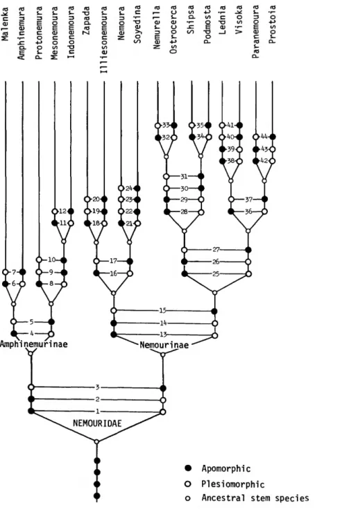
AMPHINEMURINAE, new subfamily Amphinemura is the type-genus for this sub- family, which also includes the genera Indone- moura, Malenka, Mesonemoura, and Protonemura.
The subfamily is recognized by the following char- acters:
1. Paraprocts divided into three lobes.
2. Middle and outer lobes of paraprocts usually bearing spines or prongs.
S. Epiproct elongate, generally laterally com- pressed and with ventral sclerite forming a rounded or triangular keel.
4. Cervical gills present.
5. Subgenital plate well developed in females but pregenital plate usually poorly developed.
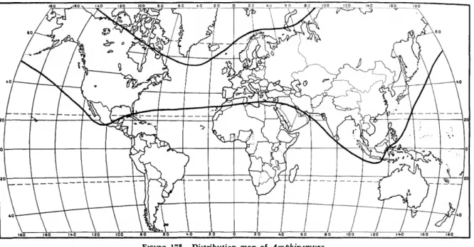
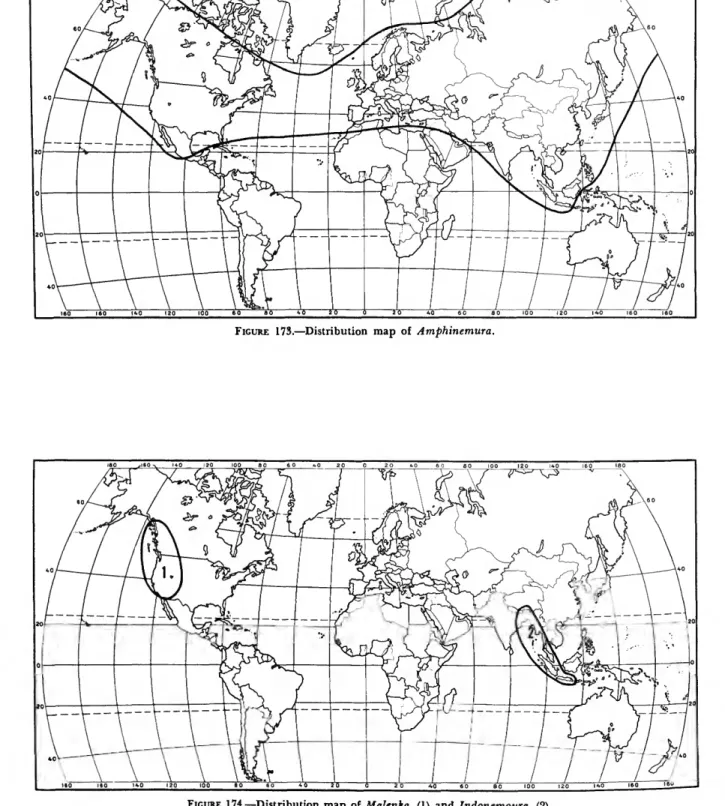
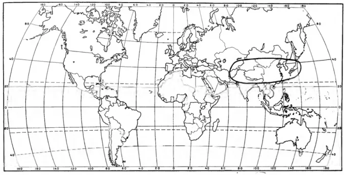
The Amphinemurinae are well represented in the Palearctic and Nearctic regions and also extend south into the Oriental region. Amphinemura is found in the Nearctic, Palearctic, and Oriental re- gions (Figure 173); Mesonemoura occurs in the Oriental and Palearctic regions (Figure 175); Pro- tonemura (Figure 176) and Indonemoura (Figure 174) are found only in the Palearctic and Oriental regions respectively; and Malenka is restricted to the western part of the Nearctic region (Figure 174).
NUMBER 211 11
Amphinemura Ris
FICURES 4-11, 140, 155, 159, 173
Nemura (Amphinemura) Ris, 1902:384. [Type-species:
Nemoura cinerea Olivier = Amphinemura sulcicollis Stephens.]
Amphinemura.—Claassen, 1940:47.
ADULT.—Length: small to medium (5-10 mm).
Wings (Figure 155): macropterous, venation typi- cal for family, clear or fumose to dark and mottled, veins dark and distinct, sometimes with light spots in cells or light bands on dark background. Gills (Figure 140): 2 branched cervical gills on each side of midline, 1 arising inside and 1 outside of lateral cervical sclerites, minimum branches 5, maximum observed 16, all branches extending to gill base.
MALE TERMINALIA.—Hypoproct (Figure 10):
broad at base, tapering to narrow apex, extending distally over inner lobes of paraprocts, tip ex- tending to base of epiproct and sometimes re- curved dorsally; vesicle present. Paraprocts (Figure 7): divided into 3 lobes; inner lobes lightly sclero- tized, naked, short and closely applied at midline, often completely hidden by hypoproct; median lobes mostly sclerotized but with membranous inner portion, bearing few to many hairs or spines, lobes large and elongate, lying alongside hypoproct and recurved dorsally parallel to epiproct; outer lobes mostly sclerotized, sometimes with small membranous portion, shape variable, often re- curved dorsally alongside cerci, usually with a few hairs or spines. Cerci (Figures 8-10): membranous, short and unmodified. Epiproct (Figures 4-6):
short and usually fairly broad in dorsal aspect and narrow with ventral projections in lateral aspect, completely recurved, bilaterally symmetrical; dor- sal sclerite, large and broad at base of epiproct, extending dorsolaterally toward apex, darkly scle- rotized lateral arms on lateral margins, variously modified, apical portion large and extending lat- erally over ventral sclerite, bearing small spines or sclerotized scales; basal sclerites, as 2 broad tri- angular patches located at basolateral margins of epiproct; ventral sclerite, heavily sclerotized, broad at base and becoming narrower toward apex, form- ing median keel-shaped ridge, apical portion in- serted between folds of dorsal sclerite and vari- ously modified, often extending to dorsal margin, sclerite bearing few to many spines which are often
very large. Tenth tergum (Figure 8) all or mostly sclerotized, forming a large flat area anterior to base of epiproct, usually bare but sometimes with spines or projections. Ninth tergum sclerotized and usually produced along distal margin, pro- duced portion bearing spines or hairs, produced area sometimes large and bizarre and can even be asymmetrical.
FEMALE TERMINALIA (Figure 11).—Seventh ster- num produced and extended at distal margin forming small pregenital plate, covering part of sternum eight, produced area lightly sclerotized.
Eighth sternum forming subgenital plate, size of plate variable but usually quite small and bifid, divided at genital opening into symmetrical scle- rotized vaginal lobes which are often quite dis- tinctive, lobes usually restricted to eighth sternum but occasionally extend over anterior margin of ninth sternum.
NYMPH.—Gills (Figure 140): 2 branched cervi- cal gills on each side of midline, 1 arising inside and 1 outside of lateral cervical sclerites, minimum branches 5, maximum observed 16, all branches originating at same place near base. Forelegs (Figure 159): femur and tibia of approximately equal length, whole leg covered with fine short hairs; femur with row of long spines along pos- terior margin and often with a few long hairs intermingled, dorsal surface with few to numerous long spines on distal half; tibia with rows of medium-sized spines along anterior and posterior surfaces, few to numerous long hairs intermingled with spines on posterior margin. Pronotum with fringe of spines along lateral margins. Abdominal terga with fringes of spines along distal margins.
Cerci with whorls of large spines at distal margins of segments, segments covered randomly with numerous small intermediate spines.
DISCUSSION.—Amphinemura is the most wide- spread genus in the family Nemouridae, ranging from the Arctic in the north to North Africa, the Sunda Archipelago and southern Mexico respec- tivly in the south. It contains 90 known species and surely many more that are unstudied. Mem- bers of the genus are easily recognized in both the nymphal and adult stages by their highly branched cervical gills (Figure 140).
It is found in a wide variety of freshwater habi- tats but is most common in cold, clear mountain streams that run throughout the year.
DISTRIBUTION AND SPECIES LIST.—Holarctic and Oriental (Europe, Asia, North Africa, North America, Orient).
amatulai Aubert 1967, Assam
apache Baumann & Gaufin 1972, Arizona arcadia (Aubert) 1956a, Greece
banksi Baumann & Gaufin 1972, Rocky Mountains bilolai Aubert 1967, Assam
bomdilai Aubert 1967, Assam
borealis (Morton) 1894, Europe, northern Asia, Mongolia chiffensis (Aubert) 1956c, Morocco
•chui (Wu) 1935, China
•claassenia (Wu) 1935, China
•claviloba (Wu) 1973, n. comb., China coreana Zwick 1973c, Korea
"cornuloba (Wu) 1973, n. comb., China
* cryptoctrcia (Wu) 1938, n. comb., China
*curvispina (Wu) 1973, n. comb., China
*decemseta Okamoto 1922, Japan delosa (Ricker) 1952, eastern United States
•dentiloba (Wu) 1973, n. comb., China dichotoma (Kawai) 1954, Japan
•falciloba (Wu) 1973, n. comb., China
•fililoba (Wu) 1973, n. comb., China
•flavicollis Klapalek 1912b, Taiwan
*flavostigma Okamoto 1922, Japan
•fleurdelia (Wu) 1949, n. comb., China
•forcipiloba (Wu) 1962, n. comb., China
•furcospinata (Wu) 1949, n. comb., China
*furcostyla (Wu) 1973, n. comb., China gressitti Kawai 1969, Viet Nam gritsayae Zhiltzova 1971, central Asia
handschini (Geijskes) 1937, Java, Sumatra, Viet Nam
*hastata (Wu) 1973, n. comb., China
•kiangsiensis (Wu) 1935, China
*lagnucula Harper 1975, Nepal
•liccnti (Wu) 1938, China
linda (Ricker) 1952, eastern and northern North America lithami Aubert 1967, Assam
*longispina (Okamoto) 1922, Japan luteipes Kimmins 1947, Himalaya, Assam
"macrolamellata (Wu) 1935, n. comb., China manipurensis Aubert 1967, Assam
*maoi (Wu) 1938, China
maracandica (McLachlan) 1875, central Asia megaloba (Kawai) 1960, Japan
mexicana Baumann 1972, Mexico minuta Kawai 1969, Borneo
mirabilis (Martynov) 1928, western Asi.i mockfordi (Ricker) 1952, Tennessee
mogollonica Baumann and Gaufin 1972, southwestern United States
•mokanshenensis (Wu) 1938, China monotuberculata (Kawai) 1956, Japan moshingi Aubert 1967, Assam
*multispina (Wu) 1973, n. comb., China
"nepalemis Harper 1975, Nepal
nigritta (Provancher) 1876, eastern North America
•nigritula Navas 1932, Formosa nongrimi Aubert 1967, Assam nowegica Tobias 1973, Norway nubila Kimmins 1950a, southern India okinawaensis Kawai 1968a, Okinawa paraluteipes Aubert 1967, Assam, Nepal
*pentagona (Okamoto) 1922, Japan
•pieli (Wu) 1938, n. comb., China pseudoluteipes Aubert 1967, Assam puebla Baumann 1972, Mexico rahungi Aubert 1967, Assam reinerti Baumann 1976, Mexico renata Kimmins 1950b, Assam
*rostroloba Wu 1962, n. comb., China
*sagittata (Okamoto) 1922, Japan schmidi (Aubert) 1959a, Pakistan
•sinensis (Wu) 1926, China sperchiana Berthelemy 1971, Greece 'spinata (Wu) 1949, n. comb., China standjussi (Ris) 1902, Europe steinmanni Zwick 1973c, Korea sulcicollis (Stephens) 1835, Europe talungdzongi Aubert 1967, Assam tetraspinosa (Kawai) 1960, Japan thienemanni (Geijskes) 1952, Java
tragula Kimmins 1950a, Turkestan, Tadzhikistan
•trassaerti (Wu) 1938, China
trialetica Zhiltzova 1957, Caucasus, Asiatic Turkey triangularis Ris 1902, Europe
tricantha (Jewett) 1958, Himalaya
•triramia (Wu) 1962, n. comb., China
*unihamata (Wu) 1973, n. comb., China varshava (Ricker) 1952, eastern United States venusta (Banks) 1911, Arizona, Mexico verrucosa Zwick 1973c, Korea
wui (Claassen) 1936, eastern North America zimmermanni Joost 1970b, Tien Shan
*zonata Okamoto 1922, Japan
Indonemoura, new genus
FIGURES 12-19, 174
TYPE-SPECIES.—Protonemura indica Kimmins, 1947:727, herein designated.
ADULT.—Length: medium to large (7-15 mm).
Wings: macropterous, venation typical for family, clear or fumose, veins dark and distinct. Gills: sin- gle short oval gills on each side of midline, located on outside of lateral cervical sclerites.
MALE TERMINALIA.—Hypoproct (Figure 18):
broad at base, tapering to narrow apex, extending to base of epiproct, often covering inner lobes-of paraprocts; vesicle present. Paraprocts (Figure 15): divided into 3 lobes; inner lobes small and lightly sclerotized, often closely affixed to large
NUMBER 211 13
median lobes; median lobes mostly sclerotized, broad at base, basal half lightly sclerotized and covered with hairs, apical half darkly sclerotized, long narrow and formed into one or more long prongs or projections, usually with a small inner membranous portion; outer lobes well developed, darkly sclerotized, long narrow and almost com- pletely encircling cerci, tip often terminating in one or more sharp prongs. Cerci (Figures 16-18):
membranous, very long and thin, extending be- yond tip of abdomen, covered with thin hairs.
Epiproct (Figures 12-14): usually long and narrow in dorsal aspect, lateral aspect narrow near base but expanded near apex, completely recurved, bi- laterally symmetrical; dorsal sclerite, large and fairly broad at base of epiproct, extending dorso- laterally toward apex, sometimes enlarged at bend, apical portion usually greatly enlarged, especially near tip; basal sclerites as 2 broad triangular patches at basolateral margins of epiproct; ventral sclerite, heavily sclerotized, broad at base and be- coming narrower toward apex, forming median keel-shaped ridge, usually with few to many spines in small rounded area near apex, tip inserted in membranous folds of dorsal sclerite and rarely with tubelike structure extending from tip. Tenth tergum (Figure 16) all or mostly sclerotized but with large membranous area under apex of epi- proct, usually forming a flat or concave plate but sometimes with 1 or 2 large upwardly directed pro- tuberances. Ninth tergum sclerotized and produced along distal margin, produced portion bearing spines or hairs, and sometimes large and bizarre in shape.
FEMALE TERMINALLY (Figure 19).—Seventh ster- num somewhat produced at distal margin, extend- ing slightly onto eighth sternum medially, pro- duced area lightly sclerotized. Eighth sternum forming subgenital plate, median area enlarged and heavily sclerotized, forming distinctive plate which covers genital opening and extends over part of vaginal lobes, shape of plate highly angu- lar and darkly sclerotized. Cerci long, thin, mem- branous, and unmodified.
NYMPH.—Unknown (included in nymphal key using adult gill characters).
DISCUSSION.—Indonemoura includes the Indica group of Aubert (1967) and two large species from the Sunda Archipelago. It probably occurs in other parts of Asia, especially Burma and Thailand. It
is similar to Mesonemoura, regarding the type of cervical gills present, but can be separated in the male by the large, highly developed and orna- mented paraprocts (Figure 15) and in the female by the large darkly sclerotized subgenital plate (Figure 19) which nearly covers the very small vaginal lobes.
The generic name Indonemoura is taken from the stem "Indo-" referring to India or the East Indies, where the genus occurs.
DISTRIBUTION AND SPECIES LIST.—Oriental (south central Asia). The following species are assigned to the genus Indonemoura:
assami (Aubert) 1967, n. comb.
dirangdzongi (Aubert) 1967, n. comb., Assam gigaoni (Aubert) 1967, n. comb., Assam indica (Kimmins) 1947, n. comb., Assam, India
jacobsoni (Klapalek) 1912a, n. comb., Bali, Java, Malaya, Sumatra
javanica (Banks) 1920, n. comb., Java kamengi (Aubert) 1967, n. comb., Assam maclachlani (Kimmins) 1950b, n. comb., Assam manipuri (Aubert) 1967, n. comb., Assam nahkui (Aubert) 1967, n. comb., Assam nyukmadongi (Aubert) 1967, n. comb., Assam quadridentata (Kimmins) 1950b, n. comb., Assam sangtii (Aubert) 1967, new comb., Assam shergaoni (Aubert) 1967, n. comb., Assam
Malenka Ricker
FIGURES 20-27, 141, 151, 158, 174
Nemoura (Malenka) Ricker, 1952:29. [Type-species: Nemoura cornuta Claasen.]
Malenka.—lilies, 1966:190.
ADULT.—Length: small to medium (5-10 mm).
Wings (Figure 151): macropterous, venation typi- cal for family, clear or slightly fumose at veins which are dark and distinct. Gills (Figure 141):
2 branched cervical gills on each side of midline, 1 arising inside and 1 outside of lateral cervical sclerites, minimum branches 5, maximum observed 8, some branches arising above gill base.
MALE TERMINALIA.—Hypoproct (Figure 26):
broad at base, tapering to narrow apex, extending distally over inner lobes of paraprocts, tip extend- ing nearly to base of epiproct; vesicle present. Para- procts (Figure 23): divided into 3 lobes; inner lobes lightly sclerotized, naked, short and closely applied to midline, often completely hidden by hypoproct; median lobes lightly sclerotized api-
cally, naked or with small hairs, base broad and membranous, apical half tapering to a narrow tip that terminates in a sclerotized point or prongs, lying alongside hypoproct and extending to or beyond tips of cerci; outer lobes sclerotized, shape variable, recurved alongside cerci, often curving in front of median lobes dorsally, nearly fused to median lobes throughout length, naked except in M. marionae (Hitchcock) which has numerous short dark spines at apex. Cerci (Figures 24-26:
with distinctive mesobasal lobes which may be membranous or heavily sclerotized, with single or bifurcate apex, main lobe of cerci membranous and unmodified. Epiproct (Figures 20-22): usually short and broad in dorsal aspect and narrow in lateral aspect, completely recurved, lightly sclero- tized, bilaterally symmetrical; dorsal sclerite, fairly large and broad at base of epiproct, extending dorsolaterally toward apex, apical portion modi- fied in structure, usually enlarged and extending over ventral sclerite laterally, covered with many tiny spines or scales; basal sderites, as 2 broad tri- angular plates located at basolateral margins of epiproct; ventral sclerite, heavily sclerotized, broadest at base, narrower toward apex, forming median keel-shaped ridge which is naked of spines, apical portion inverted between lateral folds of dorsal sclerite, extending to apex where it forms a small rounded tip. Tenth tergum (Figure 24) all or mostly sclerotized, forming a large flat sclero- tized area anterior to base of epiproct, mostly bare but often with a few spines along the anterior me- dian margin. Ninth tergum sclerotized and usually slightly produced along distal margin, produced portion bearing spines or hairs.
FEMALE TERMINALIA (Figure 27).—Seventh ster- num produced at mesoposterior margin, lightly sclerotized, bearing a nipplelike projection best seen in lateral view. Eighth sternum forming sub- genital plate which is divided at genital opening by a median notch into 2 equal sternites or vagi- nal lobes, notch often lined by heavy sclerotiza- tion, lobes often swollen or produced but do not overlap ninth sternum.
NYMPH.—Gills (Figure 141): 2 branched cervical gills on each side of midline, 1 arising inside and 1 outside of lateral cervical sclerites, minimum branches 5, maximum observed 8, some branches originating some distance from base toward apex.
Forelegs (Figure 158): femur and tibia of approxi-
mately equal length, most of leg covered with small fine hairs; femur with long hairs along pos- terior margin and bunched near distal end of dor- sal surface; tibia with rows of long spines along posterior margin, with many long hairs inter- mingled with spines, anterior margin with row of short spines. Pronotum with fringe of spines along lateral margins. Abdominal terga with spines along distal margins. Cerci with whorls of large spines at distal margins of segments, intermediate areas with randomly arranged small spines.
DISCUSSION.—Malenka is the sister genus of Amphinemura and is restricted to western North America. It is found in small streams and creeks and is also common in small springs. It usually emerges in the late summer or fall, while most other nemourid species emerge in the early spring.
It can be distinguished in the nymphal stage by its highly branched cervical gills which have some branches that do not extend to the gill base (Fig- ure 141). The male can be recognized by the presence of mesobasal lobes on the cerci (Figure 24), and the females have a nipplelike ventral projection on the seventh sternum and a distinc- tive notch on the median posterior margin of the eighth sternum (Figure 27).
DISTRIBUTION AND SPECIES LIST.—Nearctic (west- ern North America). The genus Malenka contains the following known species:
bifurcata (Claassen) 1923, northwestern United States biloba (Claassen) 1923, California
californica (Claassen) 1923, western North America coloradensis (Banks) 1897, Rocky Mountains cornuta (Claassen) 1923, northwestern North America depressa (Banks) 1898, northwestern United States flexura (Claassen) 1923, western North America marionae (Hitchcock) 1958, California
perplexa (Frison) 1936, northwestern United States tina (Ricker) 1952, western United States wenatchee (Ricker) 1965, Washington
Mesonemoura, new genus
FIGURES 28-35, 142, 161, 175
TYPE-SPECIES.—Nemoura vaillanti Navas 1922:9, herein designated.
ADULT.—Length: medium to large (7-12 mm).
Wings: macropterous, venation typical for family, clear or fumose, veins dark and distinct. Gills (Fig- ure 142): single short oval gills on each side of