A revision of the Clinid fish tribe Ophiclinini, including five new species and a definition of the family Clinidae. Revision of the clinid fish tribe Ophiclinini, including five new species, and definition of the family Clinidae. These conclusions included: (1) the synonymization of Ophiclinidae and Peronedysidae with Clinidae (also proposed by Springer, 1970), and (2) the restriction of Clinidae to include only those fishes previously classified in Ophiclinidae and Peronedy-.
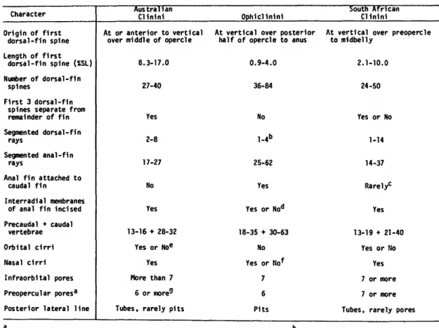
Considerable reduction in size (shorter than the orbital diameter) of the pectoral fins also occurs in the three species of the ophiclinine genus Ophiclinops, but does not occur in any other clinids. Most species of Clinidae are characterized by a hook-like process on the outer dorso-anterior edge of the cleithrum. The nature of the anal-fin attachment (and usually that of the dorsal fin) differs from that of all but one of the non-phyclinic clinids, in which there is a complete separation of the anal and caudal fins (discussion follows the exception) and the presence of a well-developed membrane joining the posterior end of the last anal-fin element to the contour of the body (if the membrane is absent, the last anal-fin ray is free from the body).
The separation of the 3 dorsal pre-fin spines from the other spines by a wide space is a common specialized condition in non-ophiclinic clinids. The specialized condition of having pits only on the posterior part of the lateral line occurs only in 2 other (Australian) clinids, Heteroclinus puellarum and H. REMARKS.—Prior to our inclusion of the Ophiclinini, 2 tribes were recognized in the Clinidae (strict sense ) : Myxodini and Clinini.
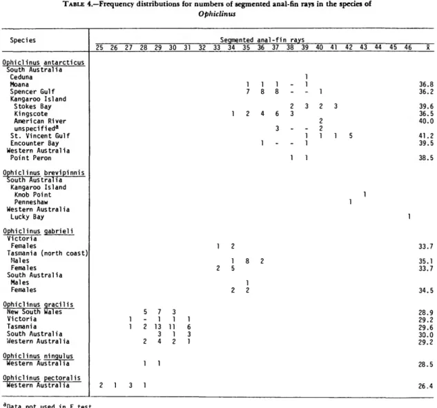
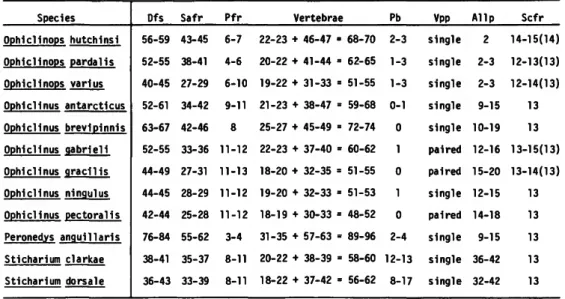
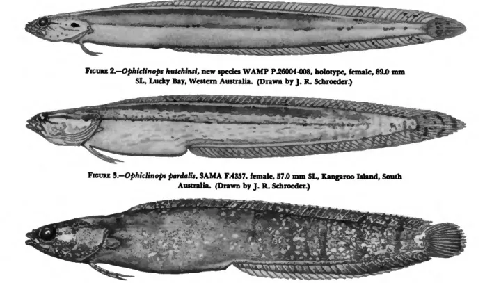
The segmental markings on fin rays in fish increase in size as the fish increases. The anal fin originates at a point below the base of the 18th spine of the dorsal fin in both specimens. Although the number of vertebrae and dorsal, anal, and caudal fin rays in 4 specimens is included in the ranges for both O.
In all three samples, the top pore of the pair is smaller than the bottom pore. The lectotype is the largest of the four syntypes and the only one with a dorsal fin vertebrae count of 60 and a vertebrae count of 21 + 46. The dorsal fin originates above the posterior i/2 of the opercle or just posterior to the opercle. in O.
One feature quickly separates most specimens of the two species: the pore position of the ventroanterior preopercular canal shows a few pores in O. The degree of arching of the lateral line is not a consistent feature, even among specimens in a single collection. COMMENTS. In the holotype, the ventroanterior-most preopercular pore position contains a few pores on the left side, but only one pore on the right side of the head.
Literature Cited
The western South Atlantic clinchfish Ribeiroclinus eigenmanni, with discussion of the intrarelationships and zoogeography of the Clinidae. Manuscripts intended for serial publication receive substantial review within their original Smithsonian museums or offices and are submitted to the Smithsonian Institution Press with the approval of the appropriate museum authority on Form SI-36. The Press's review of manuscripts and art for requirements of serial format and style, completeness and clarity of copy, and arrangement of all material, as outlined below, will, within the judgment of the Press, govern acceptance or rejection of manuscripts and art. .
The front matter (preceding the text) must contain the following: title page with only title and author and no other information, summary page with author/title/series/etc., according to the established format, table of contents with indents that reflect the main and structure of the paper. The first page of text should include the title and author at the top of the page and an unnumbered footnote at the bottom, consisting of the author's name and professional mailing address. Formal tables (numbered, with table headings, boxheads, stubs, lines) should be submitted as camera copy, but the author should contact the Series Section of the Press for editorial attention and preparation assistance before final typing of this matter.
Taxonomic keys in natural history papers should use the indented-couplet form in the zoology and paleobiology series and the multi-level indentation form in the botany series. Synonyms in the zoology and paleobiology series should use the short form (taxon, author, year:page), with a full reference at the end of the paper under "Literature Cited.". Footnotes, when few in number, regardless of whether they are annotative or bibliographical, should be written at the bottom of the text page on which the reference occurs.
If bibliographic notes are required, use the short form (author/short title/page) with the full reference in the bibliography. Use the colon system for volume/number/page citations. For alignment and order of elements, follow the format of the series for which the manuscript is intended. Legends for illustrations should not be attached to the art or included within the text, but should be submitted at the end of the manuscript - with as many legends typed, double-spaced, on one page as appropriate.
If multiple "figures" are treated as components of a single larger figure, they should be designated in small italics (underlined in copy) in the illustration, in the legend, and in text references: "Figure 9b.". If illustrations are intended to be printed separately on coated paper after the text, they should be designated as plates and any components should be lettered as in the figures: "Plate 9.b_.". Keys to all symbols in an illustration must appear on the art and not in the legend.
![Ophiclinus aethiops McCulloch and Waite, 1918:57, fig. 29 [holotype: SAMA F.481, South Australia, Kangaroo Island].](https://thumb-ap.123doks.com/thumbv2/123dok/11141726.0/19.864.112.747.185.681/ophiclinus-aethiops-mcculloch-waite-holotype-australia-kangaroo-island.webp)