ROBOTICS TRAINING ALGORITHMS FOR OPTIMIZING MOTOR LEARNING IN SPINAL CORD INJURED SUBJECTS
Thesis by Lance Lin-Lan Cai
In Partial Fulfillment of the Requirements for the Degree of Doctor of Philosophy
CALIFORNIA INSTITUTE OF TECHNOLOGY Pasadena, California
2006
(Defended on July 19th, 2006)
© 2006 Lance Lin-Lan Cai All Rights Reserved
Acknowledgement
First and foremost, I would like to thank my parents. It is their continual support and encouragement that have cultivated a life-long passion for learning in me. Even as a little child, they have never turned me away from a “why” question. In addition, they are always reminding me 跌倒不應放棄 – “never give up despite failure” and 世上無難事, 只怕有心人 – “nothing is hard in this world if you put your heart into it”. These words
are engraved in my mind and have made me not only a better scientist but a better person.
Therefore, this thesis is dedicated to my dad, 蔡星義 and my mom, 林覺華.
I would also thank my brother Franklin Cai, whom I consider to be one of my closest friends. It is his imagination and creativity that introduced me to science and engineering at an early age. I can vividly remember how we used to disassemble toys to see how they work. Instead of bugging my parents to buy me new toys, we found satisfaction from building our own toys.
I like to thank my advisors, Joel W. Burdick and V. Reggie Edgerton. Their advice and expertise has been an invaluable asset through out my graduate work. It is their innovative thinking and helpful guidance that have made my research such a success.
I am especially grateful for the generous comments and collaborations from Professor Yaser S. Abu-Mostafa, Dr. Roland Roy, Dr. Niranjala Tillakaratne and Veronica Zhong on this work. This thesis incorporates so many disciplines, it would have been nearly impossible without their expertise and facilitation.
I have also benefited from the advice, teaching, collaboration and companionship of a host of memorable individuals associated with my academic career and personal life, including but not limited to Drs. Ronald Gronsky, Luke Lee, Oliver O'Reilly, Mory
Gharib, Richard Andersen, Grégoire Courtine, Yury Gerasimenko, Igor Lavrov, Ronaldo Ichiyama, Jorge Cham, John Dabiri and Anna Hickerson; Andy Fong, Chad Otoshi, Yong-Qiang Liang, Edward Lan, Dal Mo Kang, Rebecca Rakow-Penner, Arian Forouhar, Maynor Herrera, Sharon Zdunowski, Edward Lan, Soo Kim, Rebekah Molyneux, Stephanie Enriquez and Maria Koeper; and Maggie Tang, Henry Fu, Jerald Jung, Soyen Shih, Kent Van, Amy Lin, Joseph Tan, Swati Reichmuth, Smita Pathak, Quoc Quach, Minelle David, Brian Chong, Mehnaz Khan, Johnny Fu, Wendy Cheng, Venice Calinisan, Shirley Chan, John Hu, Moty Keovisai, Kyle Yu, Emmanuell Murray, Terri Xiao and Steve Ringler.
The author will also want to recognize an incredible team of undergraduate students, Michael Andrew, Vivek Agarwal, Nikhil Daga, Armen Derian, Zac Dydek, Jee Hur, Annie Kao, Liliana Lacayo, Mary Lee, Lin Naing, Dan Popa, Traci Shiraishi, My Truong and Michael Yeranos for all of their hard works.
Lastly, and most importantly, the author will like to thank the funding sources including the Christopher Reeve Foundation, National Institute of Child Health and Human Development/National Institute of Neurological Disorders and Stroke (Grant HD44830), the National Institute of Neurological Disorders and Stroke (Grant NS16333), and the Roman Reed Spinal Cord Injury Research Funds that have made these researches possible.
Abstract
The circuitries within the spinal cord are remarkably robust and plastic. Even in the absence of supraspinal control, such circuitries are capable of generating functional movements and changing their level of excitability based on a specific combination of properceptive inputs going into the spinal cord. This has led to an increase in locomotor training, such as Body Weight Support Treadmill training (BWST) for spinal cord injured (SCI) patients. However, today, little is known about the underlying physiological mechanisms responsible for the locomotor recovery achieved with this type of rehabilitative training, and the optimal rehabilitative strategy is still unknown.
This thesis describes a mouse model to study the effect of rehabilitative training on SCI. Using this model, the effects of locomotor recovery on adult spinal mice following complete spinal cord transaction is examined. Results that indicate adult spinal mice can be robotically trained to step, and when combined with the administration of quipazine (a broad serotonin agonist), there is an interaction and retention effect. Results also demonstrate that the training paradigm can be optimized in using “Assisted-as- Needed” (AAN) training. To find the optimal AAN training parameters, a learning model is developed to test the effect of various parameters of the AAN training algorithm. Simulation results from our model show that learning is training-dependent.
In addition, the model predicts that improved motor learning can improve post-SCI by making the AAN training more adaptable.
The primary contributions of this thesis are twofold, in biology and engineering.
We develop a mouse model using novel robotic devices and controls that can be used to study SCI and other locomotor disorders in the future by taking advantage of the many
different strains of transgenic mice that are commercially available. We also further confirm that sensory integration responsible for motor control is distributed throughout the hierarchy of the neuromuscular system and can be achieved within the isolated spinal cord. Lastly, by developing a learning model, we can start looking into how variability plays a role in motor learning, the understanding of which will have profound implications in neurophysiology, machine learning and adaptive optimal controls research.
Table of Contents
Acknowledgement ... iii
Abstract... v
Table of Contents... vii
List of Figures ... x
List of Publications ... xi
CHAPTER 1: Prologue... 1
1.1 Motivation... 1
1.2 Objective ... 2
1.3 Historical Backgrounds... 2
1.4 Thesis Overview ... 3
1.4 Chapter References: ... 6
CHAPTER 2: Plasticity of functional connectivity in the adult spinal cord ... 9
2.1 Summary... 9
2.2 Introduction... 9
2.3 Some Biochemical and Electrophysiological Changes Associated with Improved Motor Performance in Spinal Animals ... 12
2.4 General Control Demands: Hierarchically Designed Networks... 16
2.5 Hierarchical Command Combined with “Smart” Sensory Control ... 21
2.6 Sensory Modulation of Motor Tasks ... 23
2.7 Implications of Synesthesia for Rehabilitation... 26
2.8 Conclusion ... 28
2.9 Chapter References: ... 30
CHAPTER 3: Experimental Background ... 38
3.1 Robotic Design... 38
3.1.1 Hardware Design ... 40
3.1.1 Software Design... 42
3.2 Animal Protocol... 44
3.2.1 Anesthetic and Surgical Procedures... 44
3.2.2 Post-Surgical Care ... 45
3.2.3 Drug Administration ... 46
3.2.4 Training Procedure... 46
3.2.5 Testing Procedure ... 48
3.2.6 Euthanization ... 48
3.3 Data Analysis... 49
3.3.1 Number of Steps ... 50
3.3.2 Step Periodicity... 51
3.3.3 Shape Consistency ... 52
3.4 Summary... 53
CHAPTER 4: Spinal Cord-Transected Mice Learn to Step in Response to Quipazine
Treatment and Robotic Training... 58
4.1 Abstract... 58
4.2 Introduction... 59
4.3 Experimental Methods ... 61
4.4 Results... 63
4.5 Conclusions and Discussions... 71
4.5.1 Quipazine facilitated spinal processing of sensory information associated with weight-bearing stepping... 71
4.5.2 Manual training did not improve stepping performance... 75
4.5.3 The robotic system significantly enhanced training effectiveness and enabled quantitative locomotion analysis... 76
4.6 Summary... 76
4.7 Chapter References: ... 78
CHAPTER 5: Implications of Assist-As-Needed Robotic Step Training after a Complete Spinal Cord Injury on Intrinsic Strategies of Motor Learning... 87
5.1 Abstract... 87
5.2 Introduction... 88
5.3 Materials and Methods... 90
5.4 Results... 94
5.5 Discussion... 96
5.5.1 All step training algorithms improved stepping beyond the level that is achieved without any step training. ... 96
5.5.2 Permitting an intrinsic network solution facilitates stepping more effectively after a SCI than imposing an extrinsic motor solution. ... 97
5.5.3 An imposed interlimb coordination pattern facilitates learning to step... 99
5.5.4 Distinction between shape consistency and quality of stepping... 100
5.6 Summary... 101
5.7 Chapter References ... 102
5.8 Chapter Appendix: Computation of the Band Vector Fields... 105
CHAPTER 6: Computational Model of Motor Learning Based on Intrinsic Variability in Stepping ... 107
6.1 Abstract... 107
6.2 Introduction... 107
6.3 Summary of the learning/training procedure ... 113
6.3.1 First Learning Rule ... 114
6.3.2 The Second Learning Rule... 116
6.3.3 Applying the Learning Model to a Biomechanical Model ... 118
6.4 Results... 119
6.4.1 First Learning Rule Simulation Results... 120
6.4.2 Second Learning Rule Simulation Results ... 121
6.4.3 Biomechanical Model Simulation Results... 121
6.5 Discussions and Conclusions... 124
6.5.1 Similar to the Animal Experiments, Learning Models Show that Motor Learning Is Dependent on the Amount of Variability the Training Allows. ... 124
6.5.2 Learning Models Allows Us to Find More Optimal Rehabilitative Training
Paradigm. ... 126
6.5.3 Principle of Neuromuscular Control and Sensorimotor Integration Resemble that of Adaptive Control Theory... 127
6.6 Summary... 128
6.7 Chapter References: ... 129
CHAPTER 7 – Concluding Remarks ... 132
7.1 Summary... 132
7.2 Future Work ... 133
List of Figures
Figure 2. 1: Force and EMG recordings from muscles of cats ... 13
Figure 2. 2: Coupling in the generation of limb movements during walking... 17
Figure 2. 3: Mean waveforms and normalize burst integral from the hindlimb ... 20
Figure 2. 4: Schematic of several features of the sensorimotor control of movement. .... 25
Figure 3. 1: Schematic of current step training system... 41
Figure 3. 2: Stepping trajectories of nontrained and trained mice... 43
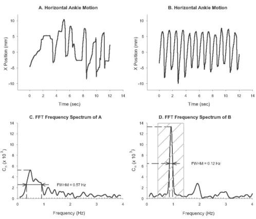
Figure 3. 3: Fast Fourier transforms analysis... 49
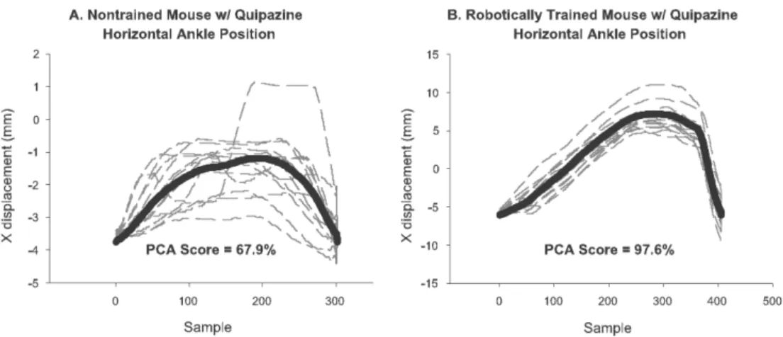
Figure 3. 4: Principle component analysis... 51
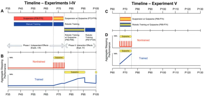
Figure 4. 1: Experimental timeline ... 61
Figure 4. 2: Summary of experimental results... 69
Figure 5. 1: AAN training paradigm I (Band). ... 91
Figure 5. 2: AAN training paradigm II (Window)... 92
Figure 5. 3: The stepping trajectories of the ankle of a neonatal mouse. ... 94
Figure 5. 4: Average number of steps performed. ... 95
Figure 5. 5: Step rhythmicity as depicted by the plot of the inverse FWHM. ... 96
Figure 5. 6: Step shape consistency as measured by PCA... 97
Figure 5. 7: Convergent velocity field of a unit circle... 105
Figure 6. 1: Block diagrams of mechanical controllers. ... 110
Figure 6. 2: Schematic of a semi-active fixed-trajectory paradigm for step training. .... 112
Figure 6. 3: Idealized one dimensional learning model...114
Figure 6. 4: A simple biomechanical model of the mouse hindlimb. ... 118
Figure 6. 5: Numerical simulation of the first learning rule. ... 119
Figure 6. 6: Numberical simulation of the second learning rule... 120
Figure 6. 7: The learning system tracking the hip angle... 122
Figure 6. 8: Application of the learning rule to the biomechanical model. ... 122
Figure 6. 9: The learning system tracking of a physiological trajectory. ... 123
Figure 6. 10: Actual ankle trajectory recordings. ... 125
Figure 6. 11: Tracking of a circular target using a variable window size... 126
List of Publications
Below is a complete list of references of publications of the author that have been included in this thesis work
1. Cai, L.L., Fong A.J., Otoshi, C.O., Liang, Y.Q., Burdick, J.W., Roy, R.R., Edgerton, V.R. “Implications of Assist-As-Needed Robotic Step Training after a Complete Spinal Cord Injury on Intrinsic Strategies of Motor Learning” J Neurosci (in review) 2. Cai, L.L., Fong, A.J., Liang, Y.Q., Edgerton, V.R., Burdick, J.W. “Assist-as-needed
Training Paradigms for Robotic Rehabilitation of Spinal Cord Injuries” Proc. Int.
Conference on Robotics and Automation 2006
3. Cai, L.L., Burdick, J.W., Fong, A.J., Courtine, G., Roy, R.R., Edgerton, V.R.,
“Plasticity of functional connectivity in the adult spinal cord” Philos. Transact. B Bio. Sci. 2005 (in press)
4. Fong, A.J., Cai, L.L., Otoshi, C.O., Reinkensmeyer, D., Roy, R.R., Burdick, J.W., Edgergon, V.R. “Spinal cord transected mice learn to step in response to quipazine administration and robotic tranining” J Neurosci, vol. 25, pp. 11738-47, 2005.
5. Cai, L.L., Fong, A.J., Otoshi, C.K., Liang, Y.Q., Cham, J.G., Zhong, H., Roy, R.R., Edgerton, V.R., Burdick, J.W. “Effects of consistency vs. variability in robotically controlled training of stepping in adult spinal mice” Proc. Int. Conference Rehab.
Robotics 2005, 9, 575-579.
6. Cai, L.L., Fong, A.J., Otoshi, C.K., Liang, Y.Q., Cham, J.G., Zhong, H., Roy, R.R., Edgerton, V.R., Burdick, J.W. “Effects of Semi-Active vs. Fixed Trajectory Robotic Training on the Stepping Ability of Adult Spinal Mice” pp. Washington, DC: Soc.
Neurosci. Abstr 2005 Program No. 396.12
7. Otoshi, C.K., Fong, A.J., Cai, L.L., Zhong, H., Roy, R.R., Tillakaratne, N.J.K., Edgerton, V.R. “5 - HT receptor distribution after spinal cord transection: effects of chronic serotonergic agonist administration and robotic training” pp. Washington, DC: Soc. Neurosci. Abstr 2005 Program No. 396.11
8. Cai, L.L., Fong, A.J., Otoshi, C.K., Liang, Y.Q., Cham, J.G., Zhong, H., Roy, R.R., Burdick, J.W., Edgerton, V.R. “Effects of varability in robotic training on the stepping ability of adult spinal mice” pp. Washington, DC: Soc. Neurosci. Abstr 2004 Program No. 418.3
9. Otoshi, C.K., Fong, A.J., Cai, L.L., Liu, C.C., Zhong, H., Roy, R.R., Tillakaratne, N.J.K., Edgerton, V.R. “Changes in 5HT-1A receptor distribution following spinal cord transaction are depending upon spinal 5HT content and peripheral sensory input in adult rats” pp. Washington, DC: Soc. Neurosci. Abstr 2004 Program No. 418.11 10. Fong, A. J., Edgerton, V. R., Cai, L. L., Otoshi, C. K., Timoszyk, W. K., Merlo, M.,
Bigbee, A. J., Zhong, H., Roy, R. R., Reinkensmeyer, D. J. & Burdick, J. W. “Effects of quipazine and robotic training on spinal mice” pp. Washington, DC: Soc Neurosci Abstr 2003 Program No. 498.20.
CHAPTER 1: Prologue 1.1 Motivation
“The frog instantly dies when the spinal cord is pierced; and previous to this it lived without head, without heart or any bowels or intestines or skin; and here therefore it would seem lies the foundation of movement and life.” – Leonardo da Vinci.
Spinal cord injury (SCI) is one of the most traumatic conditions a person will have to live through, affecting every aspect of daily life, resulting in an enormous impact from psychological and social perspective (Bedbrook 1987). Spinal cord injury has an enormous economical impact as well. As of 2005, it is estimated that a person with a paraplegic spinal cord injury person will need to spend more than $250,000 during the initial year of injury and more than $25,000 each subsequent year (SCIIN 2005). The estimate is even higher for tetraplegia patients.
Currently, there are between 250,000 and 400,000 Americans suffering from spinal cord injury and an additional 11,000 Americans are struck with spinal cord injury each year (NSCIA 2006). Many of these injuries are caused by accidents such as motor vehicle accidents, falls and sport injuries. As such, the demographic group most likely to suffer a spinal cord injury is men (~80%) between 16 and 30 years old (NSCIA 2006).
Therefore, depending on the intensity of the injury, many of these people have to live with disability, and most likely paralysis, for the greater part of their adult life. Thus, any research that can improve their mobility and motor functions will not only greatly improve their quality of life, it will have significant impacts on the general population as a whole.
1.2 Objective
As the name implies, bioengineering is an interdisciplinary field that combines biology and engineering. Because of this synergy between the physical and biological sciences, many advances have been made recently in the development and application of technology and the adaptation of new engineering discoveries to biology and medicine.
This, however, should not be the only goal of bioengineering. A less explored route of bioengineering is to use engineering technologies to further investigate and contribute to the better understanding of basic biological, physiological and pathological processes.
This has been the driving force behind this thesis work. Our objective is to develop robotic devices and control algorithms for spinal cord injury rehabilitation. In the meantime, using these devices, we want to examine the neural mechanism responsible for the plasticity observed in the isolated spinal cord, thus providing an insight into motor learning and neuromuscular control in general.
1.3 Historical Backgrounds
Spinal vertebral injury has attracted the interests of the medical and scientific community ever since the dawn of civilization, with documentations dating as far back as 2500 B.C.;
however, progress in treating and curing spinal cord injury has been slow in coming (Hughes 1988). It took more then two centuries before the central role of the spinal cord was even recognized (Lifshutz and Colohan 2004). Guy de Chauliac (1300–1368), considered by many to be the father of modern surgery, had once written, “One should not labor to cure the paralysis of spinal cord injury,” a sentiment shared by many for centuries (Walker 1967; Lifshutz and Colohan 2004). However, within the last centuries,
there has been a renewed effort in the scientific community to study spinal cord injury.
Researchers from many fields are tackling the problem of spinal cord repair with a number of different approaches. Major areas of research include: neural regeneration, sometimes called neuroengineering, where researchers try to reconnect the damage neural tissues through axon regeneration (Baitinger, Cheney et al. 1983; Herdegen, Skene et al.
1997); stem cell research, where stem cells are implanted in the injured spinal cord for regrowth (Gimenez y Ribotta, Gaviria et al. 2002; Luque and Gimenez y Ribotta 2004;
Pencalet, Serguera et al. 2006); neural stimulation and epidural stimulation, where the isolated spinal cord is stimulated electrically to elicit locomotor activities (Dimitrijevic and Dimitrijevic 2002; Gerasimenko, Avelev et al. 2003; Minassian, Jilge et al. 2004);
biochemistry and pharmacology (Rossignol and Barbeau 1993; Tillakaratne, Mouria et al.
2000); and rehabilitation (de Leon, Hodgson et al. 1998; Edgerton, Leon et al. 2001).
Perhaps some of the greatest clinical advances in the care of patients with SCI during this century have been in physical therapy and rehabilitation (Lifshutz and Colohan 2004;
Edgerton, Kim et al. 2006). This advancement resulted from our progressive understanding of spinal cord plasticity as well as neural control of locomotion post-SCI, which is reviewed in detail in the following chapter.
1.4 Thesis Overview
This thesis is comprised of peer-reviewed articles from archival journals in biological science and engineering. The author is either the lead author or co-author of these articles. The organization of this thesis is meant to highlight the interdisciplinary nature
of the topics at hand while providing a clear emphasis on the discipline-specific contributions of this work.
Chapter 2 will give an indepth review of spinal cord plasticity and neural control of locomotion post spinal cord injury (SCI), which is critical in understanding the significance of this thesis work. This review brings together perspectives from various disciplines to emphasize the importance of variability in neural plasticity even at the spinal cord level.
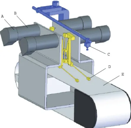
Chapter 3 consists of experimental background information including description of robotic design, surgical procedures, animal care and data analysis techniques that will be the basis of studies described in the subsequent chapters, mainly Chapter 5 and 6. The critical contributions of this chapter will be on the development of a mouse model to study SCI as well as other neuromuscular disease, and on the implantation of quantitative assessment of locomotor performance rather than relaying on frequently used qualitative methods such as the Basso, Beattie and Bresnahan (BBB) locomotor rating scale.
Chapter 4 and 5 together consist of two animal studies, the first of which is used to examine the feasibility of our animal model. Using this model, we have examined: 1) Whether adult mice with a complete spinal cord transaction can be robotically trained to step; 2) the effect of pharmacological agents such as quipazine (a broad serotonin against) has on the locomotor recovery post-SCI; 3) the combining effect of robotic training and quipazine administration. In the second animal study, we develop various forms of robotic training to see if rehabilitative training will be more optimal if the training is done in an assist-as-needed manner, thereby challenging the injured subject to use the intrinsic neural circuitries that reside within the isolated spinal cord. The results
of this study have great implications on understanding the underlying mechanism behind locomotor recovery.
Chapter 6 consists of the theoretical contribution of this thesis, which examines how optimal robotic-facilitated rehabilitative training can be achieved, giving the intrinsic properties of neuromuscular control of locomotion. It provides a learning model that captures pheromones such as “learned helplessness” and indicates how motor learning can be best achieved.
Lastly, Chapter 7 consists of concluding remarks that will discuss relevance and contribution of this thesis. In addition, it will touch upon the future direction of this research.
1.4 Chapter References:
Baitinger, C., R. Cheney, et al. (1983). "Axonally transported proteins in axon development, maintenance, and regeneration." Cold Spring Harb Symp Quant Biol 48 Pt 2: 791-802.
Bedbrook, G. M. (1987). "The development and care of spinal cord paralysis (1918 to 1986)." Paraplegia 25(3): 172-84.
de Leon, R. D., J. A. Hodgson, et al. (1998). "Locomotor capacity attributable to step training versus spontaneous recovery after spinalization in adult cats." J Neurophysiol 79(3): 1329-40.
Dimitrijevic, M. M. and M. R. Dimitrijevic (2002). "Clinical elements for the neuromuscular stimulation and functional electrical stimulation protocols in the practice of neurorehabilitation." Artif Organs 26(3): 256-9.
Edgerton, V. R., S. J. Kim, et al. (2006). "Rehabilitative therapies after spinal cord injury." J Neurotrauma 23(3-4): 560-70.
Edgerton, V. R., R. D. Leon, et al. (2001). "Retraining the injured spinal cord." J Physiol 533(Pt 1): 15-22.
Gerasimenko, Y. P., V. D. Avelev, et al. (2003). "Initiation of locomotor activity in spinal cats by epidural stimulation of the spinal cord." Neurosci Behav Physiol 33(3):
247-54.
Gimenez y Ribotta, M., M. Gaviria, et al. (2002). "Strategies for regeneration and repair in spinal cord traumatic injury." Prog Brain Res 137: 191-212.
Herdegen, T., P. Skene, et al. (1997). "The c-Jun transcription factor--bipotential mediator of neuronal death, survival and regeneration." Trends Neurosci 20(5):
227-31.
Hughes, J. T. (1988). "The Edwin Smith Surgical Papyrus: an analysis of the first case reports of spinal cord injuries." Paraplegia 26(2): 71-82.
Lifshutz, J. and A. Colohan (2004). "A brief history of therapy for traumatic spinal cord injury." Neurosurg Focus 16(1): E5.
Luque, J. M. and M. Gimenez y Ribotta (2004). "Neural stem cells and the quest for restorative neurology." Histol Histopathol 19(1): 271-80.
Minassian, K., B. Jilge, et al. (2004). "Stepping-like movements in humans with complete spinal cord injury induced by epidural stimulation of the lumbar cord:
electromyographic study of compound muscle action potentials." Spinal Cord 42(7): 401-16.
NSCIA. (2006). "National Spinal Cord Injury Association." Retrieved 31 May, 2006.
Pencalet, P., C. Serguera, et al. (2006). "Integration of genetically modified adult astrocytes into the lesioned rat spinal cord." J Neurosci Res 83(1): 61-7.
Rossignol, S. and H. Barbeau (1993). "Pharmacology of locomotion: an account of studies in spinal cats and spinal cord injured subjects." J Am Paraplegia Soc 16(4): 190-6.
SCIIN. (2005). "Spinal Cord Injury Information Network." Retrieved 20 September 2005.
Tillakaratne, N. J., M. Mouria, et al. (2000). "Increased expression of glutamate decarboxylase (GAD(67)) in feline lumbar spinal cord after complete thoracic spinal cord transection." J Neurosci Res 60(2): 219-30.
Walker, A. E. (1967). A history of neurological surgery. New York,, Hafner.
CHAPTER 2: Plasticity of functional connectivity in the adult spinal cord
Submitted on March 07, 2006 to the Philosophical Transactions of the Royal Society B:
Biological Sciences – accepted for publication 2.1 Summary
This chapter emphasizes several characteristics of the neural control of locomotion that provide opportunities for developing strategies to maximize the recovery of postural and locomotor function after a spinal cord injury (SCI). The major points of this chapter are:
1) the circuitry that controls standing and stepping is extremely malleable and reflects a continuously varying combination of neurons that are activated when executing stereotypical movements; 2) the connectivity between neurons is more accurately perceived as a functional rather than as an anatomical phenomenon; 3) the functional connectivity that controls standing and stepping reflects the physiological state of a given assembly of synapses, where the probability of these synaptic events is not deterministic;
4) rather, this probability can be modulated by other factors such as pharmacological agents, epidural stimulation, and/or motor training; and 5) the variability observed in the kinematics of consecutive steps reflects a fundamental feature of the neural control system.
2.2 Introduction
The title of this chapter may induce a myriad of perceptions, most of which will imply physiological mechanisms related to how the adaptation of neural events within the central nervous system (CNS) respond to a spinal cord injury (SCI). Clearly, after an injury of any part of the neuromuscular system, there are changes in the connectivity of
those sensorimotor circuits that generate a motor task. Changes also occur during the subsequent adaptive events that follow the injury. In this chapter, emphasis will be placed on the concept of functional rather than anatomical connectivity within the spinal cord. The term “functional connectivity” will be used to indicate that the likelihood of a given neuron being activated is dependent on its physiological state of “readiness” rather than merely on the existence of an anatomical connection. This emphasis is not to imply that changes in the actual number of synaptic connections cannot or do not occur in response to SCI. In fact, there is good evidence for the presence of such adaptations and that these changes can be associated with an improvement in motor performance capacity following a SCI (Bregman, Diener et al. 1997; Raineteau and Schwab 2001; Bregman, Coumans et al. 2002). Instead, this chapter will focus on the importance of rapid, and sometimes persistent, changes in functional connectivity between a given combinations of spatially and temporally linked sensory and motor circuits that are involved in the generation of posture and locomotion. A measure of functional connectivity, in the context of how we are using this term, is the probability of a specific set of neurons being activated for a given physiological state.
Many correlations have been drawn between anatomical connections and functional recovery post-SCI (Hase, Kawaguchi et al. 2002; Lee, Lin et al. 2004).
However, the variability in normal stepping, even under well-controlled conditions, demonstrates the versatility and complexity in the activation of the associated spinal circuitry. We propose that as the physiological states change, the continuous adaptation in functional connectivity brings about routine variability in the activation patterns during repetitive movements, such as stepping. As a result, no two steps are generated by the
same combination and sequence of neuronal activation. As the limb trajectory varies from step to step, the precise pattern of activation of the involved motor pools also must vary (Figure 2.1). This variation is reflected in the electromyographic (EMG) signals from normal (Courtine, Roy et al. 2005) and complete spinal animals (Lovely, Gregor et al. 1990; Edgerton, Roy et al. 1992) stepping on a treadmill. Thus, even within the robust size principle of recruitment of motor neurons (Burke and Edgerton 1975; Henneman and Mendell 1981), there remains a significant level of variability in the exact combination and order of motor neurons activated within a given motor pool, and certainly across synergistic motor pools (Cope and Sokoloff 1999), to generate a specific movement.
The source of this variability in stepping is undoubtedly derived from both supraspinal as well as spinal neuronal networks. It also is reasonably obvious that the variability in limb kinematics will be greater following a SCI as recovery of stepping occurs either spontaneously or as a result of motor training. After a SCI, however, the variability in stepping is significantly reduced by motor training (de Leon, Hodgson et al.
1998). We have proposed that this variability reflects the intrinsic probability of a given assembly of neurons being activated at any given time (Edgerton, Roy et al. 2001). Thus, the underlying explanation for the presence of variability in the limb kinematics during stepping under normal conditions is that whether or not an assembly of neurons is activated is not deterministic at any given instant.
After a SCI, the probability that an appropriate combination of neurons is activated in the appropriate sequence is markedly altered. During the “reorganization” of the spinal circuitry following SCI, these probabilities can become lower or higher depending, to a large degree, on the frequency with which the sensorimotor circuits
experience the specific patterns of activity. For example, the repetitive performance of a motor task, such as stepping, over a period of weeks increases the probability of completing a successful step (Lovely, Gregor et al. 1986; Barbeau and Rossignol 1987;
de Leon, Hodgson et al. 1998). It appears from the results of virtually all of the studies involving motor training after a SCI that the benefits of step training can be manifested as an increased probability of generating a successful step. At the systems level, a number of motor training-induced biochemical and electrophysiological changes in the spinal cord are associated with improved motor performance after a SCI (de Leon, Tamaki et al.
1999; Tillakaratne, Mouria et al. 2000; Tillakaratne, de Leon et al. 2002; Cote, Menard et al. 2003; Cote and Gossard 2004).
2.3 Some Biochemical and Electrophysiological Changes Associated with Improved Motor Performance in Spinal Animals
Prior research has identified a number of biochemical and physiological changes in the spinal cord after a complete thoracic spinal cord transection in response to training of a specific motor task. Many of these changes have been reviewed recently (Edgerton, Tillakaratne et al. 2004). Briefly, the biochemical changes generally reflect an up- regulation of both the glycinergic and GABAergic (Gamma-aminobutyric acid) neurotransmitter systems within the lumbosacral spinal cord. The biochemical indicators consist of an increased number of glycinergic receptors (Edgerton, Leon et al. 2001), an increased responsiveness to strychnine administration, an agent that blocks the glycinergic receptor (de Leon, Tamaki et al. 1999), an increased level of glutamic acid decarboxylase (GAD67) (Tillakaratne, Mouria et al. 2000), and improved locomotion when blocking GABAergic inhibition with biccuculine (Robinson and Goldberger 1986;
Robinson and Goldberger 1986). As importantly, however, is the observation that the increased level of inhibition in the spinal cord after a SCI can be countered by motor training (Edgerton, Leon et al. 2001; Tillakaratne, de Leon et al. 2002; Edgerton, Tillakaratne et al. 2004).
Figure 2.1 Force and EMG recordings from the soleus (SOL, top) and medial gastrocnemius (MG, bottom) muscles of a normal, intact cat and an adult spinal cat during stepping on a treadmill at 0.8 m/sec. Bold bars indicate the period of contralateral support. Compared to normal, the spinal cat exhibited a longer cycle period, a steeper decline in force beginning at mid-support, a delay in the onset of flexion at the ankle (Fa), lower peak EMG forces, and clonus in the EMG and force records of both muscles. PC, paw contact.
(taken from Lovely et al. 1990)
It appears that repetition of a specific motor task reduces the level of persistent inhibition present in the neural networks that normally generate the motor task. These effects have been demonstrated in spinal animals that have been trained to step (de Leon, Hodgson et al. 1998) or stand (de Leon, Hodgson et al. 1998). However, it remains unclear as to how specific excitatory vs. inhibitory neural pathways are modulated by repetitive use. Repetitive use of the extensor musculature may down-regulate the GAD67
associated with extensor motor neurons, and simultaneously enhance the levels of GAD67
by increasing the inhibition of flexor motor neurons (Tillakaratne, de Leon et al. 2002).
A limitation of these observations is that linking the level of excitation vs. inhibition of specific neural pathways to specific adaptations within the different neurotransmitter systems has not been possible to date. Furthermore, there has been relatively little identification of the receptor subtypes that may be associated with the observed level of behavioral performance. These data are further limited in that it is not certain whether the observed biochemical changes are simply correlated with changes in motor performance as opposed to there being a cause and effect relationship. Further studies are needed to address these issues.
Electrophysiological changes also have been observed in chronic spinal animals, and there is strong evidence that the efficacy of selected neuromotor pathways can be modified by repetitive training of a motor task. For example, there is improved coordination of motor pools controlling the hindlimb musculature following step training in spinal animals, as shown by EMG bursting patterns (Lovely, Gregor et al. 1990).
Likewise, step training greatly improves the transmission in polysynaptic excitatory group I load pathways (Cote, Menard et al. 2003) that convey locomotor drive to extensor
motor neurons, and thus could contribute to improved recovery of weight-bearing during stepping in spinal animals. The mean amplitude of excitatory postsynaptic potentials has similarly been reported to increase in response to activation of skin sensory receptors located under the paw of chronic spinal cats that have been trained to step (Cote and Gossard 2004). Recent observations also indicate that improved stepping following training in complete spinal rats correlates with the peak amplitude of the segmental excitatory post-synaptic potential and action potential afterhyperpolarization depth of those motor neurons recruited during locomotor activity (Petruska, Ichiyama et al. 2004).
Based on the results of these electrophysiological studies, it appears that in chronic spinal animals, a number of spinal neural pathways can respond specifically to step training. Accordingly, it is likely that after repetitive exposure of the spinal cord to a given motor task, significant alterations may occur in the transmission of many, if not all, sensorimotor pathways caudal to the lesion. Such task-dependent functional plasticity of the spinal motor infrastructure could, in turn, contribute to the observed decrease in the intrinsic variability in performing a motor task (de Leon, Hodgson et al. 1998), i.e., there would be an increase in the probability of activating specific motor pathways, and therefore specific functional sets of neurons, to accomplish the required task (Edgerton, Roy et al. 2001). These results suggest an improved efficacy of the interneurons that are responsible for coordinating motor pools, e.g. Ia interneurons that provide reciprocal inhibition between antagonistic motor pools. Direct measurements of decreases followed by increases in the excitability of synapses associated with Ia interneurons in response to spinal cord transection have been observed, but not the training-related changes (Valero- Cabre, Fores et al. 2004; Valero-Cabre, Tsironis et al. 2004). On the other hand,
significant increases in the excitability of lumbar monosynaptic reflexes have been reported (Thompson, Parmer et al. 1998).
Thus, it seems reasonable to hypothesize that the observed biochemical and electrophysiological adaptations of spinal animals associated with step training are not due to an induction of a specific set of synaptic events required for the acquisition of stepping ability. Rather, these adaptations result from the increased probability of the neural circuitries within the spinal cord of generating a successful step.
2.4 General Control Demands: Hierarchically Designed Networks
Several observations demonstrate that supraspinal control can be, and probably often is, relatively nonspecific. Supraspinal input can instruct the spinal cord to walk by providing a relatively nonspecific tonic input, leaving the detailed decisions of which neuronal systems have to be activated at the spinal level. Stepping can be induced in decerebrated animals via tonic stimulation of the mesencephalic locomotor region (Shik, Severin et al. 1966). Fictive locomotion can be generated via stimulation of the dorsal roots of the spinal cord (Sjostrom and Zangger 1976). Locomotion can be induced pharmacologically in complete spinal animals (Rossignol and Barbeau 1993; de Leon, Tamaki et al. 1999; Antri, Orsal et al. 2002; Orsal, Barthe et al. 2002), and chronic complete, low thoracic spinal animals can be trained to step even without any pharmacological enhancement (Lovely, Gregor et al. 1986; Barbeau and Rossignol 1987;
de Leon, Hodgson et al. 1998). Another illustration of the nonspecific nature of the signals (in this case from the periphery) that generate walking is the locomotor-like movements that can be elicited in humans in a recumbent position by applying non- specific tonic vibration to the relaxed leg musculature (Gurfinkel, Levik et al. 1998).
These widely different manipulations represent an impressive array of different techniques that change the physiological state of the spinal cord, all having a remarkably similar effect, i.e. they induce or improve stepping ability.
Humans
-40 60 40
-50 60
-60
stanc
e
swing
St
Sw
Monkeys
Foot
Thigh (deg) Shank -40
-40 60
-60 60
-60
St
Sw stance
sw
ing
A B C
60 80 100
Explained variance (%)
1POST (weeks)12
PRE 2 6
* * *
Figure 2.2: Coupling in the generation of limb movements during walking in humans and monkeys. (A) When plotted in a 3-D space, the angular oscillation of thigh, shank, and foot segment with respect to the direction of gravity, i.e. elevation angles, covaries close to a plane, both during human (A) and monkey (B) locomotion. The gait loop evolves in the counterclockwise direction. The onset of stance (St) and swing (Sw) are indicated. (C) The degree of coupling among limb movements is evaluated by applying a principal component (PC) analysis on the elevation angles of hindlimb segments (thigh, shank, foot). Mean (SD) values of the variance explained by the first PC during treadmill locomotion performed pre-lesion (PRE) and 1, 2, 6, and 12 weeks after a unilateral lesion to the thoracic dorsolateral column (POST) is shown for 3 monkeys. *, significant difference between pre- and post-lesion values. The high variance accounted for by the first PC reflects the high degree of coupling in the neuronal systems that generate the oscillation of the limbs during stepping both in intact and spinal cord-injured animals. (adapted from Courtine et al., 2005a)
The generality of the supraspinal, and even spinal, commands also is apparent from the strong interrelationships among the kinematics of multiple joints within and across limbs during locomotion in intact, as well as in complete and incomplete SCI animals (Figure 2.2). This stereotypical output implies a close link between the neuronal systems controlling each of these joints and all of the musculature associated with their dynamics. Such a high intrinsic coupling in the generation of limb oscillation also simplifies the details required by the brain to generate a complex motor task, such as stepping at a range of speeds (Bianchi, Angelini et al. 1998), grades (de la Torre and
Goldsmith 1990), and directions (Courtine and Schieppati 2004). Furthermore, this stereotypical output supports the concept of automaticity in the control of locomotion (Orlovsky and Feldman 1972). Briefly, automaticity is the ability to generate a range of motor tasks, such as stepping and standing, in response to highly predictable ensembles of sensory stimuli from the periphery and motor commands from the brain. Considerable automaticity remains within the spinal cord in the absence of supraspinal input.
One source of such automaticity is found in the organization of the spinal circuits generating the motor patterns for walking. Stimulation of such neural circuits, often referred to as central pattern generators (CPGs), produces rhythmic alternating flexor and extensor activity in several vertebrate models, e.g., lamprey eels, neonatal rats, or adult cats (Arshavsky, Deliagina et al. 1997; Grillner 2002; MacKay-Lyons 2002) that mimics locomotion. The structural organization of these neural circuitries in mammals, however, are unknown, but even within this most automated action from CPGs, automatic adjustments can be made by varying levels of hierarchical control. For example, extensor or flexor muscle activity can be altered independently during fictive locomotion without affecting the ongoing locomotor rhythm (Lafreniere-Roula & McCrea, 2005). This observation suggests that the spinal-generated rhythmical input drives multiple pattern formation modules, and that the activity of these modules can be modified by other sources, e.g. sensory or supraspinal inputs. Such hierarchical control would introduce both stereotypical and ongoing adjustments (variability) in the motor output to adjust locomotor kinematics. This hierarchal control can range from volitional circuits to extreme automaticity, i.e., CPGs. Although there is still no direct evidence for the existence of locomotor spinal circuits that display CPG properties in humans,
Dimitrijevic et al. report that non-patterned electrical stimulation of the posterior structures of the lumbar spinal cord in subjects with complete, long-term SCI can induce rhythmic, alternating stance and swing phases of the lower limbs (Dimitrijevic, Gerasimenko et al. 1998).
This predictability of a stereotypic stepping pattern may seem contradictory to the concept noted above that variability in stepping reflects an important feature of the neural control system. However, it should be recalled that spinal interneurons receive input from supraspinal, as well as from spinal, sources, and from the periphery. Nevertheless, given the apparent hierarchical organization of the neuronal systems that generate motor patterns and interpret sensory information as implied above, there are multiple combinations and levels of neuronal control systems that underlie the variability observed during stepping while simultaneously maintaining a very high probability of success from step to step (Figure 2.3).
It is worth noting that recovery of locomotion kinematics after an incomplete SCI in the monkey to levels observed pre-lesion (Figure 2.3) does not imply re-establishment of pre-lesion muscle synergies, but instead reflects novel activation patterns of interneurons and motor pools that may be possible as a result of the hierarchal features noted above (Courtine, Roy et al. 2005). It also has been found that new motor patterns underlie the learning of foot kinematics that are similar to those of non-disabled individuals during treadmill stepping following training in humans with an incomplete or complete SCI (Wernig, Muller et al. 1995; Grasso, Ivanenko et al. 2004). Such findings provide evidence that successful stepping, as defined kinematically, can be achieved
through activation of a variety of spinal motor neurons, and that there is no fixed locomotor circuitry for the generation of stepping in mammals, including primates.
A
PAW DRAG
0 25 50 75 100 Hip (deg)KneeMTPAnkle
Cycle duration (%) stance
FlexFlexFlexFlex
Post-lesion 1 wk Pre-lesion
Post-lesion 12 wks
*
Normalized Burst Integral
0.5 1 1.5
0 1 2
0 0.5 1 1.5
0.5 1 1.5
0 2 4 6
0.5 1 1.5
VL Sol
MG
FHL FDL
TA 1
POST (weeks) 12 PRE
*
*
*
* *
*
* *
B
Figure 2.3: (A) Mean (SD) waveforms of each joint angle for the hindlimb ipsilateral to the lesion side recorded during treadmill locomotion (0.45 m/s) before (Pre-lesion) and 1 and 12 wks after (Post-lesion) a unilateral interruption of the lateral corticospinal tract in the thoracic spinal cord of adult Rhesus monkeys (n = 2). The horizontal bars at the bottom indicate the mean (SD) value of the stance phase duration. (B) Mean (SD) values of EMG burst integrals for all recorded muscles. Sol, soleus; MG, medial gastrocnemius;
VL, vastus lateralis; FHL, flexor hallucis longus; EDL, extensor digitorum longus; TA, tibialis anterior.
Values are normalized to the Pre-lesion baseline (dashed lines) computed as the mean value of muscle activity during Pre-lesion locomotion. *, significant difference between pre- and post-lesion values.
(Adapted from Courtine et al. 2005b)
Besides being able to accommodate the control that can be exerted by the brain on specific interneuronal assemblies or motor pools, even more direct neural connections must be present to control specific muscle units or combinations of units (Figure 2.4). It is generally accepted, at least in primates, that there are direct projections from the corticospinal tract to spinal motor neurons (cortico-motoneuronal connections), although a small portion of the corticospinal projections actually represent a direct target to motor neurons (Lacroix, Havton et al. 2004). The cortical projection to the spinal cord can be an important source of modulation in the production of skilled locomotor movements, such as stepping over an obstacle or the precise positioning of the paw during walking (Lawrence and Kuypers 1968; Georgopoulos and Grillner 1989; Drew, Jiang et al. 2002;
Courtine, Roy et al. 2005; Courtine, Roy et al. 2005). Theoretically, corticospinal input to subsets of interneurons that control specific combinations of muscle units in specific motor pools (Drew et al., 2002 Fetz, E. E., S. I. Perlmutter, Current Op Neuro, 2000;
Lemon et al., Prog Brain Res. 2004) could allow for precise voluntary activation and frequency control of groups of motor units (Kuypers 1978).
2.5 Hierarchical Command Combined with “Smart” Sensory Control
The progression from a supraspinal motor command to the detailed control of hundreds of thousands of muscle fibers reflects a phenomenal rostral-to-caudal anatomical and physiological divergence. Whereas convergence of inputs from cortical cell populations onto motor neuron assemblies exists, there is also a remarkable divergence in how the supraspinal drive can affect various interneuronal circuits (Figure 2.4). Similarly, projections of the signals from specific sensory receptors in the periphery
to the spinal cord networks and brain are highly divergent. For example, a single muscle spindle alerts thousands of neurons within the spinal cord (and even more in the brain) that a signal related to the physical environment of that mechanoreceptor has been generated (Scott and Mendell 1976). It is inevitable that all of the sensory information from the periphery projects to networks of neurons within the spinal cord. At the same time, it is apparent that the signals from multiple receptors merge and project at some level to common, as well as unique, combinations of neurons within the spinal cord.
Every unique combination of neurons can, in turn, readily recognize complex and very specific sensory patterns that can trigger the appropriate motor responses for a given pattern of sensory information. In other words, a given pattern of sensory information provides very specific and recognizable information to the neurons that eventually generate the appropriate motor response to that given ensemble of sensory information.
To what degree this extensive divergent and convergent information from the periphery is processed and integrated within the spinal networks prior to relaying this information to the brain is unknown, but it is readily apparent that these complex patterns of sensory input provide a continuous stream of critical information for the ongoing control of specific motor responses such as stepping and standing.
These observations are not meant to imply that the neural circuits within the spinal cord segments have a lesser role than the brain circuits in controlling stepping. For example, the ability of the spinal circuitry to generate rhythmic motor patterns that mimic locomotion without any sensory input, i.e., fictive locomotion, is clear (Grillner 2003).
This fictive central pattern generation, however, cannot make any adjustments to changes in its environment and, therefore, there are no mechanisms to change stepping frequency,
to modulate the appropriate level of load bearing, or to adjust to any, even slight, perturbation. In fact, not only does the afferent input provide the spinal locomotor circuits with information related to unexpected events, but the ongoing sensory flux also contributes substantially to the activation of motor neurons, even under normal walking conditions (Pearson 2004). On the other hand, this central pattern generation, combined with its massive online sensory information processing capability, can effectively generate motor tasks such as stepping and standing without input from the brain (Edgerton, Tillakaratne et al. 2004).
2.6 Sensory Modulation of Motor Tasks
What is the evidence that the sensory information derived from the limbs during posture and locomotion represent a critical and primary influence on motor control in complete spinal animals? Several experiments demonstrate that sensory information can define most details of all postural and locomotor movements. Administration of strychnine, which blocks glycinergic inhibition, at a dose that did not generate spontaneous rhythmic motion of the hindlimbs, facilitated consistent, full weight-bearing treadmill stepping of the hindlimbs in chronic complete spinal cats that otherwise could not step (de Leon, Tamaki et al. 1999). In addition, the rate of stepping was modulated to accommodate the speed of the treadmill belt. These results demonstrate that the spinal cord was not induced to generate rhythmic activity and stepping by strychnine itself, but that strychnine changed the physiological state of the spinal neural circuits so that the sensory information could be processed and transformed with sufficient accuracy to control locomotion over a range of speeds and levels of loading. Similar observations
have been made after the administration of quipazine, a serotonergic agonist, to mice having a complete SCI (Fong, Edgerton et al. 2003; Fong, Cai et al. 2005). Combined, these results clearly demonstrate that the sensory input associated with standing and stepping generates successful and remarkably adaptive control of posture and locomotion in the absence of supraspinal input. Under these conditions, this adaptive control cannot be solely attributed to central pattern generation, i.e., repetitive cycles of flexion and extension. A very important additional feature of the neural circuitry that generates these patterns is its ability to interpret the sensory input in a manner that becomes meaningful to the success of the hindlimbs in responding to its environment.
Other observations support the importance of the interaction between CPG and sensory processing. Results similar to those described for strychnine above were observed when the dorsum of the lumbosacral spinal cord of complete mid-thoracic spinal rats (Ichiyama, Gerasimenko et al. 2005) and cats (Gerasimenko, Avelev et al.
2003; Gerasimenko, Lavrov et al. 2005) was stimulated via epidural electrodes. In this case, a tonic general stimulation of modest intensity applied to the dorsum of the spinal cord did not induce any rhythmic, step-like motion. When the hindlimbs were placed on a moving treadmill belt, however, the animals stepped at a rate consistent with the speed of the treadmill belt. Previous experiments also have demonstrated that complete spinal cats receiving tonic electrical stimulation of the dorsum of the lumbar spinal cord can step backwards when their hindlimbs are placed on a treadmill belt moving forward (Gerasimenko, Avelev et al. 2003). These data demonstrate that detailed complex signals that drive motor pools in a highly coordinated fashion can be derived from very general patterns of stimuli to the lumbosacral spinal cord. Furthermore, these experiments clearly
indicate that the sensory information provided to the spinal cord essentially defines the type of motor task that will be performed, as well as the characteristics of the motor pattern associated with the task.
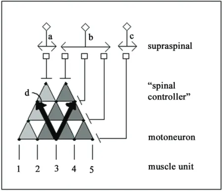
Figure 2.4: Cartoon depicting several features of the sensorimotor control of movement. The cartoon illustrates the possibility of a supraspinal control center with neurons projecting to control level neurons (“spinal controllers” of movements of differing complexities) that would project to a group of synergistic motor pools, muscles and muscle units. In cases illustrated by the projection of neuron a or neuron b, specific control of a small group of motor units might be unnecessary in executing a generalized motor program to control stepping. The numbers 1-5 denote five muscle units. The dots embedded in the triangles represent individual neurons. Activation of neuron a would result in muscle units 1-4 being recruited. Neuron b would recruit muscle units 2-5, whereas neuron c would recruit only muscle unit 5.
On the other hand, there can be even more selective control of motor units as illustrated with neuron c. At least for some muscle groups in some species, there may be direct supraspinal connections to some motor pools as well as the more generalized command neurons that exert more general control signals among motor pools. Two sets of divergent triangles are illustrated to point out the flexibility in modulating the set of muscles may be recruited for a given movement. One also can view the upright triangles in the reverse direction (see arrows projecting upward, labeled as d), symbolizing a single sensory receptor projecting rostrally and diverging markedly, thus illustrating a single sensory receptor that could provide excitatory or inhibitory input to a large number of neurons within the spinal cord. This sensory information, in turn, may further diverge or even converge to specific supraspinal locations. The diverging circuits that enable different levels of control of multiple muscles also provide a means of detailed conscious control of fine movements, while also providing mechanisms for executing more general and predictable tasks, even when they are considerably complex.
2.7 Implications of Synesthesia for Rehabilitation
Synesthesia is the merging of different modes of sensation received by the nervous system. Each mode of sensation, e.g., hearing, seeing, or touching, is generally thought to be very closely linked with specific types of sensory receptors providing information to areas of the brain that have the capability to process sound, light or mechanical perturbation, respectively. There are many examples of how sensory modes can be merged or exchanged with respect to a sensor generating a predictable perception.
For example, Cytowic (Cytowic 2002) described a subject who was born blind, but later regained vision. After his vision was restored, this individual had difficulty seeing an object without touching it with his hands. For example, when he saw a gorilla at a zoo, he could not understand its posture and movements until he had felt a statue of a gorilla.
There also are impressive examples of utilizing this synesthetic capability to rehabilitate individuals that had their vestibular system destroyed by medication. Individuals that have extreme difficulty in standing and walking as a result of a pharmacologically induced loss of vestibular function can rapidly regain excellent control by substituting the vestibular information with the output from an accelerometer placed on the head. In these cases, the electrical output from the accelerometer was passed via a wire leading to the surface of the tongue (Tyler, Danilov et al. 2003). In some way, the subject’s tongue was able to “calibrate” the accelerometer output with visual and, presumably, head, neck, trunk and lower limb proprioceptive signals, functionally merging the information so that virtually normal posture and locomotion could be sustained. Furthermore, it is interesting that once the accelerometer device was removed, the renewed control of posture and movement was maintained for days or even weeks. Essentially the brain of this patient
was able to substitute electrical signals derived from an accelerometer and “plug” this information into the circuitry that coordinates the musculature of the head, neck, trunk and lower limbs that performs postural and locomotor tasks.
With respect to the topics of the present chapter, the concept of synesthesia may be important in several ways when developing strategies to recover sensorimotor function. Perhaps the most important point from these observations on synesthesia is the degree to which the brain can reorganize its function, even in individuals without any detectable neural dysfunction. This raises the question as to what extent we can learn to substitute one sensory mode for another in facilitating recovery of function after a SCI.
Following a severe, functionally incomplete SCI, for example, to what extent can the brain reorganize itself to utilize the small number of fibers preserved that can functionally project signals to the spinal cord below the lesion? In other words, can a residual source of control from the brain be modified to control a function that is different from its normal action? A second important point that can be derived from these examples of synesthesia is that it appears that two modes of sensory information can be substituted, or at least merged, to improve sensorimotor function.
Another example of functional sensorimotor reorganization after an injury is the perception of the phantom limb, with a subject sensing the presence, and even the touch, of an arm that has been amputated (Kuiken, Dumanian et al. 2004). Subjects that have had an arm amputated can learn to control prosthetic devices using the EMG signals derived from intact muscles of the shoulder or from shoulder muscles that have been re- innervated with nerve branches that originally innervated muscles controlling hand and wrist movement. Interestingly, touching the skin overlying these re-innervated muscles
gives the subject the sensation of touching the skin overlying the hand or wrist, i.e., the region that it normally innervates.
All of these observations indicate that the potential for reorganization of sensorimotor function after a SCI has not been fully realized as a rehabilitative strategy.
Combining this potential for plasticity with new technologies, such as virtual reality and smart robotic devices, seems to be a feasible and logical direction for future efforts in enhancing recovery of sensorimotor function following a wide range of neuromotor disorders. For example, robotic devices can be used to provide more precise and versatile training to SCI subjects (see Chapter 3).
2.8 Conclusion
In the present chapter, we have emphasized the high degree of plasticity of the functional connectivity within the spinal sensorimotor infrastructure in response to an injury and/or step training. We have pointed out that the neural processes involved in the generation of standing and stepping are extremely flexible functionally, and are unlikely to be due to a hardwired, fixed neuronal architecture. Instead, there are many possible pathways and combinations of circuits that can generate movement. This view implies that locomotor-related neural circuits are better defined as the probability of a given assembly of synapses to fire appropriately to produce a successful step that simply by the presence of anatomical connectivity. Such functional flexibility in the activation of the sensorimotor circuits for stepping, in turn, would be responsible for the variability inherent to gait patterns. This variability, in turn, reflects a fundamental feature of the neural control system that should be recognized and accommodated in developing
strategies designed to enhance motor performance by motor training using robotic devices after a SCI.
2.9 Chapter References:
Anderson, J. R., R. S. a. Michalski, et al. (1983). Machine learning : an artificial intelligence approach. Los Altos, Calif., M. Kaufmann.
Antri, M., D. Orsal, et al. (2002). "Locomotor recovery in the chronic spinal rat: effects of long-term treatment with a 5-HT2 agonist." Eur J Neurosci 16(3): 467-76.
Arshavsky, Y. I., T. G. Deliagina, et al. (1997). "Pattern generation." Curr Opin Neurobiol 7(6): 781-9.
Barbeau, H., J. Fung, et al. (2002). "A review of the adaptability and recovery of locomotion after spinal cord injury." Prog Brain Res 137: 9-25.
Barbeau, H. and S. Rossignol (1987). "Recovery of locomotion after chronic spinalization in the adult cat." Brain Res 412(1): 84-95.
Barto, A. G. (1994). "Reinforcement learning control." Curr Opin Neurobiol 4(6): 888- 93.
Bianchi, L., D. Angelini, et al. (1998). "Kinematic coordination in human gait: relation to mechanical energy cost." J Neurophysiol 79(4): 2155-70.
Bregman, B. S., J. V. Coumans, et al. (2002). "Transplants and neurotrophic factors increase regeneration and recovery of function after spinal cord injury." Prog Brain Res 137: 257-73.
Bregman, B. S., P. S. Diener, et al. (1997). "Intervention strategies to enhance anatomical plasticity and recovery of function after spinal cord injury." Adv Neurol 72: 257- 75.
Burke, R. E. and V. R. Edgerton (1975). "Motor unit properties and selective involvement in movement." Exerc Sport Sci Rev 3: 31-81.
Cope, T. C. and A. J. Sokoloff (1999). "Orderly recruitment tested across muscle boundaries." Prog Brain Res 123: 177-90.
Cote, M. P. and J. P. Gossard (2004). "Step training-dependent plasticity in spinal cutaneous pathways." J Neurosci 24(50): 11317-27.
Cote, M. P., A. Menard, et al. (2003). "Spinal cats on the treadmill: changes in load pathways." J Neurosci 23(7): 2789-96.
Courtine, G., R. R. Roy, et al. (2005). "Kinematic and EMG determinants in quadrupedal locomotion of a non-human primate (Rhesus)." J Neurophysiol 93(6): 3127-45.
Courtine, G., R. R. Roy, et al. (2005). "Performance of locomotion and foot grasping following a unilateral thoracic corticospinal tract lesion in monkeys (Macaca mulatta)." Brain Epub ahead of print.
Courtine, G. and M. Schieppati (2004). "Tuning of a basic coordination pattern constructs straight-ahead and curved walking in humans." J Neurophysiol 91(4): 1524-35.
Cytowic, R. E. (2002). Synesthesia : a union of the senses. Cambridge, Mass., MIT Press.
de la Torre, J. C. and H. S. Goldsmith (1990). "Collagen-omental graft in experimental spinal cord transection." Acta Neurochir (Wien) 102(3-4): 152-63.
de Leon, R. D., J. A. Hodgson, et al. (1998). "Full weight-bearing hindlimb standing following stand training in the adult spinal cat." J Neurophysiol 80(1): 83-91.
de Leon, R. D., J. A. Hodgson, et al. (1998). "Locomotor capacity attributable to step training versus spontaneous recovery after spinalization in adult cats." J Neurophysiol 79(3): 1329-40.
de Leon, R. D., M. D. Kubasak, et al. (2002). "Using robotics to teach the spinal cord to walk." Brain Res Brain Res Rev 40(1-3): 267-73.
de Leon, R. D., D. J. Reinkensmeyer, et al. (2002). "Use of robotics in assessing the adaptive capacity of the rat lumbar spinal cord." Prog Brain Res 137: 141-9.
de Leon, R. D., H. Tamaki, et al. (1999). "Hindlimb locomotor and postural training modulates glycinergic inhibition in the spinal cord of the adult spinal cat." J Neurophysiol 82(1): 359-69.
Dimitrijevic, M. R., Y. Gerasimenko, et al. (1998). "Evidence for a spinal central pattern generator in humans." Ann N Y Acad Sci 860: 360-76.
Drew, T., W. Jiang, et al. (2002). "Contributions of the motor cortex to the control of the hindlimbs during locomotion in the cat." Brain Res Brain Res Rev 40(1-3): 178- 91.
Edgerton, V. R., R. D. Leon, et al. (2001). "Retraining the injured spinal cord." J Physiol 533(Pt 1): 15-22.
Edgerton, V. R., R. R. Roy, et al. (2001). Neural Darwinism in the mammalian spinal cord. Boston, Kluwer Academic Publishers.
Edgerton, V. R., R. R. Roy, et al. (1992). "Potential of adult mammalian lumbosacral spinal cord to execute and acquire improved locomotion in the absence of supraspinal input." J Neurotrauma 9 Suppl 1: S119-28.
Edgerton, V. R., N. J. Tillakaratne, et al. (2004). "Plasticity of the spinal neural circuitry after injury." Annu Rev Neurosci 27: 145-67.
Fong, A. J., L. L. Cai, et al. (2005). "Spinal cord-transected mice learn to step in response to quipazine treatment and robotic training." J Neurosci 25(50): 11738-47.
Fong, A. J., V. R. Edgerton, et al. (2003). "Effects of quipazine and robotic training on spinal mice, pp." Washington, DC: Soc Neurosci Abstr Program No. 498.20.