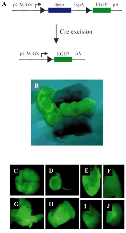
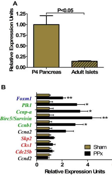
Surprisingly, global deletion of Foxm1 in mice (Foxm1-/-) does not have catastrophic results, as gross embryological morphology is intact (Figure 12B) (Krupczak-Hollis et al., 2004). Polyploid cardiomyocytes were also observed (Figure 11B) (Korver et al., 1998), likely due to endoreduplication (DNA synthesis without subsequent cytokinesis) (Wonsey and Follettie, 2005). One of the initial groups to identify FoxM1 (WIN) found that it was highly expressed in the early embryonic pancreas and to a lesser extent in the later embryonic and neonatal pancreas, with undetectable levels of expression in the whole adult pancreas from northern blot analysis (Figure 13) (Yao et al., 1997).
Cre recombinase
The insulin content of each fraction was measured using the rat insulin RIA kit (Millipore/Linco) according to the manufacturer's protocol. For paraffin embedding, tissues were fixed in 4% paraformaldehyde in PBS, pH 7.4 for 1-4 hours at 4oC, washed in PBS at 4oC, stored in 70% ethanol at 4oC, dehydrated in an ascending ethanol series at room temperature, cleared twice with xylene at room temperature, infiltrated with xylene:paraffin (1:1, v/v) and then 100% paraffin at 56oC, embedded in paraffin and sectioned at 5 µm. For frozen embedding, tissue was fixed in 4% paraformaldehyde in PBS, pH 7.4 for 1-4 hours at 4oC, washed in PBS, partially dehydrated with 30% [w/v] sucrose overnight at 4oC, embedded in Tissue-Tek Optimal Cutting Temperature (OCT) compound (Sakura) on dry ice and sectioned at 7 µm.
Detection of phospho-histone H3 required microwave antigen retrieval at 1200 W in 10 mM sodium citrate, pH 6.5 for 4 min and permeabilization with 0.2% (v/v) Triton-X-100 in PBS for 10 min. Coverslips were mounted with Permount (Fisher) for bright-field microscopy or with anti-fade mounting medium (50% glycerol [v/v] and 2% N -propyl gallate [w/v] in PBS, pH 7.4) for fluorescent microscopy. Sections were then postfixed in 4% paraformaldehyde in PBS, pH 7.4 for 20 min on ice, treated with 15 µg/ml Proteinase K in PBS for 7 min at room temperature, postfixed again in.
Plasmid maxi preparations were performed using the Plasmid Maxi Kit (Qiagen) according to manufacturer's protocol. Reactions were incubated for 5 min at room temperature and chilled on ice before use.
1 day
IPGTT
0 day
3 days 5 days 7 days1 day
IPGTT, sac
8 weeks old
BrdU in drinking waterA
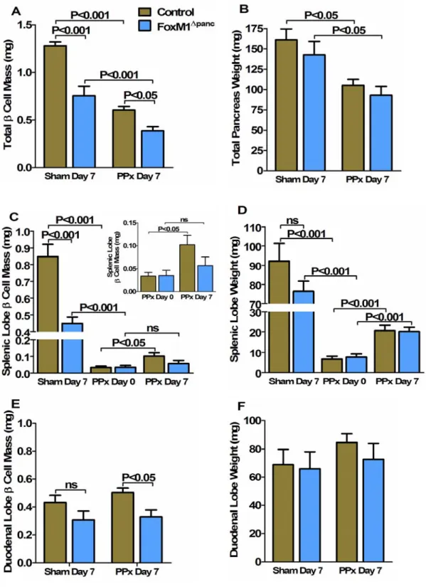
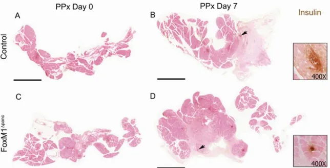
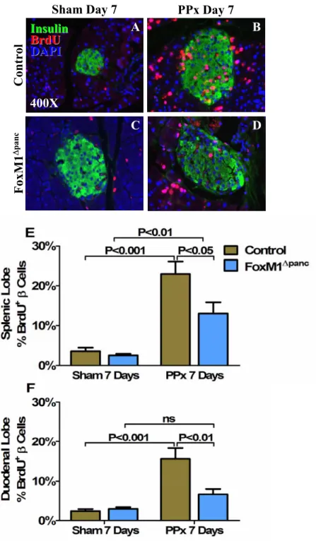
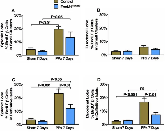
Regeneration of β-cell mass within the splenic lobe was specifically impaired in FoxM1∆panc mice. Splenic lobe regeneration was observed within 7 days of 60% PPx in Control and FoxM1∆panc mice. Furthermore, β-cell proliferation within the duodenal lobe of FoxM1∆panc mice was not significantly increased after PPx.
Summary of β-cell proliferation results for Control and FoxM1∆panc mice after Sham versus PPx. In contrast, proliferation of acinar (Figure 32B) and ductal cells (Figure 32C) was not impaired in FoxM1∆panc mice compared to control mice after PPx. BrdU incorporation into α cells (A), acinar cells (B) and duct cells (C) was measured in Control and FoxM1∆panc mice after PPx or Sham.
After a Sham operation, FoxM1∆panc mice showed increased β-cell size in both the splenic (A) and duodenal (B) pancreatic lobes. Islet density was significantly less in the splenic lobe of FoxM1∆panc mice versus controls after Sham surgery (Figure 35A), but no differences were observed in the duodenal lobe between the two genotypes (Figure 35B).
Ngn3 Ngn3 CK CK
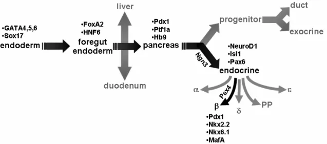
FoxM1 is highly expressed in pancreatic endocrine cells during embryogenesis and is downregulated with age (Figure 14A-C) (Zhang H et al., 2006), corresponding to reduced β-cell proliferation. Because neither β-cell mass at birth ( Zhang H et al., 2006 ) nor embryonic β-cell proliferation were altered in FoxM1∆panc mice, it is clear that β-cell neogenesis during development also does not require FoxM1. Peshavaria et al., 2006) showed increased small endocrine cell clusters 2 days after PPx, which normalized by 7 days. Compared to results published for ductal ligation (Xu et al., 2008), relatively few ductule cells were found to express Ngn3 7 days after PPx (0–3 positive cells per 200X field of view).
-Castellano et al., 2006a), β-cells express all cell cycle "brakes" studied so far, but only some "accelerators". Spontaneous mutations in the genes encoding leptin (ob/ob) (Lindstrom, 2007) or the leptin receptor (db/db) (Chen et al., 1996) also resulted in obesity and insulin resistance. In rodents, the expansion of β-cell mass in response to insulin resistance occurs primarily through increased proliferation ( Zhong et al., 2007 ) and secondarily through β-cells.
In contrast, obese humans undergo only an approximate 50% expansion of β-cell mass compared to lean individuals ( Butler et al., 2003 ). While FoxM1 has been identified as an important regulator of β-cell proliferation and post-weaning β-cell mass growth in mice under normal conditions (Zhang H et al., 2006), it was not known whether FoxM1 was similarly required for β-cell and β-cell proliferation. diet-induced obesity-stimulated mass expansion and insulin resistance.
IPITT, NMR
Insulin resistance results in an increased demand for insulin, which stimulates increased β-cell insulin-secretory function and expansion of β-cell mass. Four-week-old mice were placed on either a high-fat/carbohydrate diet or a moderate-fat chow diet for 12 weeks. Insulin secretion was measured at 0 and 30 min during the IPGTT at 8, 12, and 16 weeks of age.
Mice were sacrificed at 16 weeks of age, after 12 weeks on the respective diets, and whole pancreatic tissue was harvested.
Age (weeks)
High-fat (59%) or Chow (24%) Diet
However, these mice were crossed with the C57Bl/6 strain, and other groups have observed diet-induced β-cell mass expansion in mouse strains other than C57Bl/6 (Gupta et al., 2007). However, the majority of male FoxM1∆panc mice develop glucose intolerance and diabetes under normal conditions at six to nine weeks of age, associated with islet necrosis and fatty infiltration of the pancreas (Zhang H et al., 2006), which may could mask complications. diet-induced effects. However, an alternative approach would be to use FoxM1∆islet male mice (Foxm1flox/flox;Pdx1PB-CreERTM), where Foxm1 is specifically deleted in islet endocrine cells (mainly β-cells) after injection of tamoxifen (Zhang H et al. , 2005).
Delaying Foxm1 deletion until just before the start of the study and limiting Foxm1 deletion to islets could avoid the confounding effects of pre-existing hyperglycemia and exocrine defects (Zhang H et al., 2006), which may allow for a better analysis of the role of FoxM1 in β-cell mass expansion as response to diet-induced obesity. These results are interesting, especially because mice with a global deletion of Skp2 (Skp2-/-), a direct transcriptional target of FoxM1, also showed reduced insulin secretion in response to glucose without a reduction in insulin protein expression per β-cell mass, as also impaired β-cell proliferation in response to diet-induced obesity (Zhong et al., 2007). Several studies have shown that replication of pre-existing β cells is the primary mechanism by which expansion and regeneration of β cell mass occurs in adulthood (Dor et al., 2004; Teta et al., 2007; Meier et al., 2008). (Chapter III).
Most recently, adult duct cells were labeled by CreERTM-mediated recombination of the Rosa26 reporter locus, under the control of the carbonic anhydrase II (CAII) promoter, and labeled islet cells were observed after duct ligation (Bonner-Weir et al., 2008). However, several caveats existed in Dor et al. 2004) study that could have confounded the results.

PPx or Sham
BrdU in drinking water
1 day 0 day
Tamoxifen or
2 days 3 days 4 days1 day
5 weeks old
The original experimental design included Foxm1floc/-;Pdx1PB-CreERTM;R26R and Foxm1flox/+;Pdx1PB-CreERTM;R26R mice in order to address both goals of this study with one set of experiments. A protocol of 3 subcutaneous injections of 8 mg tamoxifen in 5-week-old female Pdx1PB-CreERTM;R26R mice resulted in ~90% recombination of R26R in β-cells by 9 weeks of age, as assessed by double labeling with anti-insulin and anti- β-galactosidase. PPx experiments were designed to account for the slow clearance of tamoxifen, with Pdx1PB-CreERTM;R26R mice subjected to PPx or Sham two weeks after tamoxifen.
Pdx1PB-CreERTM;R26R mice were injected 3 times with 8 mg tamoxifen and underwent Sham surgery or 60% PPx. In contrast, Pdx1PB-CreERTM;R26R mice injected with vehicle (corn oil) alone did not show any significant amount of nuclear-localized Cre (Figure 60B,B'). To determine if there was a time point at which Cre was no longer localized to the nucleus, time course experiments were performed in which Pdx1PB-CreERTM;R26R mice were administered 3 injections of 8 mg tamoxifen each and the mice were sacrificed at various time points hereafter for Cre labeling of pancreatic sections.
Five weeks after two injections, Pdx1PB-CreERTM;R26R mice showed similar results to those seen at three or five weeks after three injections, with the majority of β cells showing strong nuclear Cre labeling (Figure 62A,A'). Cre was primarily localized to islet nuclei 3 weeks after Pdx1PB-CreERTM mice; R26R injected 8 mg tamoxifen 3 times.
3 weeks - Sham
3 weeks
Insulin Cre DAPI
5 weeks
8 weeks
2 injections, 5 weeks
1 injection, 5 weeks
Pdx1PB-CreERTM;R26R mice after only one or two tamoxifen injections showed a reduced percentage of β-galactosidase+ β cells compared to that observed after three injections (Figure 63A,A',B,B'). Al Powers (Vanderbilt University) is planning experiments to determine the pharmacokinetic profile of tamoxifen in mice and to determine whether Pdx1PB-CreERTM;R26R islets will undergo recombination when transplanted into mice previously injected with tamoxifen. Although lineage tracing experiments were inconclusive and were postponed due to possible confounding effects of tamoxifen lifespan, data from studies performed in Foxm1flox/flox;Pdx15.5kb-Cre (FoxM1∆panc) and Foxm1flox/flox mice indicated that β-cell neogenesis occurred after 60% PPx and was not impaired in the absence of FoxM1 (see Chapter III).
Supporting experiments were designed to determine whether FoxM1 is required in non-endocrine cells to generate new β cells after 60% PPx, using Foxm1flox/-;Pdx1PB-CreERTM (FoxM1∆islet) and Foxm1flox/+;Pdx1PB-CreERTM littermates. . These mice were exposed to 60% PPx or Sham surgery and the regeneration of β cell mass was evaluated. In addition, to determine the extent of regeneration of β-cell mass in these mice, they were compared with that of FoxM1∆panc mice.
Insulin β-gal
2 injections, 5 weeks