The Role of Immune Regulation In Host and Parasite Fitness
By
Justin Thomas Critchlow
Dissertation
Submitted to the Faculty of the Graduate School of Vanderbilt University
in partial fulfillment of the requirements for the degree of
DOCTOR OF PHILOSOPHY in
Biological Sciences December, 16, 2023 Nashville, Tennessee
Approved:
Julián F. Hillyer, Ph.D.
Patrick Abbot, Ph.D.
Megan Behringer, Ph.D.
Eric Skaar, Ph.D.
Ann Tate, Ph.D.
DEDICATION
This dissertation is dedicated to my parents, for their unconditional love and encouragement. To my brother, for being the best role model I could ask for. To my wife, Jeanette, for your limitless capacity to love, listen, and encourage me. To my son, Kai, sitting here with you next to me I realize that every hardship I overcame was for you. I love you all now and always.
ACKNOWLEDGMENTS
I would like to express my deepest gratitude to my fellow Tate lab members. Over the past six years, you have been my constant companions through every challenge and triumph. Your collaborative spirit, countless laughs, willingness to help, and shared passion for our work have made even the most
daunting problems surmountable.
To Dr. Ann Tate, my advisor, thank you for your endless patience and unwavering belief in me. I will miss our weekly chats about our dogs (and science) and how excited you get when I tell you a tornado might be nearby. Over the past six years, I've not only benefited from your academic guidance but have also learned invaluable lessons in how to be a leader. You treated every person that came through your door with the same level of respect, no matter if they are a freshman at office hours or a visiting professor. I hope to do the same.
I want to thank Dr. Kurt Vandegrift, as my first scientific advisor, without your mentorship I would never have had the courage to pursue this path. Cheers.
Lastly, I extend my sincere thanks to my committee members. The opportunity to learn from and work alongside each of you has been a privilege, and your guidance has been instrumental in molding me into the scientist I am today.
To all of you, thank you for being such a positive part of this incredible journey.
TABLE OF CONTENTS
Page
DEDICATION ... ii
ACKNOWLEDGEMENTS ...iii
LIST OF TABLES ... ix
LIST OF FIGURES ... x
CHAPTER 1 Introduction ... 1
Navigating the costs and benefits of immunity when facing infectious diseases .... 1
Natural variation in immune systems vary among life stages in infection history . 4 Parasite trade-offs and evolution to host immunity... 5
Research aims and thesis structure ... 6
CHAPTER 2 The legacy of larval infection on immunological dynamics over metamorphosis ... 10
Preface ... 10
Abstract ... 11
Introduction... 12
Methods... 19
Gregarine infections ... 19
Flour beetle rearing and sample collection ... 20
Quantification of immune gene expression via RT-qpcr ... 21
Statistical analyses of gene expression ... 23
Results ... 24
The impact of gregarine infection on pupal development ... 24
Immune gene expression differs by tissue ... 24
The effect of developmental stage and parasite exposure on immune gene expression ... 27
Discussion ... 28
CHAPTER 3 Exploration of immune regulators in the Tribolium castaneum Toll and IMD pathways 33 Preface ... 33
Abstract ... 34
Introduction... 35
Materials and Methods ... 40
Beetle sources, rearing, and experimental grouping ... 40
Primer sequences ... 40
DsRNA synthesis ... 41
RNAi injections ... 41
Microbial challenge ... 42
Immune gene expression via RT-qpcr ... 43
Statistical analyses ... 43
Results ... 44
Combinatorial knockdowns of tollip with either dnr1 or pgrp-sc2 do not significantly alter constitutive AMP transcription ... 44
RNAi knockdown of dnr1 does not significantly alter AMP transcription in response to microbial challenge ... 46 DsRNA-mediated knockdown of pgrp-sc2 and tollip does not
alter humoral immune dynamics in beetle guts ... 48
10-day RNAi-mediated knockdown of skpA decouples T. castaneum Toll and IMD signaling ... 49
Discussion ... 52
SkpA functions as a pathway-dependent regulator in T. castaneum humoral immune signaling ... 53
Pgrp-sc2 silencing may influence the peak expression of IMD signaling ... 54
Pgrp-sc2 silencing does not alter gut-specific IMD signaling ... 55
Dnr1 silencing does not alter humoral immune signaling... 55
CHAPTER 4 Mapping the functional form of the trade-off between infection resistance and reproductive fitness under dysregulated immune signaling ... 57
Preface ... 57
Abstract ... 58
Introduction... 59
Results ... 63
Cactus RNAi increases constitutive and total transcriptional activation and delays decay of Toll signaling ... 63
Amplification of Toll Signaling via cactus RNAi increases total circulating hemocytes ... 66
Uninhibited Toll signaling enhances antibacterial activity ... 68
The magnitude of Toll pathway activation depends on the dose of cactus dsRNA ... 68
Elevated Toll pathway signaling increases survival to septic bacterial infection ... 70 Increased Toll pathway signaling has severe fitness related costs 71
Discussion ... 74
Small increases in Toll signaling have massive costs to reproductive potential ... 74
Dose dependent Cactus depletion regulates Toll signaling ... 76
Toll signaling increases circulating hemocytes after microbe challenge ... 77
Increasing Toll signaling amplifies damage to host health ... 77
Conclusion ... 79
Materials and Methods ... 80
Beetle rearing and experimental groups ... 80
DsRNA synthesis ... 80
RNAi treatments ... 81
Microbial infections ... 82
Immune gene expression and Bt load quantification via RT-qpcr ... 83
Hemocyte proliferation quantification ... 84
Antibacterial activity ... 85
Host survival experiments... 86
Resistance to live Bt infection ... 87
Female reproductive output ... 87
Body mass and fat body dissections ... 88
Gut/intestinal integrity quantification (Smurf assay) ... 88
CHAPTER 5 Efficient methods for experimental evolution of bacillus thuringiensis isolated against variable immune defenses in flour beetles ... 90
Preface ... 90
Abstract ... 91
Introduction... 92
Methods ... 94
Representative Results ... 96
Serial passaged strains evolve lower virulence to their passaged population ... 97
Serial passaging in a low-susceptibility host increases in vitro lag time……. ... 100
Discussion ... 102
FUTURE DIRECTIONS ... 104
REFERENCES ... 106
APPENDIX ... 121
LIST OF TABLES
Table Page
2.1 The interaction of metamorphosis and immune function across holometabolous insect orders. ... 15 2.2 Primers used to assay immune gene expression in T. castaneum. ... 22 2.3 Summary of statistical results for the impact of stage, larval parasite exposure,
or their interaction on immune gene expression in the gut and whole body. ... 27
LIST OF FIGURES
Figure Page
2.1 The proposed functional roles of T. castaneum immune genes quantified in
chapter II. ... 19
2.2 The influence of tissue type and gregarine parasite exposure on immune gene expression across developmental stages of the flour beetle T. castaneum. ... 25
2.3 Gene expression correlations suggest partial decoupling of immune genes between tissues and among life stages. ... 26
3.1 Simplified view of Toll and IMD signaling parameters. ... 37
3.2 DsRNA-mediated knockdown of dnr1, pgrp-sc2, and tollip in combination does not constitutively increase cec-2 nor def-2 expression. ... 46
3.3 DsRNA-mediated knockdown of dnr1 results no change in Toll nor IMD signaling. ... 47
3.4 DsRNA-mediated knockdown of tollip and pgrp-sc2 results no change in Toll nor IMD signaling in beetle guts... 49
3.5 10-day incubation after dsRNA injection significantly silences gene expression for dnr1, skpA, and pgrp-sc2. ... 51
3.6 DsRNA-mediated knockdown of skpA significantly increases IMD signaling while decreasing Toll signaling. ... 52
4.1 Simplified overview of the flour beetle Toll signaling pathway. ... 61
4.2 DsRNA-mediated knockdown of cactus results in increased Toll signaling ... 65
4.3 Toll signaling increases functional metrics of cellular immunity and antibacterial activity ... 67
4.4 Quantitative knockdown of cactus transcripts benefits resistance and survival during infection. ... 69
4.5 The costs of immune over-activation to fitness-associated traits. ... 72
5.1 Bt killing rate after serial passaging. ... 99
5.2 Shifts in Bt growth parameters after serial passaging. ... 101
CHAPTER 1
Introduction
Navigating the costs and benefits of immunity under the threat of infectious diseases
Infectious diseases are a pervasive threat to the well-being and survival of human, animal, and plant populations. In humans, diseases such as tuberculosis (TB) continue to be a major driver of morbidity and mortality worldwide (Chakaya et al., 2021). Emergence of infectious diseases to new susceptible populations can even endanger and destabilize entire species, such as white-nose syndrome, which is responsible for killing over 5 million bats in Eastern USA and Canada (C. L. Frank et al., 2014). Similarly, Rust fungi represent one of the most serious threats to agriculture and wood production, causing extensive damage to plants and threatening food security (Lorrain et al., 2019).
Given these widespread threats, the heavy burden of infectious diseases necessitates substantial efforts in hosts to control infections.
In response to these pervasive threats, hosts have fortunately evolved immune systems to recognize and expel foreign invaders. Site-specific defenses, such as barriers, act as the first line of defense by preventing colonization, which is complemented by wound healing mechanisms like blood clotting and resistance mechanisms triggered by the recognition of foreign invaders. Subsequently,these early defenses signal for immune cells and signaling cascades that induce the expression of infection- specific immune factors (Hamilton et al., 2008). In the context of TB, for example, cilia and mucus in the lung help trap TB to prevent colonization, apoptosis mechanisms help eliminate infected cells, and the activation of macrophages contain the bacteria by initiating the production of cytokines and
chemokinesthat draw other additional immune cells to the site of infection (de Martino et al., 2019;
Guirado et al., 2013). While indispensable for eliminating parasites and protecting host health, immune
defenses require substantial resources and can themselves inflict significant damage to self, leading to immunopathology and autoimmunity (Graham et al., 2021).
Allocating resources to immunity inherently requires the diversion of energy from other vital processes like development or reproduction (Schwenke et al., 2016). In flax plants with the L6 resistance mutations against Rust fungi, for example, energy is diverted away from growth and reproduction before there is even an infection (Howles et al., 2005). Constitutive defenses like these prevent infections from colonizing and replicating, reducing the impacts of parasite-induced pathology and curbing parasite transmission (Boots & Best, 2018). Even in the absence of infections, however, constitutive defenses require the allocation of precious resources. In contrast, inducible defenses limit immunological costs absent infection but risk being overwhelmed by fast-growing parasites if the response is not fast and strong enough (Hamilton et al., 2008). Yet, failure to dampen an induced immune response can have similar dire consequences that threaten the health and lifespan of a host (Duneau et al., 2017). For example, it is crucial for macrophages to recognize and respond to TB infection, but their increased sensitivity also increases the risk of chronic inflammation leading to rheumatoid arthritis (Buscher et al., 2017). To prevent such runaway immunological costs, hosts utilize multiple layers of of control (S. A.
Frank & Schmid-Hempel, 2019; Luecke et al., 2021).
The layers of regulation on inducible immune signaling through the insect Toll and IMD pathways provide particularly well documented case studies of the costs and benefits of controlling the flux of immune signaling after infection.(Mitchell et al., 2016). For the Toll pathway, circulating immune cells recognize a microbial invasion by identifying microbe-associated molecular patterns (MAMPs) like Lys-type peptidoglycan. This recognition initiates peptidase cascades to activate the Toll receptor ligand SPZ to move the signal through the cell membrane (Arnot et al., 2010). SPZ-ligand binding recruits intracellular adaptors to form the Myddosome signaling complex (Balka & De Nardo,
2019). Signaling kinases then act as regulatory hubs that manage the initiation, amplification, and termination of downstream effectors through phosphorylation (Luecke et al., 2021). This
phosphorylation unlocks the transcription factors Dif and Dorsal (Engström, 1999; Whalen & Steward, 1993), which facilitates their translocation into the nucleus for transcription. To regulate this pathway, some insects use extracellular serine protease inhibitors (serpins) to inhibit peptidase cascades (Gulley et al., 2013; Ligoxygakis et al., 2002). As the signal passes through the cell membrane, intracellular
negative regulators, like Tollip in mammals, dampen the signal by inhibiting signaling complexes (P. H.
Wang et al., 2013). As the signal continues, the protein Cactus binds to the transcription factors (TFs) Dif and Dorsal to prevent them from moving to the nucleus to induce AMP transcription (AMPs) (Roth et al., 1991; Whalen & Steward, 1993).
To respond to the vast diversity of parasitic threats while mitigating fitness costs, immune systems can modify their regulatory elements at each level of the signal transduction cascade to create unique immune phenotypes tailored to specific threats (Luecke et al., 2021). By altering the localization and abundance of receptors, hosts can finely tune the rate of transmembrane signaling (Chen et al., 2017; Roberts et al., 2017). Additionally, variations in the characteristics of signaling complexes, such as the speed of Myddosome scaffolding and the half-life of the complex, offer another layer of
modulation (Luecke et al., 2021). The signaling process is further refined through the addition or removal of ubiquitin chains to adaptors, which controls the speed of scaffolding and the proteasomal degradation of various immune proteins, significantly impacting signal propagation (F. Wang & Xia, 2018). Altering the activity of phosphorylation allows hosts to adjust the speed and strength of the signal (Witzel et al., 2012). The culmination of these modifications then dictate the activation dynamics of transcription factors and post-transcriptional modifiers (Hayden & Ghosh, 2004). This malleable approach enables immune responses to produce a response appropriate for the specific parasitic attack while
balancing the costs of other fitness traits. While the field has made significant strides in identifying these regulatory levers that modulate components of immune responses, there remains a substantial gap in understanding how these components naturally vary across individuals, populations, and species.
Furthermore, the exploration of how shifts in fitness landscapes exert selective pressures on these immune mechanisms, and the identification of the evolutionary constraints that govern their adaptation, are areas ripe for further investigation.
Natural variation in immune systems vary among life stages in infection history
Constraints from life history trade-offs and early life exposure to parasites can give rise to a wide variety of phenotypes. As organisms age, their interactions with microbes shape the maturation and specialization of their immune systems. For instance, children recovering from severe Respiratory Syncytial Virus (RSV) infection undergo long-term alterations in microbiome composition, metabolism, and dendritic cell transcription and epigenetics, priming the immune system for secondary infections and increasing the risk of pulmonary diseases like asthma (Malinczak et al., 2020). Alternatively, early life exposure to microbes can train the immune system, allowing it to tolerate subsequent infections and thereby reduce the risk of destructive immunopathology (Metcalf et al., 2022). As the organism matures, life history traits like growth, development, and reproduction impose new energetic and physiological demands, leading to age-specific limitations on immune system investments (Tate & Graham, 2015a). In the case of field crickets (Gryllus campestris), an induced immune response can lead to a reduction in body condition and a decrease in sexual signaling, affecting mate choice and reducing male cricket fitness (Jacot et al., 2004). Given this trade-off, crickets that are too sensitive to avirulent, ubiquitous infections could be less reproductively successful. Recognizing why and how immune systems evolve and adapt over an organism's life course will enable more accurate predictions and interventions, especially in populations with a diverse age structure.
Parasite trade-offs and adaptation to host immunity
In 2016, 8.5 million people died from parasitic infections (Ritchie & Roser, 2018). While
microbial harm to hosts might seem counterproductive, the consensus of evolutionary theory connects the harm a parasite inflicts to its ability to transmit to a new host (Anderson & May, 1986). Since parasites need host resources to replicate and transmit, evolving quicker growth rates requires increased host exploitation resulting in higher virulence (α), represented as host mortality from infection in the following virulence-transmission trade-off model: 𝑅0 = 𝛽(𝛼)𝑁
μ + α + ν(α) (Anderson & May, n.d.; Day, 2003). Increasing host exploitation leads to increased transmission potential (β) and, consequently, increased fitness (R0), or the number of new infections from a single infection (Alizon et al., 2009; Anderson & May, 1986). The term virulence can also describe the infectiousness of a parasite (Jaenike, 1996) or the
presence of specific factors that cause pathology (Dussurget et al., 2004), but neither of these definitions capture the effect of host damage on parasite fitness.An increase in exploitation is also theorized to increase the host’s immune response to clear infection (ν) and the mortality from infection (α) both of which shorten the infection transmission period. This produces a trade-off where increasing host exploitation increases parasite density, but it also decreases the time allowed for transmission, thus an intermediate level of virulence is predicted to maximize parasite fitness.
Trade-offs between parasite traits can also manifest from energetic constraints like
overproduction of costly virulence factors. For example, Salmonella typhimurium expresses its type III secretion system, ttss-1, to induce host inflammation to outcompete resident microbiota and prevent clearance from the immune response so it can successfully infect epithelial cells and achieve
transmission (Sturm et al., 2011); however, this mechanism is costly and reduces its growth rate
allowing for competition from faster growing strains. S. typhimurium clones overcome this trade-off by
dividing labor among genetically identical cells; a subset of bacteria express ttss-1 while a different subset focus exclusively on growth, preventing the establishment of coinfecting strains (Diard et al., 2013). Task specialization is only one of many strategies (e.g. quorum-sensing, nutrient sensing-
dependent modules, or virulence factor production) that parasites take to maximize their fitness (Diard &
Hardt, 2017; Rumbaugh et al., 2009), and different strategies could have different payoffs at each step of the infection cycle, from colonization to replication and through transmission.
The host immune response is a key contributor to variation in the microenvironments that the parasite experiences over its life cycle, contributing to variation in selection pressures at each stage of the infection process (Hawley & Altizer, 2011; Long & Boots, 2011). Immune mechanisms that clear infection before the parasite can reach its transmission phase will disproportionally influence parasite fitness, which is predicted to select for increased virulence (S. A. Frank & Schmid-Hempel, 2008; Grenfell et al., 2004). However, constraints on parasite evolution can create trade-offs between traits. For
example, when Staphylococcus aureus is experimentally evolved against two AMPs, it mutates in either the pmt or nsa operons to resist the AMPs (Makarova et al., 2018). This resistance is costly though, resulting in longer in vitro lag phases. Despite the many examples of parasites evolving to overcome immune responses, our understanding of the influence of individual immune mechanisms and strategies on parasite fitness and evolution remains scarce (Armitage et al., 2022). When researchers infected Tenebrio molitor with the evolved S. aureus strains, they observed no fitness benefit for S. aureus, even though the AMPs are essential for T. molitor to survive S. aureus infection. An ideal system to study how parasites overcome immune responses would allow researchers to control individual mechanisms of the host immune response and evolve the parasites in these different immune environments in vivo.
Research aims and thesis structure
In our ever-changing world, the landscape of natural infections is continually shifting, with varying selection pressures on both hosts and parasites. Given that climate change is likely to
systematically alter ecological conditions and the resulting selection pressures on extant species as their range expands and changes, integrating context into the mechanistic study of immune systems becomes imperative to enhance our understanding and prediction of the factors influencing infectious disease outcomes. My dissertation adds to our understanding of how life-stage and infection life-history can influence immune phenotypes. Additionally, it uncovers examples of regulators of immune signaling in the model system T. castaneum. The dissertation then dives into a key T. castaneum negative regulator to reveal the fitness-related benefits and costs of failing to properly regulate an immune signaling pathway. Finally, my work outlines an in vivo serial passaging protocol that isolates the interaction between a pathogenic bacterium and inducible immune defenses to understand how varying immune environments influence parasite evolution and disease outcomes.
In chapter 2, we investigate the long-term impact on the T. castaneum immune system from parasite exposure as larva. We expose beetles to a protozoan parasite that inhabits the midgut of the larval and adult life stages, despite clearance during metamorphosis. The study then measures changes in immune gene expression across larval, pupal, and adult life-stages in the gut and whole body. Our study finds that larval exposure to the benign parasite casts a shadow over immune gene expression in the larval gut and whole body that continues through metamorphosis and into adulthood. This study
indicates that early life infection experiences can have long-lasting impacts on the maturation of immune systems, providing a source of natural immunological variation and revealing the variation in parasite selection pressures over ontogeny.
In chapter 3 I aim to uncover the mechanisms that negatively regulate the flour beetle immune response. Regulatory immune genes are known to heavily influence basal, max expression, and the
decay of an immune response, which can influence host-parasite disease outcomes. Yet, the regulatory architecture and the costs and benefits of having layered regulators is not well described despite its importance for understanding immune system evolution. Here we lay the groundwork for revealing those costs and benefits in the model organism T. castaneum while also prodding the extent to which these regulators are conserved across taxa, thus revealing more about the potential selection pressures acting on immunity. In this study I target the orthologous Drosophila negative regulators pgrp-sc2 (Guo et al., 2014), dnr1 (Guntermann et al., 2009), and skpA (Khush et al., 2002) as well as the mammalian and weevil negative regulator tollip (Anselme et al., 2008; Zhang & Ghosh, 2002a). My study found potential changes in peak AMP expression when silencing pgrp-sc2 for 10 days. The chapter also indicates that skpA may play a dual role as a negative regulator of the IMD pathway but a positive regulator of the Toll pathway. This highlights the imperative for broader investigations into non-model insects, as it can reveal diverse mechanisms and tactics employed to balance the costs associated with immune responses.
In chapter 4 we take advantage of the unique RNAi system characteristics in flour beetles to alter the expression of cactus, a key negative regulator of Toll pathway signaling, thereby modulating the activation level of the Toll pathway in the red flour beetle. Our research reveals that while
intensifying immune pathway activation bolsters immune responses and survival against bacterial infections, it imposes a notably severe penalty on female egg production, gut health, body mass, and lifespan. This insight highlights the substantial health and reproductive implications that can arise from even minor alterations in negative regulation and immune pathway activation.
Finally in Chapter 5, I introduce and implement a serial passaging protocol designed to focus on the interplay between immunity and the microbial parasite, Bacillus thuringiensis (Bt), in T. castaneum.
Our results indicate that passaging Bt through populations with varying susceptibility to infection can
alter Bt's ability to kill its host. This protocol provides a framework that distinguishes early resistance mechanisms from downstream inducible immunity, paving the way for studies to explore how immune responses shape the evolutionary trajectories of parasites.
Altogether, my dissertation delves into the consequences that parasite exposure history and variation in regulatory immune genes have on host fitness and disease outcomes. The methodologies and insights presented pave the way for a deeper understanding of immune system evolution, bridging crucial gaps in our knowledge of immune signaling network dynamics and host-parasite interactions.
The dissertation then detailspotential avenues for expanding upon the current work, emphasizing its implications for refining intervention strategies, improving theoretical models, and ultimately charting the nuanced relationship between immune investment and fitness.
CHAPTER 2
The legacy of larval infection on immunological dynamics over metamorphosis
Preface
This chapter establishes that metamorphosis partially decouples immune gene expression covariance among tissues and life stages. It also demonstrates that early-life exposure to a benign gut protozoan parasite affects host immune gene expression during the larval stage, which continues to influence immune phenotypes through metamorphosis. This finding is significant since it reveals that while metamorphosis can decouple immune gene coregulation, a seemingly harmless protozoan infection can overcome this decoupling to leave a lasting imprint on the insect's immune response. My advisor, Dr. Ann Thomas Tate, conceptualized and obtained funding for this study. Adriana Norris and I conducted the experiments and processed the samples. All authors analyzed the data and wrote the manuscript. All authors approved the final version of this manuscript. This work was supported by the National Institute of General Medical Sciences at the National Institutes of Health (grant number R35GM138007).
This chapter is adapted from “The legacy of larval infection on immunological dynamics over metamorphosis”, published in 2019 in the journal Philosophical Transactions of the Royal Society B (374) and has been reproduced with the permission of the publisher and my co-authors, Adriana Norris and Dr. Ann Thomas Tate.
Abstract
Insect metamorphosis promotes the exploration of different ecological niches, as well as exposure to different parasites, across life stages. Adaptation should favor immune responses that are tailored to specific microbial threats, with the potential for metamorphosis to decouple the underlying genetic or physiological basis of immune responses in each stage. However, we do not have a good understanding of how early-life exposure to parasites influences immune responses in subsequent life stages. Is there an developmental legacy of larval infection in holometabolous insect hosts? To address this question, we exposed flour beetle (Tribolium castaneum) larvae to a protozoan parasite that inhabits the midgut of larvae and adults despite clearance during metamorphosis. We quantified the expression of relevant immune genes in the gut and whole body of exposed and unexposed individuals during the larval, pupal, and adult stages. Our results suggest that parasite exposure induces the differential expression of several immune genes in the larval stage that continues into adulthood. We also demonstrate that immune gene expression covariance is partially decoupled among tissues and life stages. These results suggest that larval infection can leave a lasting imprint on immune phenotypes, with implications for the evolution of metamorphosis and immune systems.
Introduction
Few factors have a greater impact on the outcome of an interaction between host and parasite, or the spread of disease in a host population, than the age and stage of the host. As hosts age, cumulative exposure to microbes shapes the maturation and polarization of their immune systems. Life history priorities shift from growth to reproduction, inducing alterations in behavior, food source, and even ecological niche (Urban et al., 2013). As a result, hosts experience dynamic changes over their ontogeny in both exposure to parasites and susceptibility to infection once exposed (Tate & Graham, 2015b).
The consequences of these ontogenetic dynamics can be observed across broad swaths of the tree of life.
In plants, for example, gibberellin hormones that promote plant growth also inhibit signals related to defense against predators and parasites. At the same time, signaling from the salicylic acid pathway, which is involved in the response to biotrophic pathogens, inhibits the action of the growth hormones (Karasov et al., 2017). As a result, growing plants are susceptible to different pathogens than mature stages, and infection can influence the growth trajectories of their plant hosts (Navarro et al., 2008). In humans, lack of early-life exposure to beneficial microbes and other environmental antigen can set the stage for chronic inflammation, allergy, and other forms of immunopathology (Simon et al., 2015;
Yassour et al., 2016). From an evolutionary perspective, the risk of immunopathology in early life is predicted to favor decreased immunological sensitivity to infection later in life (Metcalf et al., 2017). In all of these examples host ontogeny is a fairly continuous process, punctuated by hormonal signals that encourage flowering or the onset of puberty but otherwise keep major organs and physiological
structures intact. In animals that undergo metamorphosis, however, developmental continuity is swapped for discrete stages characterized by transition periods of dramatic physiological restructuring that alter the calculus of host-parasite interactions.
During metamorphosis, tadpoles become frogs and caterpillars become butterflies, allowing
hosts to exploit disparate resources and environments that individually maximize particular, stage- associated traits like growth or reproduction (Haldane, 1932). Metamorphosis is not a requirement for stage-specific niche differentiation; even within insects, dragonflies and mosquitoes both have an aquatic juvenile stage and a terrestrial adult stage, but dragonflies undergo relatively continuous maturation from instar to instar while the holometabolous mosquitoes undergo pupation prior to adulthood. Why bother with metamorphosis, then? After all, the pupal stage can be a liability as it is generally sessile, poorly defended, and unable to acquire resources to fuel its energetic needs. The adaptive decoupling hypothesis suggests that the pupal stage might be the price paid for immature and mature developmental modules that can respond relatively independently to evolutionary pressures (Moran, 1994), allowing organisms to simultaneously maximize performance in multiple life stages.
The re-invention of the midgut during complete metamorphosis is a particularly potent example of adaptive flexibility achieved by decoupling one life stage from another. In most insects, the midgut comprises epithelial cells, goblet cells, and stem cells (Hakim et al., 2010; Parthasarathy & Palli, 2008;
Royet, 2011). The ratio and renewal rates of these cell types differ extensively from one life stage to another, and vary dynamically even within life stages. For example, as larvae grow larger and molt, the stem cells of the midgut undergo proliferation and differentiate into new, polyploid epithelial cells and goblet cells (Parthasarathy & Palli, 2008). This renewal process is also crucial in the host response to bacterial toxins and viruses that rely on the invasion of epithelial cells to colonize the host (Loeb et al., 2001). As insects transition to the pupal stage, however, the old somatic cells are excised into the lumen to form the yellow body, which undergoes apoptosis and autophagy to recycle the nutrients before being evacuated during eclosion of the new adult (Hakim et al., 2010). In the midgut of a new pupa only the intestinal stem cells remain, imaginal structures that proliferate and differentiate into the epithelial cells that will eventually compose the adult gut (Parthasarathy & Palli, 2008). Consistent with the adaptive
decoupling hypothesis, the relative morphologies of larval and adult epithelial cells reflect the relative feeding ecologies of each life stage. For example, in fruit flies, the polyploid epithelial cells of larvae facilitate the rapid acquisition and processing of nutrients from complex food media, while the smaller, diploid nuclei of adult midgut epithelial cells reflect the narrower breadth of adult food sources (Royet, 2011). On the other hand, the larval and adult stages of the flour beetle Tribolium castaneum both feed on the same resource (Parthasarathy & Palli, 2008), and both contain midgut epithelial cells that share a common, polyploid morphology.
Midgut remodeling during insect metamorphosis can exert complex effects on the persistence of parasites and other microbes. Protozoan trophozoites that remain embedded in the flour beetle (T.
confusum) gut when a larva enters metamorphosis, for example, are evacuated with the yellow body (THOMAS & RUDOLF, 2010), allowing the adult to eclose without a parasite burden. On the other hand, the elimination of the gut epithelia could also eliminate beneficial microiota, allowing any remaining opportunistic pathogens to exploit the pupa or colonize the new adult gut. Indeed Galleria mellonella moth pupae cooperate with a beneficial microbe (Enterococcus mundtii) to exclude pathogenic Serratia strains during metamorphosis (Johnston & Rolff, 2015). Knocking down host immune gene expression or preventing the E. mundtii strain from producing bacteriocins allowed Serratia to dominate, at a cost to pupal survival. Furthermore, the cessation of resource acquisition during pupal gut remodeling can render larvally-acquired infections hazardous during metamorphosis. The microsporidian parasite Nosema whiteii kills its flour beetle host during the pupal stage after manipulating the host into an extended larval stage during which the parasite converts acquired resources into spores (Blaser & Schmid- Hempel, 2005). Conversely, a protozoan parasite (Ophryocystis elektroscirrha) of the Monarch butterfly (Danaus plexippus) can lethally deform its host during the pupal stage if it reaches excessive spore densities in the larval stage, prematurely curtailing transmission (de Roode et al., 2008). Thus,
metamorphosis can shape parasite life history evolution while also influencing host phenotypes and fitness.
The impact of metamorphosis on the adaptive decoupling of gene expression is hypothesized to extend to the immune system (FELLOUS & LAZZARO, 2011; League et al., 2017), since life stages that use different resources or display disparate behaviors are also likely to encounter different types of parasites that requires alternate forms of immunological defense. Empirical evidence from multiple
holometabolous insect species support this hypothesis, as summarized in Table 1. For example, the larvae and adults of Drosophila melanogaster fruit flies express the antimicrobial peptide diptericin at similar levels, but fundamentally differ in their expression of the antimicrobial peptide drosomycin (FELLOUS & LAZZARO, 2011). In a similar vein, the larvae of the Anopheles gambiae mosquito, which lives in a microbe-rich aquatic environment, exhibits higher numbers of hemocytes that phagocytose bacteria and higher levels of immune gene expression than adults (League et al., 2017). These examples suggest that expression of an immune phenotype in the larval stage does not indelibly predict adult phenotypes, allowing plasticity in immunological investment over ontogeny.
Table 1. The interaction of metamorphosis and immune function across holometabolous insect orders. Notes: AMP = antimicrobial peptides, PO = Phenoloxidase.
Host
Immune
challenge Tissues Stages
Immunological Dynamics
Host Phenotype
Reference s
Manduca sexta (Lepidoptera)
none Gut
Ecdysis at the larval to pupal transition
AMPs are prophylactically excreted into gut lumen
during early metamorphosis
[46, 50]
Manduca sexta (Lepidoptera)
Photorhabdus luminescens
Fat body, hemocytes,
cell-free hemolymph
Pre- wandering and newly ecdysed
larvae
Cellular and humoral defenses reduced upon
entering metamorphosis
Older larvae succumb
faster to infection
[51]
Manduca sexta (Lepidoptera)
peptidoglycan Hemolymph
Wanderin g larvae, pupae, and new
adults
PO and AMP activity peak in larval stage, nadir in pupal stage
[52]
Galleria mellonella (Lepidoptera)
Bacteria (E. coli, M. luteus) and
fungi (S.
cerevisiae)
Hemolymph
Larvae, pupae,
adults
Antimicrobial properties highest in
pupae
Imm.
challenge shortens development
time, decreases pupal mass
[53]
Galleria mellonella (Lepidoptera)
none Hemolymph
and cuticle
Every day from last
instar larva to new adult
PO activity lowest
during late pupal stage [54]
Galleria mellonella (Lepidoptera)
Symbiotic (E.
mundii) and pathogenic
(Serratia, Staphylococcus)
bacteria
Gut
Multiple stages of larval to pupal molt;
adults
Lysozyme and symbiont interaction important for excluding
pathogens as pupae
Pathogenic bacteria in
pupal microbiota
increased mortality hazard
[14, 63]
Bombyx mori (Lepidoptera)
S. aureus, E. coli
bacteria Gut
Multiple stages of the larval to pupal
molt
Toll pathway AMPs highly expressed
during ecdysis
[25]
Bombyx mori
(Lepidoptera) none Gut
Feeding and wandering
stage larvae;
pupae
AMP expression increased just prior to
pupation; changes in midgut morphology
[55]
Danaus plexippus (Lepidoptera)
Ophyrocystis elektroscirrha
(protozoan)
Hemolymph Larvae, adults
Hemocyte count higher in larvae but PO activity higher in adults
Individuals infected as larvae had shorter lifespans as
adults
[56]
Arctia plantaginis (Lepidoptera)
none Whole body
Multiple larval and
pupal stages;
adult
Cold larval rearing temperatures increased
larval and adult body melanization
Larval body melanization trades off
with antipredator
coloration
[64]
Drosophila melanogaster
(Diptera)
none Whole body Larvae, Adults
AMPs differed in the strength of correlation
between larval and adult expression
Larval expression of
the AMP drosomycin
correlated with male offspring weight
[18]
Drosophila melanogaster
(Diptera)
Erwinia carotovora
(Ecc15)
Gut, whole body
Multiple larval and
pupal stages;
adult
Duox-controlled gene expression highly expressed in late larval
and late pupal stages but declines during
adulthood
[43]
Anopheles gambiae (Diptera)
Enterobacter or E.
coli
Hemolymph , whole
body
Larva and adult
Hemocyte metrics differed between larvae
and adults; generally higher in larvae
Larval immune challenge
increases adult susceptibility
to Plasmodium
[17, 62]
Apis mellifera (Hymenoptera
)
Lipopolysaccharid
e (LPS) Hemolymph
Multiple larval, pupal, and
adult stages
PO activity increased over development from
larva to adult
[42]
Apis mellifera (Hymenoptera
)
E. coli Hemolymph
Larva, pupa,
adult
AMP induction after bacterial exposure in pupae is much lower than other stages
Pupae fail to clear bacteria and succumb to infection
[57]
Carabus lefebvrei (Coleoptera)
none Hemolymph
Larvae, pupae,
adult
Hemocyte counts are much higher in pupae than in adults or larvae
[44]
Nicrophorus vespilloides (Coleoptera)
none Hemolymph
Multiple larval stages,
pupa, adult
Hemocyte count lower but PO activity higher in pupae than in other
stages
[58]
Despite the importance of complete metamorphosis for the outcome of host-parasite interactions, we know little about the legacy of larval infection on the immunological state of pupal and adult stages,
particularly upon remodeling of the midgut. Most of what we do know (Table 1) focuses on the response to immune challenge with bacteria. A as both beneficial endosymbionts and virulent
entomopathogens, bacteria are undoubtedly important selective factors on the adaptive decoupling of immune responses across life stages. Horizontally-transmitted, relatively avirulent parasites like eugregarine protozoa are also ubiquitous among insect populations (Clopton, 2009; Detwiler & Janovy, 2008; Rodriguez et al., 2007), and yet we know almost nothing about host immune responses to these parasites or their interaction with host metamorphosis. In this paper, we compare the expression of immune genes in the guts and whole bodies of larval, pupal, and adult flour beetles (T. castaneum) that were infected as larvae with a naturally occurring and common gregarine parasite that gets expelled with the gut epithelia during metamorphosis. We chose to assay the expression of antimicrobial peptides, recognition proteins, and other immune effectors previously associated with the insect gut,
metamorphosis, and/or protozoan infection (Fig. 1). Because larval and adult flour beetles live in the same flour medium, we predicted that, in the absence of infection, immune gene expression would not differ substantially between life stages. However, we expected that larval infection would inform the expression of immune genes in pupae and adults even after the expulsion of larvally acquired parasites, in anticipation of re-exposure as adults. We discuss the implications of our results for our understanding of the evolution of metamorphosis and innate immune systems.
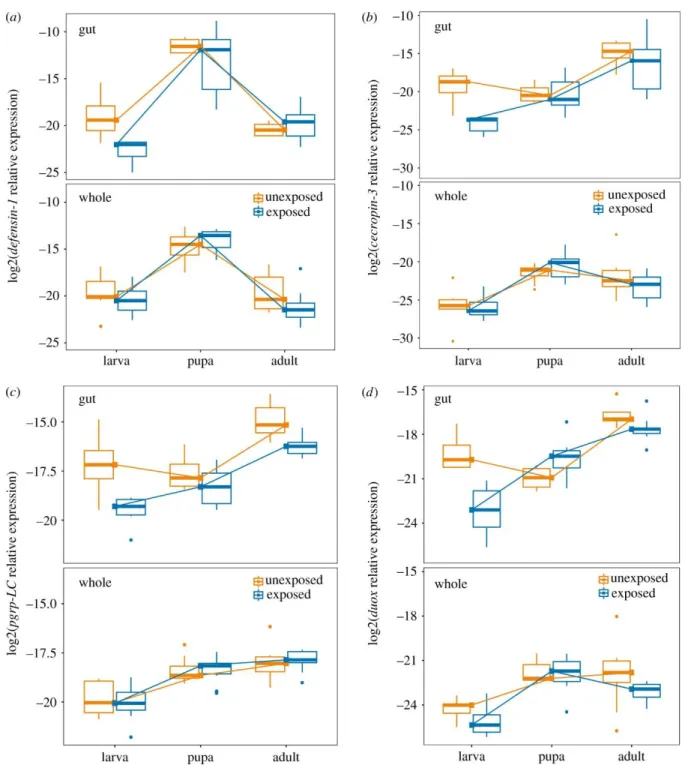
Figure 1. The proposed functional roles of T. castaneum immune genes quantified in chapter II.
Peptidoglycan recognition proteins homologs (e.g. PGRP-LC and PGRP-LA) are thought to recognize parasites and stimulate signaling cascades that result in the production of antimicrobial effectors. The immune factors in this study are involved in the melanization pathway (DDC), production of reactive oxygen species (DUOX), opsonization by phagocytes (TepB), and degradation of microbial
peptidoglycan via amidase activity (PGRP-SC2). The expression of antimicrobial peptides Defensin-1 and Cecropin-3 provide read-outs on the activation of Toll and IMD pathways.
Methods
Gregarine infections
Septate eugregarine protozoa are ubiquitous and generally avirulent inhabitants of insect midguts (Detwiler & Janovy, 2008; Janovy et al., 2007; Tate & Graham, 2015c; THOMAS & RUDOLF, 2010). The strain of Gregarina parasites used in this study was originally derived from infected T. castaneum
beetles collected at a feed store in Kentucky in June 2017 and subsequently maintained in a continuously infected colony. We have not observed any obvious disease-induced mortality or other symptoms of virulent infection with this parasite. This parasite is transmitted via the secretion of gametocysts from the infected insect gut. The gametocyst produces oocysts in the flour environment that are then ingested by the new host. Thus, the addition of beetle eggs to flour derived from a heavily infected colony is sufficient to reliably expose newly hatched larvae to the parasites (Detwiler & Janovy, 2008; Janovy et al., 2007; Tate & Graham, 2015c; THOMAS & RUDOLF, 2010). Before the start of the experiment we
confirmed infection in the source colony by dissecting the guts of 15 mature larvae and staining with a 60% iodine saline solution to visualize gregarine parasites via light microscopy (25x). We found that 7/15 larvae had visible trophozoites in the midgut, although the infection rate is likely higher as the trophozoites are hard to see until almost ready to enter syzygy. None of the 15 pupae that we dissected showed signs of infection, agreeing with previous observations (THOMAS & RUDOLF, 2010) that parasites are unlikely to survive in the pupal gut because the epithelia to which they are attached are destroyed.
Flour beetle rearing and sample collection
We set up 11 petri dishes containing all-purpose white flour, to which we added 40 T. castaneum adults from the “Snave” colony, originally collected from a Pennsylvania grain elevator in July 2013 and subsequently maintained in the lab (Tate & Graham, 2015c). Three days later we sieved approximately 300 eggs from the breeding groups and distributed them into new petri dishes, to which we added either 10 grams of gregarine-positive flour from the heavily infected T. castaneum colony or 10 grams of gregarine-free flour from a parasite-negative colony. Three weeks later we pulled 50 pupae as well as 50 larvae with an approximate length of 4mm from each treatment, and 25 newly ecolosed, virgin adults
treatment and placed them in individual wells of a 96 well plate, monitoring them daily first for pupation and then for eclosion as new adults. All beetles were kept at 29°C in the same incubator in the dark except when handled.
Individuals destined for gene expression studies were starved for approximately 24 hours prior to sample processing to eliminate non-colonized parasites and food in the guts, and then dipped in sterile water to remove excess flour immediately prior to sample collection. We dissected guts from all stages by making an incision in the abdomen and gently removing the gut with tweezers while the insect was immersed in 10uL insect saline. Guts were immediately placed in a 1.5 mL collection tube on dry ice.
After collections were complete, guts were kept at -80°C. We originally treated a subset of guts with iodine as well to visualize parasites before freezing the guts, but after finding that iodine treatment severely affected gene expression, we eliminated these samples from subsequent analyses, leaving us with 5-7 gut samples per exposure treatment per life stage. Whole individuals (8-10 per treatment/life stage) were placed in individual tubes, frozen, and kept at -80°C.
Quantification of immune gene expression via RT-qPCR
We isolated gut RNA using the Qiagen All Prep Micro Kit, and isolated whole body RNA with Qiagen All Prep and RNeasy kits. We synthesized cDNA with 0.5uL RNA (whole body) or 4uL RNA (gut) in a 5uL or 10uL reaction using the manufacturer-recommended protocol with SuperScript IV VILO master mix (ThermoFisher Scientific), and diluted the cDNA with 30-40 μL nuclease free water.
We conducted RT-qPCR on the Biosystems Quantstudio 6 Flex machine using sybr green chemistry (PowerUp SYBR green master mix from Applied Biosystems, 500nM primers, 10-50ng cDNA).
Thermal cycling conditions were 95 °C for 2 minutes, followed by 40 cycles of 95 °C (15 sec), 55 °C (10 sec), and 60 °C (1min). All samples were run in duplicate or triplicate and the average ct value was
used for subsequent analyses as long as technical replicates were within 1ct.
We used RT-qPCR to quantify the expression of immune response-associated genes (Fig. 1) including defensin-1, cecropin-3, dopa decarboxylase (DDC), thioester containing protein-B (TepB), dual oxidase (duox), and the peptidoglycan recognition proteins pgrp-LA, pgrp-LC, and pgrp-SC2 (Table 2). Defensin-1 and cecropin-3 are anti-microbial peptides (AMPs) that are thought to be activated by both the IMD and Toll pathways in T. castaneum and have orthologs that are upregulated during bacterial oral challenge in Bombyx mori and D. melanogaster (Tzou et al., 2000; Yokoi, Koyama, Minakuchi, et al., 2012a). Pgrp-LA and pgrp-LC are transmembrane receptor proteins for the IMD pathway in T. castaneum and essential for its production of AMPs (Koyama et al., 2015a). PGRP-SC2 is the T. castaneum homolog of pgrp-LB in D. melanogaster, which downregulates the IMD pathway (Paredes et al., 2011a). DDC is a precursor in the melanization pathway which kills malaria parasites in the midgut of Anopheles mosquitoes (WHITTEN et al., 2006; Wu et al., 2010). TEPs are highly
expressed in the crop and proventriculus in D. melanogaster (Bou Aoun et al., 2011). Finally, Duox synthesizes reactive oxygen species (ROS) in gut epithelial cells, and RNAi knockdown of Duox has been shown to increase susceptibility to oral bacterial infection in D. melanogaster (Ha et al., 2005).
We used ribosomal protein S18 (rps18) as a reference gene for quantification of relative gene expression (Schmittgen & Livak, 2008a), as it has been shown to be stably expressed during infection (Lord et al., 2010a) in T. castaneum.
Table 2. Primers used to assay immune gene expression in T. castaneum.
Prime
r Set Full Name Function Forward Oligo Sequence Reverse Oligo Sequence AT.
(°C) Def1 Defensin-1 Toll/IMD
AMP TTTRYCGTTGCARTAKCCTCC TCAARSTGAATCATGCCGCW
TG 55
Cec3 Cecropin-3 Toll AMP AACATGARYACCAAACTTTT CCAAYTTATMGGCTKTGGW
G 55
-LA n recognition protein LA
recognitio n
AC T
PGRP -LC
Peptidoglyca n recognition protein LC
IMD recognitio
n
ACGAAGGCCGGGGATGGAAA GTTGTTTGCAAGCCGTTATC
TG 55
PGRP -SC2
Peptidoglyca n recognition protein SC2
IMD recognitio
n
ACAGTTGGATGCKTTGAAACA
GT AACTSGTYCTGCTCCCTTG 55
DDC
Dopa decarboxyla
se
Melanin
synthesis AGAAGTCGTGATGCTKGACT CTTGRATCACGCCGCC 55
Duox Dual oxidase ROS
synthesis CGCAATTGATCGGCCACTTT AGCTCCAAGGGATTTGGTCG 55
TEP-B
Thioester containing
protein B
Cellular recognitio
n
AGGTTTCACCTCATCGCAGG GTTGAAATTGTGGCGCTGGT 55
S18 Ribosomal Protein S18
Ribosoma
l Protein CGAAGAGGTCGAGAAAATCG CGTGGTCTTGGTGTGTTGAC 55
Statistical analyses of gene expression
We calculated the Δct values for each gene for each individual sample by subtracting the target gene mean ct value from the reference gene mean ct value. Thus, the Δct value represents the log2- transformed relative expression value of the target gene (Schmittgen & Livak, 2008a). As our expression data were log-normally distributed, we retained the log2-transformed value for subsequent analyses. All statistical analyses were conducted in R (v3.5.2) To analyze the main effect of tissue on overall host gene expression, we conducted a MANOVA with our eight genes as dependent variables and tissue as the independent variable. To analyze the impact of each life stage, parasite exposure, and their
individual interactions within each tissue on gene expression, we used linear models (lm function) of the form (target relative expression ~ stage + exposure + stage*exposure). We adjusted the p values with the Benjamini-Hochberg method to control for the false discovery rate (Benjamini & Yekutieli, 2001). Finally, to analyze pairwise expression correlations between genes, we used the cor() function on gut, whole body, larva, pupa, and adult data. To get differences in covariance relationships among these datasets,
we subtracted the absolute value of one matrix from another and graphed the resulting differences using the lowerUpper (psych package) and ggcorrplot (ggcorrplot package) functions.
Results
The impact of gregarine infection on pupal development
The majority of individuals from both treatment groups took six days to develop from newly ecdysed pupae to newly eclosed adults and thus the distribution of development times was
underdispersed (dispersion parameter = 0.05). Nevertheless, individuals exposed to gregarine parasites as larvae developed significantly faster than those who were not exposed (quasi-poisson GLM, t = 2.05, p = 0.046), although the effect size was less than one day among treatments.
Immune gene expression differs by tissue
The overall effect of tissue (gut vs. whole body) on gene expression was highly significant (MANOVA, F1,89 = 31.34, p < 2e-16). Models for individual genes revealed that the gene ddc had, on average, 5.7-fold higher expression in the whole body than in the gut (F1,89 = 27.7, p < 1e-6). Genes that showed significant upregulation in the gut relative to the whole body, on the other hand, include pgrp- LC (fold change = 2.6, F1,89 = 21.4, p < 2e-5) , duox (fold change = 11.47, F1,89 = 66.2, p < 1e-11), and cecropin-3 (fold change = 14.8, F1,89 = 31.4, p < 1e-6). There was no significant tissue-driven difference in expression for the genes pgrp-LA, defensin-1, pgrp-SC2, or tepB (p > 0.05). Fig. 2 illustrates relative expression of four genes among tissues (top row in each panel = gut expression, bottom row = whole body expression).
Figure 2. The influence of tissue type and gregarine parasite exposure on immune gene expression across developmental stages of the flour beetle T. castaneum. The expression of the antimicrobial peptides defensin -1 (A), and cecropin-3 (B), the recognition protein pgrp-LC (C), and the reactive oxygen species generator duox (D) were assayed in extracted guts (top row of each panel) or whole bodies (bottom row) from larvae, pupae, or adults that were either exposed to gregarine parasites as larvae (blue) or not (orange). The expression of each gene relative to the reference gene RP18s is represented on a log2 scale. Lines have been added to visualize the developmental trajectory of median
gene expression.
Does the modularity of immune gene expression differ between tissues? Previous work on T.
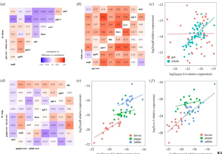
castaneum and other model insects like D. melanogaster have proposed that several of our genes are likely to be under control of common pathways like IMD and Toll (e.g. (Yokoi, Koyama, Ito, et al., 2012a)), resulting in expression patterns that covary among co-regulated genes. In our data, all genes showed a moderate to high correlation of expression with at least one other assayed gene (Fig. 3B), but these relationships were not always consistent among tissues (Fig. 3A). For example, pgrp-LA and tepB were tightly correlated at the whole body level, but show no relationship in the gut (Fig. 3C).
Figure 3. Gene expression correlations suggest partial decoupling of immune genes between tissues and among life stages. The pairwise Pearson correlation values of whole body gene expression was
subtracted from those of gut-only pairwise correlations to get the difference in correlation strength (A).
Large positive values indicate a stronger relationship in the gut, while large negative values indicate
stronger correlations in the whole body. The underlying correlations are visualized in (B) for whole body (top left) or gut only (bottom right); colors and numbers indicate the Pearson correlation coefficient. The breakdown of the correlation of pgrp-LA and tepB expression (log2 scale, relative to reference gene) in the gut relative to the whole body (C) illustrates decoupling among tissues. There was also decoupling by life stage, as illustrated by the relative magnitudes of the correlation coefficients for pupae against larvae (D, top left) and pupae against adults (D, bottom right). Stage-specific pairwise comparisons of pgrp-LC vs. tepB expression (E.) and pgrp-LA vs. cecropin-3 expression (F) illustrate different examples of differences in coefficients among stages.
The effect of developmental stage and parasite exposure on immune gene expression
To analyze the impact of developmental stage, larval gregarine exposure, and their interaction on immune gene expression (Fig. 2), we performed linear modeling on each gene. We analyzed gut and whole body datasets separately because of the complex tissue-specific genic interactions described above. In the whole body, there was no significant effect of gregarine exposure on gene expression (Table 3), but expression differed broadly by life stage. Most genes showed higher expression in pupae and adults relative to larvae (Appendix A “whole body”). Pgrp-LC, pgrp-LA and tepB increased in each subsequent life stage, while defensin-1 (Fig. 2A) and to a lesser extent cecropin-3, duox, and ddc peaked in the pupal stage. Only pgrp-SC2 expression showed no significant effect of stage.
Table 3. Summary of statistical results for the impact of stage, larval parasite exposure, or their interaction on immune gene expression in the gut and whole body. Full statistical tables for each gene are available in Appendix A. The expression of each gene was fit with the model: expression ~ stage*exposure using the lm()
function in R, where stage has three levels (larva, pupa, adult) and parasite exposure has two levels (exposed, unexposed). P values were adjusted for false discovery rate using the Benjamini-Hochberg method, and asterisks indicate the level of significance for at least one level of factor or interaction, relative to unexposed larvae: *padj < 0.05,
**padj<0.01, ***padj < 0.001. '-' indicates lack of statistical significance.
Gut
Whole Body
Gene Stage Exposure Stage*Exposure Stage Exposure Stage*Exposure Defensin-
1 *** - - *** - -
Duox * *** *** *** - -
TepB *** - - *** - -
Cecropin-
3 - * - *** - -
Ddc - - - ** - -
Pgrp-LC * ** - *** - -
Pgrp-LA - - - *** - -
Pgrp-SC2 - - - - - -
The expression of immune genes in the gut was more diverse in their responses to stage and exposure (Table 3). Larval exposure to gregarines resulted in the overall down-regulation of cecropin-3 (Fig. 2B), pgrp-LC (Fig. 2C), and duox (Fig. 2D). The expression of duox further depended on the interaction of exposure and life stage (Appendix A) as expression was suppressed in exposed larvae but upregulated in the guts of pupae that were previously exposed (Fig. 2B). Only defensin-1 was more highly expressed in pupae than in larvae and adults (Fig. 2A), but pgrp-LC, duox and tepB were significantly more highly expressed in adults relative to larvae (e.g. Fig. 2C,D). No gene was most highly expressed in larvae than in other life stages.
In whole organisms, the strength of pairwise gene expression correlations differed among life stages (Fig. 3D). For example, pgrp-LC and tepB expression was tightly and steeply correlated in larvae but less so in pupae and adults (Fig. 3E), while the strong positive relationship observed between pgrp- LA and cecropin-3 in larvae and adults broke down in pupae (Fig. 3F).
Discussion
Our data suggest that larval exposure to a relatively benign protozoan parasite can leave an imprint on gut immune system function that persists through metamorphosis. Our study also reflects a dynamic change in the immunological profile of the insects as they mature through metamorphosis into adulthood. As we know little about the insect immune response to eugregarines despite their ubiquity