ii
THE ROLE OF INSULIN SIGNALING ON DOPAMINE TRANSPORTER TRAFFICKING
By
Nicole K. Speed
Dissertation
Submitted to the Faculty of the Graduate School of Vanderbilt University
in partial fulfillment of the requirements for the degree of
DOCTOR OF PHILOSOPHY in
Pharmacology August, 2010 Nashville, Tennessee
Approved:
Dr. Aurelio Galli Dr. Randy Blakely Dr. Kevin Niswender Dr. Gregg Stanwood Dr. Brian Wadzinski
iii
To my amazing, supportive parents, for always keeping me smiling
iv
ACKNOWLEDGMENTS
This work would not have been possible without the funding that helped support it. This financial support included an individual predoctoral NRSA fellowship (F31 DA024885) from the National Institute on Drug Abuse, as well as support awarded to my mentor, Dr. Galli, from the Vanderbilt Diabetes Research and Traning Center (DK020593), and grants from the National Institute of Health (DA14684 and DK085712).
I would first like to acknowledge my advisor, Dr. Aurelio Galli. His support throughout my graduate career has been invaluable. He has encouraged my development scientifically and beyond. Thank you for pushing me, even when I pushed back. I hope to embrace science and life with the enthusiasm that he does. I would also like to thank my thesis committee members Drs. Gregg Stanwood, Brian Wadzinski, Kevin Niswender, and Randy Blakely for their helpful guidance and support throughout my graduate career. Special thanks are extended to my chair, Dr. Randy Blakely, for his help and advice, and to Dr.
Kevin Niswender for his collaborative efforts on my project.
I would also like to acknowledge my collaborators including Dr. Lynette Daws and Anthony Owens, who provided much time and effort to my project and always put a smile on my face, and Dr. Scott Russo, who provided the IRS-2 virus and helpful guidance with manuscript preparations. I am also grateful for the assistance I received from several people at Vanderbilt, including Dr. Fang Yu, Dr. Nicole Herring and Dr. Heather Gosnell, who helped me with learning
v
techniques. Also I owe a debt of gratitude to Sanaz Saadat, who may be the most pleasant and helpful person I have ever encountered. I would also like to thank the Pharmacology Department for their help and support throughout graduate school.
I am grateful to the members of the Galli lab for their support, scientifically and otherwise. I would especially like to thank Dr. Heiner Matthies for taking the time to provide training with laboratory techniques and answer my many questions, and Nicole Bibus-Christianson for her help, her patience at my disorganization and, most importantly, her friendship. She truly has a heart of gold. I would also like to thank Dr. Adeola Davis for her help with feeding rats.
Most of all, I am grateful to my incredibly supportive family and friends.
My parents have always given me faith in my abilities, and their constant encouragement and love is the reason I push myself to accomplish my goals. I cannot express how much their support means to me; it is the foundation to all of my successes. I also would like to thank my sister Alexa, who always lends an ear, and my crazy family, who always believe in me and are my rock of support.
I want to thank Leda Ramoz and Jessica Moore for their friendship while in graduate school, which kept me sane, and Laurie and Ben Ott for their friendship and hospitality. Lastly I would like to thank my fiancé, Brandon Lute. His love and faith in me are endless, and for that I am extremely grateful.
vi
TABLE OF CONTENTS
Page
DEDICATION ... ii
ACKNOWLEDGMENTS... iii
LIST OF FIGURES ... vii
LIST OF ABBREVIATIONS ... ix
Chapter I. INTRODUCTION ... 1
The Neurotransmitter Dopamine and Dopaminergic Pathways ... 1
Dopaminergic Neurotransmission and the Dopamine Transporter .... 4
The Dopamine Transporter Structure and Function ... 10
Psychostimulants and the Dopamine Transporter ... 17
DAT Regulation by Interacting Proteins ... 20
Regulation of Dopamine Transporter Surface Expression ... 23
Insulin and the Dopamine Transporter ... 30
Dopamine and Feeding Behavior ... 33
Insulin Regulation of Feeding Behavior ... 37
Insulin, Diabetes, and DA-Related Diseases ... 40
Specific Aims ... 42
II. ISOFORM SPECIFIC REGULATION OF DAT CELL SURFACE EXPRESSION BY AKT2 ... 45
Introduction ... 45
Methods ... 47
Results ... 52
Discussion ... 60
III. DIET-INDUCED CHANGES IN INSULIN SIGNAILNG REGULATES THE TRAFFICKING AND FUNCTION OF THE DOPAMINE TRANSPORTER ... 63
Introduction ... 63
Methods ... 67
Results ... 73
vii
Discussion ... 93
IV. GENERAL DISCUSSION AND FUTURE DIRECTIONS ... 100
Summary ... 100
Obesity and Diabetes ... 102
Alternative Interpretations ... 106
Future Directions ... 109
V. REFERENCES ... 117
viii
LIST OF FIGURES
Figure Page
1. Pathways of dopamine signaling in the brain ... 2
2. Biosynthesis of dopamine ... 5
3. Diagram of a dopaminergic synapse ... 7
4. Enzymatic degradation of dopamine. ... 9
5. Illustration of the dopamine transporter ... 12
6. Dopamine transporter uptake action ... 15
7. Schematic of activation of Akt by insulin... 27
8. Schematic of insulin regulation of the dopamine transporter. ... 44
9. Structures of isoform selective, allosteric Akt inhibitors. ... 53
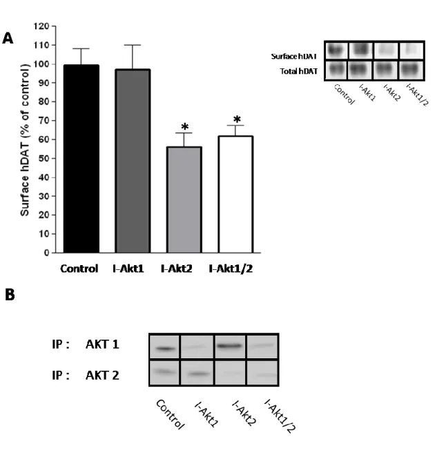
10. Inhibition of Akt2 reduces hDAT cell surface expression in hDAT cells ... 55
11. Inhibition of Akt2 reduces rDAT cell surface expression in rat striatal tissue ... 57
12. Inhibition of Akt2 reduces DAT-mediates reverse transport of DA. ... 59
13. High fat feeding results in increased food intake and weight gain ... 76
14. High fat feeding results in insulin resistance ... 77
15. Tyrosine hydroxylase levels are unchanged by high fat feeding ... 78
16. Dopamine (DA) levels in striatum are unchanged by high fat feeding ... 79
17. DIO induces a decrease in p-Akt in striatum and substantia nigra ... 81
18. DIO induces a decrease in Akt activity in the striatum ... 82
19. DIO induces a decrease in p-Akt2 ... 83
20. Dopamine transporter cell surface expression is reduced in striatal slices of high fat fed rats. ... 85
21. Dopamine clearance rate is reduced in DIO animals... 86
ix
22. AMPH-induced locomotor activity is reduced in DIO animals ... 88 23. AMPH levels in striatum after I.P. injection of AMPH remain the same
between LF and HF fed rats ... 89 24. Viral-mediated expression of HSV-GFP ... 91 25. Viral-mediated expression of IRS2 in DIO rats restores p-Akt levels ... 92 26. Viral-mediated expression of IRS2 in DIO rats restores surface expression of
DAT ... 94 27. Viral-mediated expression of IRS2 does not alter the surface expression of
the Na/K ATPase ... 95 28. Viral-mediated expression of IRS2 restores surface expression of DAT and
AMPH-induced locomotor activity ... 96
x
LIST OF ABBREVIATIONS
5-HT Serotonin
ADHD Attention-deficit hyperactivity disorder Akt Serine/threonine protein kinase B
AMPH Amphetamine
ANOVA Analysis of variance
BBB Blood-brain barrier
BMI Body Mass Index
CaMKII Calcium/calmodulin-dependent kinase II cAMP Cyclic adenosine monophosphate
COMT Catechol-O-methyl transferase
DA Dopamine
DAT Dopamine transporter
DIO Diet induced obesity
DOPA 3,4-dihydroxyphenylalanine
DR Dopamine receptor
ECL Extracellular loop
ERK Extracellular signal-regulated kinase fMRI Functional magnetic resonance imaging
GABA γ-aminobutyric acid
GFP Green fluorescent protein GSK3 Glycogen synthase kinase 3
xi
hDAT Human dopamine transporter
HEK Human embryonic kidney
HSCA High speed chronoamperometry
HSV Herpes simplex virus
IGF Insulin-like growth factor
IR Insulin receptor
IRS-2 Insulin receptor substrate 2 LeuT Leucine transporter
MAOB Monoamine oxidase B
MAPK Mitogen-activated protein kinase MPP+ 1-methyl-4-phenylpyridium
NE Norepinephrine
NET Norepinephrine transporter PBS Phosphate-buffered saline
PDK Phosphoinositol dependent kinase PET Positron emission tomography PI3K Phosphatidylinositol 3-kinase PICK1 Protein interacting with C kinase
PKA Protein kinase A
PKC Protein kinase C
PMA Phorbol 12-myristate 13-acetate PP2A Protein Phosphotase 2A
RACK1 Receptor for activated C-kinase
xii RFP Red fluorescent protein RTK Receptor tyrosine kinase
SERT Serotonin transporter
STZ Streptozotocin
SYN-1A Syntaxin 1a
TH Tyrosine hydroxylase
TMD Transmembrane domain
VMAT Vesicular monoamine transporter VTA Ventral tegmental area
1 CHAPTER I
INTRODUCTION
The Neurotransmitter Dopamine and Dopaminergic Pathways
Dopamine (3-hydroxytyramine; DA) is a catecholamine neurotransmitter that is a precursor to the synthesis of the neurotransmitter norepinephrine (NE).
DA in synthesized from tyrosine by a two step process, where tyrosine hydroxylase (TH) is the rate-limiting enzyme in the reaction. Being that DA is a precursor to NE synthesis, it was originally thought that it did not have signaling properties on its own, but instead was only an intermediate to NE production.
However, in 1958, Carlsson and colleagues demonstrated that DA had signaling properties on its own. Using 3,4-dihydroxyphenylalanine (DOPA), the precursor to DA, they showed that in rabbits depleted of catecholamines by reserpine, DOPA treatment could reverse the reserpine-mediated effects. Importantly, this reversal corresponded to an increase in DA, not NE (Carlsson, Lindqvist et al.
1958). Later work pointed to enrichments of DA in certain brain regions, namely the basal ganglia (Bertler and Rosengren 1959). Soon DA brain regions were mapped, displaying several distinct pathways of DA signaling (Fuxe 1965;
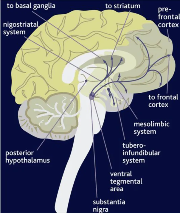
Ungerstedt 1971). There are four main dopaminergic pathways: the tuberoinfundibular pathway, the nigrostriatal pathway, the mesocortical pathway, and the mesolimbic pathway (Figure 1). The tuberoinfundibular pathway, which
2
Figure 1. Pathways of Dopamine Signaling in the Brain. Illustration of major DA projections in the central nervous system. The nigrostriatal pathway originates in the substantia nigra and projects to the dorsal striatum. The mesolimbic and mesocortical projections originate in the ventral tegmental area and project both to ventral striatum and areas in the prefrontal cortex, respectively. The final system is the tuberoinfundibular system which projects from the hypothalamus to the pituitary. This image was obtained from cnsforum.com.
3
refers to a group of DA neurons in the arcuate nucleus of the hypothalamus that project to the median eminence, controls prolactin secretion from the anterior pituitary gland (Weiner and Ganong 1978). The nigrostriatal pathway consists of neurons whose cell bodies originate in the substantia nigra and terminate in the dorsal striatum. This area is implicated in movement since degeneration of these projections has been shown to cause Parkinson’s Disease; characterized by tremors, rigidity, and overall improper movement (Barbeau 1962). Recently it has been demonstrated that this region is also important in feeding behavior (Volkow, Wang et al. 2002; Sotak, Hnasko et al. 2005; Robinson, Rainwater et al.
2007). Next, dopaminergic neurons in the mesocortical pathway project from the ventral tegmental area (VTA) to the frontal lobes of the cerebrum, particularly the prefrontal cortex, and are involved in cognition and emotion. Lastly, neurons of the mesolimbic pathway also originate in the VTA but instead innervate the ventral striatum, also known as the nucleus accumbens. This pathway is implicated in reward and pleasure.
DA is involved in a number of physiological and behavioral processes including cognition, locomotion, mood, motivation, and reward. Abnormalities in the central dopaminergic systems contribute to several neuropsychiatric diseases, including Parkinson’s disease, attention-deficit hyperactivity disorder (ADHD), schizophrenia, bipolar disorder, binge eating disorder, and addiction (Wise 1998; Horschitz, Hummerich et al. 2005; Kienast and Heinz 2006; Volkow, Wang et al. 2007; Davis, Levitan et al. 2008; Koob and Le Moal 2008). In particular, the nigrostriatal system is thought to give us the motivation to seek
4
basic needs, such as food, while the mesolimbic pathway enables us to feel pleasure from them (Palmiter 2007). Our eating behavior and our desire for food is tied in closely with these systems, which receives input from areas of the brain that monitor our nutritional need for food, such as the hypothalamus (Obici, Feng et al. 2002; Obici, Zhang et al. 2002; Niswender and Schwartz 2003; Schwartz and Porte 2005). Therefore, it is not surprising that dysfunction in DA signaling has been linked to eating disorders and obesity (Wang, Volkow et al. 2001;
Shinohara, Mizushima et al. 2004; Chen, Yang et al. 2008).
As such, DA dysregulation looks to be the basis of several neurological disorders. In order to develop effective pharmacotherapeutic approaches, it is critical to understand dopaminergic neurotransmission, the regulatory factors governing it, and how dysreguation of these factors can contribute to disease.
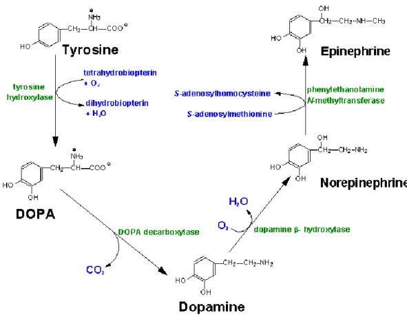
Dopaminergic Neurotransmission and the Dopamine Transporter Several factors influence dopaminergic neurotransmission, such as the amount of DA synthesized and released, the number of DA receptors (DRs) at the synapse, and the length of time DA spends in the synaptic space. As noted above, DA is synthesized through a series of enzymatic reactions, beginning with the hydration of amino acid tyrosine to DOPA via TH. DOPA is decarboxylated by aromatic amino acid decarboxylase to produce DA (Figure 2). The transmitter is then packaged into synaptic vesicles by a vesicular monoamine transporter (VMAT) and released at nerve terminals into the synapse upon stimulation.
Released DA then binds to DRs to elicit a response in the
5
Figure 2. Biosynthesis of dopamine. Synthetic enzymes and their changes to each product are labeled in green and blue, respectively. This image was obtained from
www.neurosci.pharm.utoledo.edu.
6
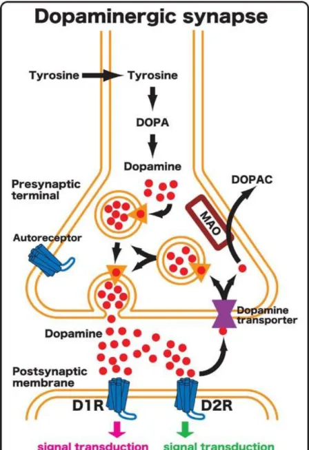
postsynaptic cell. The released DA is then cleared from the synapse primarily by the dopamine transporter (DAT), where it re-enters the presynaptic neuron to be recycled and repackaged into vesicles (Figure 3).
An important component in DA signaling is the receptor itself. DRs are a family of G protein-coupled receptors. There are five subtypes, which are divided into two groups. D1-like receptors, comprising the D1 and D5 receptors, are coupled to G proteins which stimulate adenylyl cyclase and cyclic adenosine monophosphate (cAMP) production, whereas D2-like receptors, comprising the D2 (D2R), D3 (D3R), and D4 (D4R) receptors, couple to Gi/o proteins and result in the inhibition of adenylyl cyclase and suppression of cAMP production (Kebabian and Calne 1979; Stoof and Kebabian 1981). D1-like receptors, by stimulating cAMP production, are excitatory, whereas activation of D2-like receptors is inhibitory. There are two isoforms of the D2R, a short form found presynaptically, and a long form found postsynaptically(Usiello, Baik et al. 2000).
In fact, D2R is the main presynaptic autoreceptor of the dopaminergic system (Mercuri, Saiardi et al. 1997). D2Rs are expressed throughout DA regions of the brain. D3Rs, another member of the D2-like receptor family, also inhibit cAMP production. They are found postsynaptically, and have a higher density in limbic areas of the brain, such as the nucleus accumbens (Bouthenet, Souil et al.
1991). The diversity of DRs expressed at a given synapse help to define the response elicited when DA is released. Furthermore, this response is not only dependent on the receptor
7
Figure 3. Diagram of a dopaminergic synapse. Enlarged view of a typical dopaminergic synapse. The presynaptic terminal is located at the top and the postsynaptic neuron is on the bottom. DOPA: 3,4-dihydroxyphenylalanine, DOPAC: 3,4-dihydroxyphenylacetic acid, D1R: type 1 dopamine receptor, D2R: type 2 dopamine receptor, MAO: monoamine oxidase. The dopamine transporter is shown on the presynaptic terminal in purple. The D2R subtype represents the main presynaptic autoreceptor of the dopaminergic system. The source for the image is
http://www.nibb.ac.jp/annual_report/2004/img/240-01.jpg
8
type(s) at the synapse, but also the number of receptors present, lending another element to regulation of DA neurotransmission (Figure 3).
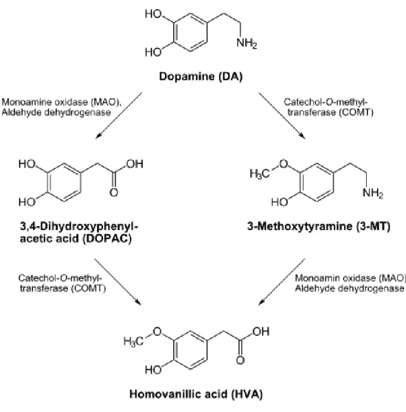
Termination of DA neurotransmission is another important component for maintaining proper dopaminergic tone. DA is degraded enzymatically by monoamine oxidase B (MAOB) and catechol-o-methyl transferase (COMT) (Figure 4), but enzymatic degradation does not account for inactivation of DA in the synapse. Instead, termination of DA neurotransmission is regulated by DAT.
DAT allows DA to be cleared out of the synapse and taken up into the presynaptic bouton (Giros and Caron 1993). The importance of this transport system was demonstrated by the creation of DAT knockout mice, where DA clearance is significantly slower than in wild type mice. DA remained in the synapse at least 100 times longer than the control animals (Giros, Jaber et al.
1996; Gainetdinov, Jones et al. 1999), leading these animals to be hyperactive.
In addition to its main role, DAT also provides a supply of DA for repackaging into vesicles for future release. This DAT-mediated recycling is the main source of DA for vesicular release in the neuron, thus decreasing the amount of synthesis needed to replenish vesicular stores of DA (Giros, Jaber et al. 1996). Indeed, when stimulated, the DA neurons of DAT knockout mice release significantly less DA than control animals (Giros, Jaber et al. 1996; Gainetdinov, Jones et al.
1999). It is thought that in DAT knockouts, there is a lack of DA available for packaging into vesicles, and consequently a reduction in the amount of DA available for vesicular release.
Furthermore, D2R expression and activity are reduced in mice lacking the
9
Figure 4. Enzymatic degradation of dopamine. Metabolic enzymes, COMT and MAO, that degrade unpackaged DA in synaptic areas.
10
DAT (Jones, Gainetdinov et al. 1999). These changes demonstrate that while DAT plays an important role in directly regulating DA signaling, it also influences several components of the dopaminergic synapse. Therefore changes in the activity and functioning of DAT can markedly disrupt DA neurotransmission.
Due to the important role of DAT in DA homeostasis, loss of proper function and regulation of the transporter has been implicated in several DA- related diseases. Decreased striatal DAT binding has been reported in first- episode schizophrenic patients (Mateos, Lomena et al. 2005), clinical depression (Laasonen-Balk, Kuikka et al. 1999), and obese individuals (Chen et al. 2008).
Differences in the genomic variable number tandem repeat have been identified as risk factors for bipolar disorder (Greenwood, Alexander et al. 2001).
Furthermore, genetic polymorphisms in the DAT coding region have been also associated with ADHD (Mazei-Robison, Couch et al. 2005; Yang, Chan et al.
2007; Binda, Dipace et al. 2008), as well as eating disorders (Shen, Hagino et al.
2004). DAT is also well-known for its role in addiction, including substance abuse of psychostimulants such as amphetamine (AMPH) (Giros, Jaber et al.
1996), which act on the transporter to elicit their behavioral effects. Due to its role in several neurological disorders and addiction, many researchers have focused on DAT structure, function, and regulation.
The Dopamine Transporter Structure and Function
The presence of a transport mechanism for biogenic amines was initially reported in the 1960s by Julius Axelrod. In 1961, Hertting and Axelrod showed
11
that that NE could be accumulated in nerve endings, and released upon stimulation (Axelrod, Whitby et al. 1961). Further characterization of catecholamine uptake regions in the brain revealed that both NE and DA could be accumulated by distinct regions of the brain, and that this accumulation could be inhibited by co-application of either tricyclic antidepressants or drugs of abuse, including cocaine and amphetamine (AMPH) (Glowinski and Axelrod 1964; Ross and Renyi 1967).
DAT is a member of the Na+/Cl--dependent neurotransmitter transporter family (SLC6) that contains high affinity transporters for NE, serotonin (5-HT), γ- aminobutyric acid (GABA), glutamate, and glycine (Kilty and Amara 1992; Torres, Gainetdinov et al. 2003). In 1991, DAT was cloned (Giros, el Mestikawy et al.
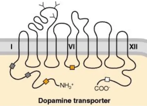
1991; Kilty, Lorang et al. 1991; Shimada, Kitayama et al. 1991; Usdin, Mezey et al. 1991), and analysis of the human DAT (hDAT) primary sequences revealed that the DAT cDNA encodes a protein of 620 amino acids. Hydropathy analysis predicts the presence of twelve transmembrane domains (TMDs) with intracellular amino and carboxyl termini (Figure 5). DAT is closely related to other catecholamine transporters, namely the NE transporter (NET), with which it shares 66% sequence identity (Blakely, Defelice et al. 2005). The structure of DAT also supports the notion that it is regulated by several signaling molecules.
The termini of the transporter contain several serine, threonine, and tyrosine residues, allowing for regulation via phosphorylation. In fact, some of these residues are found in the consensus sequences for kinases
12
Figure 5. Illustration of the dopamine transporter. The human dopamine transporter is an integral membrane protein that contains twelve transmembrane domains (TMDs) with intracellular N- and C-termini, a large extracellular loop between TMDs 3 and 4 with three N-linked glycosylation sites, and multiple phosphorylation sites located on intracellular termini and loops.
Putative glycosylation sites are indicated with Y-shaped symbols on extracellular sequences.
Possible phosphorylation sites are indicated with boxes for various protein kinases: gray boxes, protein kinase A; orange boxes, protein kinase C; white boxes, calcium-calmodulin protein kinase. This image was obtained from (Siegal 1999).
13
such as protein kinase C (PKC), protein kinase A (PKA), and calcium/calmodulin- dependent kinase II (CaMKII) (Giros and Caron 1993). Additionally, there are three glycosylation sites in the large second extracellular loop (ECL) between TMDs 3 and 4.
Chimeric studies conducted on DAT and NET in heterologous expression systems initially defined DAT function as it related to structure. These studies led to the hypothesis that TMDs 1-3 and 9-12 were important for the affinity of substrates and sodium/chloride dependence, where TMDs 4-8 were involved in transporting the substrate and inhibitor binding (Buck and Amara 1994; Giros, Wang et al. 1994; Syringas, Janin et al. 2000). Mutagenesis studies have also helped to reveal the structure/function relationship of DAT. For example, mutation of glycosylation sites on ECL 2 results in a transporter that is expressed at the plasma membrane, but has a reduction in DA uptake, as well as an increase in sensitivity to inhibitors (Li, Chen et al. 2004). This predicts that glycosylation on ECL2 is important to the transport process.
A high resolution crystal structure of the leucine transporter (LeuT), a prokaryotic sodium-dependent transporter with approximately 25% homology to DAT and related neurotransmitter transporters, is reported (Yamashita, Singh et al. 2005). The LeuT is a fellow member of the SLC6 gene family and contains all 12 TMDs. Therefore, its structure has served as a point of reference for the structure/function relationship of DAT, allowing for the initial hypotheses of DAT topology to be confirmed (Yamashita, Singh et al. 2005). In fact, a recent study used LeuT as a reference for determination of the binding sites of cocaine and
14
DA in DAT. Beuming and collegues developed models of DAT based on the LeuT structure to predict a binding site for cocaine and dopamine between TMD 1, TMD 3, TMD 6, and TMD 8 (Beuming, Kniazeff et al. 2008) They then confirmed these predictions experimentally using site directed mutagenesis and chemical cross-linking methods (Beuming, Kniazeff et al. 2008). This study, which was possible only with the hi-resolution LeuT structure, demonstrates that cocaine utilizes the same binding site as DA. This work, which was aided with the LeuT structure, is important in helping develop therapies for cocaine abuse and addiction.
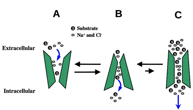
These structural studies have helped to further establish to a model of transporter action that was first proposed in 1966, known as the alternating access model (Jardetzky 1966), where external and internal gates alternately allow access to the transporter channel (Figure 6). First the gate to the extracellular space opens, allowing in the substrate and cotransported ions. This gate then closes, allowing the gate facing the intracellular space to open and release its cargo, thus ―alternating‖ the access of the transporter channel between the outside and inside of the cell. This event is powered by the electrochemical gradient generated by the plasma membrane Na+/K+-ATPase (Torres, Gainetdinov et al. 2003). DA uptake by DAT, therefore, is reliant on both sodium and chloride, where two sodium ions and one chloride ion are co- transported with each DA molecule. It is important to note that stoichiometry
15
Figure 6. Dopamine transporter uptake action. The model for how DA uptake occurs is via the alternating access model. (A) The extracellular facing conformation of the transporter binds substrate and driving ions located in the extracellular space. (B) Following a conformational change, the now intracellular facing conformation of the transporter releases the cargo into the cytosol. The transporter is also thought to act as a channel, as demonstrated in (C). Courtesy of Dr. Kris Kahlig, Vanderbilt University.
Extracellular
Intracellular
S S
S
S S S S S
S
S S S S S S
S
S S Substrate
Na+and Cl-
A B C
Extracellular
Intracellular
S S
S S S
S S
S S
S S S S S
S
S S S S S
S S S S S
S
S S S S S
S S SS
S S S S S S S S
S S S S
S S S S S S S
S
S S S
S S
S S S Substrate
Na+and Cl-
S
S Substrate Na+and Cl-
A B C
16
predicts that this transport process should be electrogenic, with a net translocation of two positive charges per transport cycle. Due to the flux in electrical charge, this phenomenon can be measured by electrophysiology in a single cell expressing DAT, providing a useful tool in studying the transporter (Sonders, Zhu et al. 1997; Sitte, Huck et al. 1998; Khoshbouei, Wang et al. 2003;
Pifl, Rebernik et al. 2004). Interestingly, these studies have found that the inward current is larger than the current predicted by the charge-to-flux ratio of the proposed stoichiometry (Lester, Cao et al. 1996; Sonders and Amara 1996). This suggests an uncoupled conductance. Therefore, the alternating access model cannot fully account for transporter actions.
Interestingly, some recordings taken of DAT currents have exhibited a large flux of DA crossing the membrane for a very brief amount of time, providing support for an alternative mode of action (Galli, Blakely et al. 1996; Galli, Blakely et al.
1998; Kahlig, Binda et al. 2005). This has led to one hypothesis to account for the uncoupled conductance of DAT; that it can act, albeit very briefly, as a channel.
This channel-like mode is similar to the open conformation of a ligand-gated ion channel, consisting of large fluxes of substrate molecules and ions crossing the membrane (Figure 6). Due to the large number of molecules that move across the membrane very quickly, this state is of interest, even if it only occurs infrequently. Other functions have also been discovered by studying the effect of psychostimulants on the transporter. AMPH, for example, is shown to promote a channel-like state of DAT that contributes to DA efflux (Kahlig, Binda et al.
17
2005). The ability of DAT to function in several modes demonstrates the dynamic nature of this protein.
Psychostimulants and the Dopamine Transporter
DA has a well established role in pleasure and reward. It is not surprising then that this system is the target of several psychostimulant drugs, many of which elicit their effects through DAT. These drugs are mainly viewed as abuse liabilities, they produce rewarding effects, alter DA tone, and lead to drug addiction. DAT is a target for these drugs, which include cocaine, methamphetamine, and AMPH (Kuhar, Ritz et al. 1991; Zaczek, Culp et al. 1991;
Giros and Caron 1993). They act to disrupt DAT’s ability to properly function, causing increases in extracellular DA that stimulate postsynaptic receptors, thereby enhancing neurotransmission. These drugs can broadly be characterized through their mode of action. For example, one mechanism is to block DAT directly. This is the mode used by the drug cocaine. By directly blocking uptake of DA, cocaine allows for an increase in synaptic levels of DA and enhanced DA neurotransmission (Ross and Renyi 1967; Giros, el Mestikawy et al.
1991). Several lines of evidence have also demonstrated that cocaine alters the surface expression of DAT. Repeated cocaine administration was shown to increase DA uptake in rats (Ng, Hubert et al. 1991; Parsons, Schad et al. 1993), and acute exposure to 10 µM cocaine for 30 minutes increased DAT transport activity and DAT cell surface expression (Daws, Callaghan et al. 2002). Studies have also examined the impact of chronic cocaine treatment on DAT by looking at postmortem
18
tissue from cocaine addicts. Striatal synaptosomes obtained from such tissue display a significant increase in both the Bmax of binding for the cocaine analog, WIN 35428, as well as the Vmax for DA uptake (Mash, Pablo et al. 2002). Furthermore, reports have shown increased binding of WIN 35428 to DAT in postmortem brains of addicts (Little, Kirkman et al. 1993; Staley, Hearn et al. 1994; Little, Zhang et al.
1999). These studies indicate a functional upregulation of DAT following chronic cocaine abuse.
Another important method of action by psychostimulant drugs is to cause effux of DA via DAT into the synapse. These drugs act as a substrate of DAT, competing with DA for uptake. AMPH is perhaps the most well-known drug to utilize this mechanism. Once inside the cell, AMPH acts to reverse the conformation of the transporter, causing efflux of DA into the synapse (Fischer and Cho 1979; Pierce and Kalivas 1997). This allows for the drug’s effects by acting as an indirect agonist of DRs, thus stimulating the postsynaptic cell and increasing DA neurotransmission.
Increases in DA signaling by either blocking DAT or by DAT-mediated DA efflux in limbic areas is thought to mediate the rewarding properties of psychostimulant drugs (Koob and Bloom 1988). Furthermore, AMPH-evoked DA efflux reveals another functional aspect of DAT beyond uptake of DA.
Modifications to DAT function can be examined by changes to AMPH-induced DA efflux. For example, in order for AMPH to elicit its effects, DAT must be on the cell surface. Therefore alterations in the surface level of DAT will also change the amount of AMPH-induced DA efflux, which allows for another method
19
to examine alterations in DAT function. As such, beyond its effects as a psychostimulant drug, AMPH can be a useful tool to examine changes to DAT function.
AMPH effects include several behavioral changes, such as restlessness, reduced appetite, and hyperlocomotion. These drug actions are believed to occur due to AMPH-induced increase in extracellular DA. Consistent with this notion, DAT knockout mice do not show an increase in locomotion when administered AMPH (Giros, Jaber et al. 1996). These findings demonstrate that DAT plays a crucial role in the hyperlocomotor effects of AMPH, and indicate that alterations in DAT function or expression can affect the ability of AMPH to elicit its behavioral effects.
In addition to causing efflux and inducing behavioral changes such as hyperlocomotion, AMPH alters the surface expression of DAT, a phenomenon that has been shown by several groups (Fleckenstein, Haughey et al. 1999;
Saunders, Ferrer et al. 2000; Carvelli, Moron et al. 2002; Gulley, Doolen et al. 2002;
Kahlig, Javitch et al. 2004; Garcia, Wei et al. 2005; Johnson, Furman et al. 2005;
Boudanova, Navaroli et al. 2008). Application of AMPH to DAT transfected cells for one hour reduced the rate of DA uptake and also decreased the surface expression of DAT as measured by biotinylation (Saunders, Ferrer et al. 2000). Similar results were obtained for the endogenous substrate, DA, as well (Saunders, Ferrer et al.
2000). Furthermore, measurements of a single transporter current after AMPH application demonstrated that the current itself was unaltered, confirming that changes to DA uptake were due to a redistribution of DAT away from the plasma
20
membrane (Kahlig, Javitch et al. 2004). Recently, live cell imaging was used to examine changes to DAT surface expression after rapid treatment (up to one minute) of AMPH in neuroblastoma N2A cells expressing DAT. These studies indicated that intially, AMPH acts to increase the expression of DAT on the cell surface, as does the endogenous substrate DA (Furman, Chen et al. 2009).
Prolonged exposure, however, led to a decrease of DAT cell surface expression.
Importantly, these results demonstrate that endogenous, as well as exogenous, substrates of DAT regulate its expression on the plasma membrane in a biphasic manner.
Due to the changes that occur to DA systems with chronic use, these drugs are often thought of in a negative manner and as an abuse liability.
However, at times they have proved useful. AMPH is used frequently to treat ADHD, under the tradename Adderall. Also, the unique properties and functions they convey on DAT and DA transmission provide useful tools for studying the transporter. Still, most exogenous compounds that act on the transporter are, in fact, addictive and can have many negative consequences with frequent use.
DAT Regulation by Interacting Proteins
Being that DAT has an important role in DA neurotransmission, it is not surprising that it is tightly regulated. Several proteins that regulate DAT also physically interact with it, forming protein complexes, and much work continues to further define these. To date, some identified DAT interacting proteins are, but not limited to, protein interacting with C kinase (PICK1) (Torres, Yao et al. 2001),
21
Hic5 (Carneiro, Ingram et al. 2002), the catalytic subunit of protein phosphatase 2A (PP2A) (Bauman, Apparsundaram et al. 2000), the PKC isoforms β1 and β2
(Johnson, Guptaroy et al. 2005), the receptor for activated C-kinases (RACK1) (Lee, Kim et al. 2004), SYN-1a (Lee, Kim et al. 2004; Binda, Dipace et al. 2008), CaMKII (Fog, Khoshbouei et al. 2006; Wei, Williams et al. 2006), and D2R (Lee, Pei et al. 2007; Binda, Dipace et al. 2008).
PICK1 was found to interact with DAT via the PDZ recognition motif found on the C-terminal tail on the transporter. PKC, a well known modulator of DAT surface expression, also interacts with PICK1, leading to the possibility that PICK1 serves at an adaptor protein for these two molecules (Torres, Yao et al. 2001). Interestingly, RACK1 have been shown to interact with the DAT’s N-terminus by a yeast-two hybrid assay (Lee, Kim et al. 2004). RACK1 also interacts with PKC, as well as several other kinases, and therefore may serve to facilitate kinase regulation of DAT by PKC and other kinases on the N-terminus (Ron and Mochly-Rosen 1994;
Rodriguez, Ron et al. 1999). Therefore, both of these interacting proteins, PICK1 and RACK1, may help to facilitate PKC modulation of DAT at two distinct regions on the transporter. This indicates that PKC may modulate DAT in several ways, depending on the area of DAT available for binding and the interacting protein available to it.
SYN-1a is another protein identified to interact with DAT (Lee, Kim et al.
2004). This is of interest due to work on SYN-1a and DAT family members, including the GABA transporter, GAT1, and NET. SYN-1a interacts with the N- terminus of GAT1 and modulates the translocation of the transporter (Wang, Deken
22
et al. 2003), and is also shown to interact with NET (Sung, Apparsundaram et al.
2003), suggesting that it might serve a similar function in regulation of DAT. Recent studies have shown an increase in association between SYN-1a and DAT with AMPH treatment (Binda, Dipace et al. 2008), allowing for a possible role for SYN-1a in AMPH-mediated DAT trafficking.
In addition to the proteins mentioned above, recent work has also identified D2R to associate with DAT. Disruption of this interaction has been shown to decrease DA uptake (Lee, Pei et al. 2007). The short isoform of D2R functions as an inhibitory autoreceptor on the presynaptic cell, with a localization similar to DAT (Centonze, Usiello et al. 2002). Previous work has demonstrated that D2R stimulation may be regulating DAT through downstream activation of second messenger signaling cascades (Mayfield and Zahniser 2001). For example, Bolan and colleagues demonstrated that D2R stimulation causes enhanced substrate clearance through an increase in DAT cell surface expression, which was dependent upon extracellular signal-regulated kinases 1 and 2 (ERK 1/2), but independent of phosphatidylinositol 3-kinase (PI3K) (Bolan, Kivell et al. 2007).
Lee and colleagues reported the first evidence of a direct association between DAT and D2R, and that disruption of this interaction affects DAT function (Lee, Pei et al. 2007). These studies suggest that there is a definite interplay, direct and indirect, between presynaptic D2Rs and DAT to help maintain DA homeostasis.
It is also quite interesting to note that DAT is thought to interact with itself.
Cross linking and mutagenesis studies, as well as studies using fluorescence
23
resonance energy transfer (FRET) imaging, demonstrated that DAT forms an oligomer (Hastrup, Karlin et al. 2001; Hastrup, Sen et al. 2003; Sorkina, Doolen et al. 2003; Torres, Carneiro et al. 2003). Recently, the substrates DA and AMPH have been shown to reduce DAT oligomerization (Chen and Reith 2008), lending yet another opportunity for regulation of DAT function. Further work is needed to examine how second messager systems may also play a role in altering DA uptake by changing the ability of DAT to form oligomers.
Regulation of Dopamine Transporter Surface Expression DA uptake capacity depends on the turnover rate of an individual transporter as well as on the number of functional transporters expressed at the plasma membrane. DAT function is dependent upon expression on the plasma membrane, and as such the surface expression of the transporter is a tightly regulated mechanism for controlling DA neurotransmission. Several signaling molecules have been identified as regulators of DAT cell surface expression, including PKC, PKA, ERK1/2, and members of the insulin signaling family (i.e.
phosphoinositol 3 kinase (PI3K) and protein kinase B (PKB, or Akt)) (Batchelor and Schenk 1998; Carvelli, Moron et al. 2002; Page, Barc-Pain et al. 2004; Garcia, Wei et al. 2005; Bolan, Kivell et al. 2007).
Possibly the most well characterized of these pathways is PKC modulation of DAT. PKC activation by phorbol esters has been shown to cause trafficking of the transporter away from the plasma membrane, resulting in reduced DA uptake (Huff et al. 1997; Vaughan et al. 1997; Zhang et al. 1997; Zhu et al. 1997; Pristupa et
24
al. 1998; Daniels and Amara 1999; Melikian and Buckley 1999; Granas et al.
2003; Loder and Melikian 2003; Kahlig et al. 2004; Foster et al. 2008). This PKC-induced trafficking of DAT from the plasma membrane to intracellular compartments is a clathrin-mediated and dynamin-dependent endocytic mechanism (Daniels and Amara 1999; Sorkina, Hoover et al. 2005; Foster, Adkins et al. 2008). Beyond this work, PKC has been also shown to modulate the functionality of DAT by shifting it to a state that is more likely to result in DA efflux (Kantor and Gnegy 1998; Kantor, Hewlett et al. 2001; Johnson, Guptaroy et al.
2005). This work displays another aspect of the role of PKC in altering DAT function by supporting the reversal of DA transport.
Although it was originally believed that phosphorylation of DAT by PKC was required for internalization (Huff, Vaughan et al. 1997; Vaughan, Huff et al.
1997; Cowell, Kantor et al. 2000; Chang, Lee et al. 2001; Foster, Pananusorn et al. 2002; Granas, Ferrer et al. 2003; Foster, Adkins et al. 2008), recent lines of evidence have shown that this is not the case. Truncation of the PKC consensus sequence in the DAT N-terminus abolishes PKC-induced phosphorylation of DAT, yet this form of the transporter is still able to traffic in response to phorbol ester treatment (Granas, Ferrer et al. 2003). These data suggest that PKC regulation of DAT may involve separate phosphorylation and trafficking components. In fact, recent work by Foster and collegues demonstrated that, with PKC activation, the loss of surface DAT occurred only in concanavalin A-sensitive, non-raft membranes (Foster, Adkins et al. 2008). However, treatment with methyl-β- cyclodextrin, which destabilizes lipid rafts by depleting cholesterol, inhibited PKC-
25
induced downregulation of DAT activity but still allowed for internalization of DAT (Foster, Adkins et al. 2008). In addition, DAT phosphorylation was found to be at a significantly greater level in cholesterol-rich lipid raft microdomains. These findings suggest that regulation by PKC in the non-raft DAT population occurs through trafficking-dependent processes. Conversely, in lipid rafts DAT regulation by PKC is achieved through trafficking-independent processes (Foster, Adkins et al. 2008). Importantly, this study suggests that the localization of DAT is important in determining how it is regulated. Further work is needed to look at the importance of DAT localization and how regulation of these distinct populations affects the transporter.
Notably, recent work has revealed another interesting aspect of regulation of DAT trafficking. Furman and collegues explored the changes to DAT cell surface expression over time after exposure to a substrate, either DA or AMPH, using live cell confocal imaging and biotinylation assays in neuroblastoma N2A cells transfected with DAT. These investigations defined a biphasic regulation of DAT cell surface expression in response to substrate binding. Initially, within seconds, there is a rapid increase of DAT on the cell surface, beginning at 10 seconds and going to 2 minutes. This is followed by a decrease of cell surface DAT upon continued exposure to the substrate (Furman, Chen et al. 2009).
Importantly, further work by this group also showed that inhibition of PKCβ blocked the initial rapid increase of cell surface DAT after exposure to DA or AMPH (Chen, Furman et al. 2009; Furman, Chen et al. 2009). Furthermore, PKCβ knockout mice were found to have a reduction in cell surface DAT
26
expression compared to wild type mice, while there was no change to the total levels of DAT protein between the two genotypes (Chen, Furman et al. 2009). In addition, the rapid increase in DAT cell surface expression upon substrate treatment seen in wild type mice was not observed in PKCβ knockout mice (Chen, Furman et al. 2009). Overall, these studies indicate that there are two phases of DAT trafficking in response to substrate binding, and that the initial rapid phase is dependent upon PKCβ. This work further displays the complexity to the regulation of DAT function via transporter trafficking.
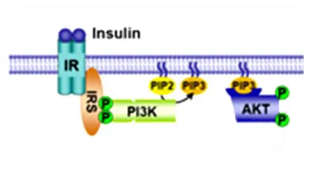
PI3K signaling in regulating DAT Surface Expression. Much work has shown that DAT activity is modulated by multiple signal transduction pathways, and often this regulation involves modifying DAT trafficking and expression at the plasma membrane. The PI3K signaling pathway has been extensively studied for its role in modulating the surface expression of DAT. PI3K phosphorylates the D-3 position of phosphoinositol-2 phosphate (PI(4,5)P2)to yield phosphoinositol-3 phosphate (PI(3,4,5)P3). The generation of PI(3,4,5)P3 at the plasma membrane upon the activation of PI3K allows for translocation of Akt (Taha and Klip 1999;
Bondy and Cheng 2004). This localization of Akt to the plasma membrane is critical for its activation (Figure 7). Once bound to PI(3,4,5)P3, Akt is phosphoylated by phosphoinositide dependent kinase 1 (PDK1), and may go on to signal to several downstream effectors (Yang, Tschopp et al. 2004).
In 2002, work by Carvelli and collegues showed that in hDAT expressing heterologous cells, inhibition of PI3K by LY294002 led to a decrease in surface
27
expression of hDAT. The investigators also reported a decrease in DA uptake with LY294002 treatment, not only in cells but also in rat striatal
Figure 7. Activation of Akt by Insulin. Upon activation of the insulin receptor (IR), insulin receptor substrate (IRS) acivates phosphoinositol-3-kinase (PI3K). PI3K goes on to
phosphorylate phosphoinositol-2 phosphate (PIP2), converting it to phosphoinositol-3 phosphate (PIP3) and allowing translocation of Akt to the plasma membrane, leading to its subsequent activation. Akt is now an active kinase and can phosphorylate downstream targets. This figure was obtained from http://www.hsph.harvard.edu/faculty/brendan-
manning/images/Insulin_Signaling.jpg
28
synaptosomes. These effects are dynamin dependent as DA uptake and DAT cell surface expression after LY294002 treatment was inhibited by expression of a dominant negative mutant of dynamin I.
PI3K is activated by receptor tyrosine kinase stimulation. Importantly, RTK inhibitors have been shown to downregulate DAT activity and plasma membrane expression (Zahniser and Doolen 2001), thus fitting the model that PI3K activation through RTKs causes an increase in surface expression of DAT.
Carvelli and coworkers also demonstrated that incubation with a RTK activator, insulin, showed increases in DAT transport capacity and surface levels (Carvelli, Moron et al. 2002).
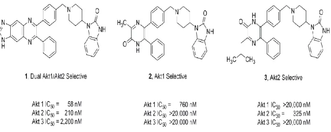
As mentioned earlier, further downstream effects of PI3K signaling include the activation of Akt (Taha and Klip 1999). Akt is an important component in insulin and growth factor signaling pathways, and is thought to regulate several cellular functions including cell growth, glucose uptake and metabolism, as well as apoptosis (Hanada, Feng et al. 2004). Three isoforms of Akt have been identified (Hanada, Feng et al. 2004), all of which contain a pleckstrin homology domain that interacts with PI(3,4,5)P3. This interaction allows for phosphorylation of Akt at residues threonine-308 and serine-473, that are required for full Akt activation (Hanada, Feng et al. 2004). Akt1 and Akt2 are ubiquitously expressed throughout the body, but Akt3 is only found to be expressed in the brain and testes. Insights
29
into the specificity of these isoforms in Akt-related functions such as glucose homeostasis and cell growth, have been provided by genetic studies where each isoform, or combinations of them, have been genetically deleted. Akt1 null mice knocked out show impaired growth, but similar glucose metabolism and maintenance of insulin levels as their wild type counterparts (Cho, Thorvaldsen et al.
2001). The opposite is true, however, for Akt2 knockout mice. These animals are normal in size, but are insulin resistant and hyperglycemic (Cho, Mu et al. 2001).
These studies suggest that Akt1 is important in proper growth factor and cell survival signaling, whereas Akt2 is critical in insulin signaling and glucose metabolism.
Clues to the specificity of Akt3 were also discovered using knock out animals.
Knockouts of Akt3 show a reduced brain size (Tschopp, Yang et al. 2005).
Interestingly, dual knockouts of isoforms 2 and 3 have both reduced brain size and impaired glucose metabolism, but a knockout of Akt1/Akt3 is embryonic lethal (Dummler, Tschopp et al. 2006). These data suggest that Akt2 and Akt3 serve distinct functions, because a dual knockout is not lethal. However, with both Akt1 and Akt3 knocked out, the mice do not survive, suggesting that one of these isoforms is needed for proper development, and Akt2 cannot serve that function.
Therefore, it is thought that Akt1 and Akt3 are critical for proper cell growth, particularly Akt3 in brain, whereas the primary role of Akt2 is maintaining insulin signaling and keeping glucose at an appropriate level in the blood.
Being that Akt is downstream of PI3K activation, it is not surprising that the role of Akt in regulating DAT has been explored (Garcia, Wei et al. 2005; Wei, Williams et al. 2006). These studies provided further evidence that PI3K signaling
30
controls DAT plasma membrane expression by demonstrating that basal Akt activity sustains DAT on the surface. Using both pharmacological means (ML9, an Akt inhibitor) and genetic manipulations (dominant negative Akt mutant), Garcia and collegues demonstrated in heterologous expressing cells that inhibiting active Akt results in a decrease of DAT on the surface. Akt can be altered to remain active as well through myristylation (myr-Akt), which keeps the protein anchored at the plasma membrane in an active state. Importantly, transfection of this constitutively active, myr-Akt increased the surface expression of DAT (Garcia, Wei et al. 2005;
Wei, Williams et al. 2006). The physiological relevance of these results was emphasized by demonstrating that DA uptake, measured in heterologous expressing cells and striatal synaptosomes, was also impaired with ML9 treatment (Garcia, Wei et al. 2005; Wei, Williams et al. 2006). These data provide compelling evidence to support the hypothesis that basal PI3K/Akt signaling is important in regulating DAT function. Furthermore, this work implies insulin regulates DAT trafficking, consistent with hormonal regulation of dopaminergic signaling. The suggestion that insulin signaling can regulate DA neurotransmission via DAT is compelling given that insulin levels fluctuate with food consumption (Niswender, Morrison et al. 2003), and DA is an important neurotransmitter in regulation of reward and motivation for feeding.
Insulin and the Dopamine Transporter
DAT is an important component in regulation of DA signaling, and therefore it is of interest that several lines of evidence suggest that insulin can
31
regulate the expression and activity of DAT. For example, chronic intracranial administration of insulin enhanced DAT mRNA in the substantia nigra (Figlewicz, Szot et al. 1994). Moreover, hyperinsulinemic rats showed increased DAT mRNA in the substantia nigra (Figlewicz, Patterson et al. 1998). Food restriction, which causes low circulating levels of insulin, results in a 32%
decrease in the Vmax of DA uptake in the striatum of rats, whereas the Km
remains unaltered, pointing to a reduction in the surface expression of DAT (Zhen, Reith et al. 2006). Furthermore, food deprivation also reduces DA uptake in striatal synaptosomes (Patterson, Brot et al. 1998). Incubation of these synaptosomes with a physiological concentration of insulin restored DA uptake to control levels (Patterson, Brot et al. 1998), suggesting that changes in circulating insulin can modulate DAT activity. As discussed previously, important components of the insulin signaling pathway, PI3K and Akt, have been shown to regulate the transporter’s surface expression and function. These studies also show that activating PI3K and Akt by stimulating heterologous cells expressing DAT with insulin causes an increase of the transporter on the cell surface, as well as an increase in DA uptake.
Perhaps the most telling studies thus far related to insuilin regulation of DAT examined the effects of streptozotocin (STZ)-induced insulin depletion on DAT surface expression and its function. STZ enters the pancreatic β-cell via the glucose transporter GLUT2 and causes DNA damage, resulting in free radical production and subsequent β-cell necrosis (Szkudelski 2001). Without β-cells, animals suffer from hypoinsulinemia and hyperglycemia (Carr 1996). Rats made
32
hypoinsulinemic by streptozotocin treatment show decreased DAT mRNA in the substantia nigra (Figlewicz, Brot et al. 1996; Patterson, Brot et al. 1998), as well as decreased DA clearance as measured by high speed chronoamperometry (HSCA) (Owens, Sevak et al. 2005). Furthermore, rats treated with STZ were found to have a reduction AMPH-induced efflux, a phenomen that requires DAT on the cell surface. Here the authors used functional magnetic resonance imaging (fMRI), a technique that displays an image that correlates to the oxygenation of the tissue, which is a representation of the activity in that brain region. Upon receiving I.P. injection of AMPH, the rats treated with STZ had significantly less activity in the striatum compared to the control animals (Williams, Owens et al. 2007). The data from the fMRI scans were confirmed by using high speed chronoamperometry (HSCA). This technique measures DA efflux in vivo by carbon fiber amperometry in the brain of anesthetized animals after microinjection of AMPH in the striatum. Using HSCA, the investigators found that STZ-treated rats had a reduction in AMPH-induced efflux. Importantly, the authors also showed STZ to reduce active Akt, linking insulin signaling and Akt to DAT cell surface expression and function.
Although these studies highlight insulin regulation of DAT, they do use pharmacological manipulations to alter insulin levels. Further work has begun to examine DAT and the effect of high fat diets, which result in insulin resistance in the brain (De Souza, Araujo et al. 2005; Posey, Clegg et al. 2009). One study found that, after 20 days on a high fat diet, DAT binding density in the ventral and dorsal striatum was reduced (South and Huang 2008). Furthermore, an inverse
33
relationship between Body Mass Index (BMI) and striatal DAT availability has been shown (Chen, Yang et al. 2008). These studies begin to point to changes in DAT with a high fat diet, but they do not provide a mechanism for such changes. However, these data are of interest for they demonstrate alterations in a component of DA signaling in obesity. Still, it is not understood whether this dysfunction is a cause, or a consequence of a high fat diet. Additionally, co- morbidity between obesity and several DA-related disorders points to a need for a better understanding of the interactions between insulin signaling and DA systems.
Dopamine and Feeding Behaviors
DA is important in modulating several behaviors, ranging from movement to cognition to motivation and pleasure, including our motivation to eat and the pleasure we receive from eating. The role DA has in feeding behavior has been demonstrated by studies that illustrate improper DA signaling in obesity. For example, upon eating a palatable meal, dopaminergic areas of the brain, such as the dorsal striatum, increase in activity (Stice et al., 2008). Interestingly, in subjects with a BMI in the obese range, this activity is dampened, suggesting a dysregulation of DA neurotransmission in obese individuals (Stice et al., 2008).
In studies on obese rats on high fat diets for up to 16 weeks, striatal DA turnover is impaired (Davis, Tracy et al. 2008), and mRNA levels of DAT, D4R, D2R, and TH are reduced (Huang, Yu et al. 2005; Huang, Zavitsanou et al. 2006). In humans, D2R occupancy as measured by positron emission tomography (PET)
34
is reduced in a BMI-dependent manner (Wang, Volkow et al. 2001), and similar results for DAT were also shown (Chen, Yang et al. 2008). Therefore, it is becoming clear that DA systems are very important in our consumption of food, as they are altered in states of excessive food consumption. The exact mechanism of this phenomenon, however, is still being explored.
DA has several roles in our consumption of food, namely motivation for seeking food and food consumption (Volkow, Wang et al. 2002; Palmiter 2007), and the reward and satiety we feel when we eat (Volkow and Wise 2005). The latter of these has been studied extensively. The DA reward system originates in the VTA, with the projections ending in the ventral striatum, also known as the nucleus accumbens. Several inputs feed into this system, including canabanoid and opiate systems, as well as signaling molecules that are regulated by our food consumption; which include insulin and leptin (Palmiter 2007). DA is well known for the role it plays as the signaling molecule for our endogenous reward system.
Activation of DRs in the reward system is thought to give us a pleasant feeling for several tasks, such as consuming food or having sex. Drugs are known to hijack this system, leading to highs when consuming the drug and cravings when not.
In fact, comparisons between drug addiction and obesity have been made (Volkow and Wise 2005). In fMRI scans, drug-addicted subjects had a reduction of activity in the striatum, a dopamine rich area, upon receiving the drug compared to non-addicted subjects. With constant drug use, their DA systems were altered, as shown by a reduction in striatal activity. This is thought to be the biological basis for addiction, causing them to crave the drug more, and also
35
need more to create a similar ―high‖. Some groups have presented a similar hypothesis for food consumption and obesity. In fact, fMRI scans of obese subjects also show a reduction in activity in the striatum when consuming a highly palatable food compared to normal subjects (Stice, Spoor et al. 2008;
Stice, Spoor et al. 2008). It is hypothesized that this disparity will override one’s metabolic signals for when food is actually needed.
As researchers continue to explore DA signaling in feeding behavior, this view becomes overly simplistic. One study examined the effects of DA on feeding by creating mice that are DA deficient in the brain (Sotak, Hnasko et al.
2005). This was accomplished by knocking out expression of TH, the enzyme needed to synthesize DA, in neurons. Interestingly, these DA-deficient mice would not eat and die shortly after birth, having no motivation to seek and consume food. To attempt to rescue the animal’s motivation to eat, the researchers rescued DA signaling by injection of a virus expressing TH in different DA regions in the brain to determine exactly which region was responsible for feeding behavior. Only restoration to the dorsal striatum caused the animals to eat and survive. This study highlights the dorsal striatum, and therefore the nigrostriatal system, in feeding behavior. The authors suggest that the dorsal striatum may be the basis for our motivation for feeding; defining our basic need for food. Without DA signaling in this region, the animals did not eat.
Therefore, alterations to DA signaling in the dorsal striatum and nigrostriatal system may have implications for our basic need for consuming food, thus altering feeding behavior. Importantly, restoration of signaling to the ventral
36
striatum had no effect, leading to the hypothesis that the dorsal striatum is necessary for food intake, and the ventral striatum may act just to fine tune feeding behavior (Sotak, Hnasko et al. 2005; Palmiter 2008).
Further work has supported the hypothesis of the importance of the dorsal striatum in feeding behavior. A study conducted by Volkow and colleagues examined extracellular DA levels using PET imaging, specifically a ratio of Bmax/Kd of D2R ligand raclopride, in hungry subjects. These subjects were presented with food they could see and smell, but not consume. They found, that the Bmax/Kd ratio of raclopride was decreased in the dorsal striatum (Volkow, Wang et al. 2002). They concluded these changes to be from an increase in extracellular DA as the hungry subjects were presented with food, which bound to the D2Rs and decreased the D2Rs available for raclopride binding.
Interestingly, they saw no change in D2R availability in the ventral striatum (Volkow, Wang et al. 2002), further pointing to the role of the dorsal striatum in desire and motivation for food.
These studies highlight the importance of DA systems, in particular the dorsal striatum, to feeding behaviors. Notably, the effects of high fat diets on DA systems, including the dorsal striatum, have also begun to be explored. Geiger and colleagues examined the effects of obesity from a ―cafeteria style‖ diet in rats, consisting of access to several different highly palatable, highly caloric foods such as meats, cheeses, cookies, sweetened condensed milk, etc., on DA systems. After 15 weeks on this diet, the rats became obese (Geiger, Haburcak et al. 2009). They then measured the amount of DA released after electrical