R E V I E W Open Access
2016 SOSORT guidelines: orthopaedic and rehabilitation treatment of idiopathic
scoliosis during growth
Stefano Negrini1,2, Sabrina Donzelli3*, Angelo Gabriele Aulisa4, Dariusz Czaprowski5,6, Sanja Schreiber7,8,
Jean Claude de Mauroy9, Helmut Diers10, Theodoros B. Grivas11, Patrick Knott12, Tomasz Kotwicki13, Andrea Lebel14, Cindy Marti15, Toru Maruyama16, Joe O’Brien17, Nigel Price18, Eric Parent19, Manuel Rigo22, Michele Romano3, Luke Stikeleather20, James Wynne21and Fabio Zaina3
Abstract
Background:The International Scientific Society on Scoliosis Orthopaedic and Rehabilitation Treatment (SOSORT) produced its first guidelines in 2005 and renewed them in 2011. Recently published high-quality clinical trials on the effect of conservative treatment approaches (braces and exercises) for idiopathic scoliosis prompted us to update the last guidelines’version. The objective was to align the guidelines with the new scientific evidence to assure faster knowledge transfer into clinical practice of conservative treatment for idiopathic scoliosis (CTIS).
Methods:Physicians, researchers and allied health practitioners working in the area of CTIS were involved in the development of the 2016 guidelines. Multiple literature reviews reviewing the evidence on CTIS (assessment, bracing, physiotherapy, physiotherapeutic scoliosis-specific exercises (PSSE) and other CTIS) were conducted. Documents, recommendations and practical approach flow charts were developed using a Delphi procedure. The process was completed with the Consensus Session held during the first combined SOSORT/IRSSD Meeting held in Banff, Canada, in May 2016.
Results:The contents of the new 2016 guidelines include the following: background on idiopathic scoliosis, description of CTIS approaches for various populations with flow-charts for clinical practice, as well as literature reviews and recommendations on assessment, bracing, PSSE and other CTIS. The present guidelines include a total of 68 recommendations divided into following topics: bracing (n= 25), PSSE to prevent scoliosis progression during growth (n= 12), PSSE during brace treatment and surgical therapy (n= 6), other conservative treatments (n= 2), respiratory function and exercises (n= 3), general sport activities (n= 6); and assessment (n= 14). According to the agreed strength and level of evidence rating scale, there were 2 recommendations on bracing and 1 recommendation on PSSE that reached level of recommendation“I”and level of evidence“II”. Three recommendations reached strength of recommendation A based on the level of evidence I (2 for bracing and one for assessment); 39 recommendations reached strength of recommendation B (20 for bracing, 13 for PSSE, and 6 for assessment).The number of paper for each level of evidence for each treatment is shown in Table 8.
(Continued on next page)
* Correspondence:[email protected]
3ISICO (Italian Scientific Spine Institute), Via R. Bellarmino 13/1, 20141 Milan, Italy
Full list of author information is available at the end of the article
© The Author(s). 2018Open AccessThis article is distributed under the terms of the Creative Commons Attribution 4.0 International License (http://creativecommons.org/licenses/by/4.0/), which permits unrestricted use, distribution, and reproduction in any medium, provided you give appropriate credit to the original author(s) and the source, provide a link to the Creative Commons license, and indicate if changes were made. The Creative Commons Public Domain Dedication waiver (http://creativecommons.org/publicdomain/zero/1.0/) applies to the data made available in this article, unless otherwise stated.
(Continued from previous page)
Conclusion:The 2016 SOSORT guidelines were developed based on the current evidence on CTIS. Over the last 5 years, high-quality evidence has started to emerge, particularly in the areas of efficacy of bracing (one large multicentre trial) and PSSE (three single-centre randomized controlled trials). Several grade A recommendations were presented.
Despite the growing high-quality evidence, the heterogeneity of the study protocols limits generalizability of the recommendations. There is a need for standardization of research methods of conservative treatment effectiveness, as recognized by SOSORT and the Scoliosis Research Society (SRS) non-operative management Committee.
Keywords:Idiopathic scoliosis, Treatment, Guidelines
Premise Mandate
This is the third edition of the guidelines promoted by the international Scientific Society on Scoliosis Orthopaedic and Rehabilitation Treatment (SOSORT). The first guide- lines were produced in Milan in 2005 and published in 2006 in Scoliosis and Spinal Deformities Journal [1, 2], followed by the guidelines update published in 2012 [3]. In the light of emerging evidence in the past 5 years on con- servative treatment for scoliosis, we revised them again.
The objective of the SOSORT Committee was to align the guidelines with the new scientific evidence and offer up- dated recommendations to assure faster knowledge transfer into clinical practice of conservative treatment of idiopathic scoliosis (CTIS). In the attempt to update each section in depth, it was decided that the next updates of the guide- lines will be divided into different section, the next update will be on 2019 and will regard the chapter of General informations on idiopathic scoliosis, then 2 years later (2021) brace chapter will be published and updating the current knowledge. The exercises chapter will follow 2 years later in 2023, and evaluations will be updated in 2025.
Committee
The Committee was open to all SOSORT Members who decided to adhere to the project, and it is now composed by a group of SOSORT member lead by Stefano Negrini, member of the SOSORT Advisory Board and Past President of the SOSORT, helped by Angelo Gabriele Aulisa, member of the SOSORT Scientific Board.
Content
The contents of the document of the 2016 SOSORT guidelines on “Orthopaedic and Rehabilitation Treat- ment of Idiopathic Scoliosis During Growth”include the following:
1. Methodology
2. Background on idiopathic scoliosis
3. Approach to conservative treatment of idiopathic scoliosis in different patients, with practical flow-charts 4. Literature review and recommendations on
assessment, bracing, physiotherapy,
physiotherapeutic scoliosis-specific exercises (PSSE) and other conservative treatments
A detailed description of the methods is presented in Additional file 1.
Scope, purpose, and applications
The aim of these guidelines was to present the evidence- based updated review and clinical recommendations on the conservative treatment for scoliosis during growth. The multiple grey areas, important for everyday clinical prac- tice, for which was not possible to provide evidence-based recommendations, were discussed in multiple structured surveys using Delphi method (Additional file 1).
The guidelines were meant to apply to all growing patients with idiopathic scoliosis. The main clinical ques- tions that they assessed include the following:
How should a patient be assessed?
Which conservative treatment should be provided, and how?
How and when should bracing be applied?
How and when should exercises be used?
Development of the guidelines
Various types of professionals engaged in the conservative treatment of scoliosis have been involved: specialty physi- cians (orthopaedics, physical and rehabilitation medicine, psychiatry) and allied health professionals (orthotists, physiotherapists, chiropractors).
These guidelines were developed by the Society on Scoli- osis Orthopaedic and Rehabilitation Treatment (SOSORT), whose focus is the conservative treatment approaches for scoliosis. The other two international scientific societies dedicated to research into, and treatment for spinal deform- ities, primarily focus on the surgical treatment (Scoliosis Research Society) or on general research (International Research Society on Spinal Deformities). The SRS and IRSSD did not participate in the development of the guide- lines, although several members of these Societies are also members of the SOSORT. Moreover, the final Consensus was held during a joint SOSORT/IRSSD meeting.
Patients have been involved in the development of the guidelines, through the US National Scoliosis Founda- tion, representing 25,000 patients with scoliosis.
Methods
Methods are outlined in detail in the Appendix (Add- itional file 1). For the treatment sections, we updated the previously performed reviews of the literature looking for all papers from December 2010 to December 2015. The search strategies, the selection criteria, and the number of retrieved papers are listed in the individual sections. We also hand-searched the abstracts of all SOSORT Meetings, from 2010 to 2015; we checked the references of the in- cluded articles and consulted personal files and knowledge of all the authors. To update these guidelines, we revised the previous ones [1–4]. The final documents, recommen- dations, and practical approach flow charts have been developed according to a Delphi procedure listed in the Appendix (Additional file 1). After a review process, the final Consensus Session was held during the 2016 Banff SOSORT and IRSSD Joint Meeting. A classical Level of Evidence (LoE) table has been adopted (Table 1). As in the Italian Guidelines and the SOSORT 2011 guidelines [2, 3],
levels V and VI have been added according to the Consen- sus session held during the SOSORT Meeting. A Strength of Recommendation Taxonomy (SoRT) has also been used (Table 2) that states the strength that each Recommenda- tion should have in the clinical world, balancing all typical factors involved in this decision (patients, professionals, social). The SoRT scale is meant to accompany and com- plement the Strength of Evidence scale and it consists of grades A, B and C.
Target users of the guidelines
These guidelines are targeted to the professionals involved in the Conservative Treatment of Scoliosis, and their patients.
Updates
We project that these 2016 guidelines will be updated by SOSORT in 3 to 5 years. If important changes in practice occur before that, an earlier update may be warranted.
Applicability
These guidelines will be published in the Open Access Journal“Scoliosis and Spinal Disorders”(http://www.scolio- sisjournal.com). Open Access will ensure the visibility and accessibility to the worldwide community of stakeholders, including researchers and practitioners interested in conser- vative treatment of scoliosis, as well as patients. The Consensus process, involving professionals from all over the world, should provide an objective document that a wide variety of interested organizations and third party payers may review to gain insight into the treatment modal- ities. In the meantime, single national adaptations should eventually be considered. The guidelines itself should serve as basis for these national documents.
Translations in different languages have been planned.
These translations will be published on the Official SOSORT website: http://www.sosort.mobi.
General information on idiopathic scoliosis Definitions
Scoliosis is a general term comprising a heterogeneous group of conditions consisting in changes in the shape and position of the spine, thorax and trunk.
Hippocrates spoke of “spina luxate”, gathering all the vertebral deviations. It is Galen who defined the first
“scoliosis”(sKolios, which means crooked or curved) [5], by meaning an abnormal lateral spinal curvature.“Struc- tural scoliosis”, or just scoliosis, must be differentiated from“functional scoliosis”that is a spinal curvature sec- ondary to known extra spinal causes (e.g. shortening of a lower limb or paraspinal muscle tone asymmetry). It is usually partially reduced or completely subsides after the underlying cause is eliminated (e.g. in a recumbent pos- ition). Functional scoliosis is not the subject of this Table 1Strength of evidence grading used in these guidelines.
Questions on effectiveness (treatment results) and diagnosis (assessment) have been considered
Strength of evidence
Question Meaning
I Effectiveness Multiple Randomized Controlled Trials or Systematic Reviews of such studies
Diagnosis Multiple Randomized Controlled Trials, or Cross-sectional Studies with verification by reference (gold) standard, or Systematic Reviews of such studies
II Effectiveness One Randomized Controlled Trial Diagnosis One Randomized Controlled Trial, or one Cross-sectional Study with verification by reference (gold) standard
III Effectiveness Multiple Controlled nonrandomized Studies or Systematic Reviews of such studies
Diagnosis Multiple Cross-sectional Studies with incomplete &
unbalanced verification with reference (gold) standard
IV Effectiveness Other studies
Diagnosis
V Effectiveness SOSORT consensus with more than 90% of agreement
Diagnosis
VI Effectiveness SOSORT consensus with 70 to 89%
of agreement Diagnosis
paper. The term idiopathic scoliosis was introduced by Kleinberg [6], and it is applied to all patients in which it is not possible to find a specific disease causing the deform- ity; in fact, it occurs in apparently healthy children and can progress in relation to multiple factors during any rapid period of growth. By definition, idiopathic scoliosis is of unknown origin and is probably due to several causes.
Etiopathogenetically, the spinal deformity caused by idio- pathic scoliosis may be defined as a sign of a syndrome with a multifactorial etiology [7–9]. Nearly always, scoli- osis manifests as a solitary deformity, but further investiga- tion may reveal other significant subclinical signs [10, 11].
Idiopathic Scoliosis has been described as a torsional de- formity of the spine, with several torsional regions joined by a junctional zone, every region including a variable number of morphologically lordotic vertebrae translated and rotated to the same side [12]. Notwithstanding, al- though the morphological lordotization (flat back), related to a secondary relative anterior spinal overgrowth is an al- most constant when looking at the middle sagittal plane of the central scoliotic region (apex), the geometry of the spine is highly variable when observing the spine on a latero-lateral radiograph (middle sagittal plane of the pa- tient), Trunk deformity and back asymmetry correlates with the spinal deformity, but there can be significant dis- crepancies in some cases [13].
The curvature in the frontal plane (AP radiograph in upright position) is limited by an “upper end vertebra”
and a “lower end vertebra”, taken both as a reference level to measure the Cobb angle. The Scoliosis Research Society (SRS) suggests that the diagnosis is confirmed when the Cobb angle is 10° or higher and axial rotation can be recognized. Maximum axial rotation is measured at the apical vertebra. However, structural scoliosis can be seen with a Cobb angle under 10° [7], with a potential for progression. Progression is more common in girls during the growth spurt at puberty, and then, it is called progressive idiopathic scoliosis. When untreated, it may lead to severe trunk deformities, which limit the capacity and functional biomechanics of the chest, exercise cap- acity, general fitness and ability to work, all factors re- lated with impairment on quality of life.
Epidemiology
In approximately 20% of cases, scoliosis is secondary to another pathological process. The remaining 80% are cases of idiopathic scoliosis. Adolescent idiopathic scoli- osis (AIS) with a Cobb angle above 10° occurs in the general population in a wide range of prevalence from 0.93 to 12% [8, 9, 14–29]: 2 to 3% is the value the most often found in the literature, and it has been suggested that the incidence changes according to latitude [15, 30].
Approximately 10% of these diagnosed cases require conservative treatment and approximately 0.1–0.3% re- quire operative correction of the deformity. Progression of AIS is much more frequently seen in females. When the Cobb angle is 10 to 20°, the ratio of affected girls to boys is similar (1.3:1), increasing to 5.4:1 for Cobb angles between 20° and 30°, and 7:1 for angle values above 30°
[31, 32]. If the scoliosis angle at completion of growth exceeds a“critical threshold” (most authors assume it to be between 30° and 50° [33], there is a higher risk of health problems in adult life, decreased quality of life, cosmetic deformity and visible disability, pain and pro- gressive functional limitations [32, 34].
Etiology
The etiopathogenesis of scoliosis has not been elucidated.
The causes of scoliosis are being sought in congenital or acquired disorders of vertebral structure. Patients with this type of deformity are usually noted to suffer from such co-existent abnormalities as asymmetrical structure of the brain stem, sensory and balance impairment, disor- ders of blood platelet and collagen function [4, 5]. The role of genetic factors in the development of spinal axial disorders is also emphasized and is confirmed by the ten- dency of scoliosis to run in families, with researchers sug- gesting a hereditary disorder of oestrogen receptor structure and function [35]. Numerous authors indicate that the causes of scoliosis are systemic disorders of, among others, mucopolysaccharide and lipoprotein syn- thesis [36, 37]. In the 1990s, a group of researchers under the guidance of Dubousset proposed that scoliosis de- velops as a result of melatonin synthesis disorder [38–42].
They produced spinal curvatures in chickens via pinealec- tomy and later ameliorated the melatonin deficiency to find decreased incidence of scoliosis in the animals. Ma- chida reported reduced serum melatonin levels in girls with rapidly progressive idiopathic scoliosis. His finding has been questioned by other authors, who found no dif- ferences between melatonin levels in scoliotic girls and those in a healthy control group [37–41]. Currently, mela- tonin is attributed only a limited role in scoliosis patho- genesis [43]. The possible role of melatonin in scoliosis etiology is also discussed in connection to age at menar- che in different geographic latitudes [15]. According to more recent studies, calmodulin may disturb melatonin Table 2Strength of recommendation grading used in these
guidelines
Strength of recommendation Meaning
A It must be applied widely and to all
patients with this specific need
B It is important, but does not have
to be applied to all patients with this specific need
C Less important, it can be applied
on a voluntary basis
D Very low importance
levels. Kindsfater et al. [44] assessed calmodulin levels in order to determine the risk of curve progression. Based on this hypothesis, melatonin plays a secondary role in the spontaneous induction of scoliosis. It is a consequence of interaction with calmodulin, a protein that has receptors for calcium ions and is thus able to influence the contract- ility of skeletal muscles; it can also be found in blood platelets (its level in platelets was higher in patients with scoliotic progression rates of more than 10° over 12 months) [35]. Other authors have evaluated the possibility that gene variants of IL-6 and MMPs might be associated with scoli- osis and suggest that MMP-3 and IL-6 promoter polymor- phisms constitute important factors for the genetic predisposition to scoliosis [45]. More recently, an increased BNC2 expression has been implicated in the etiology of AIS [44]. In summary, the etiology of scoliosis has not been fully elucidated [46, 47]. Based on the variety of opinions on idiopathic scoliosis development, we can assume a multifactorial origin. The opinions presented above are sup- plementary rather than mutually exclusive. At the same time, they explain the complex determinants of and rela- tionships between disorders of spinal development in chil- dren and adolescents.
Natural history
Idiopathic scoliosis (IS) may develop at any time during childhood and adolescence. It most commonly appears in periods of growth spurt-the first is in the first months of life, generally between 6 and 24 months, the between the age of 5 and 8 years, there is a height peak growth and at puberty the most important and rapid growth spurt, generally at age 11 to 14 years of life [2, 3, 48].
The rate of development of spinal curvature changes the most rapidly at the beginning of puberty [14, 15].
According to the Tanner scale, which assesses tertiary sex maturation characteristics, this period corresponds to stage S2 and P2 in girls and T2 and P2 in boys [16].
The pubertal growth spurt begins with accelerated longi- tudinal growth of limbs, which causes a temporary dis- proportion of the body (long limbs and short trunk).
Then, longitudinal growth is seen in the axial skeleton.
It is the period of the most marked progression of IS.
After approximately 2/3 of the period of pubescent growth spurt, girls experience menarche, which indicates that the peak of growth has been passed, with a gradual decrease in the risk of scoliosis progression. There is a much lower potential for progression of idiopathic scoli- osis after the spinal growth is complete. In adulthood, IS may intensify as a result of progressive osseous deform- ities and collapsing of the spine. This phenomenon is re- ported especially in scoliosis that is more severe than 50°, while the risk of progression starts to increase as the curve grows above 30° [17, 21, 22, 34]; less severe idio- pathic scoliosis curves often remain stable. Nevertheless, the natural history of adult scoliosis is not well known to date, and it is still possible the progression can have some peak periods [49]. Typically, in adult scoliosis, the evolution of AIS with delayed risk of rotatory dislocation is differentiated from a“de novo”scoliosis rapidly chan- ging in a few years to the rotatory dislocation [50, 51].
Classifications
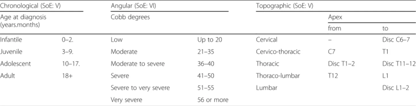
During the years, many different classifications of idio- pathic scoliosis have been proposed, but not all of them are either relevant for conservative care or currently used beyond research purposes. Recent developments in 3D reconstructions of all spine deformities using standard or digital radiography allow to deepen the analysis of the scoliosis deformity in all space planes. In the text, we present the classifications endorsed by SOSORT Consensus (Table 3).
Chronological
James [52, 53] proposed that scoliosis should be classi- fied based on the age of the child at which the deformity was diagnosed (Table 3). This classification is important since the longer the period between diagnosis of scoli- osis and completion of growth by the developing child, the greater the risk of developing a more severe and complicated deformity. Today, the general term “Early onset scoliosis” is sometimes used to classify together Infantile and Juvenile scoliosis, but we prefer the James
Table 3Classifications of idiopathic scoliosis
Chronological (SoE: V) Angular (SoE: VI) Topographic (SoE: V)
Age at diagnosis (years.months)
Cobb degrees Apex
from to
Infantile 0–2. Low Up to 20 Cervical – Disc C6–7
Juvenile 3–9. Moderate 21–35 Cervico-thoracic C7 T1
Adolescent 10–17. Moderate to severe 36–40 Thoracic Disc T1–2 Disc T11–12
Adult 18+ Severe 41–50 Thoraco-lumbar T12 L1
Severe to very severe 51–55 Lumbar Disc L1–2
Very severe 56 or more
classification, due to the fact that infantile scoliosis has a different prognosis. In fact, there are congenital postural scoliosis curves diagnosed in newborns, as a component of a syndrome usually resulting from intrauterine com- pression caused by malposition of the fetus during preg- nancy, but they represent exceptional conditions. Such curvatures are not three-plane deformities and usually undergo spontaneous remission. As the range of hip mo- tion is often asymmetrical and the child prefers to rest their head on one side only, exercises and correction of body position are usually employed. Examination usually reveals gradual remission of the curvature in these in- fants, and such scoliosis curves may thus be categorized as regressive [54].
Angular
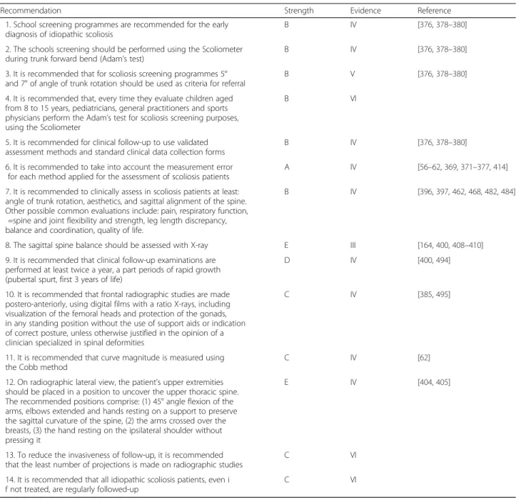
The angle of scoliosis measured on the standing frontal radiograph according to the Cobb method is one of the decisive factors in managing idiopathic scoliosis, and it is directly correlated to all treatment decisions. Many different classifications have been proposed based on these angular measurements, but no one system today has widespread validity. Nevertheless, there is an agree- ment on some thresholds [32, 34, 55–57]:
Under 10° of scoliosis, the diagnosis of scoliosis should not be made. The inter-reliability of the Cobb angle is well known, and the potential limitation of this criterion are clear. On the other hand, a clear and simple criterion is needed for a generally accepted and a simple agreed definition of structural scoliosis.
Over 30° of scoliosis, the risk of progression in adulthood increases, as well as the risk of health problems and reduction of quality of life.
Over 50°, there is a consensus that it is almost certain that scoliosis is going to progress in
adulthood and cause health problems and reduction of quality of life.
From these thresholds, and taking into account that the recognized measurement error in measuring Cobb angles is 5° [58–63], very important decisions are made.
When measured manually on the radiograph, the most commonly cited measurement error of Cobb angle is in- deed 5° [58–63]. However, new computer-assisted meas- urement methods have lesser measurement errors, ranging from 1.22° to 3.6° [64]. When making clinical decisions, measurement error thresholds of a corre- sponding method used should be taken into account.
Topographic
Most commonly used classifications of idiopathic scoliosis are based on the anatomical site of the spinal deformity in the frontal plane. A classification developed by Ponseti [65]
(based on Schulthess work [66]) distinguishes four major types of scoliosis: thoracic, lumbar, thoraco-lumbar and S- shaped. This classification is the oldest. It is reported in Table 3. It is used both in conservative treatment and in the pre-operative classification of scoliosis [67]. Two other clas- sification systems of idiopathic scoliosis based on the ana- tomical site of spinal deformity have been proposed and used in preoperative planning [68–73]. The most widely used for operative treatment is Lenke classification [69].
This classification however uses some objective criteria that make it not applicable to be used for non-operative treat- ment. Mild scoliosis with indication for non-operative treat- ment, specific exercises or bracing, cannot be properly classified according to Lenke objective criteria. Patients under non-operative treatment rarely are prescribed a side bending radiograph, and even in that case, the criterion of
“finding a residual coronal curve on side-bending radio- graphs of at least 25° in the proximal thoracic, main thor- acic, thoracolumbar or lumbar regions, as a definition of a structural curve”, is not applicable to scoliosis in the range of 15° to 30°. Since these guidelines concern conservative treatment, the abovementioned classification is not dis- cussed beyond here. Moreover, efforts were made to clinic- ally evaluate the third dimension, mainly for surgical purposes; recently, several 3D classifications have been pro- posed [74–82], but the most useful one in clinical practice is yet to be defined [83].
Rigo classification
Many clinicians and brace developers base the treatment on some general and individualized criteria [84, 85], rather than to a classification able to guide brace fitting and con- struction as in the Rigo Cheneau brace and in the Spinecor System [73, 86]. The Rigo classification has been accepted (LoE VI) by these guidelines. They have been developed specifically to correlate with Rigo-Chenau brace design and treatment. The Rigo Cheneau classification was devel- oped in order [72] to define specific principles of correc- tion required for efficacious brace design and fabrication.
The classification includes radiological as well as clinical criteria. The radiological criteria are utilized to differenti- ate five basic types of curvatures including (I) imbalanced thoracic (or three curves pattern), (II) true double (or four curve pattern), (III) balanced thoracic and false double (non 3 non 4), (IV) single lumbar and (V) single thoracol- umbar. In addition to the radiological criteria, the Rigo Classification incorporates the curve pattern according to SRS terminology, the balance/imbalance at the transitional point, and L4-5 counter-tilting. This classification has been evaluated for intra-and inter-observer reliability: the intra- observer Kappa value was 0.87 (acceptance > 0.70); the inter-observer Kappa values fluctuated from 0.61 to 0.81 with an average of 0.71 (acceptance > 0.70) [72].
Evidence-based clinical practice approach to idiopathic scoliosis during growth
Goals of conservative treatment General goals
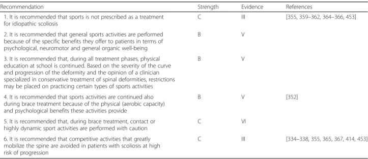
A SOSORT 2005 Consensus paper, titled“Why do we treat adolescent idiopathic scoliosis? What do we want to obtain and to avoid for our patients” [34], can serve as reference for specific insights on this topic. In the present guidelines, the most general goals of treatment are presented in Table 4 [34].
The goals of conservative treatment of idiopathic scoli- osis may be divided into two groups: morphological and functional. The first aspect is related to aesthetics which was defined as the first goal of treatment by SOSORT experts. Both aspects are related to patients’quality of life, psychological well-being and disability (defined as the second, third and fourth goals according to the SOSORT experts) [34]. For didactic reasons, the goals will be present here in a different order. The basic objectives of comprehensive conservative treatment of Idiopathic Scoliosis are as follows:
1. To stop curve progression at puberty (or possibly even reduce it)
2. To prevent or treat respiratory dysfunction 3. To prevent or treat spinal pain syndromes 4. To improve aesthetics via postural correction
To stop curve progression at puberty (or possibly even reduce it) Recently, a multi-centre RCT demon- strated that bracing is effective at preventing progression to the surgical range (defined as ≥50°) [87], although on average the curves did not improve. Moreover, a long-term RCT found that PSSE improved Cobb angles at skeletal maturity in patients with AIS [88]. Current evidence sug- gests that conservative treatment for scoliosis is effective at stopping curve from progression, as well as improving the curves at skeletal maturity.
It is possible and usually sufficient to prevent further pro- gression, even if recent research papers conducted accord- ing to the SRS criteria have shown that it is also possible to obtain some amount of curve correction [89–93].
To prevent or treat respiratory dysfunctionsThe mor- phological aspect of the deformity is closely related to the effects on bodily function. Depending on its degree and location, the curvature may affect respiratory function. The most prominent changes within the respiratory system are produced by curvatures of the thoracic spine [94–97].
To prevent or treat spinal pain syndromes Statistically significant differences in pain prevalence are already noted in people with scoliosis between 20 and 30 years of age. In a follow-up study of over 40 years’duration, a three-fold higher prevalence of chronic pain-related complaints and over 20-fold higher incidence of severe and darting pain were observed in a group of people with untreated idiopathic scoliosis compared to a control group. The occurrence of pain-related complaints is probably multi- factorial in origin [33, 50, 98–101].
In adult with spinal deformities, sagittal parameters influ- ence pain the most as compared to the magnitude of scoli- osis curve [102]. The assessment of regional and global alignment parameters in full-length standing postero- anterior and lateral, as well as pelvic parameters, is strongly recommended due to their relation with pain and disability [103]. In addition, pain is significantly correlated to three dimensional olysthesis, L3 and L4 endplate obliquity angles, loss of lumbar lordosis, and thoracolumbar kyphosis [102].
The SRS-Schwab classification based on curve type and magnitude associated with specific index based on sagittal pelvic and spine parameters has been showed to be reli- able and to correlate with quality of life in adults with spinal deformities [104]. This new classification suggests that there are specific parameters able to predict the risk of pain and disability, in adulthood. Currently, no studies have confirmed if it is possible to treat sagittal alterations during growth, or if the conservative treatment play a role in creating unbalanced spine in adults previously braced, nor if the same treatment is able to prevent future alter- ation of the sagittal profile of the spine and pelvis. Despite this knowledge gap, there is a general agreement among experts that the best possible treatment should take into account not only the correction of the spine in the coronal plane but also the maintenance or the restoration of the normal sagittal profile of the spine.
To improve the appearance via postural correction Quality of life is significantly affected by aesthetic self- perception and appearance. Therefore, visual correction of scoliosis-related external trunk deformity is an important Table 4Goals of treatment according to the SOSORT
consensus paper. Only the goals that reached 80% of agreement are listed here, starting from the most important Esthetics
Quality of life Disability Back pain
Psychological well-being Progression in adulthood Breathing function Scoliosis Cobb degrees
Need of further treatments in adulthood
issue in conservative treatment. Therapeutic outcomes may be subjectively visually assessed using specifically de- signed questionnaire or objectively assessed using surface topography and photographic methods [13, 105–111].
Specific goals of conservative treatment during growth Specific goals of conservative treatment for patients during growth should be set at baseline (X-ray before treatment).
These goals should be considered as a dynamic continuum, which can be adapted during treatment according to the change in the patient clinical status (change in deformity, compliance with the treatment, proposed therapies, etc.). In this respect, we can define the following goals:
Absolute goal: these are the minimum expected goals of conservative treatment. If not anything else, at least these goals should be reached.
Primary goals: these are the“best possible”goals for patients starting treatment in each specific clinical situation.
Secondary goals: these are the compromise goals that come when it becomes clear that it is not possible to reach the primary goals
According to this approach, SOSORT has reached a Con- sensus (Strength of Evidence VI—Strength of Recommen- dation C) shown in Table 5. This table has been organized with a minimum and a maximum of primary and second- ary goals that can be reached for each clinical situation.
The absolute goals for all patients in every clinical situation are to avoid fusion surgery. A first similar scheme had been proposed in 2007 [112]: these goals were applied in some studies [90, 91, 112] and proved to be useful. Accordingly, we propose these goals of treatment here to be applied in clinical studies of conservative treatment of idiopathic scoliosis.
Evidence-based clinical practice approach
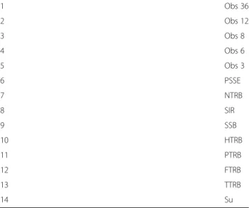
This section is constituted mainly by a Practical Ap- proach Scheme (PAS) (Table 6) that has been prepared
through the Consensus Procedure reported in (Add- itional file 1). The PAS constitutes an Evidence-Based Clinical Practice approach to idiopathic scoliosis. The Level of Evidence of PAS is VI, while the Strength of Recommendation is B.
Here, we present a Strength of Treatments Scheme (STS) (Table 7) that reports all the possible treatments that can be proposed for Idiopathic Scoliosis starting from the least to the most demanding (both in terms of challenge for the patient, and possible efficacy). In addition, the STS is Consensus based (Level of Evidence V—Strength of Recommendation B). Starting from the STS, it is possible to state, for each single clinical situ- ation of the PAS, a minimum and a maximum of possible treatments that could be proposed: conse- quently, all treatments that in the STS are reported between this minimum and maximum can be considered for that specific clinical situation. Tables 8 and 9 show the number of paper for each Level of Evidence and the Strength of recommendation for each treatment.
The PAS has some main characteristics that constitute its strength and justification:
PAS is proposed to resolve the differences in treatment decisions between different clinicians in their clinical practice. PAS guards against
presumably wrong clinical decisions (above maximum: overtreatment, below minimum:
undertreatment).
It reports a real approach, since most clinicians usually choose a variety of treatments for a single patient; the final decision comes after discussion with the patient, and weighting the various risk Table 5Specific aims of conservative treatment during growth
(strength of evidence VI–strength of recommendation C) at least 70% of agreement (SoE VI)
Absolute aim of treatments Percentage
Avoid surgery 90.70
Improve aesthetics 86.05
Improve quality of life 82.56
Degree of curve Primary aim Secondary aim
Low Remain below 20° Remain below 45°
Moderate Remain below 30° Remain below 45°
Severe Remain below 45° Postpone surgery
Table 6Practical approach scheme (PAS) for an evidence-based clinical practice approach to idiopathic scoliosis (strength of evidence VI–strength of recommendation B)
1 Obs 36
2 Obs 12
3 Obs 8
4 Obs 6
5 Obs 3
6 PSSE
7 NTRB
8 SIR
9 SSB
10 HTRB
11 PTRB
12 FTRB
13 TTRB
14 Su
factors involved in the clinical situation. In fact, the PAS has been developed according to the“Step by Step”Sibilla’s theory [92,112–115], which states that for each patient, it is mandatory to choose the correct step of treatment, where the most efficacious is also the most demanding. Accordingly, coming to a wrong decision means facing one of the two main mistakes in conservative treatment of idiopathic scoliosis, overtreatment (too much burden on the patient, without improved efficacy) or
undertreatment (treatment that leads to little or no efficacy).
Evidence-Based Clinical Practice is by definition the best integration between the knowledge offered by Evidence-Based Medicine, individual clinical expertise and patients’preferences [116–118]. Consequently, different clinicians will treat a patient with the same
clinical problem differently; the variation can be due to the patient’s preferences or because of the specific expertise of the clinician. Therefore, proposing a definitive clinical approach for a certain clinical situation is problematic. Rather, a range of options should be considered.
Conservative treatments
All the treatment approaches below are listed in the STS (Table 7) and are presented from the treatments having least impact to those having greatest impact. For more details about each approach, it is possible to refer to the Brace Technology and the Rehabilitation Schools for Scoliosis Series [119, 120] and the Consensus paper on Terminology [121], published by theScoliosis and Spinal disordersjournal.
Nothing (No): No treatment is needed.
Table 7Strength of treatments scheme (STS) (strength of evidence V–strength of recommendation B): it reports all the possible treatments that can be proposed for idiopathic scoliosis graduated from the less to the most demanding (both in terms of burden on
Low Moderate Severe
Min Max Min Max Min Max
Infantile Obs3 Obs3 Obs3 TTRB TTRB Su
Juvenile Obs3 PSSE PSSE FTRB HTRB Su
Adolescent Risser 0 Obs6 SSB HTRB FTRB TTRB Su
Risser 1 Obs6 SSB PSSE FTRB FTRB Su
Risser 2 Obs6 SSB PSSE FTRB FTRB Su
Risser 3 Obs6 SSB PSSE FTRB FTRB Su
Risser 4 Obs12 SIR PSSE FTRB FTRB Su
Adult up to 25 y Nothing PSSE Obs12 SIR Obs6 Su
Adult No Pain Nothing PSSE PSSE SIR Obs12 HTRB
Pain PSSE SSB PSSE HTRB PSSE Su
Elderly No Pain Nothing PSSE Obs36 PSSE Obs12 HTRB
Pain PSSE SSB PSSE HTRB PSSE Su
trunk decompensation Obs6 SSB PSSE PTRB PSSE Su
Table 8Level of evidence of recommendations: the table shows the number of papers according to the level of evidence for each treatment
I II III IV V VI Total
Bracing 2 3 3 6 12 1 25
Specific exercises to prevent scoliosis progression during growth
1 1 1 0 8 1 12
Specific exercises during brace treatment and surgical therapy
0 3 0 0 3 0 6
Other conservative treatments 0 0 0 0 2 0 2
Respiratory function and exercises 0 0 0 0 3 0 3
Sports activities 0 0 2 0 3 1 6
Assessment 0 0 1 9 1 3 14
Total 3 7 7 15 32 6 68
Observation (Ob): It is the first step of an active ap- proach to idiopathic scoliosis, and it consists of regular clinical evaluation with a specific follow-up period. Tim- ing of this follow-up can range from 2 to 3 to 36–
60 months according to the specific clinical situation.
Clinical evaluation does not need to include taking ra- diographs: radiographs are usually performed during al- ternate clinical evaluations.
Physiotherapeutic scoliosis-specific exercises (PSSE): PSSE include all forms of outpatient physiotherapies with evi- dence of having an effect on some scoliosis outcomes and which will gradually be published in the Rehabilitation Schools for Scoliosis Series [120] in theScoliosis and Spinal Disordersjournal. They have been listed in the 3rd part of these guidelines. The frequency of therapeutic sessions var- ies from twice to 7 days a week depending on the complex- ity of the techniques, motivation and the ability of the patient to carry out the treatment. Long-term outpatient physiotherapy sessions usually take place two to four times a week if the patient is willing to cooperate fully. The actual form of exercise depends mainly on the character of the se- lected therapeutic method.
Special Inpatient Rehabilitation (SIR): If SIR is recom- mended, patients spend several weeks (usually 3–6) at a specialized health centre (hospital department, sanatorium or a similar form of health care) where they undergo an in- tensive PSSE treatment (several hours per day).
Bracing: It consists of using a brace (a corrective orth- osis) for a specified period of time each day. Usually, it is worn until maturity. The main therapeutic goal is to halt the scoliosis curves from progression. According to SOSORT, the use of a rigid brace implies the use of ex- ercises when out of the brace. Bracing includes the following:
Night Time Rigid Bracing(8–12 h per day)(NTRB):
wearing a brace mainly in bed.
Soft Bracing (SB): it includes mainly the SpineCor brace [122,123] but also other similar designs [124,125].
Part Time Rigid Bracing(12–20 h per day)(PTRB):
wearing a rigid brace mainly outside school and in bed.
Full Time Rigid Bracing(20–24 h per day)or cast (FTRB): wearing a rigid brace all the time (at school, at home, in bed, etc.). Casts have been included here as well. Casts are used by some schools as the first stage to achieve correction to be maintained afterwards with rigid brace [126–128]; a cast is considered a standard approach in infantile scoliosis [129–132]. Recently, a new brace has been
developed that has been claimed to achieve same results as casting [91,133,134].
A common feature of all forms of conservative treatment is the need to actively involve the patient and caregivers [135]. Therefore, education, psychotherapy, systematic monitoring of outcomes, assessment of patient’s compli- ance, and verification and modification of methods in the course of the therapy are deemed crucial elements of con- servative treatment. In order to achieve the best possible outcome, conservative treatment should be delivered by an experienced therapeutic team including a physician, a physiotherapist, an orthotist and possibly a psychologist [135]. Support groups and Internet forums are also import- ant in conservative treatment.
Prognostic factors
Prognostic factors should be used with PAS, to select options appropriately between the minimum and max- imum strength of treatment. The following factors have been suggested as possible determinants of a higher risk of scoliosis progression: positive family history, laxity of skin and joints (connective tissue defect), flattening of physio- logical thoracic kyphosis (impedes efficient bracing), angle of trunk rotation exceeding 10°, and growth spurt [136].
Bunnell reported that the risk of progression at the be- ginning of puberty is 20% in 10° scoliosis, 60% in 20° scoli- osis, and as much as 90% in 30° scoliosis [55, 137]. At the age of peak height growth (13 years of osseous age in girls), Table 9Strength of recommendations: the table shows the strength of recommendation for each treatment
A B C D E Total
Bracing 2 20 3 0 0 25
Specific exercises to prevent scoliosis progression during growth
0 7 5 0 0 12
Specific exercises during brace treatment and surgical therapy
0 2 4 0 0 6
Other conservative treatments 0 0 2 0 0 2
Respiratory function and exercises 0 1 2 0 0 3
Sports activities 0 3 3 0 0 6
Assessment 1 6 4 1 2 14
Total 3 39 23 1 2 68
the risk of progression is 10, 30 and 60%, in the curve se- verity threshold categories above, respectively. During the final stage of puberty (at least Risser grade II), the risk of deformity progression becomes considerably lower, falling to 2% in 10° scoliosis, 20% in 20° scoliosis and 30% in 30°
scoliosis. The prognosis regarding IS progression seems to be more optimistic for boys [138].
Considering that the sagittal spine profile of mild (10°–20°) scoliotic curves was found to be similar to the lateral spine profile of their healthy controls [139], it has been proposed that thoracic hypokyphosis, coupled with axial rotation, could be compensatory rather than etio- logical in IS pathogenesis [140].
Scoliosis can affect the spine not only through transla- tion in the frontal, and rotation about horizontal plane, but also through changes in the sagittal profile of the spine. Different types of scoliosis present with different sa- gittal profiles; one example is the typical association of flat back in thoracic scoliosis. Although the etiology of scoli- osis is unknown, some authors have hypothesized that pa- tients with certain sagittal spinal profiles seem more prone to developing scoliosis than others [141–145]. It has been demonstrated that the sagittal profile of the spine depends on the pelvic placement playing a major role in determining the sagittal balance of the spine [146–149].
The pathologic mechanism of progression of IS curve is described in recent publications [46, 47, 150, 151].
The factors that contribute to progression include the effect of gravity, the muscle action, the reactive forces causing increased lordosis, the human gait, and the growth induced torsion. The intervertebral disc could be included as an additional morphological factor involved in the progression of an IS curve [120, 152, 153].
Recently developed genetic assessment, with 53 identi- fied loci [56, 154], can now help predict the risk of IS progression. The determination of the polymorphism of selected genes is meant to facilitate the assignment of a patient to a progressive or stable group [155–157]. Un- fortunately, the data originating from one population often are not confirmed in replication studies involving other populations [158, 159]. A prognostic genetic test, known as ScoliScore, has also been developed [160]. Al- though these initial results have been promising, their generalizability is still uncertain [161].
Finally, during recent years, there have been several prognostic formulas that have been proposed [48, 162, 163]. The previous SOSORT guidelines [3] were based on the Lonstein and Carlson factor of progression [48]
for the assessment of the risk of idiopathic scoliosis.
Since there are no formulas that have been applied in specific studies after their development to verify their real accuracy, we do not apply them in these guidelines.
The wide range of normative values, already demon- strated in large population of healthy children, and the
recognized changes of pelvic and sagittal parameters during growth [164, 165] can significantly affect these results and make it very difficult to reach definite con- clusion. In addition, curve magnitude influences the sa- gittal profile of the spine. Therefore, some differences may be related to the mean Cobb angle of the popula- tion included in each study. Even though there still re- main many unanswered questions, it appears that the sagittal parameters are correlated with the development of the spinal deformities, and we recommend they be monitored during therapy.
Brace treatment Methods
In November 2015, we performed a search in MEDLINE from its inception, with no language limitations. We used the following search strategies:
“Braces”[Mesh] AND“Scoliosis”[Mesh] AND (has abstract[- text] AND (Clinical Trial[ptyp] OR Meta-Analysis[ptyp] OR Practice Guideline[ptyp] OR Randomized Controlled Trial[p- typ] OR Review[ptyp])) (198 papers).
(“Scoliosis/therapy”[Mesh]) AND“Braces”[Mesh] AND compliance (100 papers)
“Scoliosis”[Mesh] AND “Braces”[Mesh] AND (“infant, newborn”[MeSH Terms] OR“infant”[MeSH Terms:noexp]
OR“child, preschool”[MeSH Terms]) (194 references).
We selected from the titles a total of 250 references, and looking at the abstracts, 102 were selected and retrieved in full text. We also searched the following: the abstracts of all SOSORT meetings, from the first one in 2003 to 2010; the personal files and knowledge of all the authors;
the articles retrieved with all the other searches listed in these guidelines; and the references sections of all re- trieved papers. The selection criteria used in all these searches were as follows: pertinence for the topic “Brace treatment”; presence of the abstract; numerical results in relation to scoliosis; retrievability in full text; all languages.
Results
SOSORT has published in Scoliosis and Spinal Disorders Journaltwo Consensus Papers on bracing titled“SOSORT consensus paper on brace action: TLSO biomechanics of correction (investigating the rationale for force vector selection)”[131] and“Guidelines on‘Standards of manage- ment of idiopathic scoliosis with corrective braces in every- day clinics and in clinical research’: SOSORT Consensus 2008” [135]; in addition, previously published guidelines are also freely available in the Journal web page [3], which can serve as reference for specific insights.
Efficacy in adolescents
Recently, a Cochrane review and its update [166–168]
found that there is very low-quality evidence in favour of
using braces, making generalization very difficult. This review included seven articles:
five were planned as RCTs [93, 123, 169–171] and two as prospective controlled trials [90, 172, 173]. One of the RCTs failed due to very low recruitment of participants [174], while another [93] introduced a preference arm for the same reason.
Nachemson multicenter prospective international obser- vational study provided very low-quality evidence in favour of the efficacy of bracing [173]. Nachemson evalu- ated 240 patients with thoracic or thoracolumbar curves between 25° and 35°, aged between 10 and 15 years, of which 129 were only observed and 111 treated with thora- columbar braces. Progression of 6 or more degrees at any of 2 radiographic follow-ups was considered failure of the selected treatment (observation versus brace treatment).
At 4 years of follow-up, the success rate for brace treat- ment was 74% (range, 52—84%), whereas the rate for ob- servation was 34% (range, 16—49%).
In prospective trials, the results were in favour of brace [90]: Lusini reported that the rate of success (no progression of 5° or more, no fusion, or no waiting list for fusion) was 25/33 in the brace group and 0/10 in ob- servation group in the per-protocol analysis (RR 15.21, 95% CI 1.00 to 230.23) and 31/39 in the brace group and 8/18 in the observation group in the ITT analysis (RR 1.79, 95% CI 1.04 to 3.07).
A randomized controlled trial demonstrated with very low-quality evidence that a plastic TLSO brace is more effective than an elastic brace [171]. Wong randomized 43 subjects to SpineCor or rigid orthosis group. Al- though it has been stated that the authors were not trained to fit the SpineCor brace [175], the authors con- cluded that 68% of the subjects in the SpineCor group and 95% of the subjects in the rigid orthosis group did not show curve progression, with a significant difference in favour or rigid braces. The two groups had similar re- sponses to a patient acceptance questionnaire.
In a randomized controlled trials with a 2 years’follow- up (116 participants from the randomized cohort), Wein- stein found that the mean PedsQL did not differ signifi- cantly between bracing [87] and observation groups and found that the rate of success (curves remaining below 50°) was 38/51 in the brace group and 27/65 in the obser- vation group (RR 1.79, 95% CI 1.29 to 2.50).
The Cochrane review concluded that bracing pre- vented curve progression. The presence of failure of RCTs due to parents rejecting randomization of their children demonstrates important difficulties in conduct- ing RCTs in the field of conservative treatment for scoli- osis. Future research should focus on participant outcomes, adverse effects, methods to increase compli- ance, and usefulness of physiotherapeutic scoliosis- specific exercises added to bracing.
RCTs and prospective cohort studies should be con- ducted according to pre-defined criteria such as the Scoliosis Research Society (SRS) and the international Society on Scoliosis Orthopedic and Rehabilitation Treatment (SOSORT) criteria.
In fact, beyond the previously reported studies, the SRS defined some methodological criteria to be followed during brace cohort studies [176]. The optimal inclusion criteria consist of: age 10 years or older when brace is prescribed, Risser 0–2, primary curve angles 25°–40°, no prior treatment, and, if female, either pre-menarche or less than 1 year post menarche. Assessment of brace ef- fectiveness should include (1) the percentage of patients who have ≤5° curve progression and the percentage of patients who have ≥6° progression at maturity, (2) the percentage of patients with curves exceeding 45° at ma- turity and the percentage who have had surgery recom- mended/undertaken, and (3) 2-year follow-up beyond maturity to determine the percentage of patients who subsequently undergo surgery. All patients, regardless of subjective reports on compliance, should be included in the results (intent to treat). Every study should provide results stratified by curve type and size grouping. Cohort studies respecting the SRS criteria can be considered of high methodological quality. Until now, 12 papers have been published with these characteristics and 6 of them in the last 4 years [123]. Recently, a consensus statement aimed to establish a framework for research with clearly delineated inclusion criteria, methodologies, and out- come measures to allow better and easier, future meta- analysis or comparative studies was organized in con- junction with the SOSORT and SRS society [177].
Together with these criteria, SOSORT offered the“Stan- dards of management of idiopathic scoliosis with correct- ive braces in everyday clinics and in clinical research”
[135] that include 14 recommendations, grouped in 6 do- mains (Experience/competence, Behaviours, Prescription, Construction, Brace Check, Follow-up). Cohort studies using the SOSORT criteria can be considered of high quality in terms of patient and treatment management.
Until now, six papers have been published with these characteristics [89, 90, 92, 178–185].
With regard to the studies that were conducted using the SRS and/or SOSORT criteria we found:
Janicki et al. [179], following the SRS criteria, retro- spectively compared in an “intent-to-treat” analysis the effectiveness of the custom thoracolumbosacral (TLSO) worn 22 h/day and the Providence orthosis worn 8–
10 h/night. There were 48 patients in the TLSO group and 35 in the Providence group. In the TLSO group, only 7 patients (15%) did not progress (≤5°), whereas 41 patients (85%) progressed by 6° or more, including the 30 patients whose curves exceeded 45°. Thirty-eight pa- tients (79%) required surgery. In the Providence group,
11 patients (31%) did not progress, whereas 24 patients (69%) progressed by 6° or more, including 15 patients whose curves exceeded 45°. Twenty-one patients (60%) required surgery. Nevertheless, the two groups consid- ered were not fully comparable at baseline.
Coillard et al. [178], following the SRS criteria, studied prospectively a cohort of 254 patients treated with the Spi- neCor brace. Successful treatment (correction > 5° or stabilization ± 5°) was achieved in 165 of the 254 patients (64.9%). Forty-six immature patients (18.1%) required sur- gical fusion while receiving treatment. Two patients out of 254 (0.7%) had curves exceeding 45° at maturity.
Negrini et al. [92], following both the SRS and SOSORT criteria, retrospectively studied a cohort of 42 females and four males treated according to individual needs, with Risser casts, Lyon or SPoRT braces (14 for 23 h per day, 23 for 21 h/d, and seven for 18 h/d at start). No patient progressed beyond 45°, nor was any patient fused, and this remained true at the 2-year follow-up for the 85% that reached it. Only two patients (4%) worsened, both with single thoracic curve, 25–30°
Cobb and Risser 0 at the start.
Aulisa et al. [89], following both the SRS and SOSORT criteria, retrospectively reviewed a cohort of 50 adolescent females with thoraco-lumbar curves treated with the Pro- gressive Action Short Brace (PASB). Curve correction was accomplished in 94% of patients, whereas a curve stabilization was obtained in 6% of patients. No patient re- quired surgery, nor anyone progressed beyond 45°.
Aulisa et al. [184] following both the SRS and SOSORT criteria retrospectively reviewed a cohort of 40 adolescent females with lumbar curves treated with the Progressive Action Short Brace (PASB). Curve correc- tion was accomplished in 82.5% of patients, whereas curve stabilization was obtained in 17.5% of patients.
None of the patients experienced curve progression.
Gammon et al. [180], following the SRS criteria, com- pared treatment outcomes of two cohorts of patients treated via either a conventional rigid thoracolumbosacral orthoses (TLSO: 35 patients) or a SpineCor non-rigid orthosis (32 patients). No significant difference was found using the more strict outcome measure (≤5° curve pro- gression) as the success rates were 60% for TLSO and 53%
for SpineCor. Looking at patients who reached 45°, the success rates were 80% for TLSO and 72% for SpineCor with no significant difference. Guo et al. [186] following SRS criteria studied two groups: SpineCor (n= 20) or rigid brace (n= 18). Before skeletal maturity, 7 (35.0%) patients in the SpineCor Group and 1 (5.6%) patient in the Rigid brace group had curve progression > 5°.
Zaborowska-Sapeta et al. [187], including patients ac- cording to the SRS criteria, prospectively followed 79 pa- tients treated with Cheneau brace. At 1 year after weaning the brace, they found improvement in 25.3%, stabilization
in 22.8%, progression of the Cobb angle up to below 50°
in 39.2% and progression beyond 50° in 12.7%; the latter was considered surgical indication.
Aulisa et al. [183] following both the SRS and SOSORT criteria, studied prospectively 163 patients treated with PASB, Lyon brace and Milwaukee affected by juvenile idio- pathic scoliosis. Curve correction was accomplished in 88 patients (77.8%); stabilization was obtained in 18 patients (15.9%). Seven patients (6.19%) have a progression and 4 of these were recommended for surgery. Of 26 patients who abandoned the treatment, at the time of abandonment (12.5 age), 19 cases (70.0%) have achieved curve correction, 5 cases (19%) stabilized, and 3 cases (11%) progressed.
Negrini et al. [181], in a prospective cohort study of 73 patients, treated with the Sforzesco brace, following both the SRS and SOSORT criteria, reported 34 patients (52.3%) improved; seven (9.6%) worsened, of which 1 pro- gressed beyond 45° and was fused and employing intent- to-treat analysis, there were failures in 11 patients (15.1%).
Finally, Aulisa et al. [182] following both the SRS and SOSORT criteria, studied a cohort of 102 patients treated with Lyon Brace, who were drawn from a pro- spective database and found the following: 69 patients had a definite outcome, 17 have abandoned treatment and 16 are still in treatment. Curve correction was ac- complished in 85.5% of patients, curve stabilization was obtained in 13% of patients and curve progression was evident in only 1.5%. None of the patients were recom- mended surgery post-bracing. Of 17 patients who aban- doned the treatment, at the time of abandonment (14.4 age), 13 cases (77%) have achieved curve correction, 53 cases (18%) stabilized, and 1 case (5%) progressed.
In summary, these studies show a high variability among the results of bracing [90, 92, 178–184, 187, 188]. This is particularly high for rigid bracing [90, 92, 178–184, 187, 188] despite the results of the treatment being better in the recent studies [90, 181–184]. The soft braces [122, 123, 178, 180] can have a high variability of results, from better to worst [179, 180], as compared to some types of rigid braces; the best results have been achieved with the rigid once, when applying the SOSORT criteria [92, 181–184, 187, 188]. It must also be noted that high variability can be found between dif- ferent publications in the type of scoliosis treated, thus a different outcome in treatment.
Recently, Weinstein et al. [87] performed a randomized controlled study, but the trial was stopped early owing to the efficacy of bracing, the rate of treatment success was 72% after bracing, as compared with 48% after observation.
The authors concluded that bracing significantly decreased the progression of high-risk curves to the threshold for sur- gery in patients with adolescent idiopathic scoliosis. This study is in contrast with results [189] of a systematic review published earlier by Dolan. The systematic review included