SECONDARY METABOLITES ISOLATED FROM PEROVSKIA ATRIPLICIFOLIA AND THEIR IN VITRO BINDING AFFINITY FOR HUMAN OPIOID AND
CANNABINOID RECEPTORS
by
Ashli Fitzpatrick
A thesis submitted to the faculty of The University of Mississippi in partial fulfillment of the requirements of the Sally McDonnell Barksdale Honors College
Oxford, MS December 2014
Approved by
____________________________
Advisor: Dr. Stephen Cutler
____________________________
Reader: Dr. Francisco Leon ____________________________
Reader: Dr. Donna West-Strum
©2014 Ashli Fitzpatrick ALL RIGHTS RESERVED
ACKNOWLEDGEMENTS
I would like to thank my advisor Dr. Stephen Cutler and Dr. Francisco Leon for all of their devoted time, encouragement, and guidance through this meticulous process. I greatly appreciate Dr. Cutler for allowing me to work in his laboratory and giving me the resources I needed to complete this thesis project. I would like to thank Dr. Leon for guiding me through the experimental procedures involved and explaining all of the scientific literature and procedures in great detail.
I would like to thank Dr. Debra Young and the Sally McDonnell Barksdale Honors College for giving me the opportunity to complete such an interesting research project and providing resources and encouragement during stressful times.
I would lastly like to thank my family and friends for their unyielding support. I could not have completed this thesis without them.
ABSTRACT
ASHLI FITZPATRICK: Secondary Metabolites Isolated from Perovskia atriplicifolia and Their In Vitro Binding Affinity for Human Opioid and Cannabinoid Receptors
(Under direction of Dr. Stephen Cutler)
Plants and their natural products have been utilized for thousands of years in the treatment of many human diseases. Because of their structural diversity, natural products are ideal lead compounds for drug development. In this thesis, the natural products of the plant Perovskia atriplicifolia were investigated, specifically their in vitro binding affinity to receptors in opioid and cannabinoid classes. Extracts that demonstrated good selective opioid and cannabinoid receptor binding were subjected to bioassay-guided fractionation.
Four active compounds were identified from the crude extract in Perovskia atriplicifolia:
1) 5-hydroxy-3’,4’,6,7-tetramethoxy flavone, 2) 5,7-dihydroxy-3’,4’,6,-trimethoxy flavone, 3) 5-hydroxy-4’,6,7-trimethoxy flavone, and 4) 5,7-dihydroxy-4’,6,-dimethoxy flavone. Each compound exhibited good selective δ-opioid receptor binding affinity and was characterized as a flavanoid.
TABLE OF CONTENTS
LIST OF TABLES AND FIGURES………...vi
LIST OF ABBREVIATIONS……….viii
BACKGROUND………...1
MATERIALS AND METHODS………..30
RESULTS………..35
CONCLUSION………..47
BIBLIOGRAPHY………..48
APPENDIX………53
LIST OF TABLES AND FIGURES
Figure 1 Chemical structure of paclitaxel 1………...………3
Figure 2 Some important natural products……….4
Figure 3 Chemical structure of penicillin core………..5
Figure 4 Chemical structure of some terpenoids and sterols isolated from Perovskia species………...10
Figure 5 Photo of Perovskia atriplicifolia………....11
Figure 6 Major monoterpenes identified in P. atriplicifolia………....12
Figure 7 Perovstatone A isolated from P. atriplicifolia………...……12
Figure 8 Selective opioid receptor agonists and antagonists………16
Figure 9 Opioid receptor mechanism………...18
Figure 10 Major sites of opioid receptors and endogenous opioid production…..20
Figure 11 Chemical structure of Δ9-tetrahydrocannabinol (THC)………....23
Figure 12 Cannabinoid receptor agonists and antagonists……….23
Figure 13 Location of cannabinoid receptors in the human brain………..25
Figure 14 Structure of cannabinoid receptors, CB1 and CB2……….26
Figure 15 Initial steps and activity for P. atriplicifolia………...36
Figure 16 Fractionation of P. atriplicifolia………38
Figure 17 TLC plate for fraction 4………39
Figure 18 Fractionation for fraction 5 of P. atriplicifolia………..41
Figure 19 1H NMR spectrum of 5-hydroxy-3’,4’,6,7-tetramethoxy flavone…….42
Figure 20 13C NMR spectrum of 5-hydroxy-3’,4’,6,7-tetramethoxy flavone…...43
Figure 21 5-hydroxy-3’,4’,6,7-tetramethoxy flavone, compound A……….44
Figure 22 5,7-dihydroxy-3’,4’,6,-trimethoxy flavone, compound B………….…44
Figure 23 1H NMR spectrum of 5-hydroxy-4’,6,7-trimethoxy flavone…………45 Figure 24 5-hydroxy-4’,6,7-trimethoxy flavone, compound C……….45 Figure 25 5,7-dihydroxy-3’,4’,6,-trimethoxy flavone, compound D…………....46 Table 1 Selective opioid receptor ligands………...17 Table 2 % of displacement of radioligands for subfractions obtained for fraction 4 of P. atriplicifolia………39
LIST OF ABBREVIATIONS ADH Antidiuretic Hormone
BCE Before Christian Era CNS Central Nervous System COSY Correlation Spectroscopy
DEPT Distortionless Enhancement by Polarization Transfer DMEM Dublecco’s Modified Eagle Medium
DNA Deoxyribonucleic acid DOR Delta opioid receptors EtOAc Ethyl Acetate
FDA Food and Drug Administration GPCRs G-protein Coupled Receptors
HMBC Heteronuclear Multiple Bond Coherence HMQC Heteronuclear Multiple Quantum Coherence HMRS High-resolution mass spectra
IASP International Association for the Study of Pain KOR Kappa opioid receptor
MeOH Methanol
MOR Mu opioid receptor NCE New Chemical Entity NCI National Cancer Institute NIH National Institute of Health NMR Nuclear Magnetic Resonance
NOESY Nuclear Overhauser Effect Spectroscopy NOR Nociceptin opioid receptor
OGFr Opioid growth factor receptor THC Δ9-tetrahydrocannabinol TLC Thin-layer Chromatography TMS Trimethylsilane
TOSCY Total Correlated Spectroscopy VLC Vacuum Liquid Chromatography WHO World Health Organization
Background
I. Natural Products as a Resource for Drug Discovery
Natural products, also known as secondary metabolites, refer to chemical compounds or substances produced by living organisms such as plants, animals, or microbes (Clement, 2005). Primary metabolites, which include glucose, DNA, and amino acids, are chemically identical in nearly all organisms and are required for normal
development and growth of the organisms (Clement, 2005). Unlike primary metabolites, secondary metabolites are not directly required by an organism, are typically unique to a specific species or subset of a species, and can offer novelty in structure or function (Amsler et al., 2011). Secondary metabolites are synthesized primarily from the compounds acetate, mevalonate, shikimate, and various amino acids (Gao, 2010).
Because these compounds are also used in the synthesis of essential primary metabolites, it is theorized that the excess compounds were utilized to create non-essential natural products (Gao, 2010). Natural products are created in nature and have naturally evolved with their producers, resulting in biological activity that is increasingly useful and effective (Gao, 2010). Natural product structures are extremely diverse and have high binding affinities to their specific receptor type (Gao, 2010). Because of their structural diversity and receptor binding activity, natural products are ideal bioactive lead
compounds for drug development (Kingston, 2012).
Plants and their natural products have formed the foundation of complex traditional medicine systems and have been a major source of curative products for thousands of
years. Records indicate that hundreds of medicinal plants were utilized in Mesopotamia, dating back to the year 2600 BCE (Cragg and Newman, 2013). Several of these plant- derived substances, ranging from Glycyrrhiza glabra (licorice) to Papaver somniferum (poppy juice) to Cupresses sempevirens (cypress), are still used today as treatments for coughs, colds, parasitic infections, and inflammation (Cragg and Newman, 2013).
According to data retrieved by the World Health Organization (WHO), it is estimated that 80% of the world, primarily in undeveloped countries, depends on traditional medicines for their primary health care, with 65% of that number specifically relying on plant- derived medications (Cragg and Newman, 2005).
Natural products are also utilized in developed countries. Estimates indicate that over 50% of the most-prescribed drugs in the United States owe their beginnings to natural products, either through direct use of the plant or as a precursor in the design or synthesis of the drug (Cragg and Newman, 2005). Most anticancer and anti-infective drugs, however, are estimated to contain an even larger proportion of natural products (Kingston, 2012). The most popular natural-source anticancer drug, paclitaxel (Fig. 1), was isolated from the bark of the Pacific yew tree, Taxus brevifolia, and was approved for use against ovarian cancer by the Food and Drug Administration (FDA) in 1992 (NIH-NCI). Since then, the plant-derived drug has been utilized for the treatment of Kaposi’s sarcoma as well as breast and lung cancer and has become the best-selling cancer drug ever manufactured (NCI). Because of its tremendous success, many researchers attempted to develop similar chemical relatives of paclitaxel 1, resulting in the synthesis of two analogues, docetaxel and cabazitaxel (Cragg and Newman, 2013).
Chemical structure of paclitaxel 1 Figure 1
Many other important drugs have been exploited from traditional medicinal plants.
Vinblastine and vicristine, derivatives of from the Madagascar periwinkle, Catharanthus roseus, are also utilized in the treatment of cancer (Cragg and Newman, 2013). Ephedrine (1), a plant isolated from the plant Ephedra sinica, is the basis for anti-asthma agents salbutamol and salmetrol (Cragg and Newman, 2013). Reserpine (2), an antihypertensive agent isolated from the flowering plant Rauwolfia serpentina, is utilized for the treatment of snakebite (Crag and Newman, 2013). Tubocurarine (3), the muscle relaxant isolated from Chondrodendron and Curarea species, is used as an arrow toxin, curare, by indigenous groups in the Amazon (Cragg and Newman, 2013).
Ephedrine (1) Reserpine (2)
Tubocurarine (3)
Some important natural products Figure 2
It is apparent that natural products will continue to be an important source of novel structural leads and drugs, but despite their prevalence in the pharmaceutical industry, the surface of natural products has barely been scratched. Out of nearly 300,000 species of vascular plants, only 15% have been investigated phytochemically and only 6% have been explored pharmaceutically (Cragg and Newman, 2013). The vast ocean (covering nearly 70% of the earth’s surface) and its countless, unique marine plants and microorganisms remain virtually uncharted as sources for novel medicines (Cragg and Newman, 2013). With the advent of scuba diving techniques, the ocean waters have started to become a worthwhile source of novel drugs, with thousands of novel bioactive compounds emerging from this marine environment (Cragg and Newman, 2013).
Microorganisms, which consist of bacteria, archaea, and almost all protazoa, are also an extremely productive source of structurally diverse natural products and have produced drugs that are prevalent in the pharmaceutical industry. Anti-bacterial agents such as penicillins and cephalosporins are derived from the species of microorganisms
Penicillium and Cephalosporim acremonium, respectively (Fig. 3) (Cragg and Newman, 2013). Microorganisms have also yielded other products including immunosuppressive agents, cholesterol lowering agents, and antiparasitic drugs (Cragg and Newman, 2013).
Chemical structure of penicillin core Figure 3
While natural products remain a vital component in novel drug discovery, the number of new chemical entities (NCE) discovered in recent years has declined significantly (Cragg and Newman, 2013). According to the Food and Drug Administration, a new chemical entity is a “drug that contains no active moiety that has been approved by FDA” (FDA). From 2001-2010, an average of 23 NCE’s were discovered per year, down approximately 62% from the 1980’s (Cragg and Newman, 2013). Despite innovative chemotype breakthroughs, out of 35,000 plant samples processed by the National Cancer Institute (NCI) over a span of twenty years, only two compounds became commercial products--paclitaxel being one of them (Cragg and Newman, 2013).
Although natural products are invaluable in drug discovery, traditional drug innovation has its issues including funding and sustainability. Sources of finance for natural product drug discovery are extremely limited. The few large pharmaceutical companies that utilize natural products will not even discuss funding unless there is activity in a Phase I clinical trial (study conducted to determine drugs various side effects and how it is metabolized and excreted), preferably Phase II clinical trial (study to determine effectiveness of drug) (Cragg and Newman, 2013; FDA). Some companies have opted to employ new, exciting chemical techniques such as combinatorial chemistry to produce novel medicines (Cragg and Newman, 2013). Combinatorial chemistry requires the synthesis of a large amount of compounds (tens to thousands to millions), which are then screened together for specific biological activity (Pandeya and Thakkar, 2005). These techniques reduce the cost and time associated with creating competitive and effectual new drugs (Pandeya and Thakkar, 2005). Despite these advantages, it is not
evident that these techniques are more successful than natural product use in drug discovery, with very few medicines emerging via combinatorial chemistry (Kingston, 2012). To compare natural products and products synthesized via combinatorial chemistry, a variety of molecular properties were investigated to determine structural diversity. These properties included a number of rotatable bonds, degrees of unsaturation, atom types, number of chiral centers, and number of rings and chains (Gao, 2010). Based on the results of this investigation, it was determined that natural products are significantly more diverse than combinatorial compounds (Gao, 2010). Furthermore, natural products have superior drug-like properties compared to samples of compounds synthesized by combinatorial chemistry (Kingston, 2012).
Because the earth’s resources are limited, long-term sustainability is also an important issue when dealing with plant-based traditional medicines. Although there are undoubtedly enough resources for the current provision of natural and synthetic drugs, 20 or 30 years from now those resources will most likely not be available or affordable for a rapidly rising universal population (Cordell and Colvard, 2012). With climate change, deforestation in biologically diverse areas, and indiscriminate collection and overharvesting of plant samples, many important medicinal plants are under threat, or in extreme cases, no longer available (Cordell and Colvard, 2012). Because of this, access to medications has become a worldwide concern, with record supply shortages of anti- cancer drugs reported by the FDA for the United States in 2011 (Cordell and Colvard, 2012). To address supply problems inherent with plant materials, researches must exploit species with similar chemical properties or attempt to produce the product semi- synthetically (Cragg and Newman, 2005).
Despite declining interests and issues with funding and sustainability, it is undeniable that natural products have had a significant impact in the discovery of novel drugs. Not only are natural products the forebears of many anti-cancer drugs, they have also been important in therapeutic areas such as cholesterol management, arthritis, diabetes, and depression (Rouhi, 2003). Barry A. Berkowitz, vice president at Albany Molecular Research, comments on the importance of natural products, stating, “We would not have the top-selling drug class today, the statins; the whole field of angiotensin antagonists and angiotensin-converting-enzyme inhibitors; the whole area of immunosuppressives; nor most of the anticancer and antibacterial drugs. Imagine all of those drugs not being available to physicians or patients today" (Cragg and Newman, 2013).
II. Terrestrial plants of the genus Perovskia
The genus Perovskia belongs to the Family Labiatae, Order Lamiales, Class Magnoliopsida, and is made up of seven different species (Perveen et al., 2009). Native to Central Asia, Afghanistan, Iran, and Pakistan, the plants comprising the Perovskia genus are aromatic sub-shrubs featuring extremely branched stems, paniculate leaves, and small flowers (Saleem, 2000). The most common species are Perovskia artemisoides, Perovskia artiplicifolia, and Perovskia abrotanoides (Saleem, 2000).
Various phytochemical studies focusing on secondary metabolites have been performed on species in the genus Perovskia. This genus has been found to be a source of a variety of secondary metabolites, including essential oils and terpenoids. Many species produce volatile oils that exhibit antibacterial activity (Beikmohammadi, 2011).
Pentacyclic triterpenoids of ursane, oleane and lupane skeleton are usually isolated from plants of Perovskia species, such as; germanicol 2, lupeol 3, oleanolic acid 4, and β- amyrin 5, peradione 6 and sterols like: β-sitosterol 7 and stigmasterol 8 (Fig. 2), have been isolated from the members of the Perovskia genus. Sesquiterpenoids such as betulenol 9 and α-aromdendrene 10 have also been reported in this genus (Saleem, 2000) (Fig. 4).
Chemical structure of some terpenoids and sterols isolated from Perovskia species Figure 4
Species within the Perovskia genus have myriad of uses. In Pakistan, Perovskia abrotanoids is utilized as a refrigerant; in China, the species is employed in treating atherosclerosis, cardiovascular diseases, and liver fibrosis (Beikmohammadi, 2011). The sap of Perovskia abrotanoides and Perovskia artemisioides has even been utilized to treat leishmaniasis, a skin disease caused by protozoan parasites, due to the presence of abundant amounts of fat-soluble leishmanicidal constituents found in the roots of the plants (Sairafianpour et al., 2001).
III. Terrestrial plant Perovskia atriplicifolia, “Russian Sage”
Photo of Perovskia atriplicifolia Courtesy of Prescott Park
Figure 5
Perovskia atriplicifolia, more commonly known as Russian Sage, is a silver-grey shrubby plant originating and flourishing in the rocky areas of Pakistan, Afghanistan, Iran, and Tibet (Zamfirache et al., 2011). The ornamental plant reaches about 1.5 meters high and is comprised of airy spires that bear small, edible lavender flowers (Zamfirache et al., 2010). Like other species located in the Lamiaceae plant family, Perovskia atriplicifolia possesses glandular secretory trichomes (hairs) positioned on its aerial vegetative organs that produce essential oils (Saleem, 2005). Various studies have been performed to determine the chemical composition of the oil. According to Jassbi et al.
(1999), almost 50% of the oil is comprised of monoterpenes, δ-3-carene 11 and eucalyptol 12 (1,8-cineole). Boz et al. (2011) found the oil to be comprised mainly of limonene 13 (18%), cymene 14 (18%), borneol 15 (15%), cis-β-ocimene 16 (8%), and γ- terpinene 17 (7%) (Fig. 6).
Major monoterpenes identified in P. atriplicifolia Figure 6
The extremely aromatic volatile oil has been utilized medicinally as a sedative, febrifuge, expectorant, analgesic, and antibacterial drug (Boz et al., 2011). Secondary metabolites extracted from the Russian sage plant have been found to fight virosis, specifically Perovskatone A 18 (Fig. 7), a novel terpenoid that exhibits superior inhibitory hepatitis B virus activity in vitro (Jiang et al., 2013).
Perovskatone A isolated from P. atriplicifolia Figure 7
Despite its medicinal application, Perovskia atriplicifolia is mainly known for its ornamental and flavoring qualities and is also smoked as a euphoriant (Zamfirache et al., 2011). Recently, the Russian sage plant is being explored as a possible remediative plant as a means to improve air and soil quality (Zamfirache et al., 2011). P. atriplicifolia possesses many characteristics that qualify it as a remediate plant including: high rates of growth (even in highly polluted, dry areas), pest resistance, good germination, and heavy metal and radioactivity resistance (Zamfirache et al., 2011). Because of its ability to tolerate and accumulate heavy metals without transferring these toxic substances to its essential oils, P. atriplicifolia can help clean up contaminated soil areas without significant yield reduction (Zamfirache et al., 2011). Furthermore, the presence of monoterpenes, which are known for their aerosol emitting capacity, in the volatile oil of P. atriplicifolia allows the plant to improve the ambient air quality (Zamfirache et al., 2011). In this thesis, we sought to investigate P. atripliciolia because of its abuse in other countries for its psychotropic effects. Plant samples were collected from various parts of the world including Pakistan. Due to the abuse of these plants, we investigated the biological effects of their secondary metabolites. Extracts that demonstrated a desired biological activity profile were subjected to bioassay-guided fractionation. The bioassays included receptor-binding studies for receptors in the cannabinoid and opioid classes. The secondary metabolites that possessed strong binding affinity were subjected to spectroscopic evaluation in order to elucidate their chemical structures.
IV. Opioid and Cannabinoid Receptors
Opioid and cannabinoid receptors are G-protein coupled receptors and are located mainly within the central nervous system (CNS) (Gao et al., 2011). G-protein coupled receptors (GPCRs) are a group of signaling receptors that are involved in the recognition and transduction of messages across a membrane (Bockaert and Pin, 1999). Located only in eukaryotes including plants, animals, and yeast, GPCRs are the most profuse receptor type in the human body and have been involved extensively in drug therapy (Gao, 2010).
A variety of different ligands, ranging from neurotransmitter to hormones, bind to and active the g-protein receptors (Gao, 2010). Various subtypes of opioid and cannabinoid g- protein receptor systems have been recognized; the opioid receptor system involved mainly µ (mu), κ (kappa), and δ (delta) receptors, while the cannabinoid receptor system includes CB1 and CB2 receptors. Agonists of opioid and cannabinoid receptors are known to produce powerful analgesia and have been explored pharmacologically for the treatment of various neuropathic pains (Gao et al., 2011).
IV.1 Opioid Receptors and Ligands
Opium, an opioid receptor agonist acquired from the poppy Papaver somniferum, has been utilized medicinally as an analgesic and anti-diarrheal drug for more than 5000 years (Dhawan et al., 1996). Even Hippocrates, the famous ancient Greek physician, acknowledged the therapeutic benefits of the poppy extract (Dhawan et al., 1996). In 1805, morphine, the prominent psychoactive element in opium, was isolated and used in pain modulation therapy (Dhawan et al., 1996). Despite its effectiveness as an analgesic, morphine and many of its subsequent derivatives have the potential for detrimental side
effects such as respiratory depression, constipation, tolerance and abuse (Ananthan, 2006). The human brain, specifically the hypothalamus and pituitary glands, also produces endogenous opioids during pain, excitement, and even strenuous exercise (Koneru et al., 2009). Both exogenous opiates (opium and morphine) and endogenous opioid peptides (endorphins) have helped localize opioid system receptors in the human body (Dhawan et al., 1996). Many other opioid agonists and antagonists have been identified (Fig. 8).
Selective opioid receptor agonists and antagonists Figure 8
IV.2 Opioid Receptor Subtypes
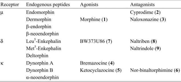
As mentioned previously, opioid receptor systems are comprised of at least three subtypes: µ, κ, and δ receptors. Each receptor is named based on its affinity towards a specific ligand (Table 1). The µ receptor is characterized by its acute affinity to morphine, while κ and δ receptors have high affinities for ketocyclazocine and enkephalins, respectively (Koneru et al., 2009).
Table 1: Selective opioid receptor ligands (Gao, 2010)
Receptor Endogenous peptides Agonists Antagonists
µ Endomorphin Cyprodime (2)
Dermorphin Morphine (1) Naloxonazine (3)
β-endorphin
β-neoendorphin
δ Leu5-Enkephalin BW373U86 (7) Naltriben (8)
Met5-Enkephalin Naltrindole (9)
Deltorphin
κ Dynorphin A Bremazocine (4)
Dynorphin B Ketocyclazocine (5) Nor-binaltorphimine (6)
α-neoendorphin
Although some opioid receptors are located in the peripheral nervous system, the majority accumulate in regions of the central nervous system associated with noxious sensory input: the spinal cord, brainstem, medial thalamus, hypothalamus, and limbic system (Koneru et al., 2009). Upon activation, opioid receptors can produce analgesia via an inhibitory mechanism (Koneru et al., 2009). When an agonist binds to an opioid receptor (consisting of seven trans-membrane proteins) (Fig. 9) on a presynaptic neuron,
two major changes occur within the neuron: calcium channels are closed and potassium channels are opened (Koneru et al., 2009). These changes lead to neuronal hyperpolarization, presynaptic inhibition, and eventually a decrease in neurotransmitter release into the synaptic cleft (Koneru et al., 2009).
Figure 9
IV.3 µ Opioid Receptor (MOR)
The µ-opioid receptors (MOR) are a class of opioid receptors which have high affinity for the exogenous agonist morphine and endogenous opioids such as endomorphin and dermorphin (Gao, 2010). The µ receptor is located in the brain (laminae III and IV of the cortex, thalamus, periqueductal gray), and spinal cord (substantia gelatinosa) (Koneru et al., 2009). Upon activation, the µ receptor causes supraspinal analgesia, physical dependence, respiratory depression, miosis, euphoria, and reduced gastrointestinal motility (Koneru et al., 2009). Two subtypes of µ receptors have been identified, µ1 and µ2 (Koneru et al,. 2009).
IV.4 κ Opioid Receptor (KOR)
The κ opioid receptors (KOR) are a class of opioid receptors which have high affinity toward endogenous dynorphins and the exogenous agonist Ketocyclazocine (Gao, 2010). KORs are located in the brain (hypothalamus, periaqueductal gray, and claustrum), spinal cord (substantia gelatinosa), and in pain neurons (Gao, 2010). κ receptor activation causes spinal analgesia, sedation, miosis, and inhibition of antidiuretic hormone release (ADH) (Koneru et al., 2009). Because of their psychotogenic and dysphoric central effects, KORs are primarily utilized as peripheral analgesics (Ananthan, 2006). Currently, very few pharmacologic drugs on the market target κ receptors (Gao, 2010). Two subtypes of KORs have been identified, κ1 and κ3 (Koneru et al., 2009).
IV.5 δ Opioid Receptor (DOR)
The δ opioid receptors (DOR) are a class of opioid receptors which have a high affinity for endogenous enkephalins (Gao, 2010). Currently, two subtypes of DORs have been identified, DOR1 and DOR2 (Rijn et al., 2013) δ opioid receptors are located in the brain (pontine nucleus, amygdala, olfactory bulbs, deep cortex) (Koneru et al., 2009).
Upon activation, DORs cause euphoria, physical dependence, and analgesia (Koneru et al., 2009). Because of their analgesic and euphoric properties, DORs have been considered as a potential drug target to treat pain, depression, and anxiety disorders (Rijn et al., 2013). Additionally, DORs are known to modulate ethanol and food consumption (Rijn et al., 2013). Despite their ability to modulate many physiological responses, only a small number of DOR-selective drugs are in clinical trials, and no DOR-selective drugs have been approved by the FDA (Rijn et al., 2013).
Figure 10
IV.6 Other Opioid Receptors
Other than the µ, δ, and κ opioid receptors, a variety of other opioid receptors have been identified. The nociceptin opioid receptor (NOR) is a g-protein coupled receptor that was discovered recently in 1994 (Gao, 2010). It is located in many regions of the brain, including the cortical areas, olfactory regions, and thalamus, and in the spinal cord (Gavioli and Calo, 2013). The endogenous peptide ligand associated with the NOR is nociceptin, also known as orphanin FQ (Redrobe, 2002). Nociceptin has been
associated with responses dealing with pain, fear and anxiety, learning, memory, feeding, drug addiction, and circadian rhythms (Redrobe, 2002).
The opioid growth factor receptor (OGFr), also known as the ζ (zeta) receptor, a λ (lambda) receptor, and a β-endorphin-sensitive ε (epsilon) opioid-binding site have also been identified (Waldhoer, 2004). Despite their identification, little is known about the characterization of these receptors, and evidence of their existence through identification of their genes is deficient (Waldhoer, 2004).
IV.7 Structure of Opioid Receptors
Opioid receptors belong to the Rhodopsin family of the g protein-coupled receptors (Gao, 2010). Each opioid receptor includes an intracellular C-terminal tail, seven transmembrane helical domains, and an extracellular N-terminal domain (Gao, 2010). This intricate formation allows for the binding of a variety of endogenous and exogenous opioid ligands (Gao, 2010). The µ, δ, and κ opioid receptors are relatively similar in structure, with about 60% overall likeness and the greatest homology in the seven transmembrane helical domains (Gao, 2010).
IV.8 Cannabinoid Receptors and Ligands
The discovery of another class of G-protein coupled receptors, cannabinoid receptors, stemmed from the desire to comprehend the effects of Δ9-tetrahydrocannabinol 19 (THC, Fig. 11), the primary psychoactive constituent of marijuana (Cannabis sativa) (Mackie, 2008).
Chemical structure of Δ9-tetrahydrocannabinol (THC).
Figure 11
Interest in the medicinal properties of Cannabis sativa began thousands of years ago, around 2600 BCE, when the people of China used the plant for the relief of cramps, rheumatic pain, and menstrual pain (Gao, 2010). Despite its utilization for many years, the marijuana plant was not scientifically studied in the Western world until the nineteenth century (Gao, 2010). In 1964, THC was first isolated by Yechiel Gaoni and Raphael Mechoulam of the Weizmann Institute of Science in Rehovot, Israel (Gao, 2010). Currently, two pharmacologic drugs based on THC are being used clinically.
Nabilone (12) is a synthetic THC analogue and is given to cancer and AIDs patients to combat nausea and as an appetite-stimulant (Gao, 2010). Dronabinol (II), a drug that contains synthetic THC, is also utilized in the treatment of nausea and appetite stimulation in AIDS-related anorexia (Gao, 2010).
The human body also produces endogenous cannabinoids. Anandamide (15) and 2- arachidonoylglycerol are the two most examined endocannabinoids (16) (Gao, 2010).
Cannabinoid receptor agonists and antagonists Figure 12
IV.9 Cannabinoid Receptors
In 1988, Allyn Howlett et al. definitively established the existence of cannabinoid receptors and further characterized a possible mechanism (Mackie, 2008). The majority of cannabinoid receptors are located in areas of the brain associated with movement, cognition, and sensory functions (Glass et al., 1997). The high density of receptors in these regions of the brain correlates with the widespread effects of cannabinoids which include: augmentation of the senses, altered emotions, errors in judgment of time and space, impulses, hallucinations, and decreased motor activities (Glass et al., 1997).
IV.9 Cannabinoid Receptor Subtypes
Agonists of cannabinoid receptor systems can bind to at least two receptor types in mammalian tissues: CB1 and CB2. CB1 receptors are located primarily at nerve terminals, while CB2 receptors have a more restricted distribution in immune cells and a few neurons (Mackie, 2008). When cannabinoid receptors are activated, neuronal excitability and the amount of neurotransmitter released into the synaptic cleft is decreased (Mackie, 2008). Like ligands of opioid receptors, ligands of cannabinoid receptors have therapeutic capability including the treatment of inflammation and neuropathic pain (Pertwee, 2006).
IV.10 CB1 Receptor
CB1 receptors are found predominately on nerve terminals in the brain, with high levels associated with the hippocampus, in basal ganglia output nuclei (globus pallidus and substantia nigra), and the cerebellum (Fig. 13) (Gao, 2010). The locations of CB1
receptors are consistent with the known behavioral and physiological effects (diminished short term memory, anxiety, and altered locomotion) of cannabinoids such as THC (Gao, 2010). Although they are most prominent in the CNS, CB1 receptors have also been reported in peripheral tissues such as fat (adipocytes), the pancreas, the liver, and skeletal muscle (Mackie, 2008). CB1 receptor agonists demonstrate therapeutic capability.
Inflammation, neuropathic pain, nausea, certain types of cancer, glaucoma, and motor dysfunction associated with multiple sclerosis and spinal cord injury can be treated with agonists of CB1 receptors (Pertwee, 2006). Furthermore, CB1 receptor agonists can be utilized as appetite stimulants (Pertwee, 2006).
Figure 13 IV.11 CB2 Receptor
CB2 receptors are located primarily in immune cells such as T cells, B cells, macrophages, and hematopoietic cells (Gao, 2010). Although less prevalent, CB2
receptors are also present in keratinocytes (Gao, 2010). Evidence suggests that CB2
receptor agonists can treat neuropathic pain and inflammation, however, their role still remains unclear (Pertwee, 2006).
IV.12 Structure of Cannabinoid Receptors
Like other g-protein coupled receptors, CB1 and CB2 receptors consist of seven transmembrane domains, an intracellular C-terminal, and an extracellular N-terminal (Galiegue et al., 1995). The amino acid sequences of CB1 and CB2 receptors are about 44% similar (Gao, 2010).
Figure 14 IV.13 Cannabinoid Receptors in Drug Discovery
Evidence suggests that the endocannabinoid system is involved in a variety of human health problems. Agonist and antagonists of the cannabinoid system have
demonstrated promising treatment against obesity, neurodegeneration, treatment of drug abuse, neuropathic pain, and even cancer (Gao, 2010).
IV.13.1 CB1 Receptor and Obesity
Obesity is an extremely prevalent health issue that causes other severe problems including diabetes, cardiovascular diseases, and some cancers (Gao, 2010). More than 70% of people in the United States and the United Kingdom suffer from obesity
according to the World Health Organization (WHO) (Gao, 2010). Although significantly altering lifestyle and diet can manage and treat obesity, many researchers are attempting to develop drugs to combat the debilitating disease. As mentioned previously, CB1
agonists have the potential to act as appetite stimulants (Pertwee, 2006). Therefore, current studies support the concept that antagonists of CB1 receptors can lead to long- term weight loss and improve lipid metabolism (Gao, 2010). Because of the severe side effects of CB1 drugs acting in the CNS (depression, anxiety, stress disorders), researchers are attempting to target CB receptors that are located in the peripheral system (fat and
liver) to alleviate the harmful side effects (Gao, 2010).
IV.13.2 CB1 Receptor and Neurodegenerative Diseases
Neurodegenerative diseases such as Huntington’s disease, Parkinson’s disease, Alzheimer’s disease, multiple sclerosis, and epilepsy involve the loss of structure or function and final death of neurons (Tian et al., 2014). Because of their prevalence and localization in the brain, CB1 receptors and agonists have been studied to determine their relationship with neurodegenerative diseases (Gao, 2010). For example, it is accepted that one of the earliest molecular changes known in human Huntington’s disease is associated with the loss of CB1 receptors in certain parts of the brain (globus pallidus and striatum) (Gao, 2010). In patients with Alzheimer’s disease, abnormal CB1 and CB2 receptors were highly expressive near the senile plaques associated with the disease (Gao, 2010).
Although there is evidence supporting the relationship between CB receptors and neurodegenerative diseases, much more research must be conducted to understand the roles of the cannabinoid system in these diseases.
IV.13.3 CB1 Receptors and Drug Abuse
The endocannabinoid receptor system has been closely linked to drug abuse, and CB1 receptor antagonism has been shown to decrease the craving for certain drugs including alcohol and cigarettes (Gao, 2010). For example, blocking CB1 receptors causes a decrease in the consumption of alcohol, while activation of the receptors results in increased alcohol consumption (Gao, 2010). Furthermore, a 10 week clinical trial demonstrated positive results when using CB1 antagonists to suppress smoking cravings (Gao, 2010). During the trial, participants (moderate cigarette smokers) took a CB1
antagonist drug (rimonabant) and experienced increased abstinence from smoking
compared to participants who took a placebo (Gao, 2010). Although there are positive outcomes resulting from the blockage of CB1 receptors, it has yet to be determined if the antagonism will be an effective long-term approach for the treatment of craving disorders (Gao, 2010).
IV.13.4 CB1 and CB2 Receptors and Neuropathic Pain
Neuropathic pain is defined as ‘‘initiated or caused by a primary lesion or dysfunction in the nervous system’’ according to the International Association for the Study of Pain (IASP) (Dworkin et al., 2007). Neuropathic pain affects millions of people all over the world and is caused by a variety of problems including diabetic neuropathy, multiple sclerosis, nerve compression syndromes, stroke, spinal cord injury, and cancer (Gao, 2010). Treatment of neuropathic pain is complex and is accompanied by many adverse side effects (Dworkin et al., 2007). The cannabinoid system is known to be associated with pain modulation (Ikeda et al., 2013). CB1 receptor activation in the CNS and peripheral tissues has been shown to inhibit pain responses (including neuropathic pain) and suppress inflammatory hypersensitivity (Ikeda et al., 2013). Because of their distribution in the brain, however, CB1 receptor agonists have produced negative side effects such as depression and anxiety (Ikeda et al., 2013). CB2 receptor agonists, located predominantly in spinal cord, have also been shown to reduce neuropathic pain and inflammation (Gao, 2010).
IV. 13.5 CB1 and CB2 Receptors and Cancer
Although CB1 and CB2 receptor agonists were found to inhibit tumor growth in certain cancers in 1975, the mechanism of the agonists and their anticancer activity was not studied until recently (Gao, 2010). It has been discovered that CB1 and CB2 receptor ligands inhibit the growth of cancer cells and reduce cancer cell viability through a variety of mechanisms including mitosis and apoptosis (Gao, 2010).
Materials and Methods:
I. General experimental procedures
1H and 13C Nuclear Magnetic Resonance (NMR) spectra were obtained on Bruker model AMX 500 NMR and 400 Avance NMR spectrometers with standard pulse sequences, operating at 500 MHz and 400 MHz, respectively in 1H and 100 MHz and 125 MHz in 13C, respectively. The chemical shift values were reported in parts per million units (ppm) from trimethylsilane (TMS) using known solvent chemical shifts. Coupling constants were recorded in Hertz (Hz). Standard pulse sequences were used for Correlation Spectroscopy (COSY), Heteronuclear Multiple Quantum Coherence (HMQC), Heteronuclear Multiple Bond Coherence (HMBC), Total Correlated Spectroscopy (TOCSY), Nuclear Overhauser Effect Spectroscopy (NOESY) and Distortionless Enhancement by Polarization Transfer (DEPT). High-resolution mass spectra (HRMS) were measured on a Micromass Q-Tof Micro mass spectrometer with a lock spray source. Column chromatography was carried out on silica gel (70-230 mesh, Merck), SPE reverse phase C-8 column and Sephadex LH-20 (Mitsubishi Kagaku, Tokyo, Japan). TLC (silica gel 60 F254) was used to monitor fractions from column chromatography. Visualization of the TLC plates was achieved with a UV lamp (λ = 254 and 365 nm) and anisaldehyde/acid spray reagent (MeOH-acetic acid-anisaldehyde- sulfuric acid, 85:9:1:5). Solvents were certified grade for Fischer Scientific. All chemicals used were purchased from Sigma-Aldrich (St. Louis, Mo) with the following exceptions: for the binding experiments, [3H]-CP-55,940 (144 Ci/mmol), was purchased
from Perkin-Elmer Life Sciences Inc. (Boston, MA, U.S.A.). CP-55,940 was purchased from Tocris Bioscience (Ellisville, Missouri, U.S.A.).
II. Plant material
The plant Perovskia atriplicifolia was collected in the region of Quetta, Pakistan in July of 2012 and identified by Dr. Arsala Mansoor from the Medicinal Plant Garden at the University of Balochistan, Quetta, Pakistan. A voucher specimen (UM 072012) has been deposited in the culture collection of the Department of Medicinal Chemistry, University of Mississippi. The plant material was separated into three parts: leaves, stems and roots.
III. Extraction and isolation
The dried plant material was ground into a powder yielding 370 g (leaves), 200 g (stems) and 120 g (roots), then the different parts of the plant were extracted with ethanol after maceration for three days, separately. Removal of the solvent afforded a viscous residue of 39.5 g (leaves), 6.22 g (stems) and 1.44 g (roots). The ethanolic extract from leaves showed moderate radioligand displacement binding affinity for opioid receptors (42.8 % for δ and 52.5% for µ). The ethanolic extract (39.5 g) was chromatographed on Silica gel flash column with stepwise fractions from hexanes to methanol, yielding five fractions (100% hexane, 100% dichloromethane, 100% EtOAc, 80% EtOAc in MeOH, and 100% MeOH). The EtOAc fraction showed strong binding affinities for δ opioid receptors (88.4 %), and this fraction was chromatographed on a SPE silica gel reverse phase column C-8, eluted with water- methanol to yield six fractions (100% water, 80 % water in methanol, 60 % water in methanol, 40% water in methanol, 20% water in
methanol, 100% methanol); all fractions were submitted for opioid binding activity. The fourth (40% water in methanol) and fifth (20% water in methanol) fractions showed strong binding affinities for δ opioid receptors with values of 62.8 % and 78.5 %, respectively. The fourth fraction (40% water in methanol) was rechromatographed in silica gel column eluted with hexane to EtOAc in step gradient to yield fifteen subfractions, and these were combined based on the TLC (CH2Cl2/EtOAc; 8:2). A precipitate was obtained from subfractions 10 and 12 and crystallized separately to obtain the flavones: 5-hydroxy-3’,4’,6,7-tetramethoxy flavone (Compound A) and 5,7- dihydroxy-3’,4’,6,-trimethoxy flavone (Compound B), respectively. The fifth fraction (20% water in methanol) was rechromatographed in silica gel column eluted with hexane to EtOAc in step gradient to yield thirteen subfractions. A precipitate was also obtained from subfractions 5-7. Based on TLC (CH2Cl2;/EtOAc; 8:2), two major spots were found in all the precipitates, which were combined and subject to a column of Sephadex LH-20 eluted with CH2Cl2/MeOH (1:1) to furnish 5-hydroxy-4’,6,7-trimethoxy flavone (Compound C) and 5,7-dihydroxy-4’,6-dimethoxy flavone (Compound D). All purified compounds were submitted for opioid binding affinity.
IV. Cell culture
Human Embryonic Kindney-293 (HEK-293) cells stably transfected with opioid receptor subtypes µ, δ, and κ were used to perform the opioid receptor binding assays.
These cells were maintained at 37°C and 5% CO2 in a Dulbecco’s modified Eagle medium (DMEM) nutrient mixture supplemented with 2 mM L-glutamine, 10% fetal bovine serum, penicillin−streptomycin, and either G418 or hygromycin B antibiotic
solutions. Membranes for the radioligand binding assays were prepared by scraping the cells in a 50 mM Tris-HCl buffer, followed by homogenization, sonication, and centrifugation for 40 min at 13650 rpm at 4°C. These were kept at -80 °C until used for bioassays. Protein concentration was determined via Bio-Rad Protein Assay (Bradford, 1976).
V. Radioligand Binding for Cannabinoids and Opioid Receptor Subtypes
All the fractions and pure compounds from P. atriplicifolia were run in competition binding assays against both cannabinoid receptor subtypes and all three opioid receptor subtypes (León et al., 2013). Cannabinoid binding took place under the following conditions: 10 µM of each compound was incubated with 0.6 nM [3H] CP 55.940and 10 µg of CB1 or CB2 membranes for 90 minutes in a silanized 96-well plate.
The reaction was terminated via rapid vacuum filtration through GF/B filters presoaked with 0.3% bovine serum albumin (BSA) using a Perkin-Elmer 96-well Unifilter followed by 10 washes with 50 mM Tris-HCl. Plates were read using a Perkin-Elmer Topcount.
Opioid binding assays were performed under the following conditions: 10 µM of each compound was incubated with [3H]-DAMGO (µ), [3H]-U-69,593 (κ), or [3H]-enkephalin (δ) for 60 min in a 96-well plate. Percent binding was calculated as the average of the triplicate tested at 10 µM. Each sample concentration point of the compounds tested in dose response was in triplicate, and each compound showing activity was tested at least three times. The reaction was terminated via rapid vacuum filtration through GF/B filters presoaked with 0.3% bovine serum albumin (BSA) using a Perkin-Elmer 96-well Unifilter followed by 10 washes with 50 mM Tris-HCl. Plates were read using a Perkin- Elmer Topcount. Total binding was defined as binding in the presence of 1.0% DMSO.
Nonspecific binding was the binding observed in the presence of 10µM DAMGO (µ), nor-binaltorphimine (κ), or DPDPE (δ). Specific binding was defined as the difference between total and nonspecific binding. Percent binding was calculated using the following formula: (binding of compound - nonspecific binding) x 100/specific binding.
Results:
I. Extraction and Initial Bioassay-guided Fractionation
The powdered sample (370 grams) of Perovskia atriplicifolia leaf was extracted in ethanol, yielding 39.57 grams of extract. The powdered sample of the plant stem (200 grams) and root (120 grams) were also extracted in ethanol, yielding 6.22 grams and 1.44 grams, respectively. The extracts were sent to the lab to test radioligand displacement binding affinity for opioid receptors and cannabinoid receptors. The leaf extract exhibited the best radioligand displacement affinity of opioid receptors, selectively inhibiting 42.8% of specific binding of δ receptors, 37.6% of κ receptors, and 52.5% of µ receptors.
Because of this activity, the extract was further subjected to silica gel vacuum liquid chromatography (VLC) in silica flash, which resulted in five fractions. These fractions were also submitted for assay and their activities are shown in the figure 15, as well as their weights.
Initial steps and activity for P. atriplicifolia Figure 15
II. Isolation and Purification/biological evaluation
The initial fractions were tested for their binding against opioid and cannabinoid receptor subtypes, and the EtOAc fraction selectively inhibited 88.4% of the specific binding of δ opioid receptors. The EtOAc fraction was then subjected to reversed-phase chromatography and yielded six fractions, which were also tested for opioid and cannabinoid receptor competitive binding (Fig. 16).
Fractionation of P. atriplicifolia Figure 16
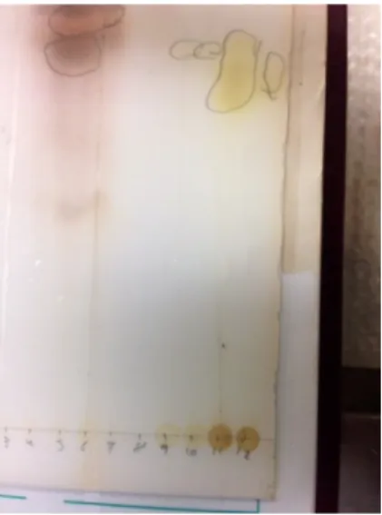
Fraction 4 was rechromatographed on silica gel column in a gradient step, increasing polarity starting with hexane to 100% EtOAC and 25 fractions were obtained. When these fractions were compared to TLC (CH2Cl2/EtOAc; 8:2) (Fig. 17), they were combined to yield 13 different fractions.
TLC plate for fraction 4 Figure 17
Subfractions 10-12 were submitted for biassays, and subfractions 10 and 11 showed a high affinity for δ opioid receptors. (Table 2)
Table 2. % of displacement of radioligands for subfractions obtained for fraction 4 of P.
atriplicifolia.
CB1
%inhibition
CB2
%inhibition
Delta
%inhibition
Kappa
%inhibition
Mu
%inhibition Fraction 4
Subfract. 10
0.3 4.2 86.0 7.5 9.5
Fraction 4 Subfract. 11
19.7 7.2 84.6 8.6 17.4
Fraction 4 Subfract. 12
22.1 5.4 60.8 27.8 54.3
Naloxone CP55,940
-- 104.3
-- 102.6
106.40 --
101.59 --
96.95 --
A precipitate was obtained from subfractions 10 and 12 and crystallized separately to obtain the compounds 5-hydroxy-3’,4’,6,7-tetramethoxy flavone (Compound A) and 5,7-dihydroxy-3’,4’,6,-trimethoxy flavone (Compound B), respectively.
Fraction 5 was subjected to chromatography on silica gel column in a gradient step, increasing polarity starting with hexane to 100% EtOAC yielding 25 fractions. When these fractions were compared to TLC (CH2Cl2/EtOAc; 8:2), they were combined to yield 13 different subfractions that were submitted for bioassays (Fig. 18).
Fractionation for fraction 5 of P. atriplicifolia Figure 18
A precipitate was obtained from subfractions 5 to 7, and by TLC (CH2Cl2;/EtOAc; 8:2), two major spots were found in all the precipitates; these were combined and subject to a column of Sephadex LH-20 eluted with CH2Cl2/MeOH (1:1) to furnish the flavones 5- hydroxy-4’,6,7-trimethoxy flavone (Compound C) and 5,7-dihydroxy-4’,6-dimethoxy flavone (Compound D). The pure compounds were then submitted for bioassays and showed 31.6% and 79.9% radioligand displacement affinity of δ opioid receptors, respectively.
III. Structural Elucidation
Compounds 5-hydroxy-3’,4’,6,7-tetramethoxy flavone (Compound A) and 5,7- dihydroxy-3’,4’,6,-trimethoxy flavone (Compound B) were isolated from the subfraction 4 as a yellowish, amorphous solid. For Compound A, a flavone skeleton was evident from the coupling pattern ABC of the B-ring protons in its 1H NMR spectrum, δH 7.65 (1H, dd, J = 2.1, 8.5 Hz), 7.52 (1H, d, J = 2.3 Hz) and 7.07 (1H, d, J = 8.6 Hz) and two singlets signals at δH 6.95 and 6.88 ppm, corresponding to the protons in C-3 and C-8, respectively. The spectrum also showed signals of four methoxy groups between 3.80 to 3.98 ppm (Fig. 18).
1H NMR spectrum of 5-hydroxy-3’,4’,6,7-tetramethoxy flavone Figure 19
Analysis of the 13C NMR spectrum and DEPT experiments confirmed a flavone moiety
with the presence of a typical ketone carbonyl signal at δC 182.6 in total fifteen signals plus four additional methyl signals at δC 60.4, 56.8, 56.2, and 56.1 ppm, corresponding with the methoxy groups (Fig. 19).
13C NMR spectrum of 5-hydroxy-3’,4’,6,7-tetramethoxy flavone Figure 20
The 1H NMR and 13C NMR spectroscopic data were found to be similar to those of 5- hydroxy-3’,4’,6,7-tetramethoxy flavone (Zhao et al., 2012). The nature and identity of this flavone was deduced from the 2D NMR experiments (COSY, NOESY, HMBC, and HSQC) which are located in the appendix.
5-hydroxy-3’,4’,6,7-tetramethoxy flavone, Compound A Figure 21
The 1H NMR spectrum of 5,7-dihydroxy-3’,4’,6,-trimethoxy flavone (Compound B) showed absence of one methoxy group, and the spectroscopic data is in agreement with reported data from Suleimenov et al., 2005. 1H, 13CNMR, HSQC, and HMBC spectra are shown in the appendix.
5,7-dihydroxy-3’,4’,6,-trimethoxy flavone, Compound B Figure 22
Compounds 5-hydroxy-4’,6,7-trimethoxy flavone (Compound C) and 5,7-dihydroxy- ,4’,6,-dimethoxy flavone (Compound D) were isolated from subfraction 5 as a yellowish, amorphous solid. For 5-hydroxy-4’,6,7-trimethoxy flavone (Compound C), its 1H NMR spectrum showed an AB pattern in ring B of the flavone, corresponding to a
monosubstitution in the position 4’, and signals for three methoxy groups (Fig. 22). 13C NMR and DEPT spectra are located in the appendix.
1H NMR spectrum of 5-hydroxy-4’,6,7-trimethoxy flavone Figure 23
5-hydroxy-4’,6,7-trimethoxy flavone, Compound C
5,7-dihydroxy-4’,6,-dimethoxy flavone, Compound D, was identified and its structure elucidated following the same analysis as the first compound, Compound A (1H NMR,
13CNMR, and DEPT spectra are showed in the appendix). The spectroscopic data for the last compound is in agreement with Horie et al., 1998.
5,7-dihydroxy-4’,6,-dimethoxy flavone, Compound D Figure 25
Conclusion
Plants and their natural products have been an undeniable resource in the
development of effective novel drugs utilized in the treatment of many human diseases.
In this thesis, we sought to investigate terrestrial plant Perovskia atripliciolia and the biological effects of its secondary metabolites. Extracts that demonstrated good selective opioid and cannabinoid receptor binding were subjected to bioassay-guided fractionation.
The secondary metabolites that possessed strong binding affinity were subjected to spectroscopic evaluation in order to elucidate their chemical structures. Four active compounds were identified from the crude extract in Perovskia atriplicifolia: 1) 5-
hydroxy-3’,4’,6,7-tetramethoxy flavone, 2) 5,7-dihydroxy-3’,4’,6,-trimethoxy flavone, 3) 5-hydroxy-4’,6,7-trimethoxy flavone, and 4) 5,7-dihydroxy-4’,6,-dimethoxy flavone.
Each compound exhibited good selective δ-opioid receptor binding affinity and was characterized as a flavonoid, a plant secondary metabolite consisting of three-ring structure with various substitutions (Middleton, 2000). Flavonoids have been found in a myriad of foods in the human diet, from citrus fruits to nuts to tea and red wine, and the compounds are also known to be the pigments responsible for giving leaves their vibrant colors in autumn (Middleton, 2000). Although the structures of these four compounds were isolated and their chemical structures elucidated, further studies need to be
performed to determine each compounds mechanism of action, find out how compounds fit into receptors, etc.
BIBLIOGRAPHY
Amsler, C. D.; McClinktock, J. B.; Baker, B. J. Secondary Metabolites as Mediators of Trophic Interactions Among Antarctic Marine Organisms. Amer. Zool. 2001, 41, 17-26.
Ananthan, S. Opioid Ligands with Mixed Mu/Delta Receptor Interactions: An Emerging Approach to Novel Analgesics. AAPS Jour. 2006, 8, 118-125.
Beikmohammadi, M. Ethno Pharmacology and the Investigation of the Most Important Secondary Materials and the Comparison of Chemical Combinations of Essential Oil of Different Organs. Mid.-East. J. Sci. Res. 2011, 9, 486-495.
Bockaert, J.; Pin, J. P. Molecular Tinkering of G Protein-Coupled Receptors: An Evolutionary Success. EMBO J. 1999, 18, 1723-1729.
Boz, I.; Padurariu, C.; Zamfirache, M.; Burzo, I,; Dunca, S.; Stefan, M.; Ivanescu, L.;
Olteanu, Z.; Badea, M. L.; Gostin, I,; Andro, A. Chemical Composition and Antimicrobial Activities of Volatile Oils in Some Lamiaceae Species. J. Bio. Eco.
Sci. 2011, 1, 21-27.
Clement, J. A. Studies of Bioactive Natural Products and Mechanism-based Bioassays.
PH.D. Thesis, Virginia Polytechnic Institute, Blackburg, VA, 2005.
Cordell, G. A.; Colvard, M. D. Natural Products and Traditional Medicine: Turning on a Paradigm. J. Nat. Prod. 2012, 75, 514-525.
Cragg, G. M.; Newman, D. J. In Drug Discovery and Development from Natural Products: The Way Forward, Proceedings of the 11th NAPRECA Symposium, Hôtel Panorama, Antananarivo, Madagascar, August 9-12, 2005; Midiwo, J. O., Yenesew, A., Derese, S., Eds.; NAPRECA Publication: Nairobi, 2006.
Cragg, G. M.; Newman, D. J. Natural Products: A Continuing Source of Novel Drug Leads. Bioch. Biophy. Acta. 2013, 1830, 3670-3695.
Dhawan, B. N.; Cesselin, F.; Raghubir, R.; Reisine, T.; Bradley, P. B.; Portoghese, P. S.;
Hamon, M. International Union of Pharmacology. XII. Classification of Opioid Receptors. Pharm. Rev. 1996, 48, 567-592.
Dworkin, R. H.; O’Connor, A. B.; Backonya, M.; Farrar, J. T.; Finnerup, N. B.; Jensen, T.; Kalso, E. A.; Loeser, J. D.; Miaskowski, C.; Nurmikko, T. J.; Portenoy, R. K.;
Rice, A.; Stacey, B. R.; Treede, R.; Turk, D. C.; Wallace, M. S. Pharmacologic Management of Neuropathic Pain: Evidence-based Recommendations. Pain. 2007, 132, 237-251.
Gao, J. Fungi Derived Natural Products with Binding Affinities for Human Opioid Receptors or Cannabinoid Receptors. PH.D. Thesis, University of Mississippi, Oxford, MS 2010.
Gao, J.; León, F.; Radwan, M. M.; Dale, O. R.; Husni, A. S.; Manly, S. P.; Lupien, S.;
Wang, X.; Hill, R. A.; Dugan, F. M.; Cutler, H. G.; Cutler, S. J. Benzyl Derivatives with in Vitro Affinity for Human Opioid and Cannabinoid Receptors From the Fungus Eurotium repens. J. Nat. Prod. 2011, 74, 1636-1939.
Gavoli, E. C.; Calo, G. Nociceptin/orphanin FQ Receptor Antagonists as Innovative Antidepressent Drugs. Pharm. & Thera. 2013, 14D, 10-25.
Glass, M.; Dragunow, M.; Faull, R. L. M. Cannabinoid Receptors in the Human Brain: A Detailed Anatomical and Quatitative Autoradiographic Study in the Fetal, Neonatal, and Adult Human Brain. Neuroscience. 1997, 77, 299-318.
Horie, T.; Ohtsuru, Y.; Kenichi, S.; Yamashita, K.; Tsukayama, M.; Kawamura, Y. 13C NMR Spectral Assignment of the A-Ring of Polyoxgenated Flavones. Phtyochem.
1998, 47, 865-874.
Ikeda, H.; Ikegami, M.; Kai, M.; Ohsawa, M; Kamei, J. Activation of Spinal Cannabinoid CB2 Receptors Inhibits Neuropathic Pain in Streptozotocin-Induced Diabetic Mice.
Neuroscience. 2013, 250, 446-454.
Jassbi, A. R.; Ahmad, V. U., Tareen, R. B. Constituents of the Essential Oil of Perovskia atriplicifolia Benth. Flavour Fragr. J. 1999, 14, 38-40.
Jiang, Z. Y.; Huang, C. G.; Xiong, H. B.; Tian, K.; Liu, W. X.; Hu, Q. F.; Wang, H. B.;
Yang, G. Y.; Huang, X. Z. Perovskatone A: A Novel C23 Terpenoid from Perovskia atriplicifolia. Tetrahedron Lett. 2013, 54, 3886-3888.
Kingston, D. Modern Natural Products Drug Discovery and Its Relevance to Biodiversity Conservation. J. Nat. Prod. 2011, 74, 496-511.
Koneru, A.; Satyanarayana, S.; Rizman, S. Endogenous Opioids: Their Physiological Role and Receptors. Glob. J. Pharm. 2009, 3, 149-153.
Leon, F.; Gao, J.; Dale, O.; Wu, Y.; Habib, E.; Husni, E.; Hill, R. A.; Cutler, S. J.
Secondary Metabolites from Eupenicillium parvum and Their in Vitro Binding Affinity for Human Opioid and Cannabinoid Receptors. Thieme. 2013, 79, 1756- 1761.
Mackie, K. Cannabinoid Receptors: Where They Are and What They Do. J.
Neuroendocrinol. 2008, 20, 10-14.
Middleton, E.; Kandaswami, C.; Theoharides, T. C. The Effects of Plant Flavonoids on Mammalian Cells: Implications for Inflammation, Heart Disease, and Cancer.
Pharm. Rev. 2000, 52, 673-751.
National Cancer Institute. Success Story: Taxol.
http://dtp.nci.nih.gov/timeline/flash/success_stories/S2_taxol.htm (accessed Oct 23, 2014).
New Chemical Entity Exclusivity Determinations for Certain Fixed-Combination Drug Products; U.S. Department of Health and Human Services, U.S. Food and Drug Administration: Silver Spring, MD, 2014.
Pandeya, S. N.; Thakkar, D. Combinatorial Chemistry: A Novel Method in Drug Discovery and Its Application. Ind. J. Chem. 2005, 44B, 335-348.
Pertwee, RG. The Pharmacology of Cannabinoid Receptors and Their Ligands: An Overview. Int. J. Obe. 2006, 30, 513-518.
Perveen, S.; Malik, A.; Tareen, R. B. Phytochemical Studies on Perovskia atriplicifolia.
J. Chem. Soc. Pak. 2009, 31, 314-318.
Redrobe, J. P.; Calo, Girolamo, C.; Regoli, D.; Quirion, R. Nociceptin Receptor
Antagonists Display Antidepressant-like Properties in the Mouse Forced Swimming Test. Arch. Pharmacol. 2002, 365, 164-167.
Rouhi, A. M. Chemical and Engineering News. Rediscovering Natural Products.
http://pubs.acs.org/cen/coverstory/8141/8141pharmaceuticals.html (accessed Nov, 4 2014).
Sairafianpour, M.; Christensen, J.; Staerk, D.; Budnik, B. A.; Kharazmi, A.;
Bagherzadeh, K.; Jarosewski, J. W. Leishmanicidal, Antiplasmodial, and Cytotoxic Activity of Novel Diterpenoid 1,2-Quinones from Perovskia abrotanoides: New Source of Tanshinones. J. Nat. Prod. 2001, 64, 1398-1403.
Saleem, M. Chemical and Biological Screening of some Relatives of Lamiaceae
(Labiatae) Family and Marine Alga Condium Iyengari. PH.D. Thesis, University of Karachi, Karachi, Pakistan, 2000.
Suleimenow, E. M.; Smagulova, F. M.; Morozova, O. V.; Raldugin, V. A.;
Bagryanskaya, Y.; Gatilov, Y. V.; Yamovoi, V. I.; Adekenov, S. M. Sesquiterpene Lactones and Flavonoids from Artemisia albida. Chem. Nat. Comp. 2005, 41, 689- 691.