A Comparative Study of
the Appendicular Musculature of Penguins (Aves: Sphenisciformes)
DONALD O. SCHREIWEIS ,
SMITHSONIAN CONTRIBUTIONS TO ZOOLOGY • NUMBER 341
SERIES PUBLICATIONS OF THE SMITHSONIAN INSTITUTION
Emphasis upon publication as a means of "diffusing knowledge" was expressed by the first Secretary of the Smithsonian. In his formal plan for the Institution, Joseph Henry outlined a program that included the following statement: "It is proposed to publish a series of reports, giving an account of the new discoveries in science, and of the changes made from year to year in all branches of knowledge." This theme of basic research has been adhered to through the years by thousands of titles issued in series publications under the Smithsonian imprint, commencing with Smithsonian Contributions to Knowledge in 1848 and continuing with the following active series:
Smithsonian Contributions to Anthropology Smithsonian Contributions to Astrophysics
Smithsonian Contributions to Botany Smithsonian Contributions to the Earth Sciences Smithsonian Contributions to the Marine Sciences
Smithsonian Contributions to Paleobiology Smithsonian Contributions to Zoo/ogy Smithsonian Studies in Air and Space Smithsonian Studies in History and Technology
In these series, the Institution publishes small papers and full-scale monographs that report the research and collections of its various museums and bureaux or of professional colleagues in the world of science and scholarship. The publications are distributed by mailing lists to libraries, universities, and similar institutions throughout the world.
Papers or monographs submitted for series publication are received by the Smithsonian Institution Press, subject to its own review for format and style, only through departments of the various Smithsonian museums or bureaux, where the manuscripts are given substantive review. Press requirements for manuscript and art preparation are outlined on the inside back cover.
S. Dillon Ripley Secretary
Smithsonian Institution
S M I T H S O N I A N C O N T R I B U T I O N S T O Z O O L O G Y • N U M B E R 3 4 1
A Comparative Study of
the Appendicular Musculature of Penguins (Aves: Sphenisciformes)
Donald 0. Schreiweis
SMITHSONIAN INSTITUTION PRESS City of Washington
1982
A B S T R A C T
Schreiweis, Donald O. A Comparative Study of the Appendicular Muscu-
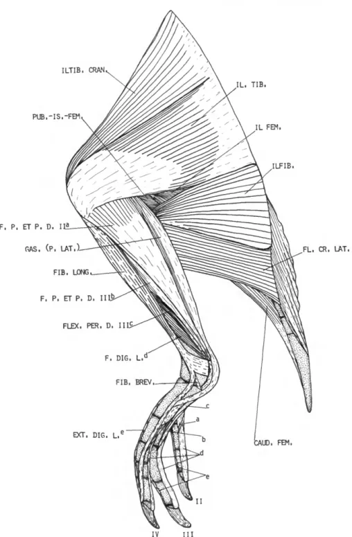
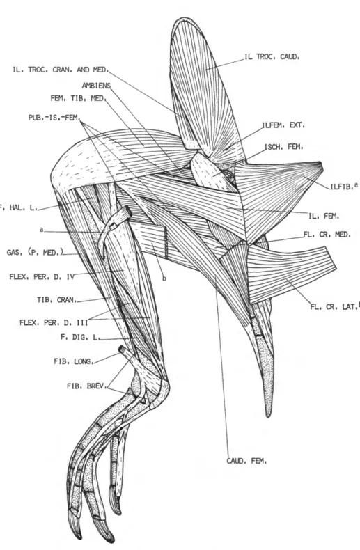
lature of Penguins (Aves: Sphenisciformes). Smithsonian Contributions to Zoology, number 341, 46 pages, 19 figures, 1982.—The gross anatomy of all the appendicular muscles is compared in 14 species, representing the 6 living genera of Sphenisciformes. Particular emphasis is placed on the interrelation- ships indicated by variations in the taxa. Eudyptes pachyrhynchus is used as a type for which all the appendicular muscles are described and most are illustrated. The salient features of other taxa, as they differ from the condition found in E. pachyrhynchus, are given. Similarities and differences are compared quantitatively in respect to 47 items in the wing and and 41 in the leg, using the method of Hudson et al. (1966:9-11). The results are given in the form of cumulative scores of differences and correlation coefficients.
The results of the comparison of the wing musculature support those for the leg. The cumulative scores and correlation coefficients give closely parallel results.
The Sphenisciformes constitute a rather uniform group of birds. The results of this study support the present classification of the order, in which 14 species are grouped into 6 genera. A tentative phylogeny of penguins is projected on the basis of variations in the wing and leg muscles.
OFFICIAL PUBLICATION DATE is handstamped in a limited number of initial copies and is recorded in the Institution's annual report, Smithsonian Year. SERIES COVER DESIGN: The coral Montastrea cavemosa (Linnaeus).
Library of Congress Cataloging in Publication Data Schreiweis, Donald O.
A comparative study of the appendicular musculature of penguins (Aves: Sphenisciformes) (Smithsonian contributions to zoology ; no. 341)
Bibliography: p.
Includes index.
Supt. of Doc. no.: SI 1.27:341
1. Penguins—Anatomy. 2. Wing—Muscles. 3. Leg—Muscles. 4. Anatomy, Comparative.
I. Title. II. Series.
QL1.S54 no. 341 [QL696.S473] 591s 81-607820 [598.4'41] AACR2
Contents
Page
Introduction 1 Materials and Methods 1 Acknowledgments 2 Muscles of the Pectoral Appendage 2 Muscles of the Pelvic Appendage 14 Position of the Flexor Tendons Passing the Intertarsal Joint 21 Discussion and Conclusions 25 Appendix: Interesting Features of the Appendicular Muscles 29 Literature Cited 31 Abbreviations on Illustrations 33 Figures 1-19 34 Index 45
m
A Comparative Study of
the Appendicular Musculature of Penguins (Aves: Sphenisciformes)
Donald 0. Schreiweis
Introduction
Penguins are an ancient group of birds that had become flightless and highly specialized for underwater locomotion by the Eocene. Simpson (1975:35) considered penguins related to Procel- lariiformes, basing this relationship on the struc- ture of the skull and mandible. In the oldest known fossil penguins, the structure of the man- dible, the shape of the foramen magnum, and the arrangement of palatal bones are similar to the procellariiform type. Sibley and Ahlquist (1972) recently concluded that the egg-white protein patterns of penguins were more like those of the Procellariiformes than those of any other group, although they also resembled several other groups of aquatic birds. The descriptive myology of pen- guins presented in this paper may help to resolve the problem of phylogeny when comparable stud- ies on other water birds become available.
Although the appendicular muscles have been studied in a few sphenisciform birds by various investigators, no one has made a concerted at- tempt to work out in detail the similarities and differences between the various genera and spe- cies. Such differences and similarities may serve as a basis for a study of functional morphology,
Donald 0. Schreiweis, Premedical Studies Program, Saint Louis University, Saint Louis, Missouri 63103.
as well as a study of phylogenetic and systematic relationships among these taxa.
A detailed study of all appendicular muscles, using descriptive and numerical methods, was made on a series of 28 specimens, representing 6 genera and 14 species. The pectoral musculature is unlike that of any other group of birds. Most of the muscles in the wing are reduced to tendons, a condition not encountered in any other group, while the leg muscles deviate less from the general avian pattern. The more interesting features of the appendicular muscles of penguins are sum- marized in the appendix.
MATERIALS AND METHODS.—The classification followed is that of Falla and Mougin (1979). I dissected the following species: Aptcnodytes patagon-
icus (2); A.forsteri (2); Pygoscelis papua (2); P. adeliae (3); P. antarctica (2); Eudyptes pachyrhynchus (2); E.
chrysocome (2); E. chrysolophus schlegeli (1); E. c.
chrysolophus (2); Megadyptes antipodes (1); Eudyptula minor minor (1); E. m. albosignata (1); Spheniscus demersus (2); S. humboldti (1); S. magellanicus (2); S.
mendiculus (2).
Fresh specimens were initially injected in all parts with a 1:10 solution of formalin. After several weeks, specimens were opened and washed in tap water for 3 to 5 days to remove most of the formalin. Subsequently, all were stored in 65%
ethyl alcohol.
To facilitate study of the musculature in most
1
SMITHSONIAN CONTRIBUTIONS TO ZOOLOGY
specimens, the coracoid was separated from the clavicle proximally and from the sternum distally, and the femur was disarticulated at the aceta- bular joint. The extremities of the limb bones were exposed by partially dissecting the joint capsules without complete disarticulation. Each muscle was exposed from origin to insertion by cutting bellies or tendons of certain muscles at prescribed points.
Descriptions of the origin, belly, and insertion of each muscle are given for Eudyptes pachyrhynchus.
The positional (anatomical) axes utilized are those related to the functional axes: for the wing, the extended position of underwater flight; for the leg, the normal standing position. The loca- tion of muscle attachments (e.g., "at about 0.63 tibiotarsus") were obtained by measuring the point of attachment from the proximal end of the bone holding the calipers parallel to the long axis of the bone. These measurements were then di- vided by the length of the bone.
Following each description, a comparison of the muscle is given for other species as these differ from E. pachyrhynchus. Included in the comparison are remarks concerning observations made by other workers. In many instances, observations made in this study clarify those of earlier workers.
Numerical comparisons were made using the method of Hudson et al. (1966:9-11). Tables of quantitative data are available from the author on request. Anatomical nomenclature follows that of the Nomina Anatomica Avium (Baumel et al.,
1979).
ACKNOWLEDGMENTS.—I wish to express my ap- preciation and thanks to Dr. George E. Hudson under whose guidance and direction this study was completed. He made available many of the specimens and, through him, various specimens were loaned from other institutions. I also wish to thank Drs. Herbert L. Eastlick, C. M. McNeil, and Richard A. Parker of Washington State Uni- versity, who critically read the manuscript and offered suggestions.
Specimens examined for this study are part of an extensive alcoholic collection at Washington State University, representing all living orders of
birds. Dr. Richard L. Zusi of the National Mu- seum of Natural History, Smithsonian Institu- tion, loaned 12 specimens. Dr. Dean Amadon of the American Museum of Natural History loaned one specimen each of Eudyptes pachyrhynchus and Spheniscus humboldti. Mr. Warwich M. Howe of the New Zealand Department of Internal Affairs sup- plied one specimen each of Eudyptes pachyrhynchus, Megadyptes antipodes, a n d Eudyptula minor albosig- nata. Mr. T. H. Barry of the South African Mu- seum supplied a specimen of Spheniscus demersus.
This study was supported in part by a grant- in-aid (GB-6088) from the National Science Foundation.
Muscles of the Pectoral Appendage
M . LATISSIMUS DORSI
The latissimus dorsi consists of three distinct parts: latissimus dorsi cranialis, latissimus dorsi caudalis, and latissimus dorsi metapatagialis.
Latissimus dorsi cranialis
(LAT. DOR. CRAN.)
FIGURES 1, 2, 8
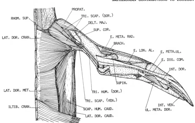
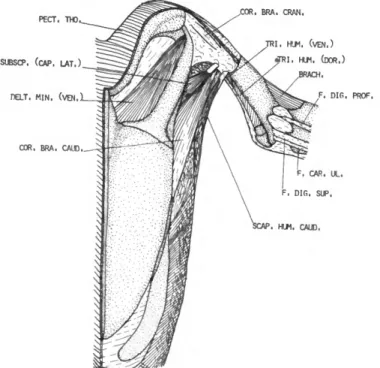
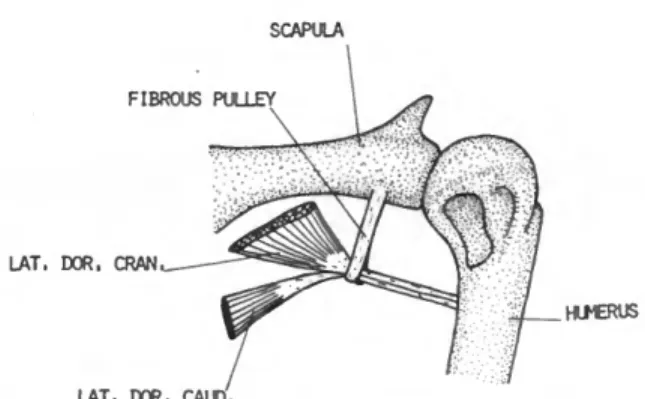
DESCRIPTION.—This large flat muscle (Figure 1) arises from the dorsal spines of about the last cervical and first five thoracic vertebrae. The origin is aponeurotic. Fleshy fibers of this muscle converge as they pass forward and outward, and terminate on a long, narrow tendon. The tendon is about one-fourth the length of the humerus. It passes through a fibrous pulley (Figure 8) much like a thread passes through the eye of a needle.
This pulley, unique to the Spheniscidae, is at- tached to the axillary border of the scapula just behind the glenoid fossa. Insertion is on the pos- terior border of the humeral shaft, near the distal border of the pneumatic fossa.
COMPARISON.—An accessory slip from the cra- nial edge of the latissimus dorsi cranialis occurs in Aptenodytes and Pygoscelis. This accessory slip
NUMBER 341
inserts on the dorsal surface of the dorsal head of the triceps scapularis.
and Eudyptula and intermediate in Eudyptes, Sphen- iscus, and Megadyptes.
Latissimus dorsi caudalis (LAT. DOR. CAUD.)
FIGURES 1, 2, 8
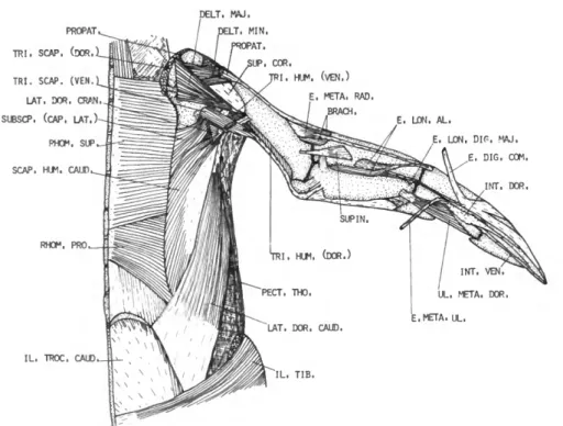
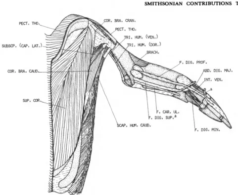
DESCRIPTION.—The latissimus dorsi caudalis (Figure 2) is distinct and separated by a wide interval from the latissimus dorsi cranialis. It arises by means of a delicate aponeurosis from the inferior edge of the ilium and the abdominal muscles immediately posterior to the last thoracic rib. Fleshy fibers on the band-like belly pass forward toward the shoulder and terminate on a long tendon which, after passing through the fibrous pulley mentioned above, is inserted on the posterior border of the humeral shaft adjacent to the insertion of the latissimus dorsi cranialis. The tendons of the latissimus dorsi cranialis and latis- simus dorsi caudalis are partly fused.
COMPARISON.—The latissimus dorsi cranialis and latissimus dorsi caudalis are fused along their contiguous borders in Aptenodytes and Pygoscelis.
All other genera have these two parts widely separated.
Latissimus dorsi metapatagialis (LAT. DOR. MET.)
FIGURE 1
DESCRIPTION.—This muscle is quadrilateral in form. It arises by a narrow aponeurosis from the dorsal spines of thoracic vertebrae 4, 5, and 6.
Insertion is along a broad line on the skin of the lateral line of the trunk. The belly of the latissi- mus dorsi metapatagialis is much wider than that of the latissimus dorsi caudalis and nearly as wide as the belly of the latissimus dorsi cranialis.
COMPARISON.—The latissimus dorsi metapata- gialis exhibits a wide range of development in penguins. In Aptenodytes this muscle is very wide (1.01 scapula), reaching from the base of the neck to the lumbar region. It is narrowest in Pygoscelis
M . RHOMBOIDEUS SUPERFICIALIS (RHOM. SUP.)
FIGURES 1, 2, 7
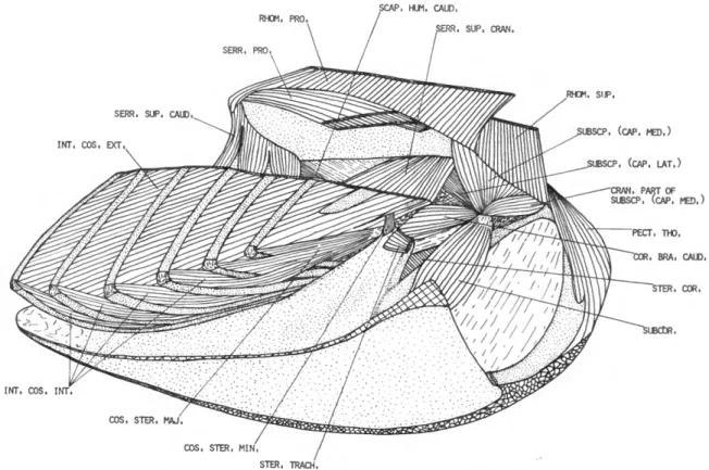
DESCRIPTION.—This wide, flat muscle (Figure 2) arises by means of a narrow aponeurosis from the dorsal spines of the last one or two cervical vertebrae and the first three or four thoracic vertebrae. The fibers pass transversely and slightly forwards; they are inserted on about the anterior half of the vertebral border of the scapula and the scapular process of the clavicle.
COMPARISON.—This muscle is very similar in all species. In Eudyptes chrysocome, Watson (1883:
76) found this muscle arising from the dorsal spines of the three anterior thoracic vertebrae and the last two cervical vertebrae. In this penguin, as well as in Pygosceles taeniatus ( = Pygoscelis papua) and Spheniscus minor (= Eudyptula minor), he found the insertion to be confined to the cranial two- thirds of the vertebral border of the scapula. The insertion is not this extensive in any of the pen- guins studied. Gervais and Alix (1877:444) found the rhomboideus superficialis in Eudyptes chrysolo- phus attached to the dorsal spines of the last two cervical vertebrae, as well as to the dorsal spines of thoracic vertebrae 1 through 5. Such a wide origin was not present in any specimen of penguin studied.
M . RHOMBOIDEUS PROFUNDUS (RHOM. PRO.)
FIGURES 2, 3, 7
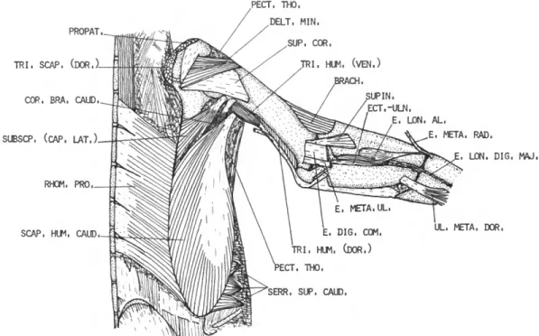
DESCRIPTION.—The rhomboideus profundus (Figure 3) arises from the dorsal spines of about five or six thoracic vertebrae (thoracic vertebrae 2-7) by means of an aponeurosis. Fleshy fibers pass transversely and somewhat obliquely back- wards to a fleshy insertion on about the caudal three-fourths of the vertebral border of the scap-
SMITHSONIAN CONTRIBUTIONS TO ZOOLOGY
ula. Rhomboideus superficialis covers about the cranial two-thirds of the rhomboideus profundus.
M . SERRATUS PROFUNDUS
(SERR. PRO.)
FIGURE 7
DESCRIPTION.—The serratus profundus consists of four large, flat fleshy fascicles superimposed on one another. These fascicles arise from the last two cervical and first two thoracic ribs, dorsal to the uncinate processes. The fascicles pass postero- dorsally and insert on about the caudal half of the costal surface of the scapula near the vertebral border.
COMPARISON.—The small cranial fascicle is usu- ally absent in Spheniscus and Megadyptes. Apteno- dytes, Pygoscelis, Eudyptes, and Eudyptula generally possess four fascicles. The caudal fascicle is some- times absent. Watson (1883:79) reports four mus- cular plates in Eudyptes chrysocome and P. papua, which arise from the second, third, fourth, and fifth ribs. In Aptenodytes patagonicus and Eudyptula minor he found only three digitations, the first from the second rib, the second from the third and fourth ribs, and the third from the fifth rib.
In no instance was the origin from the third, fourth, and fifth ribs in any penguin examined in this study. Schoepss (1829:97) has the serratus profundus arising from the transverse processes of the first and second dorsal vertebrae, as well as from the caudal border of the second rib. Gervais and Alix (1877:443) found five fleshy slips in Eudyptes chrysolophus of which the most cranial arises from the transverse process of the last cer- vical vertebra, the second from the first rib be- neath the transverse process, and the other three from the following ribs dorsal to the uncinate processes. None of the specimens in this study had five slips, nor has any other worker reported this many muscular slips for the serratus profundus.
M . SERRATUS SUPERFICIALIS CRANIALIS
(SERR. SUP. CRAN.)
FIGURE 7
DESCRIPTION.—This muscle arises by means of two fleshy fascicles from the last cervical and first thoracic ribs. The fascicles fuse near the origin.
The fibers pass obliquely anterior and dorsal.
Insertion is mostly fleshy on the axillary border of the scapula. The insertion partially separates the origin of the subscapularis caput laterale from that of the subscapularis caput mediale.
COMPARISON.—My observations agree with those of Schoepss (1829:96), who found this mus- cle in S. demersus attached to the outer surfaces of the last cervical rib and to the lower part of the first true rib. Gervais and Alix (1877:443) describe this muscle as arising from the third and fourth ribs in E. chrysolophus. Watson (1883:78) gives the origin as the second, third, and fourth vertebral ribs. In one specimen of Pygoscelis antarctica, I found the origin to be from the last cervical and first two thoracic ribs.
M . SERRATUS SUPERFICIALIS CAUDALIS
(SERR. SUP. CAUD.)
FIGURES 3, 7
DESCRIPTION.—This muscle (Figure 7) has three fascicles arising from thoracic ribs 2, 3, and 4, a little below the uncinate processes. The fibers pass nearly at a right angle to the long axis of the scapula. Insertion is mostly fleshy on the ventro- lateral border of about the posterior fifth of the scapula.
COMPARISON.—The origin of this muscle is gen- erally from the second, third, and fourth thoracic ribs. Variations to this pattern occur. One speci- men of A. patagonicus has only two fascicles, which arise from the third and fourth thoracic ribs. This pattern is also present in one specimen of Pygoscelis adeliae. The posterior fascicle in one specimen of P. antarctica arises from both the fourth and fifth thoracic ribs. Megadyptes has only two fascicles,
NUMBER 341
which arise from the second and third thoracic ribs. Watson (1883:78) found this muscle arising by two digitations from the fourth and fifth ribs in P. papua. Schoepss (1829:94) reports this muscle arising from four ribs in penguins. Watson (1883:
78) never found the serratus superficialis caudalis arising from more than three ribs, and I found it arising from three ribs in all except one specimen in which the posterior slip arose from both the fourth and fifth ribs.
M . SCAPULOHUMERALIS CAUDALIS (SCAP. HUM. CAUD.)
FIGURES 1-3, 5-7
DESCRIPTION.—The scapulohumeralis caudalis (Figure 3) has an extensive origin from about the posterior two-thirds of the dorsal and lateral sur- faces of the scapula. The fibers pass obliquely forward and end on a strong tendon, which is inserted on the posterior border of the humerus (0.33 humerus).
COMPARISON.—The scapulohumeralis caudalis is a very uniform muscle in the Spheniscidae. The cranial end of the origin shows only slight varia- tion between genera of penguins, ranging from 0.33 scapula in Aptenodytes to 0.44 scapula in
Eudyptula.
of the sternum, the interosseus membrane be- tween this process and the corpus sterni, a line along the entire length of the sternal keel ventral to the origin of the supracoracoideus, the lateral surface of the clavicle below the shoulder joint, the cranioventral edge of the membrana sterno- coraco-clavicularis, and the aponeurosis covering the supracoracoideus. The belly is composed of three distinct parts. Fibers of the clavicular part pass caudolaterally. Those of the caudolateral part pass craniolaterally, while those of the large middle part pass obliquely at increasing angles from the sternum. The clavicular fibers end on a specialized part of the tendon of insertion, which ends on about the proximal fifth of the cranial border of the humeral shaft, joining the tendon of the propatagialis near the proximal end of the humerus. The remaining fibers insert by means of a wide, curving tendon on the proximal third of the cranioventral edge of the humerus.
COMPARISON.—The pectoralis thoracica shows no significant variations in the Spheniscidae.
Reid (1835:140) described the muscle in Apteno- dytes patagonicus as arising from "the cartilages of the ribs, and from the anterior part of the cora- coid bone" in addition to the origin given above.
Such is not the case in any of the penguins examined in this study or in the study by Watson (1883:81).
M . PECTORALIS
This muscle consists of three slips: pectoralis thoracica, pectoralis propatagialis (which is not present in penguins), and pectoralis pars subcu- tanea abdominalis.
Pectoralis thoracica (PECT. THO.)
FIGURES 2-7
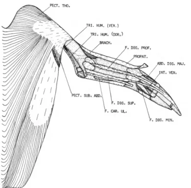
DESCRIPTION.—The pectoralis thoracica (Fig- ure 4) is a very powerful muscle which arises from the lateral edge of the sternum posterior to the coracobrachialis caudalis, the trabecula lateralis
Pectoralis pars subcutanea abdominalis (PECT. SUB. ABD.)
FIGURE 4
DESCRIPTION.—Pars subcutanea abdominalis is a wide, flat, ribbon-like muscle that arises from the skin along the flank. The caudal end of the origin is opposite the region of the acetabulum.
The muscle passes cranially and somewhat dor- sally to insert on the dorsal border of the pecto- ralis thoracica caudal to the insertion of the latter on the humerus.
COMPARISON.—The maximum width of this muscle relative to the humeral length shows some variation between genera of penguins. It is nar-
SMITHSONIAN CONTRIBUTIONS TO ZOOLOGY
rowest in Aptenodytes and Eudyptes, and interme- diate in Megadyptes, Spheniscus, and Pygoscelis. Wat- son (1883:81) describes the pectoralis pars sub- cutanea abdominalis as inserting on the anterior margin of the humerus. This is not the case in any species of penguin examined in this study.
M . SUPRACORACOIDEUS (SUP. COR.) FIGURES 1-3, 5
DESCRIPTION.—The supracoracoideus (Figure 5) arises from the entire surface of the sternum not occupied by the pectoralis thoracica, from the proximal part of the trabecula lateralis of the sternum, from the keel of the sternum, and from the basal end of the coracoid. It also has a consid- erable attachment to the membrana sterno-cor- aco-clavicularis. From these origins the fibers con- verge to a stout, flattened tendon which, after passing through the canalis triosseus, inserts (Fig- ures 1-3) on an oblique ridge on the proximodor- sal part of the humerus between the insertions of the deltoideus major and deltoideus minor. The supracoracoideus is very well developed in com- parison to the pectoralis thoracica, a fact noted by every anatomist who has worked on any mem- ber of the group.
COMPARISON.—The small, separate anterior part of the supracoracoideus described by Schoepss (1829:124), Gervais and Alix (1877:
447), and Watson (1883:82) is the long head of the deltoideus minor and will be described later with that muscle. The supracoracoideus is a very strong, bipennate muscle in penguins and is very similar in all.
M . CORACOBRACHIALIS CRANIALIS (COR. BRA. CRAN.)
FIGURES 5, 6
DESCRIPTION.—This muscle (Figure 5) is small and embedded in a very dense investment of fascia. It arises from the cranioventral surface of
the distal apex of the coracoid. The belly is very weak. Insertion is tendinous on the extreme prox- imal end of the cranioventral humeral shaft and is covered by the pectoralis thoracica.
COMPARISON.—The coracobrachialis cranialis is present in all species of penguins studied and shows the same relative degree of development.
It is not described in the major treatises on pen- guin myology.
M . CORACOBRACHIALIS CAUDALIS (COR. BRA. CAUD.)
FIGURES 3, 5-7
DESCRIPTION.—The coracobrachialis caudalis has a fleshy origin from a short, narrow line along the cranial third of the craniolateral edge of the corpus sterni and from about the proximal third of the lateral surface of the coracoid (Figure 6).
The belly is large and tapered to a point at each end. Insertion is by a strong tendon on the dorsal surface of the internal tuberosity of the humerus (Figure 3).
COMPARISON.—This muscle shows remarkable uniformity in penguins.
M . STERNOCORACOIDEUS (STER. COR.)
FIGURE 7
DESCRIPTION.—This muscle has a fleshy origin from the craniolateral sternal spine and the cra- nial border of the sternum. The sternocoracoideus is a strong, triangular muscle. Insertion is fleshy on a large triangular area of the proximal end of the dorsal surface of the coracoid.
COMPARISON.—A very uniform muscle among penguins but of varying length. It is longest in Pygoscelis and shortest in Spheniscus. The remain- ing genera are of intermediate length. This mus- cle is not described by Watson (1883).
NUMBER 341
M . SUBCORACOIDEUS (SUBCOR.)
FIGURE 7
DESCRIPTION.—The subcoracoideus is a large, powerful muscle arising fleshy from the dorsal surface of the caudal end of the spina externa of the sternum and partly tendinous from the adja- cent cranial edge of the corpus sterni. As it passes toward the shoulder, it is further attached to the dorsomedial surface of the coracoid and adjacent membrana sterno-coraco-clavicularis. Near the shoulder joint the subcoracoideus fuses with the subscapularis. Insertion is by a common, short but strong, tendon at the proximal end of the bicipital crest of the humerus. The tendon is strongly attached to the adjacent joint capsule.
COMPARISON.—The subcoracoideus is very sim- ilar in all penguins.
M . SUBSCAPULARIS (SUBSCP.) FIGURES 2, 3, 5-7
DESCRIPTION.—The subscapularis is divided into two heads, which are partially separated by the serratus superficialis cranialis (Figure 7). The subscapularis caput mediale (SUBSCP. (CAP. MED.))
arises fleshy from about the proximal third of the costal surface of the scapula, from a very small area on the medial surface of the scapular process of the clavicle, and a small cranial part from the medial surface of the coracoid distal to the sub- coracoideus. Hudson et al. (1969:464) refer to that part of the subscapularis arising from the coracoid, clavicle, and adjacent scapula as a cra- nial head of the subcoracoideus. Only the slightest indication of a division of this muscle mass exists in penguins. Because of the very intimate associ- ation with the caput mediale, I have chosen to consider it a part of the latter. The fibers of the caput mediale almost at once unite with those of the subcoracoideus and subscapularis caput lat- erale (SUBSCP. (CAP LAT.)). The caput laterale
(Figure 3) arises fleshy from about the cranial two-fifths of the lateral surface of the scapula.
Insertion of the subscapularis is by means of a tendon common to it and the subcoracoideus.
This tendon inserts on the proximal end of the bicipital crest of the humerus. The caput mediale is smaller than the caput laterale.
COMPARISON.—The caput mediale reaches far- thest distally on the scapula in Megadyptes and is shortest in Pygoscelis; the remaining genera are intermediate. Gervais and Alix (1877:445) state that the caput laterale {petit rond) inserts above the caput mediale (sous-scapulaire). Every speci- men in this study and those studied by Watson (1883:85) have the caput laterale and caput me- diale inserted by means of a common tendon.
M . PROPATAGIALIS (PROPAT.) FIGURES 1-4
DESCRIPTION.—The propatagialis (Figure I) arises fleshy from the dorsal apex of the coracoid, the craniolateral edge of the clavicle, and the intervening coracoclavicular ligament. Its belly is stout and partly separable into two layers. Inser- tion is by means of a very heavy tendon along nearly the entire length of the cranial edge of the humerus. The tendon continues along the cranial border of the ventral surface of the radius and contributes to the formation of the alar aponeu- rosis. Beyond the wrist, the tendon becomes pro- gressively less distinct.
COMPARISON.—The propatagialis exhibits little variation among penguins. No indication of a division into superficial and deep layers is present
in Eudyptula, P. antarctica, P. adeliae, and one spec-
imen of E. pachyrhynchus. Watson (1883:88) de- scribes a deep and superficial part to this muscle.
The superficial part {deltoides posterieur of Gervais and Alix) corresponds to the deltoideus major in Watson's opinion. Schoepss (1829:82, 86) figures a cranial and a caudal belly for the tensor patagii longus of penguins. The caudal belly of his de- scription most definitely corresponds to the del-
8 SMITHSONIAN CONTRIBUTIONS TO ZOOLOGY
toideus major. Meckel (1828:337-343) also de- scribes two bellies for the tensor longus, one of which can with difficulty be separated from the pectoralis major. These obviously correspond to the two partial layers described above for E.
pachyrhynchus. Watson describes an accessory slip to the tensor patagii. This slip corresponds to the long head of the deltoideus minor.
M . DELTOIDEUS MAJOR (DELT. MAJ.)
FIGURES 1, 2
DESCRIPTION.—The deltoideus major (Figure 2) is a very thin, triangular muscle. It arises fleshy from the distal apex of the clavicle, the dorsal surface of the coracoscapular ligament, and the acromial process of the scapula. The fibers con- verge distally onto a short tendon, which inserts on the caudodorsal edge of the humerus very close to the insertion of the latissimus dorsi.
COMPARISON.—The belly is triangular in most genera. It is strap-like in Spheniscus. The belly is shortest in Aptenodytes and Megadyptes, and longest in Eudyptes; intermediate in the remaining genera.
The tendon is shortest in Eudyptes and Spheniscus
and is longest in Megadyptes and Pygoscelis. Apteno-
dytes and Eudyptula are intermediate. Descriptions by other workers are very confused.
M . DELTOIDEUS MINOR (DELT. MIN.)
FIGURES 2, 3, 6
DESCRIPTION.—This muscle has two distinct heads in the Spheniscidae. The short dorsal head (Figure 3) arises fleshy from the acromial process of the scapula and from the coraco-scapular lig- ament. The short head fuses with the long head almost from the origin of the former. The long ventral head (Figure 6) arises from the caudal third of the membrana sterno-coraco-calvicularis, from the cranial edge of the manubrial spine, and from a long, narrow line along most of the ven-
tromedial edge of the coracoid. The fibers con- verge on a short, strong tendon, which inserts in common with the short head on the external tuberosity of the humerus deep to the propatagi- alis.
COMPARISON.—In most penguins the origin also arises from the clavicular process of the coracoid inside the canalis triosseum. Eudyptula and most Eudyptes lack this part of the origin.
M . TRICEPS BRACHII
DESCRIPTION.—The triceps brachii consists of two distinct parts, a triceps scapularis from the scapula and clavicle and a triceps humeralis from the caudal surface of the humerus.
Triceps scapularis
(TRI. SCAP.)
FIGURES 1-3
The triceps scapularis is very large and com- posed of a dorsal and ventral head. The much larger dorsal head (Figure 1) arises fleshy from the acromial process of the scapula, the adjacent joint capsule, and a long, narrow line on the medial and dorsal surfaces of the clavicle dorsal to the origin of the propatagialis. This head is partially divided into a superficial and deep layer.
At about 0.35 humerus the dorsal head ends on a strong tendon common to it and the ventral head. The smaller ventral head (Figure 2) arises fleshy from the axillary border of the scapula immediately behind the glenoid fossa. This belly is firmly attached to the fibrous loop through which the tendons of the latissimus dorsi caudalis pass. The vental head ends on the common ten- don at 0.37 humerus. The triceps scapularis ten- don almost immediately fuses with the tendon of the ventral head of the triceps humeralis. The resulting tendon is attached to the caudal surface of the dorsal head of the triceps humeralis and can be traced to the elbow. At the elbow two very large sesamoids develop in relation to the two parts of the triceps tendon. Distal to these sesa-
NUMBER 341
moids two tendinous slips are evident, one from each of the sesamoids. These short, strong tendons insert on the dorsoposterior edge of the ulna proximal to 0.17 ulna.
Triceps humeralis (TRI. HUM.)
FIGURES 1-6
The triceps humeralis is less massive than the triceps scapularis but is also composed of two heads. The shorter ventral head (Figure 3) arises fleshy from the region of the pneumatic foramen of the humerus. Its fibers end on a tendon at about 0.46 humerus. The very long, slender dorsal head (Figure 1) arises fleshy from the caudal border of the humerus distal to about 0.39 hu- merus. Its proximal end lies dorsal to the distal end of the ventral head. Fibers of the dorsal head pass caudodistally, ending on the common tendon of the triceps scapularis and triceps humeralis. A few of the distal fibers end on the inner sesamoid of the elbow.
COMPARISON.—In all specimens studied there was some exchange of fleshy fibers between the two heads of the triceps humeralis. Watson (1883:
91) decribes such interchange of fibers for Eudyptes chrysolophus. The dorsal head of the triceps hu- meralis is longest in Aptenodytes, Eudyptula, and Spheniscus; shortest in Pygoscelis; intermediate in Eudyptes and Megadyptes.
M . BICEPS BRACHII
This muscle is generally absent in the Sphen- iscidae. A vestigial biceps brachii was present in a single specimen of Megadyptes antipodes. It arose by a short delicate tendon from the dorsal surface of the humerus deep to the pectoralis thoracica.
A very small, fleshy belly was present near the distal end of the humerus. Insertion was by a delicate tendon on the deep surface of the pro- patagialis tendon over the brachialis. Filhol (1885) does not describe this muscle for any of the penguins he studied, including M. antipodes.
M . BRACHIALIS (BRACH.)
FIGURES 1-6
DESCRIPTION.—The brachialis (Figure 3) arises fleshy from the cranioventral border of the hu- merus distal to about 0.59 humerus. The belly is quadrilateral and well developed for this part of the wing. Insertion is mainly fleshy on the proxi- mal border of the radius, extending onto the ventral surface and slightly onto the dorsal sur- face.
COMPARISON.—The brachialis is very well de- veloped in all penguins. There has been a fair amount of controversy over the correct designa- tion for this muscle. Gervais and Alix (1877:450) consider this muscle not homologous with the brachialis anticus (M. brachialis inferior of Ga- dow and Selenka, 1891) of other birds because of its insertion on the radius. Nor do they consider it representative of a biceps, since it does not insert on the interosseus border of the radius.
Schoepss (1829:141) describes a muscle in Spheniscus demersus that arises from the lower part of the cranial border of the humerus and inserts on the radial border of the ulna near the liga- mentous capsule of the elbow joint. Watson (1883:92) failed to recognize the presence of this muscle in any species of penguin that he dissected.
Gervais and Alix (1877:450) omit all reference to it in their description of E. chrysolophus. Such a muscle slip arising from the brachialis is some- times present in penguins. Of the 26 specimens examined in this study, only 8 specimens of seven species had such a muscle slip. This slip ocurred in one specimen each of Aptenodytes forsteri ( I S ) , A. patagonicus (1 S), Eudyptes chrysolophus schlegeli ( 2 W ) , £ c. chrysolophus (1 S), E. pachyrhynchus (1 S), Spheniscus demersus (2 W), S. mendiculus ( 1 W ) , and S. magellanicus (2 W). This small slip of muscle is a definite part of the brachialis. It separates from the caudal border of the brachialis and inserts by means of a short tendon on the radial border of the ulna near the proximal end. The presence of this slip in some specimens reinforces
10 SMITHSONIAN CONTRIBUTIONS TO ZOOLOGY
the idea that the entire muscle is indeed the brachialis of other birds. It has shifted its point of insertion, a result of the extreme dorsoventral compression of the sphenisciform wing.
M . FLEXOR CARPI ULNARIS (F. CAR. UL.)
FIGURES 4-6
DESCRIPTION.—The flexor carpi ulnaris (Figure 4) is entirely tendinous. It arises from the distal apex of the epicondylus medialis humeri, imme- diately distal to the origin of the flexor digitorum superficialis. The tendon passes along the caudal border of the ventral surface of the ulna. Insertion is near the middle of the ventral surface of the os ulnare.
COMPARISON.—Similar in all penguins studied.
Watson (1883:93) also found this muscle to be similar in all penguins he studied.
M . FLEXOR DIGITORUM SUPERFICIALIS (F. DIG. SUP.)
FIGURES 4-6
DESCRIPTION.—This muscle (Figure 4) is en- tirely replaced by tendon. It arises from the distal edge of the epicondylus medialis humeri imme- diately proximal to the flexor carpi ulnaris. The tendon passes along the ventral surface of the ulna adjacent to the flexor carpi ulnaris. Just before reaching the wrist, it divides into two branches. One of these is very short and inserts on the cranioventral edge of the os ulnare, while the other passes obliquely across the carpometa- carpus to the anterior border of the wing and fuses with the tendon of the flexor digitorum profundus. Insertion is on the cranioventral edge of phalanx 2, digiti majoris.
COMPARISON.—The flexor digitorum superfici- alis is similar in all penguins studied. A small fleshy belly is present in only one of the specimens examined (A. patagomcus).
M . FLEXOR DIGITORUM PROFUNDUS (F. DIG. PROF.)
FIGURES 4-6
DESCRIPTION.—The flexor digitorum profundus (Figure 4) is represented by a single tendon, which arises from the interosseous borders of the radius and ulna. The tendon is indistinct and fused with the interosseous membrane proximal to about 0.75 radius. After passing the wrist, the tendon continues along the ventral surface of os metacar- pale majus, the proximal phalanx of digiti ma- joris, and finally inserts along the cranioventral edge of the proximal and distal phalanges of digiti majoris. The tendon fuses with the flexor digitorum superficialis near the base of the prox- imal phalanx of digiti majoris.
COMPARISON.—The flexor digitorum profundus is similar in all penguins studied. A very small belly is present in one wing of Eudyptula minor, but absent in the other wing of this specimen. None of the other specimens have fleshy fibers associ- ated with the flexor digitorum profundus.
M . ULNIMETACARPALIS VENTRALIS
DESCRIPTION.—When present, the ulnimetacar- palis ventralis is usually entirely tendinous and arises from the ventral surface of the ulna near the cranial border. Insertion is on the tendon of the propatagialis near the wrist.
COMPARISON.—The ulnimetacarpalis ventralis is absent in all specimens of Aptenodytes, Eudyptula,
a n d Spheniscus. In Eudyptes chrysolophus schlegeli a
very small belly is present. The ulnimetacarpalis ventralis is present but entirely tendinous in one specimen of E. pachyrhynchus and one specimen of E. c. chrysolophus. One specimen of Pygoscelis papua has an ulnimetacarpalis ventralis that arises like that in E. pachyrhynchus. The insertion in P. papua is on the tendon of the flexor digitorum profundus at about 0.33 carpometacarpus. In Megadyptes the origin is from the adjacent edges of the radius and ulna near the distal end of these bones. It is entirely tendinous and inserts on the caudoven-
NUMBER 341 11 tral edge of the carpometacarpus near the proxi-
mal end of the fused pollex. Schoepss (1829:152) describes this muscle in S. demersus as having an origin from the inner border of the ulna and
inserting into the inner side of the base of the first radial phalanx. Neither Watson (1883:97) nor I found this muscle in specimens of S. demersus.
Gervais and Alix (1877) and Filhol (1885) gave no reference to such a muscle in penguins.
M . EXTENSOR METACARPI RADIALIS
(E. META. RAD.)
FIGURES 1-3
DESCRIPTION.—This is a small, weak, proxi- mally situated muscle (Figure 1) arising tendinous from the cranial border of the dorsal surface of the humerus immediately proximal to the bra- chialis. The origin is strongly attached to the tendon of the propatagialis. The muscular fibers are short and end on a strong tendon which, after passing through a shallow groove near the dor- socranial border of the radius, inserts on the proximal end of the carpometacarpus near the cranial border. Insertion is in common with the extensor longus alulae.
COMPARISON.—The muscle is longest in Eudyp- tula (0.21 radius) and shortest in Pygoscelis (0.02 radius). In the remaining genera it is of interme- diate length. Schoepss (1829:145) reports that this muscle arises from the humerus by two distinct heads in penguins. I failed to find this arrange- ment in any species of penguin, nor has any other worker reported two heads of origin in penguins.
Watson (1883:94) reports a separate insertion for the extensor metacarpi radialis longus (exten- sor metacarpi radialis) and extensor metacarpi radialis brevis in all the specimens that he dis- sected. Such an arrangement was not present in any specimen examined in this study. Gervais and Alix (1877:499) and Filhol (1885:176) are in agreement with my findings.
M . SUPINATOR
(SUPIN.)
FIGURES 1-3
DESCRIPTION.—This very weak, triangular mus- cle (Figure 1) arises by means of a short, delicate tendon from the heavy tendon of the extensor digitorum communis, which arises from the dor- sal surface of the distal end of the humeral shaft.
The muscular fibers pass distally and cranially to insert on the dorsal surface of the radius proximal to 0.29 radius.
COMPARISON.—The supinator is longest in Spheniscus, intermediate in Pygoscelis a n d Eudyp- tula, a n d shortest in Megadyptes, Aptenodytes a n d Eudyptes. This muscle is absent in one specimen of P. antarctica. It lacks muscular fibers in two spec- imens of P. adeliae.
M . EXTENSOR DIGITORUM COMMUNIS
(E. DIG. COM.)
FIGURES 1-3
DESCRIPTION.—This muscle (Figure 1) is rep- resented by a strong tendinous band, which arises from the cranial edge of a wide tendinous sheet from the epicondylus lateralis humeri. There is no branch to the region of the fused pollex.
Insertion is on the craniodorsal surface of os metacarpale major near the distal end, and on both phalanges of digiti majoris nearly to the tip of the wing. The tendon is completely fused with the tendon of the extensor longus digiti majoris near the middle of the major metacarpal.
COMPARISON.—A very small, fleshy belly is pre- sent in only two of the specimens examined, one of Eudyptes pachyrhynchus a n d one of Spheniscus humboldti. In one specimen of P. papua the tendon sends a small branch to the region of the fused pollex. This branch ends in the fascia over the pollex.
12 SMITHSONIAN CONTRIBUTIONS T O ZOOLOGY M . EXTENSOR METACARPI ULNARIS
(E. META. UL.)
FIGURES 1-3
DESCRIPTION.—The extensor metacarpi ulnaris (Figure 1) is entirely tendinous. It arises from the caudal edge of the tendinous sheet in common with the extensor digitorum communis. The ten- don passes along the craniodorsal edge of the ulna and, after crossing the wrist, inserts on the caudal edge of the major metacarpal at about 0.40 car- pometacarpus.
COMPARISON.—In most specimens the insertion is confined to the major metacarpal. Watson (1883:96) found this muscle in Eudyptes crestatus to insert only on the cranial border of os metacarpale minus. None of the specimens examined in this study have this arrangement. In all other speci- mens, he found the muscle inserting on the caudal border of the major metacarpal. Schoepss (1829:
150) reports the insertion on both the major and minor metacarpals. Such an arrangement is oc- casionally present. I have found an insertion on both metacarpals in Megadyptes antipodes (IS), Aptenodytes forsteri (IS), Eudyptula minor (IS), Eu- dyptes c. chrysolophus (IS), and Spheniscus mendiculus (IS).
M . ECTEPICONDYLO-ULNARIS
(ECT.-ULN.)
FIGURE 3
DESCRIPTION.—The ectepicondylo-ulnaris is en- tirely tendinous. It arises from the middle of the tendinous sheet in common with the extensor digitorum communis and extensor metacarpi ul- naris. The tendinous sheet arises from the dorsal surface of the distal end of the humerus. Insertion is on the cranial border of about the proximal half of the ulnar shaft.
COMPARISON.—This muscle is shortest in Eu- dyptes and longest in Eudyptula and Pygoscelis.
There is a small belly in A. patagonicus (IS), Eu- dyptes chrysolophus schlegeli (2W), P. papua (IS),
Eudyptula m. albosignata ( 1 W ) , S. demersus (1W), and S. humboldti (1W); tendinous in all others.
The ectepicondylo-ulnaris (anconaeus) muscle described by Reid (1835:142) and "l'ancone ex- terne" described by Gervais and Alix (1877:449) is either a ligament of the elbow joint or a part of the triceps tendon. The structure described by these authors cannot be homologized with any muscle in other birds. They completely omit ref- erence to the tendon that I call "ectepicondylo- ulnaris." Watson (1883:92) did not find this mus- cle in any species of penguin he dissected, and Filhol (1885:175) reports the muscle to be absent in Eudyptes crestatus.
M . EXTENSOR LONGUS ALULAE
(E. LON. AL.)
FIGURES 1-3
DESCRIPTION.—The extensor longus alulae (Figure 2) arises by means of two heads; a small radial head from the caudodorsal surface of the radius beginning at about 0.43 radius and a larger ulnar head from about the proximal half of the cranial border of the ulnar shaft. The two bellies fuse about midway the forearm, and the common belly extends obliquely across the radius ending on a tendon at about 0.80 radius. After crossing the wrist, this tendon fuses with that of the exten- sor metacarpi radialis. Insertion is on the proxi- mal end of the carpometacarpus near the cranial border of this bone.
COMPARISON.—The radial head is absent in both wings of E. m. minor and one wing of E. m.
albosignata. Whether the absence of the radial head in Eudyptula is significant cannot be deter- mined on the basis of only two specimens. All other species are similar to Eudyptes.
M . EXTENSOR LONGUS DIGITI MAJORIS
(E. LON. DIG. MAJ.)
FIGURES 2, 3
DESCRIPTION.—This muscle (Figure 2) is re-
NUMBER 341 13 placed by a tendon that arises from the distal half
of the caudal surface of the radius between about 0.53 radius and 0.82 radius. The tendon then passes between the distal ends of the radius and ulna, crosses the wrist, and fuses with the tendon of the extensor digitorum communis about mid- way of the major metacarpal. There is no trace of a distal head.
COMPARISON.—The extensor longus digiti ma- joris is absent in one specimen of A. forsteri and one of A. patagonicus. It is small but partly fleshy in one specimen of A. forsteri and very small and tendinous in the other specimen of A. patagonicus.
In one specimen of Eudyptes pachyrhynchus there are a few fleshy fibers associated with this muscle.
It is entirely tendinous in all other penguins studied.
Watson (1883:97) described the extensor lon- gus digiti majoris as being a very slender muscle arising from the contiguous borders of the radius and ulna and inserting into the outer side of the second or terminal radial phalanx. Such an ar- rangement does not occur in any of the specimens examined in the current study. There is a con- nection to the posterior edge of the major meta- carpal, as well as to the extensor digitorum com- munis in Eudyptula. Gervais and Alix (1877:451) describe the extensor longus digiti majoris in Eudyptes chrysocome as a very small fleshy bundle that arises from the distal half of the interosseus space between the radius and ulna.
My findings are in agreement with those of Meckel and Schoepss. According to Meckel (1828:344), this muscle is represented entirely by tendon. Schoepss (1829:159) found that its origin was confined to the distal end of the radius.
M . ULNIMETACARPALIS DORSALIS (UL. META. DOR.)
FIGURES 1-3
DESCRIPTION.—The ulnimetacarpalis dorsalis (Figure 1) arises by means of a strong, flat tendon near the distal end of the caudodorsal edge of the ulna. The belly is single and one of the strongest
distal to the elbow. Insertion is fleshy along an extensive area of the caudal edge of the minor metacarpal. The belly ends at about 0.83 carpo- metacarpus.
COMPARISON.—Watson (1883:96) indicated the insertion as confined to the proximal half of the ulnar metacarpal bone in Eudyptes chrysocome and Spheniscus demersus. The shortest belly is in Mega- dyptes (0.76 carpometacarpus). The ulnimetacar- palis dorsalis is a very uniformly developed mus- cle in the Spheniscidae.
M . ABDUCTOR DIGITI MAJORIS (ABD. DIG. MAJ.)
FIGURES 4, 5
DESCRIPTION.—This very weak muscle (Figure 5) arises from the ventral surface of the major metacarpal near the cranial border of this bone and from the adjacent fused pollex. The belly is flat and bipennate. Its fibers converge distally on a short, flat tendon. Insertion is on the base of the proximal phalanx of the major digit.
COMPARISON.—The abductor digiti majoris is longest in Eudyptula (0.18-0.54 carpometacarpus) and shortest in Aptenodytes (0.32-0.78 carpometa- carpus). The muscle is present and fleshy in all specimens studied. However, Watson (1883:100) reported that the muscle is represented by a tendon in all penguins he studied, except in Aptenodytes longirostris ( = A. patagonicus), P. papua, and Eudyptes chrysocome, in which there was a
"distinct but weak muscular belly." According to Meckel (1828:350) and Schoepss (1829:170) this muscle is entirely absent in Sphenisciformes. Ger- vais and Alix (1877:452) found it represented by a tendon in E. chrysocome.
M . INTEROSSEUS DORSALIS (INT. DOR.)
FIGURES 1, 2
DESCRIPTION.—The interosseus dorsalis (Figure 1) is entirely tendinous. It arises from the caudal
14 SMITHSONIAN CONTRIBUTIONS TO ZOOLOGY border of the major metacarpal and the cranial
border of the minor metacarpal. The tendinous sheet that replaces the belly gives rise to a small tendon near the distal end of the carpometacar- pus. Insertion is on the middle of the dorsal surface of the base of the distal phalanx of the major digit.
COMPARISON.—A small, very weak belly is pre- sent in one specimen of A. patagonicus and one of S. mendiculus. Watson (1883:101) reports the mus- cle entirely absent in one specimen of A. patagon- icus a n d Eudyptula minor. In P. papua he found it represented by a tendon without a muscular belly. According to Schoepss (1829:172) this mus- cle is absent in S. demersus. Gervais and Alix (1877:
453) found "the muscle, seldom fleshy."
M . INTEROSSEUS VENTRALIS (INT. VEN.)
FIGURES 1, 2, 4, 5
DESCRIPTION.—The interosseus ventralis (Fig- ure 5) is a weak, fleshy, bipennate muscle that arises from the caudal border of the major meta- carpal and the cranial border of the minor met- acarpal on the ventral side of the carpometacar- pus. The muscular fibers end on a slender tendon that passes to the dorsal surface and then inserts along the entire caudal border of the distal pha- lanx of the major digit.
COMPARISON.—The belly is longest in Apteno- dytes, Megadyptes, a n d Eudyptula; shortest in Pygos- celis.
M . FLEXOR DIGITI MINORIS (F. DIG. MIN.)
FIGURES 4, 5
DESCRIPTION.—This muscle (Figure 5) has a fleshy origin from the ventral surface proximally and the caudal surface distally of the minor meta- carpal. The tendon is mainly on the ventral sur- face of the belly. Insertion is mostly tendinous on
the proximally projecting tubercle on the caudal edge of the minor digit.
COMPARISON.—The belly is longest in Apteno- dytes a n d Eudyptes; shortest in Megadyptes. T h e location of the distal end of the muscle is very uniform, at 0.92 and 0.94 carpometacarpus.
The following muscles are absent in the sphen- isciform wing: M. serratus superficialis metapa- tagialis, M. scapulohumeralis cranialis, M. ex- pansor secundariorum. M. pronator superficialis, M. pronator profundus, M. entepicondylo-ul- naris, M. abductor alulae, M. flexor alulae, M.
adductor alulae, and M. extensor brevis alulae.
Muscles of the Pelvic Appendage
M . ILIOTROCHANTERICUS CAUDALIS (IL. TROC. CAUD.)
FIGURES 2, 10, 11
DESCRIPTION.—The iliotrochantericus caudalis (Figure 10) arises fleshy from most of the prea- cetabular ilium (ala preacetabularis). The belly is very large. Its fibers converge caudally and end on a short broad tendon that inserts on the prox- imocranial edge of the external surface of the femoral trochanter. The iliofemoralis externus is fused to the iliotrochantericus caudalis except for its extreme distal end and tendon of insertion.
COMPARISON.—The iliotrochantericus caudalis is widest in Aptenodytes, Pygoscelis, a n d Eudyptula;
narrowest in Spheniscus, Megadyptes, a n d Eudyptes.
According to Watson (1883:103), this muscle
"arises from the whole of the external surface of the iliac bone as far back as the posterior border of the acetabulum, as well as from the adjoining hollowed surface formed by the fifth, sixth, and seventh lumbo-sacral vertebrae." In no instance did I find the iliotrochantericus caudalis attached to the lumbo-sacral vertebrae, nor has such an arrangement been reported by any other worker.
NUMBER 341 15
M . ILIOTROCHANTERICUS CRANIALIS
(IL. TROC. CRAN.)
FIGURES 10-12
DESCRIPTION.—This muscle (Figure 12), much smaller than the iliotrochantericus caudalis, arises partly fleshy from about the caudal two-thirds of the ventrolateral edge of the preacetabular ilium.
Carnially the belly is strongly adherent to a fi- brous septum, which separates it from the iliotro- chantericus caudalis. The iliotrochantericus cran- ialis is fused to the iliotrochantericus medius, the two muscles being barely distinguishable. Inser- tion is by means of a short, flat tendon on the lateral surface of the femoral trochanter, in com- mon with the iliotrochantericus medius.
COMPARISON.—Only in Eudyptula is the origin separate from that of iliotrochantericus medius.
In this genus the insertion, though separate, is just distal to that of the iliotrochantericus medius.
Watson (1883:104) observed a tendency in Apteno- dytes for the iliotrochantericus cranialis to divide into two distinct portions, an upper and a lower, with a cellular interval lying between them. He failed to recognize these as representing the ili- otrochantericus cranialis and iliotrochantericus medius. According to Gervais and Alix (1877:
454), the iliotrochantericus cranialis in Eudyptes chrysolophus attaches to the external border of the ilium, "et, sur la face interne du femur, au dela du trochanter"
M . ILIOTROCHANTERICUS MEDIUS (IL. TROC. MED.)
FIGURES 10-12
DESCRIPTION.—The iliotrochantericus medius (Figure 12) arises fleshy from the ventrolateral edge of the preacetabular ilium immediately cau- dal to the iliotrochantericus cranialis. The iliotro- chantericus medius and iliotrochantericus crani- alis are fused and nearly indistinguishable. Inser- tion is in common with the iliotrochantericus cranialis on the lateral surface of the femoral
trochanter. The femorotibialis medius partly cov- ers this insertion.
COMPARISON.—Watson (1883:104), referring to the iliotrochantericus medius, states, "Of this muscle the Penguins do not possess the slightest trace." In Aptenodytes he observed a tendency for the iliotrochantericus cranialis "to divide into two distinct portions, an upper and a lower, a cellular interval lying between them." His two portions of the iliotrochantericus cranialis probably rep- resent the iliotrochantericus cranialis and iliotro- chantericus medius. The bellies are fused to some degree in all penguins. In Eudyptula the origins are separate.
M . ILIOFEMORALIS EXTERNUS
(ILFEM. EXT.) FIGURES 10, 11
DESCRIPTION.—The iliofemoralis externus (Fig- ure 11) is very weakly developed. It arises fleshy from the lateral dorsal ridge of the ilium imme- diately caudal to the iliotrochantericus caudalis.
The origin is situated cranial to the acetabulum, an arrangement unusual among birds. The bellies of iliofemoralis externus and iliotrochantericus caudalis are almost completely fused, but the extreme distal end of the belly and the tendon of insertion are not fused to iliotrochantericus cau- dalis. The short, flat tendon of insertion passes over the iliotrochanterici tendons before inserting on the femoral trochanter. The tendon barely reaches the ischiofemoralis, which covers the dis- tal end of the insertion of iliofemoralis externus.
COMPARISON.—The belly of this muscle is sep- arate from that of iliotrochantericus caudalis in only one specimen (Megadyptes antipodes) exam- ined. Gervais and Alix (1877:454) found the in- sertion to be in common with the iliotrochanter- icus caudalis, but the insertions were separate in all penguins that I examined.
16 SMITHSONIAN CONTRIBUTIONS TO ZOOLOGY M . ILIOFEMORALIS INTERNUS
(ILFEM. INT.)
FIGURE 14
DESCRIPTION.—The iliofemoralis internus is a small muscle arising fleshy from the ventral edge of the preacetabular ilium just medial to the iliotrochantericus medius. It passes caudodistally to insert fleshy on the medial surface of the femur near the proximal end of the femorotibialis inter- nus.
COMPARISON.—This muscle is very uniformly developed, showing no significant variations in penguins. However, Meckel (1828:353) thought it to be absent in the Spheniscidae.
M. AMBIENS
FIGURES 10-14
DESCRIPTION.—The ambiens (Figure 13) is a large muscle, arising mostly fleshy from the ven- tral border of the acetabulum. The flattened belly lies on the medial side of the thigh, and terminates on a strong, flat tendon at about 0.86 femur. The tendon passes in a shallow groove (Figure 14) on the cranial surface of the patella, then through the patellar tendon to the lateral side of the knee.
The tendon ends opposite the proximal part of the fibula by inserting (Figure 12) on the tendon of origin of the cranial head of the perforated flexors.
COMPARISON.—The ambiens is shortest in Py-
goscelis and Eudyptula, and is longest in Aptenodytes.
M . ILIOTIBIALIS CRANIALIS (ILTIB. CRAN.)
FIGURES 1,9, 13
DESCRIPTION.—The iliotibialis cranialis (Figure 9) is a very powerful muscle, making up the anterior limit of the thigh. The origin is aponeu- rotic from the caudal end of the fused thoracic spinous ridge and fleshy from the cranial and cranio ventral edges of the ilium. There is a strong
tendinous connection with the aponeurosis of the iliotibialis. Insertion (Figure 13) is fleshy on the medial and cranial surfaces of the patella and patellar tendon.
COMPARISON.—No major differences were noted other than variations in relative width, the belly being broadest in Eudyptula and Megadyptes.
M. ILIOTIBIALIS
(IL. TIB.)
FIGURES 2, 9, 13
DESCRIPTION.—The iliotibialis (Figure 9) is a weakly developed, thin sheet of muscle that arises by means of an aponeurosis from the spinous processes opposite the acetabulum. This aponeu- rosis is attached to the belly of the iliotibialis cranialis cranially and the biceps femoris cau- dally. The postacetabular part of the iliotibialis is absent. The belly is thin along its cranial margin and much thicker along its caudal border.
The center of this muscle is aponeurotic distally.
Insertion is tendinous on the crista cnemialis cran- ialis of the tibia in common with the femorotibi- alis medius and femorotibialis externus. The iliotibialis tendon forms a part of the patellar tendon.
COMPARISON.—The iliotibialis is a uniformly developed muscle in all species examined. Ac- cording to Garrod (1873:643), the postacetabular portion of this muscle is absent in penguins, which is consistent with my observations. Watson (1883:
112) reports the presence of this portion, although reduced to a minimum size, in every species he examined.
M . FEMOROTIBIALIS EXTERNUS (FEM. TIB. EXT.)
FIGURE 12
DESCRIPTION.—The femorotibialis externus (Figure 12) is a very small muscle. It arises from the caudolateral surface of the femur beginning
NUMBER 341 17 at about 0.65 femur. Insertion is in common with
the caudal margin of the patellar tendon.
COMPARISON.—In most penguins the femoroti- bialis externus can be partially separated at either its proximal or distal end. Eudyptes pachyrhynchus and Megadyptes antipodes have the most distinct femorotibialis externus. The muscle is not distin- guishable in Eudyptula and is either absent or very vague in Aptenodytes and Pygoscelis.
M . FEMOROTIBIALIS MEDIUS (FEM. TIB. MED.)
FIGURES 10, 11, 13, 14
DESCRIPTION.—The femorotibialis medius (Fig- ure 10) consists of a large fleshy mass which arises from a very extensive area on the lateral and cranial surfaces of the femoral shaft from the trochanter to the distal condyles. The belly is strong and notched proximally by the insertion of the iliotrochantericus cranialis and iliotrochan- tericus medius. Insertion is mostly fleshy on the proximal surface of the patellar tendon.
COMPARISON.—The femorotibialis medius is similar in all forms examined.
M . FEMOROTIBIALIS INTERNUS (FEM. TIB. INT.)
FIGURES 13, 14
DESCRIPTION.—This long, slender muscle (Fig- ure 14) arises from the whole length of the medial surface of the femoral shaft, extending proximally to the insertion of the iliofemoralis internus. The belly is not divided into superficial and deep parts. Insertion is by a short, flat tendon on the cranial cnemial crest of the tibia.
COMPARISON.—The femorotibialis internus is similar in all forms examined.
M . CAUDO-ILIO-FEMORALIS
This muscle complex is composed of two mus- cles: M. caudofemoralis and M. iliofemoralis,
which are separate for most of their extent but have a common insertion.
M . CAUDOFEMORALIS (CAUD. FEM.)
FIGURES 9, 10
DESCRIPTION.—The caudofemoralis (Figure 10) arises from about the cranial half of the pygostyle and from the last one or two free coccygeal ver- tebrae. The caudal end of the origin is tendinous.
The flat, spindle-shaped belly passes craniodis- tally toward the femur. Just before fusing with the iliofemoralis it forms a short tendon. Insertion is in common with the iliofemoralis on the caudal surface of the femur between about 0.50 and 0.76 femur.
COMPARISON.—The caudofemoralis is very sim- iar in all penguins examined in this study and by other workers.
M . ILIOFEMORALIS (IL. FEM.)
FIGURES 9, 10
DESCRIPTION.—The iliofemoralis (Figure 10) arises from the ventrolateral surface of the crista iliaca dorsolateralis. The origin is mainly tendi- nous but includes a small fleshy area posteriorly.
The belly is a broad, thin sheet, distinctly nar- rower at the distal end. Insertion is in common with the caudofemoralis as described above.
COMPARISON.—The origin of the iliofemoralis varies somewhat among genera. It is entirely fleshy in Spheniscus, mostly fleshy in Aptenodytes, mainly tendinous in Eudyptes, and entirely tendi- nous in Pygoscelis, Eudyptula, and Megadyptes.
I was unable to find the variation that Watson (1883:106) described for the insertion of the caudo-ilio-femoralis. He reported that the inser- tion is confined to the middle third of the femoral shaft in S. demersus and S. magellanicus, to the lower third in Eudyptes chrysolophus, and to the lower two-thirds in Aptenodytes. The means for the distal